Báo cáo khoa học: Tetracysteine-tagged prion protein allows discrimination between the native and converted forms pptx
Bạn đang xem bản rút gọn của tài liệu. Xem và tải ngay bản đầy đủ của tài liệu tại đây (697.82 KB, 13 trang )
Tetracysteine-tagged prion protein allows discrimination
between the native and converted forms
Jernej Gas
ˇ
pers
ˇ
ic
ˇ
1
, Iva Hafner-Bratkovic
ˇ
1
, Michel Stephan
2
, Peter Veranic
ˇ
3
, Mojca Benc
ˇ
ina
1
,
Ina Vorberg
4
and Roman Jerala
1,5
1 Department of Biotechnology, National Institute of Chemistry, Ljubljana, Slovenia
2 Department of Organic and Medicinal Chemistry, National Institute of Chemistry, Ljubljana, Slovenia
3 Faculty of Medicine, Institute of Cell Biology, Ljubljana, Slovenia
4 Institute of Virology, TU Munich, Germany
5 Faculty of Chemistry and Chemical Technology, University of Ljubljana, Slovenia
Introduction
Prion diseases belong to a group of conformational
diseases characterized by the structural conversion of
native protein to alternative conformations [1]. The
protein-only hypothesis states that prions are com-
posed predominantly of abnormally folded prion pro-
tein (PrP), the scrapie pathogenic form of PrP (PrP
Sc
)
[2]. This form of PrP forms amyloid, which can be
detected by compounds that bind to these types of
ordered protein aggregate. Molecules that bind specifi-
cally to amyloids include thioflavin T (ThT) [3], Congo
Keywords
biarsenical; conversion; fibril; prion;
tetracysteine
Correspondence
R. Jerala, Department of biotechnology,
National Institute of Chemistry, Hajdrihova
19, 1000 Ljubljana, Slovenia
Fax: +386 1 476 0300
Tel: +386 1 476 0335
E-mail:
(Received 5 October 2009, revised 19
January 2010, accepted 17 February
2010)
doi:10.1111/j.1742-4658.2010.07619.x
The conformational conversion of prion protein (PrP) from a native con-
formation to the amyloid form is a hallmark of transmissible spongiform
encephalopathies. Conversion is usually monitored by fluorescent dyes,
which bind generic amyloids and are less suited for living cell imaging. We
report a new method for the synthesis of membrane-permeable and mem-
brane-impermeable biarsenical reagents, which are then used to monitor
murine PrP (mPrP) misfolding. We introduced tetracysteine (TC) tags into
three different positions of mPrP, which folded into a native-like structure.
Whereas mPrPs with a TC tag inserted at the N-terminus or C-terminus
supported fibril formation, insertion into the helix 2–helix 3 loop inhibited
conversion. We devised a quantitative protease-free method to determine
the fraction of converted PrP, based on the ability of the fluorescein arseni-
cal helix binder reagent to differentiate between the monomeric and fibril-
ized form of TC-tagged PrP, and showed that TC-tagged mPrP could be
detected on transfected cells, thereby expanding the potential use of this
method for the detection and study of conformational diseases.
Structured digital abstract
l
MINT-7709757: Prp (uniprotkb:P04925) and Prp (uniprotkb:P04925) bind (MI:0407)byelec-
tron microscopy (
MI:0040)
l
MINT-7709744: Prp (uniprotkb:P04925) and Prp (uniprotkb:P04925) bind (MI:0407)by
circular dichroism (
MI:0016)
l
MINT-7709730: Prp (uniprotkb:P04925) and Prp (uniprotkb:P04925) bind (MI:0407)byfluor-
escence technology (
MI:0051)
Abbreviations
BME, b-mercaptoethanol; CrAsH, carboxy fluorescein arsenical helix binder; EDT, ethane dithiol; FlAsH, fluorescein arsenical helix binder;
GPI, glycosylphosphatidylinositol; H, helix; mPrP, murine prion protein; PrP, prion protein; PrP
Sc,
scrapie pathogenic form of prion protein;
TC, tetracysteine; TCEP, Tris(2-carboxyethyl)phosphine; ThT, thioflavin T.
2038 FEBS Journal 277 (2010) 2038–2050 ª 2010 The Authors Journal compilation ª 2010 FEBS
red [4], 2-[1-(6-[(2-fluoroethyl)(methyl)amino]-2-naph-
thyl)ethylidene]malononitrile [5], curcumin [6], and
luminescent conjugated polymers [7]. Of these, ThT is
the most frequently used dye for detecting the forma-
tion of prion fibrils in vitro [8,9].
Localization of different proteins within living cells
became possible with the fusion of a fluorescent protein,
e.g. green fluorescent protein, to the protein of interest
[10]. Fluorescent protein domains may interfere with
protein folding or ligand binding and, more particularly,
with protein packing into ordered arrays. Although the
green fluorescent protein fusion does not influence PrP
trafficking [11], it prevents prion infection [12], similarly
to the PrP–Fc fusion protein [13]. A tetracysteine (TC)
tag that specifically binds biarsenic fluorescent dyes rep-
resents an alternative fluorescent labeling technique.
A TC tag is a short peptide motif defined by the
sequence pattern CCXXCC, where C is a cysteine and
X any amino acid except cysteine [14]. Fluorescein
arsenical helix binder (FlAsH) is an organoarsenical
compound that covalently binds to the TC tag at
subnanomolar concentrations [14]. FlAsH is based on
fluorescein with two arsenic (III) atoms at the 4 ¢-posi-
tion and 5¢-position, and is membrane-permeable
(Fig. 1). This compound by itself is nonfluorescent [14].
Rigid spacing between both arsenic atoms in FlAsH
enables it to bind with high affinity and specificity to the
TC motif introduced into a variety of different proteins
[15]. The binding of FlAsH reagent and also the quan-
tum yield of the formed FlAsH-peptide adduct depends
on the conformation of the peptide backbone adopted
by the tetracysteine tag [16]. Tagging a protein with a
TC motif has been used for the detection of proteins
of interest in vivo [17,18], in purification [19], and for
detection on gel electrophoresis [15].
We decided to investigate the potential of introduc-
ing TC peptide tags at different positions on PrP in
order to monitor conformational changes in PrP. We
show that insertion of a TC tag into the N-terminal
and C-terminal segments of a protein does not inter-
fere with fibril formation. Fluorescent biarsenical
reagents do not label the converted forms of PrP, but
rather the native and denatured forms. This property
is at the core of our new quantitative fluorescent con-
version assay for PrP.
Results
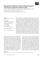
Synthesis of FlAsH and carboxy FlAsH (CrAsH)
The fluorescent reagents FlAsH and CrAsH, which
bind to the TC peptide tag, have been previously
reported on. We modified the synthetic method
described by Griffin et al. [20] (Fig. 1). The new practi-
cal procedure limits the amounts of toxic reagents
needed, and reduces the formation of byproducts by
minimizing the number of manipulation steps during
work-up. Thus, starting from 4¢,5¢-bis(trifluoroacetoxy-
mercuri)fluorescein, analytically pure FlAsH-ethane
dithiol (EDT)
2
was obtained in 29% yield (> 99%
purity) after trituration with a minimal amount of
CH
2
Cl
2
. In addition, following the simplified proce-
dure and starting from an approximately 1 : 1 mixture
of 4¢,5¢-bis(trifluoroacetoxymercuri)-5-carboxy-fluores-
cein and 4¢,5¢-bis(trifluoroacetoxymercuri)-6-carboxy-
fluorescein [21], an analytically pure mixture of
5-CrAsH-EDT
2
and 6-CrAsH-EDT
2
was obtained in
21% yield (> 99% purity). Both reagents were active
and bound effectively to peptides containing the TC
tag, as described below.
Production, secondary structure analysis and
thermal stability of the TC tag-modified protein
Insertion of even a small peptide tag could affect the
ability of PrP to convert to PrP
Sc
. We wanted to
Fig. 1. Scheme of improved method of
chemical synthesis of FlAsH and CrAsH
reagents. NMP, N-methyl-pyrolidinone;
RT, room temperature.
J. Gas
ˇ
pers
ˇ
ic
ˇ
et al. PrP conversion monitored by biarsenical reagent
FEBS Journal 277 (2010) 2038–2050 ª 2010 The Authors Journal compilation ª 2010 FEBS 2039
investigate which positions within the tertiary
structure of PrP allow the introduction of a peptide
tag without compromising protein folding and
conversion into the fibrillar structure. We selected
three positions: one at the disordered N-terminal
segment of PrP (TCN), another at the C-terminal
segment, which connects PrP to the glycosylphos-
phatidylinositol (GPI) anchor (TCC), and a third at
a position within the PrP structure between
helix (H)2 and H3 that is surface-exposed, provides
the geometry that would allow binding of the
biarsenical reagent via the thiol groups, and requires
the most conservative amino acid replacements
(TCL) (Fig. 2A). All recombinant proteins were
produced in Escherichia coli in the form of inclusion
bodies, and successfully refolded into the native-like
conformation as judged by the far-UV CD spectra.
Native murine PrP (mPrP) has a high content of
a-helical secondary structure, with characteristic
minima at 210 nm and 222 nm in the far-UV CD
spectrum. The spectra of TCC, TCN and TCL
overlap the spectra of mPrP, indicating that the
secondary structure is conserved in all TC-tagged
mPrPs (Fig. 2B, left). The thermal stabilities of
TC-tagged mPrPs determined by CD spectroscopy,
when we introduced a TC tag at the C-terminus
(64 ± 0.5 °C), at the N-terminus before octare-
peats (67 ± 0.5 °C), or in the loop between H1 and
H2 (63 ± 0.5 °C), were not substantially different
from the thermal stability of mPrP alone
(65 ± 0.5 °C) (Fig. 2B, right).
Conformational dependence of the fluorescence
of the FlAsH–TC-tagged mPrPs
The fluorescence of the FlAsH reagent increased upon
binding to the TC-tagged mPrP. Addition of the native
PrP without a TC tag did not cause an increase in
FlAsH fluorescence, demonstrating that this increase is
due to specific binding to the TC tag. In prion disease,
the native a-form of PrP is converted to oligomers and
amyloid fibrils with a high b-sheet content [22,23]. We
investigated whether the introduction of a TC tag into
PrP allows discrimination between the conformational
states of PrP. The fluorescence of the FlAsH–TC-
tagged mPrP complex increased with increasing con-
centration of urea for TCN (Fig. 3A), TCL (Fig. 3B),
and TCC (Fig. 3C), suggesting that the fluorescence of
the protein–FlAsH adduct is stronger when it is in the
denatured conformation. This was in contrast to the
measurements on the nonstructured TC-containing
peptide, which retained approximately the same
121
Octarepeats
TCN
23
A
B
230
TCC
α2
α1
α3
β3
β1
TCL
Fig. 2. TC tag insertion does not signifi-
cantly affect protein secondary structure or
stability. (A) Structural representation of TC
tags inserted into mPrP based on the solu-
tion structure (Protein Data Bank code:
1XYX [62]), with the unstructured domain
represented by dots. (B) Left: comparison of
far-UV CD spectra of mPrP and its
TC-tagged counterparts shows that the
secondary structure is conserved in
TC-tagged mPrPs. Right: the thermal
stability of TC-tagged mPrPs demonstrates
similar melting temperatures. Scans were
obtained at protein concentrations of
0.1 mgÆmL
)1
in MilliQ water, with a
temperature scan rate of 1 °CÆmin
)1
,
monitored by the ellipticity at 215 nm.
PrP conversion monitored by biarsenical reagent J. Gas
ˇ
pers
ˇ
ic
ˇ
et al.
2040 FEBS Journal 277 (2010) 2038–2050 ª 2010 The Authors Journal compilation ª 2010 FEBS
fluorescence regardless of the concentration of the dena-
turing agent, ruling out the solvent effect of the denatur-
ing agent on the FlAsH fluorescence (Fig. 3D).
Several studies have shown transient interactions
between the flexible N-terminus and globular domain
[24–26] and ordering of the N-terminal octarepeats
above pH 6.5 [27], so the increase in fluorescence of
the FlAsH–TCN adduct with increasing denaturant
concentration could result from the change in the local
chemical environment of the PrP. As a negative con-
trol, FlAsH fluorescence was measured in the presence
of increasing concentrations of denaturants with
(Fig. 3E) or without (Fig. 3F) mPrP. One possible
explanation for the increase in FlAsH fluorescence
with increasing concentration of denaturant might be
that FlAsH binds more efficiently to the denatured
PrP than to the folded protein and that the higher flu-
orescence occurs because of additional binding under
denaturing conditions. However, we obtained the
same effect when FlAsH was initially bound to the
protein under denaturing conditions, and a lower final
concentration of denaturing agent was obtained by
dilution (Fig. 3A–C). This demonstrates that TC tags
on PrP and FlAsH represent a conformation-sensitive
probe.
A TC tag at the N-terminus or C-terminus of PrP
does not prevent conversion
We investigated whether the TC tag interferes with
PrP conversion. Fibril formation under mildly denatur-
ing conditions [28] was monitored with the amyloid-
specific dye ThT. We showed that TCC and TCN
formed fibrils, whereas TCL, which also folded into a
A
B
C
D
E
F
Fig. 3. FlAsH fluorescence of TC-tagged
mPrP depends on the conformational state
of the protein. The fluorescence of TCN (A),
TCL (B) and TCC (C) in the presence of
FlAsH was measured as a function of con-
centration of urea (s). In parallel, conjugates
of PrPs with FlAsH were formed in 8
M
urea, and diluted to the final concentration
of urea as indicated (
). The fluorescence of
FlAsH bound to the short TC peptide
DDCCPGCCDD did not depend on the pres-
ence of denaturant (D). Control reactions
with mPrP without TC tag in the presence
of FlAsH (E) and FlAsH itself (F) do not
exhibit fluorescence.
J. Gas
ˇ
pers
ˇ
ic
ˇ
et al. PrP conversion monitored by biarsenical reagent
FEBS Journal 277 (2010) 2038–2050 ª 2010 The Authors Journal compilation ª 2010 FEBS 2041
native-like conformation, did not form fibrils within
4 days (Fig. 4A). mPrP, TCN and TCC changed into
the b-structured conformation, whereas TCL remained
in the a-monomeric conformation (Fig. 4B). TCN and
TCC fibril formation were also confirmed by transmis-
sion electron microscopy (Fig. 4C), which showed
fibrils similar in size and morphology to those of wild-
type mPrP.
Detection of TC-tagged mPrPs in cell culture
We introduced TC-tagged and non-TC-tagged mPrP
into the PrP-deficient cell line HpL3-4. Introduced
mPrPs additionally harbored mutations L108M and
V111M, enabling recognition by antibody 3F4.
Although both TCC (Fig. 5Ab) and TCN (Fig. 5Ac)
were expressed at the cell surface, we had difficulties in
obtaining specific labeling with FlAsH using previously
published protocols [16,20,29–33]. However, with a
modified protocol by Taguchi [34], the specific labeling
of TCC (Fig. 5Bb) and TCN (Fig. 5Bc) with FlAsH was
achieved. We also used the CrAsH, which is similar to
FlAsH but contains an additional charged group and is
not membrane-permeable, to label only surface-exposed
TCC (Fig. 5Cb) and TCN (Fig. 5Cc) [35]. No surface
FlAsH or CrAsH labeling was observed in HpL3-4
transduced with PrP without a TC tag (Fig. 5Ba,Ca).
FlAsH fluorescence discriminates native from
fibrillar mPrP
As we had demonstrated that TCC and TCN have the
ability to be converted into fibrils, we investigated
FlAsH fluorescence in combination with the native or
fibrillar form of TC-tagged PrP. FlAsH binding and
its fluorescence was followed in parallel with ThT fluo-
rescence during fibril formation. We found that FlAsH
selectively bound to the native forms of TCN and
TCC and exhibited fluorescence. In the presence of
TCN and TCC fibrils, however, FlAsH exhibited no
fluorescence (Fig. 6A,C). The fluorescence of ThT was
inversely proportional to the fluorescence of FlAsH.
When amyloid started to form, ThT fluorescence
increased, and at the same time FlAsH fluorescence
decreased along with the amount of monomeric PrP.
In the case of TCL, which does not form fibrils, both
ThT and FlAsH fluorescence remained unchanged
(Fig. 6B). This phenomenon could be explained in two
ways: either FlAsH does not bind to the fibrillar form
of TCN and TCC, because the TC tag becomes inac-
cessible to the reagent; or the cysteines of the TC tag
become oxidized during fibril formation. However, the
addition of a reducing agent to PrP fibrils did not
result in enhanced FlAsH fluorescence intensity (data
not shown), and the addition of FlAsH to nonconvert-
ing TCL resulted in fluorescence under the same reac-
tion conditions. Thus, we conclude that the TC tag in
fibrils becomes inaccessible to FlAsH. This is also sup-
ported by the finding that fibrils made from TCC pre-
labeled with FlAsH under native conditions retained
their fluorescence (Fig. 6D).
Quantitative fluorescent PrP conversion assay
We showed that TC-tagged PrP and FlAsH represent
a sensor for PrP conversion. This phenomenon could
A
C
B
Fig. 4. Conversion of TC-tagged PrPs. (A)
The kinetics of fibril formation of mPrP (
),
TCC (
), TCN (•) and TCL (.) in microtiter
plate assays were monitored by the fluores-
cence of ThT. TCC and TCN formed amy-
loid, whereas TCL did not undergo
conversion. (B) CD spectra of converted PrP
show that mPrP, TCC and TCN were
converted to the b-form, whereas TCL
remained in the a-conformation. (C)
Formation of PrP fibrils of TCC, TCN and
mPrP was confirmed by transmission
electron microscopy.
PrP conversion monitored by biarsenical reagent J. Gas
ˇ
pers
ˇ
ic
ˇ
et al.
2042 FEBS Journal 277 (2010) 2038–2050 ª 2010 The Authors Journal compilation ª 2010 FEBS
be used to quantify PrP conversion without the need
to perform proteolytic digestion of converted PrP,
which is the basic principle of most conversion assays.
We have devised an assay to determine the fraction of
converted PrP, based on the difference in fluorescence
between the monomeric and fibrillar forms of TCC.
FlAsH detects only the amount of nonconverted
TC-tagged PrP. In order to obtain an accurate result
for the conversion efficiency, we needed to normalize
the reading of the nonconverted TC-tagged PrP
against the total amount of TC-tagged PrP in the sam-
ple. This can be determined by solubilizing all, i.e. con-
verted and nonconverted, TC-tagged PrP into the
soluble denatured form by using 6 m guanidine hydro-
chloride and 10 mm dithiothreitol. The denatured form
was fluorescent, owing to the binding of FlAsH, and
was used to determine the amount of total TC-tagged
PrP from the calibration curve for the unfolded pro-
tein (Fig. 7C). We showed that mixtures of different
ratios of converted and native TC-tagged PrP showed
a linear response over the whole range (Fig. 7D), thus
providing a method for detecting TC-tagged PrP
conversion without the need to employ proteolytic
digestion.
Discussion
Different fluorescent tags have revolutionized protein
science and cell biology. However, large and slow-fold-
ing fluorescent proteins may influence the folding of
their fusion partner, and, in the case of prion disease
and other conformational diseases, they may interfere
with conformational conversion, as demonstrated for
PrP [11,12]. Therefore, small, genetically encoded tags
may prove more useful in prion research, particularly
with cell culture assays for testing prion infectivity
[36,37].
We introduced a TC tag, enabling the selective
incorporation of biarsenical fluorophores into different
positions on PrP. Ideally, tags should not affect
0µm 25
0µm25
0µm 25
0µm25
0µm25
0µm25
A
B
C
Fig. 5. Fluorescent biarsenical compounds
specifically label TC-tagged PrPs expressed
in the PrP-deficient cell line HpL3-4. (A)
mPrP (a), TCC (b) or TCN (c) are expressed
at the cell surface, as judged from flow
cytometry analysis using 3F4 as primary
antibody (white). Cells stained only with
Cy2-conjugated secondary antibodies were
used as controls (gray). Cells were stained
with FlAsH (B) or CrAsH (C), and imaged
under the confocal microscope. In cells that
express TCC (Bb and Cb) and TCN (Bc and
Cc), FlAsH and CrAsH selectively stain the
cell surface, whereas there is no staining of
the cells expressing mPrP (Ba and Ca)
protein.
J. Gas
ˇ
pers
ˇ
ic
ˇ
et al. PrP conversion monitored by biarsenical reagent
FEBS Journal 277 (2010) 2038–2050 ª 2010 The Authors Journal compilation ª 2010 FEBS 2043
protein stability or trafficking, and should not interfere
with prion misfolding. We avoided introducing TC
tags into segments around the residues that had been
previously shown to contribute to the species barrier
[38] or that show a different susceptibility to prion
strains, with the aim of developing a test with broad
applicability. None of the three introduced TC tags
substantially affected protein secondary structure.
mPrPs with TC tags inserted at the N-terminus or
C-terminus were able to undergo conversion, whereas
introduction of a TC tag into the loop between H2
and H3 prevented fibril formation. TCN and TCC
seem to undergo fibrillization even faster than mPrP,
and we are currently investigating the mechanism of
this phenomenon. Although antibody studies [39] and
some PrP
Sc
models [40,41] do not predict that the seg-
ment in the loop between H2 and H3 will undergo a
conformational change, some human pathogenic muta-
tions, such as E196K [42], indicate that this segment
might influence PrP misfolding. We speculate that this
segment forms contacts with other monomers in the
core of the fibrils, although PrP has been previously
shown to be sensitive to point mutations throughout
its compact domain.
Biarsenical reagents have great potential as molecu-
lar sensors, particularly as improved cell-staining pro-
cedures allow a high signal-to-background noise ratio.
The new simplified synthetic procedure reported here
may allow researchers to prepare reagents to stain
either all cellular TC-tagged proteins or only a subset
exposed at the cell surface. We used a modified cell-
labeling method [34] to show that FlAsH and CrAsH
specifically label TC-tagged PrPs. CrAsH is particu-
larly suitable for following the trafficking of PrPs and
other TC-tagged extracellular proteins, as it does not
cross the cell membrane, owing to the presence of
charged groups, and its fluorescence is more stable at
physiological pH [35].
FlAsH fluorescence increased with protein denatur-
ation, showing that FlAsH in the context of PrP is a
conformation-sensitive probe. This is similar to what
was found in a previous study, where FlAsH fluores-
cence increased upon unfolding of TC-tagged cellular
retinoic acid-binding protein I [16]. On the other hand,
under PrP conversion conditions, FlAsH fluorescence
decreased with an increase in ThT fluorescence, which
marks the formation of amyloid fibrils. As the pre-
sence of reducent did not reconstitute FlAsH binding
to fibrils (data not shown), the best explanation is that
the TC tag in fibrils of TCN and TCC is not accessible
to FlAsH, and that both the N-terminal and C-termi-
nal regions are buried within the formed fibrils. This is
in agreement with the results of antibody-binding stud-
ies [43,44]. Additionally, a GPI anchor attached to the
C-terminus of PrP
Sc
cannot be cleaved by phospholi-
pase C, indicating that this PrP segment is protected
after formation of the protease-resistant form of PrP
[45], which is in accordance with our results. An MS
study showed that the flexible N-terminus is highly
protected in fibrils in comparison to native PrP [46].
0µm25
AB
DC
Fig. 6. FlAsH follows the conversion of TCC
and TCN in an inversely proportional manner
relative to ThT. Structural conversion of TCN
(A), TCL (B) and TCC (C) was followed by
ThT (
) and FlAsH fluorescence (s).
Aliquots of the fibril formation mixture were
taken at the indicated times. Samples were
incubated with FlAsH for 2 h at room tem-
perature, with 1 m
M TCEP, 1 mM BLE and
50 m
M Hepes (pH 7.5) prior to fluorescence
measurement. The fluorescence intensity at
emission maxima (k
FlAsH
= 528 nm,
k
ThT
= 474 nm) is plotted against conversion
time. The FlAsH–TCC adduct was prepared
prior to fibril formation, and left to undergo
conversion for 48 h. Fluorescent aggregates
were visualized by confocal microscopy (D).
PrP conversion monitored by biarsenical reagent J. Gas
ˇ
pers
ˇ
ic
ˇ
et al.
2044 FEBS Journal 277 (2010) 2038–2050 ª 2010 The Authors Journal compilation ª 2010 FEBS
Recently Coleman et al. [47] observed an increase in
FlAsH fluorescence when it was bound to oligomeric
PrP as compared with a-structured PrP. In their con-
struct, a TC tag was introduced into the hydrophobic
domain of PrP, N-terminally to the structured domain.
This region has been shown to participate in the amy-
loid core [41,48–50], so it is possible that their result
was a consequence of observing b-oligomers, as the
presence of mature fibrils has not been demonstrated.
We used the selectivity of FlAsH fluorescence
between the native and converted forms of TC-tagged
PrP as the basis of a new quantitative fluorescent PrP
conversion assay (Fig. 7B). Most assays for PrP conver-
sion are based on the proteolytic degradation of PrP by
proteinase K, which is time-consuming and, above all,
has to be carefully calibrated for the amount of the
active protease and the duration and reaction condi-
tions of digestion, which can significantly affect the
result [36,51]. In our assay, we can quantify the amount
of nonconverted TC-tagged PrP by measuring the fluo-
rescence after the addition of FlAsH. The total initial
amount of TC-tagged PrP in the sample required for
the normalization and comparison between different
samples can be determined from the FlAsH fluores-
cence of the solubilized and unfolded PrP (Fig. 7A).
The principle of our described assay is similar to that of
the conformation-dependent immunoassay [52], where
one set of antibodies is used to detect the amount of
23
23
A
BC
D
Fig. 7. Quantitative fluorescent PrP conver-
sion assay. (A) Schematic illustration of the
principle of the assay: under native condi-
tions, only native PrP exhibits fluorescence
(indicated by a star), whereas after the
addition of denaturant, all forms of PrP are
solubilized and exhibit FlAsH fluorescence.
(B) Fluorescence emission spectra of the
native and converted form of TC-tagged PrP
(TCC) in the presence of FlAsH. (C) Fluores-
cence of converted and nonconverted PrP in
the presence of 6
M guanidine hydrochloride
and 10 m
M dithiothreitol. (D) Samples with
different fractions of converted PrP were
prepared by mixing converted and native
TCC. FlAsH fluorescence was determined
under native conditions, and normalized by
the fluorescence under the denaturing con-
ditions of each sample divided by the fluo-
rescence of native PrP under the denaturing
conditions, displaying a linear response.
J. Gas
ˇ
pers
ˇ
ic
ˇ
et al. PrP conversion monitored by biarsenical reagent
FEBS Journal 277 (2010) 2038–2050 ª 2010 The Authors Journal compilation ª 2010 FEBS 2045
nonconverted PrP, and the total amount of PrP,
unfolded in the presence of denaturing agent, is deter-
mined using the second set of antibodies. The described
method could be particularly useful in research requir-
ing fast results, such as in kinetic and structural studies
of PrP intermediates, for in vitro conversion, such as in
an amyloid seeding assay [9] and protein misfolding
cyclic amplification [53,54], and potentially also in diag-
nostics in cell-based assays based on the reporter cell
line producing the TC-tagged PrP [55].
Experimental procedures
Materials
The 3F4 antibodies were purchased from Dako (Glostrup,
Denmark), Cy2-conjugated anti-mouse IgG from Dianova
(Hamburg, Germany), pRSET A plasmid from Invitrogen
(Madison, WI, USA), the Quikchange kit from Stratagene
(La Jolla, CA, USA), and Ni
2+
–nitrilotriacetic acid resin
from Qiagen (Hilden, Germany). Guanidine hydrochloride
and urea were purchased from Fluka (Buchs, Switzerland).
All other chemicals were purchased from Sigma (St Louis,
MO, USA).
Preparation of TC-tagged PrP constructs
TC tags were introduced into three positions on 3F4-tagged
murine PrP (L108M ⁄ V111M). In TCC, CCPGCC was
inserted after Ser231. Mutations in TCL were T192C,
T193C, K194P, E196C, and N197C. Mutations in TCN
were R37C, Y38C, Q41C, and G42C (Fig. 2A).
Insertions were introduced into the 3F4-tagged mPrP
ORF (from 23 to 230 amino acids) cloned into plasmid
pRSET A (Invitrogen) with a Quikchange kit (Stratagene),
using specific sense and complementary antisense oligonu-
cleotides, as follows: for TCC sense, 5¢-GGG CGT CGT
TCC AGC TGT T GT CCG GGT TGT TGT TAA GAA
TTC GAA GC-3¢; for TCC antisense, 5¢-GC TTC GAA
TTC TTA ACA ACA ACC CGG ACA ACA GCT GGA
ACG ACG CCC-3¢; for TCN sense, 5¢-AAC ACC GGT
GGA AGC TGT TGT CCT GGT GTT GTA GCC CTG
GAG GCA AC-3¢; for TCN antisense, 5¢-GT TGC CTC
CAG GGC TAC AAC ACC AGG ACA ACA GCT TCC
ACC GGT GTT-3¢; for TCL sense, 5¢-CAC ACG GTC
ACC ACC TGC TGC CCG GGG TGC TGC TTC ACC
GAG ACC GAT-3¢; and for TCL antisense, 5¢-ATC
GGT CTC GGT GAA GCA GCA CCC CGG GCA GCA
GGT GGT GAC CGT GTG-3¢.
Protein expression, purification, and refolding
Plasmid pRSET A, encoding mPrP (or TC-tagged mPrP:
TCN, TCL, or TCC), was transformed into competent
E. coli BL21(DE3) pLysS, and mutated PrPs were expressed
in the form of inclusion bodies. The protein was purified and
refolded on an Ni
2+
–nitrilotriacetic acid column, using a
similar procedure to one previously described [6,56]. The pur-
ity of the isolated protein was checked by SDS ⁄ PAGE.
Conversion to the fibrillar form of PrP
Proteins were denatured overnight in 6 m guanidine hydro-
chloride at 4 °C. The amyloid form of mPrP was produced
by diluting denatured mPrP and TC-tagged mPrP with 1 m
guanidine hydrochloride, 3 m urea and NaCl ⁄ P
i
(pH 6.8) at
protein concentrations of 20 lm, and shaking at 37 °C [28].
The amyloid form of PrP in 96-well microtiter plates was
produced by an identical protocol, and three 3 ⁄ 32 teflon
balls were also added to each well for better shaking [57].
FlAsH cannot bind to the formed fibrils; however,
labeled PrP fibrils can be prepared by conversion of the
FlAsH-labeled PrP (TCC), as the label does not hinder con-
version. FlAsH was added to native TCC at a 2 : 1 ratio,
in a reaction mixture containing 50 mm Hepes, 1 mm
Tris(2-carboxyethyl)phosphine (TCEP), and 1 mm b-mer-
captoethanol (BLE). The reaction solution was incubated
at room temperature for 2 h, protected from light. Fibrils
made from FlAsH-labeled TCC were made using an identi-
cal protocol to the one described above.
Electron microscopy
Holey formvar carbon-coated copper grids (SPI Supplies)
were coated with 0.1% poly(l-lysine) and placed on one
drop of the protein sample for 3 min. Samples on grids
were negatively stained with 1% (w ⁄ v) aqueous uranyl ace-
tate, and observed on a Jeol 100CX electron microscope
operating at 80 keV.
CD spectroscopy
CD spectra were recorded on an Applied Photophysics
Chirascan spectropolarimeter under nitrogen flow. Far-UV
CD spectra for protein secondary structure determination
were recorded between 190 nm and 260 nm in a 0.1 cm
pathlength cuvette at a protein concentration of
0.1 mgÆmL
)1
, using steps of 0.5 nm with 1 s per point.
The temperature stability of proteins was recorded in a
0.1 cm pathlength cuvette (300 lL) at a protein concentra-
tion of 0.1 mgÆmL
)1
with a temperature scan rate of
1 °CÆmin
)1
at 215 nm. Spectra were smoothed by software
supplied with the instrument.
Fluorescence spectroscopy
For fluorescence measurements, a Perkin Elmer LS55 fluo-
rimeter was used. ThT emission (460–535 nm) was tracked
PrP conversion monitored by biarsenical reagent J. Gas
ˇ
pers
ˇ
ic
ˇ
et al.
2046 FEBS Journal 277 (2010) 2038–2050 ª 2010 The Authors Journal compilation ª 2010 FEBS
by excitation at 442 nm with a protein concentration of
1 lm and 10 lm ThT.
The fluorescence of FlAsH was monitored between
510 nm and 560 nm with excitation at 508 ± 5 nm in a
0.3 cm pathlength cuvette. FlAsH (5 lm) was incubated at
room temperature for 2 h with 5 l m protein or peptide in
the presence of 1 mm BME and 1 mm TCEP in 50 mm
Hepes (pH 7.5) [16].
Conversion calculation
The amount of converted PrP was calculated from the fluo-
rescence intensity values of the sample after the 1 h incuba-
tion period in the presence of 10 lm FlAsH, 1 mm TCEP,
1mm BME, and 50 mm Hepes (pH 7.5), and after reading
of the same sample after the addition of 6 m guanidine
hydrochloride, 10 mm dithiothreitol, 50 mm Hepes
(pH 7.5), and 10 lm FlAsH. The amounts of native and
denatured PrP were read from the calibration curve
obtained with different amounts of PrP under native and
denaturing conditions in the presence of FlAsH, in both
cases with subtracted fluorescence of sample with FlAsH
without TC-tagged prion protein.
The percentage of converted PrP was calculated using the
equation
fraction converted ¼ðPrP
tot
À PrP
nat
Þ=PrP
tot
where PrP
nat
represents the nonconverted amount and
PrP
tot
the total amount (converted plus nonconverted) of
PrP.
Construction of retroviral plasmids, production of
retrovirions and transduction of the HpL3-4 cell
line, and flow cytometry
TC tags were introduced into the 3F4-tagged mPrP ORF in
pcDNA3.1zeo(+) by Quikchange mutagenesis (Stratagene).
Whereas the N-terminal TC tag was prepared using the
same oligonucleotides as given above, sense (5¢ -TCC
CAG GCC TAT TAC TGT TGT CCA GGA TGT TGT
GAC GGG AGA AGA TCC-3¢) and antisense (5¢-GGA
TCT TCT CCC GTC ACA ACA TCC TGG ACA ACA
GTA ATA GGC CTG GGA-3¢) oligonucleotides were used
to insert the TC tag before the GPI attachment signal.
Mutant mPrP ORFs were subcloned into the retroviral
expression vector pSFF [58–60]. The pSFF vectors were
transfected into a coculture of packaging cell lines w2 and
PA317. When cells were more than 80% positive for PrP,
retroviral supernatants were harvested and cleared by cen-
trifugation (120 g,4°C, 10 min). HpL3-4 cells (3 · 10
5
per
well) [61] were plated into six-well microtiter plates 1 day
before transduction. Cells were incubated with polybrene
(4 lgÆmL
)1
) 2 h before addition of the retrovirions. One
milliliter of retroviral supernatant was incubated with the
cells for 2 days, after which the cells were transferred to a
6 cm or 10 cm culture plate.
We used a flow cytometry protocol adapted from
Maas et al. [37] to check whether TC-tagged proteins are
expressed at the cell surface of HpL3-4 cells similarly to
wild-type PrP. Cells (5 · 10
5
per tube) were first incubated
with FACS buffer (2.5% fetal bovine serum in NaCl ⁄ P
i
)
for 10 min at 4 °C. One hundred microliters of 3F4 anti-
body (5 lgÆmL
)1
; Dako) was added to the cells and incu-
bated for 45 min at 4 °C. After washing, the cells were
incubated with Cy2-conjugated anti-mouse IgG as second-
ary antibodies (Dianova) for 45 min at 4 °C in the dark.
Rinsed cells were analyzed by flow cytometry.
Cell labeling with biarsenical compounds
Cells were stained with FlAsH (CrAsH) using an adapted
protocol [34]. Cells were incubated in l-Slide eight-well
slides (iBidi) overnight. Prior to staining, FlAsH (CrAsH)
was preincubated with 0.8 m dithiothreitol and 5 mm EDT
for 5 min at room temperature. Cells were washed with
HBSS and stained for 1 min with 1.3 lm FlAsH (CrAsH)
and 15 mm dithiothreitol in HBSS. Unbound FlAsH
(CrAsH) was rinsed off with HBSS. Cells were fixed either
with 4% paraformaldehyde or ice cold ()20 °C) methanol
for at least 5 min.
Confocal microscopy
Images were obtained on a Leica TCS SP5 laser scanning
microscope mounted on a Leica DMI 6000 CS inverted
microscope (Leica Microsystems, Germany) with an HCX
plan apo ·63 oil (numerical aperture: 1.4) oil immersion
objective. For excitation, the 514 nm line of a 25 mW argon
laser was used. As laser power of 5% was used for the argon
laser. Fluorescence emission was detected at 530–560 nm.
Acknowledgements
We would like to thank D. Oven and R. Rost for
excellent technical assistance. We are grateful to
A. Aguzzi for the plasmids for mPrP expression,
S. A. Priola and B. Chesebro for providing the w2 and
PA317 cells and the vector pSFF, and T. Onodera for
the HpL3-4 cells. We would like to thank C. Taft for
careful reading of the manuscript. The authors
acknowledge financial support from the state budget
by the Slovenian Research Agency. This project was
supported by the 6th framework EU project, TSEUR.
References
1 Stefani M (2004) Protein misfolding and aggregation:
new examples in medicine and biology of the dark
J. Gas
ˇ
pers
ˇ
ic
ˇ
et al. PrP conversion monitored by biarsenical reagent
FEBS Journal 277 (2010) 2038–2050 ª 2010 The Authors Journal compilation ª 2010 FEBS 2047
side of the protein world. Biochim Biophys Acta 1739,
5–25.
2 Prusiner SB (1998) Prions. Proc Natl Acad Sci USA 95,
13363–13383.
3 Hobbs JR & Morgan AD (1963) Fluorescence micros-
copy with thioflavine-T in the diagnosis of amyloid.
J Pathol Bacteriol 86, 437–442.
4 Johansson GA & Pfeiffer HH (1954) On the amyloid–
Congo red complex in histological sections and the gen-
esis of the amyloid substance. Acta Anat (Basel) 20,
285–290.
5 Shoghi-Jadid K, Small GW, Agdeppa ED, Kepe V,
Ercoli LM, Siddarth P, Read S, Satyamurthy N, Petric
A, Huang SC et al. (2002) Localization of neurofibril-
lary tangles and beta-amyloid plaques in the brains of
living patients with Alzheimer disease. Am J Geriatr
Psychiatry 10, 24–35.
6 Hafner-Bratkovic I, Gaspersic J, Smid LM, Bresjanac
M & Jerala R (2008) Curcumin binds to the alpha-
helical intermediate and to the amyloid form of prion
protein – a new mechanism for the inhibition of PrP(Sc)
accumulation. J Neurochem 104, 1553–1564.
7 Sigurdson CJ, Nilsson KP, Hornemann S, Manco G,
Polymenidou M, Schwarz P, Leclerc M, Hammarstrom
P, Wuthrich K & Aguzzi A (2007) Prion strain discrimi-
nation using luminescent conjugated polymers. Nat
Methods 4, 1023–1030.
8 Bocharova OV, Breydo L, Parfenov AS, Salnikov VV
& Baskakov IV (2005) In vitro conversion of full-length
mammalian prion protein produces amyloid form with
physical properties of PrP(Sc). J Mol Biol 346, 645–659.
9 Colby DW, Zhang Q, Wang S, Groth D, Legname G,
Riesner D & Prusiner SB (2007) Prion detection by an
amyloid seeding assay. Proc Natl Acad Sci USA 104,
20914–20919.
10 Chalfie M, Tu Y, Euskirchen G, Ward WW & Prasher
DC (1994) Green fluorescent protein as a marker for
gene expression. Science 263, 802–805.
11 Barmada S, Piccardo P, Yamaguchi K, Ghetti B &
Harris DA (2004) GFP-tagged prion protein is correctly
localized and functionally active in the brains of trans-
genic mice. Neurobiol Dis 16, 527–537.
12 Barmada SJ & Harris DA (2005) Visualization of prion
infection in transgenic mice expressing green fluorescent
protein-tagged prion protein. J Neurosci 25, 5824–5832.
13 Meier P, Genoud N, Prinz M, Maissen M, Rulicke T,
Zurbriggen A, Raeber AJ & Aguzzi A (2003) Soluble
dimeric prion protein binds PrP(Sc) in vivo and antago-
nizes prion disease. Cell 113, 49–60.
14 Griffin BA, Adams SR & Tsien RY (1998) Specific
covalent labeling of recombinant protein molecules
inside live cells. Science 281, 269–272.
15 Adams SR, Campbell RE, Gross LA, Martin BR,
Walkup GK, Yao Y, Llopis J & Tsien RY (2002) New
biarsenical ligands and tetracysteine motifs for protein
labeling in vitro and in vivo: synthesis and biological
applications. J Am Chem Soc 124, 6063–6076.
16 Ignatova Z & Gierasch LM (2004) Monitoring protein
stability and aggregation in vivo by real-time fluorescent
labeling. Proc Natl Acad Sci USA 101, 523–528.
17 Andresen M, Schmitz-Salue R & Jakobs S (2004) Short
tetracysteine tags to beta-tubulin demonstrate the signif-
icance of small labels for live cell imaging. Mol Biol Cell
15, 5616–5622.
18 Rudner L, Nydegger S, Coren LV, Nagashima K, Thali
M & Ott DE (2005) Dynamic fluorescent imaging of
human immunodeficiency virus type 1 gag in live cells
by biarsenical labeling. J Virol 79
, 4055–4065.
19 Thorn KS, Naber N, Matuska M, Vale RD & Cooke R
(2000) A novel method of affinity-purifying proteins
using a bis-arsenical fluorescein. Protein Sci 9, 213–217.
20 Griffin BA, Adams SR, Jones J & Tsien RY (2000)
Fluorescent labeling of recombinant proteins in living
cells with FlAsH. Methods Enzymol 327, 565–578.
21 Ueno Y, Jiao G-S & Burgess K (2004) Preparation of
5- and 6-carboxyfluorescein. Synthesis 15, 2591–2593.
22 Gerber R, Tahiri-Alaoui A, Hore PJ & James W (2007)
Oligomerization of the human prion protein proceeds
via a molten globule intermediate. J Biol Chem 282,
6300–6307.
23 Baskakov IV, Legname G, Gryczynski Z & Prusiner SB
(2004) The peculiar nature of unfolding of the human
prion protein. Protein Sci 13, 586–595.
24 Donne DG, Viles JH, Groth D, Mehlhorn I, James TL,
Cohen FE, Prusiner SB, Wright PE & Dyson HJ (1997)
Structure of the recombinant full-length hamster prion
protein PrP(29–231): the N terminus is highly flexible.
Proc Natl Acad Sci USA 94, 13452–13457.
25 Li R, Liu T, Wong BS, Pan T, Morillas M, Swietnicki
W, O’Rourke K, Gambetti P, Surewicz WK & Sy MS
(2000) Identification of an epitope in the C terminus of
normal prion protein whose expression is modulated by
binding events in the N terminus. J Mol Biol 301, 567–
573.
26 Zahn R, Liu A, Luhrs T, Riek R, von Schroetter C,
Lopez Garcia F, Billeter M, Calzolai L, Wider G &
Wuthrich K (2000) NMR solution structure of the
human prion protein. Proc Natl Acad Sci USA 97,
145–150.
27 Zahn R (2003) The octapeptide repeats in mammalian
prion protein constitute a pH-dependent folding and
aggregation site. J Mol Biol 334, 477–488.
28 Baskakov IV, Legname G, Baldwin MA, Prusiner SB &
Cohen FE (2002) Pathway complexity of prion
protein assembly into amyloid. J Biol Chem 277, 21140–
21148.
29 Roberti MJ, Bertoncini CW, Klement R, Jares-Erijman
EA & Jovin TM (2007) Fluorescence imaging of amy-
loid formation in living cells by a functional, tetracyste-
ine-tagged alpha-synuclein. Nat Methods 4, 345–351.
PrP conversion monitored by biarsenical reagent J. Gas
ˇ
pers
ˇ
ic
ˇ
et al.
2048 FEBS Journal 277 (2010) 2038–2050 ª 2010 The Authors Journal compilation ª 2010 FEBS
30 Hoffmann C, Gaietta G, Bunemann M, Adams SR,
Oberdorff-Maass S, Behr B, Vilardaga JP, Tsien RY,
Ellisman MH & Lohse MJ (2005) A FlAsH-based
FRET approach to determine G protein-coupled recep-
tor activation in living cells. Nat Methods 2, 171–176.
31 Langhorst MF, Genisyuerek S & Stuermer CA (2006)
Accumulation of FlAsH ⁄ Lumio Green in active mito-
chondria can be reversed by beta-mercaptoethanol for
specific staining of tetracysteine-tagged proteins.
Histochem Cell Biol 125, 743–747.
32 Nakanishi J, Nakajima T, Sato M, Ozawa T, Tohda K
& Umezawa Y (2001) Imaging of conformational
changes of proteins with a new environment-sensitive
fluorescent probe designed for site-specific labeling of
recombinant proteins in live cells. Anal Chem 73, 2920–
2928.
33 Martin BR, Giepmans BN, Adams SR & Tsien RY
(2005) Mammalian cell-based optimization of the
biarsenical-binding tetracysteine motif for improved
fluorescence and affinity. Nat Biotechnol 23, 1308–1314.
34 Taguchi Y, Shi ZD, Ruddy B, Dorward DW, Greene L
& Baron GS (2008) Specific biarsenical labeling of cell
surface proteins allows fluorescent- and biotin-tagging
of APP and prion proteins. Mol Biol Cell 24, 233–244.
35 Cao H, Chen B, Squier TC & Mayer MU (2006)
CrAsH: a biarsenical multi-use affinity probe with low
non-specific fluorescence. Chem Commun (Camb)24,
2601–2603.
36 Klohn PC, Stoltze L, Flechsig E, Enari M &
Weissmann C (2003) A quantitative, highly sensitive
cell-based infectivity assay for mouse scrapie prions.
Proc Natl Acad Sci USA 100, 11666–11671.
37 Maas E, Geissen M, Groschup MH, Rost R, Onodera T,
Schatzl H & Vorberg IM (2007) Scrapie infection of
prion protein-deficient cell line upon ectopic expression
of mutant prion proteins. J Biol Chem 282, 18702–18710.
38 Vorberg I, Groschup MH, Pfaff E & Priola SA (2003)
Multiple amino acid residues within the rabbit prion
protein inhibit formation of its abnormal isoform.
J Virol 77, 2003–2009.
39 Eghiaian F, Grosclaude J, Lesceu S, Debey P, Doublet
B, Treguer E, Rezaei H & Knossow M (2004) Insight
into the PrPC–>PrPSc conversion from the structures
of antibody-bound ovine prion scrapie-susceptibility
variants. Proc Natl Acad Sci USA 101, 10254–10259.
40 Govaerts C, Wille H, Prusiner SB & Cohen FE (2004)
Evidence for assembly of prions with left-handed beta-
helices into trimers. Proc Natl Acad Sci USA 101,
8342–8347.
41 DeMarco ML & Daggett V (2004) From conversion to
aggregation: protofibril formation of the prion protein.
Proc Natl Acad Sci USA 101, 2293–2298.
42 Peoc’h K, Manivet P, Beaudry P, Attane F, Besson G,
Hannequin D, Delasnerie-Laupretre N & Laplanche JL
(2000) Identification of three novel mutations (E196K,
V203I, E211Q) in the prion protein gene (PRNP) in
inherited prion diseases with Creutzfeldt–Jakob disease
phenotype. Hum Mutat 15, 482.
43 Polymenidou M, Moos R, Scott M, Sigurdson C, Shi
YZ, Yajima B, Hafner-Bratkovic I, Jerala R, Horne-
mann S, Wuthrich K et al. (2008) The POM monoclo-
nals: a comprehensive set of antibodies to non-
overlapping prion protein epitopes. PLoS ONE 3,
e3872, doi:10.1371/journal.pone.0003872.
44 Peretz D, Williamson RA, Matsunaga Y, Serban H,
Pinilla C, Bastidas RB, Rozenshteyn R, James TL,
Houghten RA, Cohen FE et al. (1997) A conforma-
tional transition at the N terminus of the prion protein
features in formation of the scrapie isoform. J Mol Biol
273, 614–622.
45 Caughey B, Neary K, Buller R, Ernst D, Perry LL,
Chesebro B & Race RE (1990) Normal and scrapie-
associated forms of prion protein differ in their sensitiv-
ities to phospholipase and proteases in intact neuroblas-
toma cells. J Virol 64, 1093–1101.
46 Nazabal A, Hornemann S, Aguzzi A & Zenobi R
(2009) Hydrogen ⁄ deuterium exchange mass spectrome-
try identifies two highly protected regions in recombi-
nant full-length prion protein amyloid fibrils. J Mass
Spectrom 44, 965–977.
47 Coleman BM, Nisbet RM, Han S, Cappai R, Hatters
DM & Hill AF (2009) Conformational detection of
prion protein with biarsenical labeling and FlAsH
fluorescence. Biochem Biophys Res Commun 380,
564–568.
48 Wille H, Michelitsch MD, Guenebaut V, Supattapone
S, Serban A, Cohen FE, Agard DA & Prusiner SB
(2002) Structural studies of the scrapie prion protein by
electron crystallography. Proc Natl Acad Sci USA 99,
3563–3568.
49 Williamson RA, Peretz D, Pinilla C, Ball H, Bastidas
RB, Rozenshteyn R, Houghten RA, Prusiner SB &
Burton DR (1998) Mapping the prion protein using
recombinant antibodies. J Virol 72, 9413–9418.
50 Leclerc E, Peretz D, Ball H, Sakurai H, Legname G,
Serban A, Prusiner SB, Burton DR & Williamson RA
(2001) Immobilized prion protein undergoes spontane-
ous rearrangement to a conformation having features in
common with the infectious form. EMBO J 20, 1547–
1554.
51 Notari S, Capellari S, Giese A, Westner I, Baruzzi A,
Ghetti B, Gambetti P, Kretzschmar HA & Parchi P
(2004) Effects of different experimental conditions on
the PrPSc core generated by protease digestion: implica-
tions for strain typing and molecular classification of
CJD. J Biol Chem 279, 16797–16804.
52 Safar JG, Geschwind MD, Deering C, Didorenko S,
Sattavat M, Sanchez H, Serban A, Vey M, Baron H,
Giles K et al. (2005) Diagnosis of human prion disease.
Proc Natl Acad Sci USA 102, 3501–3506.
J. Gas
ˇ
pers
ˇ
ic
ˇ
et al. PrP conversion monitored by biarsenical reagent
FEBS Journal 277 (2010) 2038–2050 ª 2010 The Authors Journal compilation ª 2010 FEBS 2049
53 Castilla J, Saa P & Soto C (2005) Detection of prions
in blood. Nat Med 11, 982–985.
54 Atarashi R, Moore RA, Sim VL, Hughson AG, Dor-
ward DW, Onwubiko HA, Priola SA & Caughey B
(2007) Ultrasensitive detection of scrapie prion protein
using seeded conversion of recombinant prion protein.
Nat Methods 4, 645–650.
55 Mahal SP, Baker CA, Demczyk CA, Smith EW, Julius
C & Weissmann C (2007) Prion strain discrimination in
cell culture: the cell panel assay. Proc Natl Acad Sci
USA 104, 20908–20913.
56 Zahn R, von Schroetter C & Wuthrich K (1997)
Human prion proteins expressed in Escherichia coli and
purified by high-affinity column refolding. FEBS Lett
417, 400–404.
57 Breydo L, Bocharova OV & Baskakov IV (2005)
Semiautomated cell-free conversion of prion protein:
applications for high-throughput screening of potential
antiprion drugs. Anal Biochem 339, 165–173.
58 Mann R, Mulligan RC & Baltimore D (1983) Construc-
tion of a retrovirus packaging mutant and its use to
produce helper-free defective retrovirus. Cell 33, 153–
159.
59 Bestwick RK, Kozak SL & Kabat D (1988) Overcom-
ing interference to retroviral superinfection results in
amplified expression and transmission of cloned genes.
Proc Natl Acad Sci USA 85, 5404–5408.
60 Miller AD & Buttimore C (1986) Redesign of retrovirus
packaging cell lines to avoid recombination leading to
helper virus production. Mol Cell Biol 6, 2895–2902.
61 Kuwahara C, Takeuchi AM, Nishimura T, Haraguchi
K, Kubosaki A, Matsumoto Y, Saeki K, Matsumoto
Y, Yokoyama T, Itohara S et al. (1999) Prions prevent
neuronal cell-line death. Nature 400, 225–226.
62 Gossert AD, Bonjour S, Lysek DA, Fiorito F &
Wuthrich K (2005) Prion protein NMR structures of
elk and of mouse ⁄ elk hybrids. Proc Natl Acad Sci USA
102, 646–650.
PrP conversion monitored by biarsenical reagent J. Gas
ˇ
pers
ˇ
ic
ˇ
et al.
2050 FEBS Journal 277 (2010) 2038–2050 ª 2010 The Authors Journal compilation ª 2010 FEBS