Báo cáo khoa học: BRCA1 16 years later: risk-associated BRCA1 mutations and their functional implications pptx
Bạn đang xem bản rút gọn của tài liệu. Xem và tải ngay bản đầy đủ của tài liệu tại đây (220.63 KB, 11 trang )
MINIREVIEW
BRCA1 16 years later: risk-associated BRCA1 mutations
and their functional implications
Rebecca J. Linger
1
and Patricia A. Kruk
1,2
1 Department of Pathology and Cell Biology, University of South Florida, Tampa, FL, USA
2 H. Lee Moffitt Cancer Center, Tampa, FL, USA
Introduction
Family history is the strongest risk factor for the
development of ovarian cancer and a major risk factor
for the development of breast cancer [1]. Understand-
ing how risk-associated mutations contribute to cancer
initiation and progression will provide insight into
molecular mechanisms and aid in better risk assess-
ment, prophylaxis and treatment for carriers. The
majority of hereditary ovarian cancers and a significant
proportion of hereditary breast cancers are associated
with mutation of the breast cancer susceptibility gene 1
(BRCA1) [1,2]. The objective of this review is to pro-
vide a brief consideration of the normal functions
associated with BRCA1, followed by a discussion of
the types of risk-associated BRCA1 mutation and their
molecular and cellular impact. Lastly, we will consider
the clinical implications of these mutations for breast
and ovarian cancer patients.
BRCA1
The predominantly nuclear BRCA1 protein, which
shuttles between the nuclear and cytoplasmic compart-
ments, has multiple functions in the cell [3,4]. BRCA1
plays an important role in the DNA damage response,
as evidenced by the fact that BRCA1 null mice die
early in embryonic development and exhibit chromo-
somal aberrations that are exacerbated by a p53 muta-
tion [5] (see also [6–8]). BRCA1’s expression and
phosphorylation are cyclic, and BRCA1 plays a role
in the cell cycle as well, by regulating key cell cycle
Keywords
BRCA1; breast cancer; mutation;
ovarian cancer; risk
Correspondence
P. A. Kruk, Department of Pathology and
Cell Biology, MDC 11, University of South
Florida, 12901 Bruce B. Downs Blvd,
Tampa, FL 33612, USA
Fax: +813 974 5536
Tel: +813 974 0548
E-mail:
(Received 26 January 2010, revised 27 April
2010, accepted 4 June 2010)
doi:10.1111/j.1742-4658.2010.07735.x
Mutations in the tumor suppressor breast cancer susceptibility gene 1
(BRCA1), an important player in the DNA damage response, apoptosis,
cell cycle regulation and transcription, confer a significantly elevated life-
time risk for breast and ovarian cancer. Although the loss of wild-type
BRCA1 function is an important mechanism by which mutations confer
increased cancer risk, multiple studies suggest mutant BRCA1 proteins
may confer functions independent of the loss of wild-type BRCA1 through
dominant negative inhibition of remaining wild-type BRCA1, or through
novel interactions and pathways. These functions impact various cellular
processes and have the potential to significantly influence cancer initiation
and progression. In this review, we discuss the functional classifications of
risk-associated BRCA1 mutations and their molecular, cellular and clinical
impact for mutation carriers.
Abbreviations
BARD1, BRCA1-associated RING domain protein 1; BRAT, BRCA1 185delAG truncation; BRCA1, breast cancer susceptibility gene 1; BRCT,
BRCA1 C-terminus.
3086 FEBS Journal 277 (2010) 3086–3096 ª 2010 The Authors Journal compilation ª 2010 FEBS
controllers, including p21, and by physically interact-
ing with cell cycle regulators (reviewed in [9]). BRCA1
can also recruit chromatin modifying proteins, such
as histone acetyltransferases and histone deacetylases,
and directly interact with other transcription factors
to alter their function (reviewed in [9]). For example,
BRCA1 binds and modulates phosphorylation of
p53 to enhance its transactivation function [10,11].
Lastly, BRCA1 is capable of ubiquitin ligase activity
when heterodimerized with BRCA1-associated RING
domain protein 1 (BARD1) [12]. The loss of these
cellular functions of BRCA1 may contribute to cancer
by promoting genomic instability and accumulation
of cancer-causing mutations [6], a process further
accelerated by p53 mutation, a common characteristic
of BRCA1 mutant ovarian cancers [13]. BRCA1
mutation carriers have a 30% risk of developing ovar-
ian cancer during their lifetime [14] and a 50–80%
risk of developing breast cancer before the age of
70 years [6].
Types of BRCA1 mutation
All types of BRCA1 mutation have been reported,
including frameshift, nonsense, missense, in-frame
insertions and deletions, splice altering mutations,
mutations in the untranslated regions, as well as silent
mutations. The majority of risk-associated mutations
are frameshift or nonsense mutations that result in a
premature stop codon and truncated protein product
BRCA1
DNA damage
response
Chemosensitivity
Apoptosis
Proliferation
Tu mo r i g e ne s i s
Transcription/gene
regulation
Transactivation
BRCT
BRCT
NLS
NLS
NES
NES
domain
185delAG
5382InsC
N-terminal 602aa*
N-term 302 aa*
N-term 771 aa*
185delAG
5382InsC
5677InsA
ΔN
aa303-1863*
185delAG
185delAG
M1775K
P1749R
Y1853STOP
Q1756InsC
Δ500-1863*
Δ1314-1863*
ΔNLS*
ΔNLS/C+NLS*
Δ515-1091*
Δ BamH1
N-terminal 1313aa*
Δ Kpn1
N-terminal 771aa*
Δ EcoR1
N-terminal 302aa*
Δ500-1863*
5083del19
Δ1808-5556*
Ser1841Asn
5382InsC
M1775K
P1749R
C64G
T826K
M1775R
ΔN aa303-1863*
* Denotes synthetic mutation
1835STOP
340STOP
Δ343-1081*
Δ 515-1092*
5677InsA
ΔEcoR1
N-term 302aa*
CT-BRCA1
aa1293-1863*
N-terminal 602aa*
Δ11 splice variant
ΔN aa303-1863*
1835STOP
340STOP
Δ343-1081*
Δ 515-1092*
Δ 542*
BRCA1 tr/tr
aa1-900*
N-terminal 602aa*
ΔRING splice variant*
trBRCA1 (N-term 300aa)*
Δ11 splice variant
W1777Stop*
ΔRING splice variant*
Development
Q1756InsC
Y1853STOP
M1775K/R
P1749R
C64G
T826K
1835STOP
340STOP
Ser1841Asn
5083del19
B
A
RING
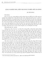
Fig. 1. BRCA1 mutations and their cellular and physiological impact. (A) Domain structure of BRCA1 protein and the location of risk-associ-
ated mutations discussed. NES, nuclear export signal; NLS, nuclear localization signal. (B) BRCA1 mutations categorized by cellular pro-
cesses in which each has been found to lack function or exhibit function different from the wild-type. The nomenclature used for each
mutation was that used in the original research article, or a structural description if designation was not descriptive of the mutation or
mutant structure.
R. J. Linger and P. A. Kruk Risk-associated BRCA1 mutations and their functional implications
FEBS Journal 277 (2010) 3086–3096 ª 2010 The Authors Journal compilation ª 2010 FEBS 3087
(NIH Breast Cancer Information Core Database,
Risk-associated trun-
cation mutations are found throughout the entire
BRCA1 coding sequence (Fig. 1) and result in mutant
proteins that vary in length and structural impairment.
For example, the nonsense mutation Y1853X, which
lacks the last 11 amino acids, is only missing a small
portion of the second BRCT (BRCA1 C-terminus)
repeat, whereas the 39 amino acid 185delAG mutant
lacks all of BRCA1’s known functional domains.
A smaller percentage of risk-associated BRCA1
mutations are point mutations classified as missense
mutations. Like truncation mutations, missense muta-
tions occur throughout the entire BRCA1 coding
sequence (Fig. 1) [15], although it is difficult to deter-
mine the clinical importance of these mutations
because of their rarity and because they do not often
result in gross structural or functional loss. Therefore,
many missense mutations remain ‘variants of unknown
significance’ [16]. The functional significance of the
RING and BRCT domains, as well as the substantial
conservation of their sequences, fuel speculation that
many missense mutations in these areas are probably
linked to cancer predisposition. Nonetheless, several
missense mutations have already been linked to breast
and ⁄ or ovarian cancer predisposition, including C61G,
M1775K and P1749R.
BRCA1 is thought to act as a classical tumor sup-
pressor and the loss of BRCA1’s cellular functions is
thought to occur through bi-allelic inactivation. Carri-
ers of mutations have one germline hit (the inherited
mutated copy of BRCA1) and, in the tumor, a second
somatic hit usually through the loss of heterozygosity
[6]. The observed phenotype of enhanced breast and
ovarian cancer risk is generally thought to result from
the loss of some or all wild-type functions of the
BRCA1 gene product.
However, countless studies have revealed the com-
plexities of signaling molecule and transcription factor
interactions, as well as cellular adaptations in response
to the unique selective pressures of tumor initiation
and progression. Therefore, it is important to investi-
gate all possible molecular mechanisms by which a
mutation may contribute to the disease phenotype.
Mutant proteins may antagonize wild-type proteins in
a dominant negative manner, resulting in the loss of
remaining wild-type function [17], or they may engage
in unique molecular interactions and manifest novel
functions independent of the loss of wild-type protein
function [18]. Likewise, BRCA1 mutations may con-
tribute to cancer risk through the loss of wild-type
BRCA1 function or through the gain of function asso-
ciated with mutant BRCA1 proteins.
Loss of function mutations
As mentioned previously, several lines of evidence sug-
gest the loss of wild-type BRCA1 function as a com-
mon mechanism for enhanced breast and ovarian
cancer risk (Table 1). Similar to BRCA1 knockout
mice and cell lines, elevated levels of aneuploidy and
loss of heterozygosity indicative of an impaired DNA
damage response have been noted in breast cancer tis-
sue from mutation carriers compared with control
breast cancers, as well as in the human BRCA1 trun-
cated breast cancer cell line, HCC1937 (reviewed in
[6]). In structural protein studies, Tischkowitz et al.
[19] suggested that structural alterations in the BRCT
phosphopeptide-binding pocket caused by the BRCA1
M1775K missense mutation contributed to enhanced
breast and ovarian cancer risk through diminished
transactivation and binding to other DNA damage
response proteins. Likewise, Williams et al. [20] found
that decreased stability of BRCA1 missense and trun-
cation mutants resulting from aberrant protein folding
contributed to the loss of BRCA1 function and
enhanced cancer risk.
Expression of mutant BRCA1 constructs in the
absence of wild-type BRCA1 frequently fails to restore
wild-type BRCA1 function. Scully et al. [21] utilized
the c radiation-sensitive HCC1937 breast cancer cell
line, which lacks wild-type BRCA1 and carries two
5382InsC BRCA1 alleles that code for a frameshift
and premature stop signal at codon 1829, and were
able to decrease c radiation sensitivity with restoration
of wild-type BRCA1. However, transfection of several
BRCA1 mutants into these cells failed to alter radia-
tion sensitivity. In agreement, the addition of wild-type
BRCA1 expression into breast cancer cell lines that
exhibit low wild-type BRCA1 expression due to the
presence of a single wild-type BRCA1 allele inhibited
growth. However, expression of the risk-associated
truncation mutants 1835STOP and 340STOP, as well
as the synthetic internal deletion mutants D343-1081
and D 515-1092, failed to alter cell growth, tumor for-
mation and tumor progression in nude mice [22].
Lastly, introduction of wild-type BRCA1 into
HCC1937 breast cancer cells and IGROV 1 ovarian
cancer cells inhibited tumor initiation and growth,
whereas a synthetic BRCA1 mutant lacking the last
542 amino acids did not [23]. Interestingly, Cousineau
& Belmaaza [24] hypothesized that reduced gene dos-
age of wild-type BRCA1 in mutation carriers is solely
responsible for altered DNA damage repair, subse-
quent mutation accumulation and increased cancer
risk. Using MCF7 breast cancer cells that harbor a
single copy of wild-type BRCA1 and exhibit enhanced
Risk-associated BRCA1 mutations and their functional implications R. J. Linger and P. A. Kruk
3088 FEBS Journal 277 (2010) 3086–3096 ª 2010 The Authors Journal compilation ª 2010 FEBS
Table 1. Studies supporting loss or gain of function mutation as mechanisms of enhanced breast cancer and ovarian cancer risk.
Mutation Result of mutation In vitro In vivo Model system Endpoint Summary Reference
Loss of function
Various X NA Number of genetic
changes
Mutant breast cancers more chromosomal gain ⁄ loss
events versus control breast cancers
59
P1749R
C64G
T826K
M1775R
Missense P>R
Missense C>G
Missense T>K
Missense M>R
X Breast cancer DNA damage Wild-type BRCA1 rescued c radiation sensitivity of
HCC1937 cells; mutants did not
21
5382InsC Truncated: 1828 amino
acids
X Breast cancer DNA damage,
chemosensitivity
Wild-type BRCA1 rescued hyper-recombination,
chemosensitivity of MCF7 cells; mutants did not
24
P1749R Q1756InsC
Y1853STOP
Missense P>R
Truncated: 1828
amino acids
Truncated: 1852
amino acids
X COS-7, colon cancer Gene regulation Wild-type BRCA1 increased p21 expression in COS-7,
cancer cells; mutants did not
26
1835STOP 340STOP Truncated: 1834 amino
acids Truncated: 339
amino acids
X X Breast cancer Cell growth, tumor
growth
Wild-type BRCA1 inhibited growth, tumor growth in
nude mice; mutants did not
22
Gain of function
5677InsA Truncated: 1852
amino acids
X Prostate cancer Proliferation Mutant inhibited proliferation more efficiently than
wild-type BRCA1
38
N-terminal 602
amino acids
Synthetic mutant: 602
amino acids
X X Mouse ovarian
epithelium
Proliferation,
chemosensitivity,
tumorigenesis
Mutant BRCA1 enhanced proliferation,
chemosensitivity, tumorigenesis; wild-type BRCA1
suppressed
41
5677InsA N-terminal
302 amino acids
N-terminal 771
amino acids
Truncated: 1852 amino
acids
Synthetic mutant: 302
amino acids
Synthetic mutant: 771
amino acids
X Prostate cancer Proliferation,
chemosensitivity
5677InsA and wild-type BRCA1 impaired
proliferation, enhanced chemosensitivity; synthetic
truncations decreased sensitivity
39
185delAG Truncated: 39 amino
acids
X Ovarian epithelium Apoptosis 185delAG decreased cIAP1, XIAP, P-Akt, and
enhanced cleaved caspase 3, apoptosis after drug
treatment
46
5382InsC 5677InsA Truncated: 1828 amino
acids
Truncated: 1852 amino
acids
X Breast, ovarian
cancer
Apoptosis Co-expression of mutants with wild-type BRCA1
inhibited wild-type BRCA1’s ability to enhance
apoptosis
50
5083del19 Truncated: 1669 amino
acids
X X HeLa Gene regulation Mutant increased periostin mRNA, protein and
mutation carrier serum, breast cancer tissue
52
R. J. Linger and P. A. Kruk Risk-associated BRCA1 mutations and their functional implications
FEBS Journal 277 (2010) 3086–3096 ª 2010 The Authors Journal compilation ª 2010 FEBS 3089
spontaneous recombination or ‘hyper-recombination’,
they showed that transfection of MCF7 cells with
wild-type BRCA1 diminished hyper-recombination and
chemosensitivity, whereas addition of the 5382InsC
BRCA1 mutation affected neither endpoint. These
studies further support a role for the loss of wild-type
BRCA1 function as a contributing factor to enhanced
breast and ovarian cancer risk.
It is important to note that many of the aforemen-
tioned studies attempted to delineate BRCA1 mutant
function in model systems lacking normal levels of
wild-type BRCA1, which makes it difficult to discrimi-
nate between the contribution of BRCA1 mutants and
the loss of wild-type BRCA1 to disease risk. However,
several studies utilizing a wild-type BRCA1 back-
ground clearly support the loss of BRCA1 wild-type
function for cancer risk. For example, although the
overexpression of wild-type BRCA1 in several wild-
type BRCA1 cancer cell lines and COS cells upregulat-
ed p21 expression, several synthetic deletion and trun-
cation mutants and risk-associated BRCA1 mutants,
including P1749R, Q1756InsC (aka 5382InsC) and
Y1853STOP (aka 5677InsA), a frameshift mutation
resulting in a premature stop codon that lacks the last
11 amino acids [25], failed to alter p21 expression [26].
Gain of function mutations
Although mutations resulting in a premature stop
codon are typically susceptible to nonsense-mediated
mRNA decay, mounting evidence suggests that mutant
mRNA and proteins are not uniformly degraded. Per-
rin-Vidoz et al. [27] found that several BRCA1 muta-
tions were unaffected by mRNA decay, including
185delAG and 5382InsC, two of the most common
risk-associated BRCA1 mutations [28]. Truncation
mutant mRNAs may avoid decay by translation re-ini-
tiation at a methionine codon downstream of the pre-
mature stop codon [29], and consequently, may
contribute aberrant gene products coding for trunca-
tion proteins exhibiting varying degrees of protein sta-
bility that may impart novel cellular functions [30]. It
is important to consider that detection of some mutant
BRCA1 proteins in clinical samples has proven unsuc-
cessful due to technical challenges such as cross-reac-
tivity of antibodies with wild-type BRCA1. However,
validation studies of mutant proteins in tissue samples
are ongoing and will provide a framework within
which to view experimental studies of mutant function.
BRCA1 mutant proteins may participate in novel
protein–protein interactions as a result of aberrant cel-
lular localization. Rodriguez et al. [31] found that
exogenous missense and truncation mutants lacking a
small portion of the BRCA1 C-terminal, including
5382InsC, exhibited aberrant cytoplasmic localization
in breast cancer cells, whereas larger truncations
resulted in enhanced nuclear localization of mutants.
Aberrant localization may result from mutation or loss
of the nuclear localization or export signals, impaired
recognition of these signals as a result of improper
protein folding, or altered interaction with binding
partners that impact BRCA1 localization, such as
BARD1 [31].
Mutant BRCA1 proteins may convey unique pheno-
types by inhibiting the normal function of wild-type
BRCA1 in a dominant negative manner by binding
BRCA1 and inhibiting its interaction with other pro-
teins, or by sequestering BRCA1 binding partners.
Likewise, mutant proteins may also convey unique
functions by interacting with novel proteins and ⁄ or
regulating alternative genes. Indeed, a significant pro-
portion of BRCA1-associated breast cancer tissue sam-
ples [32], as well as primary cells from mutation
carrier-derived ovarian cancer cell xenograft tumors
[33], exhibit loss of the wild-type BRCA1 allele con-
comitant with increased mutant allele copy number.
Consequently, mutant BRCA1 proteins have been
shown to impact a range of cellular functions, includ-
ing development, proliferation, chemosensitivity, apop-
tosis and gene regulation (Fig. 1, Table 1).
Role of gain of function mutations for
development, cellular proliferation,
chemosensitivity, apoptosis and gene
regulation
Essentially all BRCA1 knockouts are embryonic lethal
in mice (reviewed in [34]). However, mice homozygous
for a specific synthetic mutation truncating the BRCA1
protein by half are viable, although highly susceptible
to multiple tumor types, including lymphomas, sarco-
mas, and carcinomas ⁄ adenocarcinomas of the colon,
endometrium, lung, liver and mammary gland [35].
Interestingly, introduction of a synthetic BRCA1 trun-
cation mutant encoding the first 300 BRCA1 amino
acids inhibits mammary gland differentiation and
structural formation during murine development,
despite the presence of wild-type BRCA1 [36]. Like-
wise, when injected into the cleared murine mammary
fat pad, primary human breast epithelial cells trans-
fected with the BRCA1 D11 splice variant or murine
BRCA1-W1777Stop (which mimics the human
1835STOP mutation), undergo limited differentiation
and branching and develop extensive hyperplasia [37].
The 5677InsA insertion mutation, resulting in a
frameshift and premature stop signal at codon 1853,
Risk-associated BRCA1 mutations and their functional implications R. J. Linger and P. A. Kruk
3090 FEBS Journal 277 (2010) 3086–3096 ª 2010 The Authors Journal compilation ª 2010 FEBS
inhibits proliferation of DU145 human prostate cancer
cells expressing a low level of wild-type BRCA1 more
efficiently than exogenous wild-type BRCA1 [38],
whereas a synthetic N-terminal mutant was found to
inhibit physical interaction of wild-type BRCA1 and
cyclin D1 [39]. In contrast, an exogenous C-terminal
fragment of BRCA1 can enhance normal breast epithe-
lial cell growth, possibly by acting in a dominant nega-
tive manner to inhibit wild-type BRCA1’s growth
suppressive function [40]. Similarly, whereas over-
expression of wild-type BRCA1 in the ID8 mouse
ovarian epithelial cell line diminished proliferation,
chemosensitivity and tumorigenicity of intraperitone-
ally injected cells, expression of a synthetic truncation
mutant encoding the first 602 amino acids of BRCA1
yielded enhanced proliferation and chemosensitivity.
Furthermore, when injected intraperitoneally, cells
expressing the mutant were significantly more tumori-
genic [41]. It should be noted, however, that BRCA1
mutants have also been shown to exhibit some residual
wild-type growth function as a result of remaining
intact domains. For example, mouse embryonic fibro-
blasts homozygous for D11 BRCA1 exhibited a failed
G2-M checkpoint [42], whereas breast cancer cells
expressing only the 5382InsC mutant maintained an
intact G2-M checkpoint [21].
Fan et al. [39] reported that in DU145 prostate cancer
cells expressing low levels of wild-type BRCA1, overex-
pression of wild-type BRCA1 or 5677InsA increased to-
poisomerase inhibitor cytotoxicity, which could be
reversed by transfection of synthetic mutants DEcoRI
(amino acids 1-302) and DKpnI (amino acids 1-771),
yielding chemoresistant cells. Likewise, in the HCC1937
breast cancer cell model system lacking endogenous
wild-type BRCA1 , the addition of exogenous wild-type
BRCA1 enhanced chemoresistance, which was reversed
by cotransfection of DEcoRI and DKpnI [39]. This
suggests that mutants can, at least in part, overturn
wild-type BRCA1 function, thereby supporting a role
for gain of function BRCA1 mutations.
The 185delAG (BRAT) mutation, which imparts
upon carriers a 66% lifetime risk of developing ovar-
ian cancer [43], arises from the deletion of two nucleo-
tides (AG) in the second exon of the BRCA1 gene.
This deletion results in a reading frame shift that pro-
duces a premature stop signal at codon 39 and a trun-
cated protein product. Using SV-40 transfected
ovarian surface epithelial cells from women with the
BRAT mutation, we found that mutant cells exhibited
enhanced apoptosis and caspase 3 activation in
response to staurosporine [44], possibly related to
diminished levels of phospho-Akt, XIAP and cIAP1
[45]. To rule out the possible contribution of wild-type
BRCA1 haploinsufficiency to altered apoptosis in
185delAG cells, BRAT was expressed in wild-type
BRCA1 ovarian surface epithelial cells. In agreement
with our earlier studies, BRAT enhanced caspase
3-mediated apoptosis and diminished levels of
phospho-Akt, cIAP1 and XIAP [46]. In more recent
studies, we found that BRAT upregulated the expres-
sion of maspin [47], a tumor suppressor important in
apoptosis, invasion and metastasis that is uniquely
overexpressed in several tumor types, including ovarian
cancer [48]. Maspin expression has been correlated
with cisplatin sensitivity in ovarian cancer cell lines
and longer progression-free and overall survival times
in ovarian cancer patients [49], and may be involved in
BRAT-mediated enhanced chemosensitivity [47].
Lastly, Thangaraju and colleagues [50] found that
co-expression of 5382InsC and 5677InsA with wild-
type BRCA1 inhibited the wild-type protein’s ability to
enhance apoptosis in breast and ovarian cancer cells.
Several studies support a role for BRCA1 mutants
in gene regulation. For example, wild-type BRCA1
and 5677InsA inhibited exogenous estrogen receptor
alpha transactivation, but co-transfection of DBamHI,
DKpnI and DEcoRI reversed this phenomenon [39].
Similarly, the synthetic BRCA1 mutant (D500-1863),
which encodes a protein less than a third the length
of the wild-type, inhibited wild-type BRCA1-mediated
activation of a p53 reporter [10]. Likewise, using the
mouse mammary gland-specific expression of wild-type
BRCA1, a risk-associated mutation that truncates the
protein at amino acid 340, or a BRCA1 splice variant
that omits the N-terminal 72 amino acids, Hoshino
et al. [51] showed that the splice variant mediated
hyperproliferation and enhanced lobule formation in
the mammary gland. In addition, tumorigenesis and
death were accelerated in mice expressing the splice
variant. In separate studies, Quaresima and colleagues
[52] performed microarray analysis on HeLa cells
stably expressing vector, wild-type BRCA1 or the
founder mutation 5083del19, which encodes a BRCA1
protein missing the last 193 amino acids, and, conse-
quently both BRCT domains, and found differential
regulation of multiple genes, including upregulation of
periostin. Furthermore, periostin levels were also
increased in serum and breast cancer tissue from a
small number of patients carrying this mutation. In
other studies, expression of a synthetic truncation
mutant maintaining the first third of the BRCA1 pro-
tein enhanced p53 expression in 1D8 mouse epithelial
ovarian cancer cells and downregulated constituents of
the SAPK ⁄ JNK and MAPK ⁄ ERK1⁄ 2 pathways [53].
Finally, the missense mutation Ser1841Asn, which
is associated with enhanced breast cancer risk, upregu-
R. J. Linger and P. A. Kruk Risk-associated BRCA1 mutations and their functional implications
FEBS Journal 277 (2010) 3086–3096 ª 2010 The Authors Journal compilation ª 2010 FEBS 3091
lates D52 (TD52) and the folate receptor alpha
(FOL1) in HeLa cells [54]. This regulation is clinically
relevant, as expression of these genes correlates with
tumor progression in breast [55,56] and ovarian
cancers [57,58].
Taken together, these studies support a gain of func-
tion role for some mutations. The presence or absence
of a mutant function, as well as its impact on the cell,
is probably very specific to each mutation and factors
impacting mutant function, including mutant protein
size, loss ⁄ maintenance of various domains, or struc-
tural changes resulting in novel domains. These studies
must also be viewed in a cautionary manner. Gain and
loss of function experiments provide valuable insight
into the mechanism of BRCA1 mutant functions.
However, until the presence of stable mutant proteins
is validated clinically, it is necessary to remain mindful
of the limitations, as well as the promise, of this type
of experimental study.
Clinical impact of gain of function
mutations
Studies investigating the effect of BRCA1 mutant pro-
teins in the context of wild-type BRCA1 are clinically
important. They represent the genotypic and pheno-
typic state of disease-free mutation carriers before the
loss of both wild-type BRCA1 alleles. Novel functions
mediated by mutant proteins have been shown in vari-
ous model systems to significantly impact proliferation
and apoptosis and, therefore, have the potential to
influence cancer initiation, progression and, ultimately,
prognosis for patients carrying mutations. Although
some mutants may retain specific wild-type BRCA1
functions, others may enhance the risk of cancer devel-
opment by antagonizing BRCA1’s tumor suppressive
functions. Further investigation of mutant protein
function is warranted, as a better understanding of the
function of specific mutations could greatly improve
risk assessment and prognostic value for mutation
carriers.
A better understanding of BRCA1 mutant functions
may also help to identify novel drug targets for treat-
ment and prophylaxis of mutation carriers. Novel
interacting proteins and signaling pathways, as well as
downstream target genes, may reveal as yet unidenti-
fied players in BRCA1 mutation-associated breast and
ovarian cancer. Data from our laboratory suggest that
genes important for cancer initiation and progression,
such as maspin, are differentially regulated in normal
human ovarian epithelial cells expressing the BRAT
mutation [47]. Furthermore, compared with sporadic
breast cancer tissue, BRCA1 mutation-associated
breast cancer samples reveal more chromosomal
aberrations in specific regions, potentially containing
additional tumor suppressors important in BRCA1-
dependent tumor initiation and progression [59]. An
understanding of specific interacting proteins, signaling
pathways and target genes involved in the mechanism
of enhanced breast and ovarian cancer risk conveyed
by each mutation provides the opportunity for muta-
tion-specific personalized therapy for mutation carriers.
Similar mutations may also share common functions
and respond to similar therapeutic strategies. Further-
more, targeting functions of BRCA1 mutants that
probably contribute to premalignancy, cancer initiation
and the early stages of tumor growth holds great
promise for effective prophylactic measures that are
less invasive than oophorectomy and mastectomy.
It is interesting to speculate that cells heterogeneous
for risk-associated mutations, although nontumorigenic
in their current state, may represent an initial step
towards cellular transformation, although additional
changes may be necessary for these cells to become
malignant. Likewise, early changes that may promote
malignant transformation, including enhanced telo-
meric instability, have been observed in cell lines gener-
ated from normal ovarian surface epithelial cells of
women with a strong family history of ovarian cancer
[60] (reviewed in [61]). Furthermore, several studies
have found more frequent occurrence of deep invagin-
ations in the ovary surface, dysplasia, hyperplasia and ⁄
or surface papillae in high-risk prophylactically
removed ovaries versus normal ovaries [62–64], suggest-
ing that early ‘premalignant’ changes may already exist
in those carriers. The possibility of independent mutant
BRCA1 functions does not exclude the contribution
of other oncogenes, tumor suppressors or invasion ⁄
metastasis-promoting proteins. Conversely, these early
changes probably facilitate further cellular changes that
manifest in the aggressive phenotype seen clinically in
hereditary breast and ovarian cancer.
Lastly, there are salient differences between the
mechanisms of tumor initiation and progression of
breast and ovarian cancer in BRCA1 mutation carriers.
The lifetime risk for development of breast cancer is
higher than that for ovarian cancer [14], and carriers
do not always develop both types of disease. Further-
more, the importance of differential expression and
stoichiometry of transcription factors and signaling
molecules in different tissues is also well established.
The impact of specific mutants is, therefore, probably
context specific. Holt and colleagues [22] observed a
series of BRCA1 mutants to be largely ineffective in
inhibiting the growth of breast cancer cells. However,
one mutant was shown to inhibit the growth of three
Risk-associated BRCA1 mutations and their functional implications R. J. Linger and P. A. Kruk
3092 FEBS Journal 277 (2010) 3086–3096 ª 2010 The Authors Journal compilation ª 2010 FEBS
ovarian cancer cell lines. You et al. [65] also found cell
type-specific BRCA1 mutant functions. Although
expression of the 185delAG mutation in immortalized
ovarian surface epithelial cells and ovarian cancer cells
revealed multiple downstream effectors and physiologic
impacts [46,47], primary and immortalized cells derived
from normal breast tissue of a 185delAG mutation
carrier did not show a significant difference in growth,
stress response, growth in soft agar or tumorigenicity
when compared with normal breast epithelial cells
homozygous for wild-type BRCA1 [66]. Several epide-
miological studies have observed differential ovarian
and breast cancer risk based on the location of the
truncation mutation within the BRCA1 gene [67,68].
Disparate risk levels may represent tissue-specific
degrees of importance for the specific functions lost or
gained as a result of each mutation, and the interplay
of these factors.
In conclusion, it is clear from a wide range of model
systems and endpoints that BRCA1 mutations are
capable of significant physiological impacts. Further-
more, molecular and phenotypic changes are evident in
mutation carriers. These changes may result from loss
of wild-type BRCA1 function, gain of function muta-
tions or both. Consequently, further experimental and
clinical studies of mutant BRCA1 proteins are war-
ranted, and will provide a better understanding of
mutation-associated breast and ovarian cancer and
improve the strength of prognosis and efficacy of pro-
phylaxis and treatment for mutation carriers.
References
1 Lux M, Fasching P & Beckmann M (2006) Hereditary
breast and ovarian cancer: review and future perspec-
tives. J Mol Med 84, 16–28.
2 Ford D, Easton DF, Bishop DT, Narod SA & Goldgar
DE (1994) Risks of cancer in BRCA1-mutation carriers.
Breast Cancer Linkage Consortium. Lancet 343, 692–
695.
3 Thompson M (2010) BRCA1 16 years later: nuclear
import and export processes. FEBS J 277, 3072–3078.
4 Yang ES & Xia F (2010) BRCA1 16 years later: DNA
damage-induced BRCA1 shuttling. FEBS J 277, 3079–
3085.
5 Shen SX, Weaver Z, Xu X, Li C, Weinstein M, Chen
L, Guan XY, Ried T & Deng CX (1998) A targeted
disruption of the murine Brca1 gene causes
gamma-irradiation hypersensitivity and genetic
instability. Oncogene 17, 3115–3124, doi:10.1038/sj.
onc.1202243.
6 Deng CX (2006) BRCA1: cell cycle checkpoint, genetic
instability, DNA damage response and cancer
evolution. Nucleic Acids Res 34, 1416–1426,
doi:34 ⁄ 5 ⁄ 1416 [pii] 10.1093 ⁄ nar ⁄ gkl010.
7 Boulton SJ (2006) Cellular functions of the BRCA
tumour-suppressor proteins. Biochem Soc Trans 34,
633–645, doi:BST0340633 [pii] 10.1042/BST0340633.
8 Gudmundsdottir K & Ashworth A (2006) The roles of
BRCA1 and BRCA2 and associated proteins in the
maintenance of genomic stability. Oncogene 25, 5864–
5874, doi:1209874 [pii] 10.1038/sj.onc.1209874.
9 Mullan PB, Quinn JE & Harkin DP (2006) The role of
BRCA1 in transcriptional regulation and cell cycle
control. Oncogene 25, 5854–5863, doi:1209872 [pii]
10.1038/sj.onc.1209872.
10 Zhang H, Somasundaram K, Peng Y, Tian H, Bi D,
Weber BL & El-Deiry WS (1998) BRCA1 physically
associates with p53 and stimulates its transcriptional
activity. Oncogene 16, 1713–1721, doi:10.1038/
sj.onc.1201932.
11 Xu X, Qiao W, Linke SP, Cao L, Li WM, Furth PA,
Harris CC & Deng CX (2001) Genetic interactions
between tumor suppressors Brca1 and p53 in apoptosis,
cell cycle and tumorigenesis. Nat Genet 28, 266–271,
doi:10.1038/90108 90108 [pii].
12 Jhanwar-Uniyal M (2003) BRCA1 in cancer, cell cycle
and genomic stability. Front Biosci 8, s1107–s1117.
13 Prat J, Ribe A & Gallardo A (2005) Hereditary ovarian
cancer. Hum Pathol 36, 861–870, doi:S0046-8177(05)
00283-2 [pii] 10.1016/j.humpath.2005.06.006.
14 Whittemore AS, Gong G & Itnyre J (1997) Prevalence
and contribution of BRCA1 mutations in breast cancer
and ovarian cancer: results from three U.S. population-
based case-control studies of ovarian cancer. Am J Hum
Genet 60, 496–504.
15 Szabo CI, Worley T & Monteiro AN (2004) Under-
standing germ-line mutations in BRCA1. Cancer Biol
Ther 3, 515–520, doi:841 [pii].
16 Easton DF, Deffenbaugh AM, Pruss D, Frye C,
Wenstrup RJ, Allen-Brady K, Tavtigian SV, Monteiro
AN, Iversen ES, Couch FJ et al. (2007) A systematic
genetic assessment of 1,433 sequence variants of
unknown clinical significance in the BRCA1 and
BRCA2 breast cancer-predisposition genes. Am J Hum
Genet 81, 873–883, doi:S0002–9297(07)63865-8 [pii]
10.1086/521032.
17 Yudt MR, Jewell CM, Bienstock RJ & Cidlowski JA
(2003) Molecular origins for the dominant negative
function of human glucocorticoid receptor beta. Mol
Cell Biol 23, 4319–4330.
18 Song H, Hollstein M & Xu Y (2007) p53 gain-of-func-
tion cancer mutants induce genetic instability by
inactivating ATM. Nat Cell Biol 9, 573–580,
doi:ncb1571 [pii] 10.1038/ncb1571.
19 Tischkowitz M, Hamel N, Carvalho MA, Birrane G,
Soni A, van Beers EH, Joosse SA, Wong N, Novak D,
R. J. Linger and P. A. Kruk Risk-associated BRCA1 mutations and their functional implications
FEBS Journal 277 (2010) 3086–3096 ª 2010 The Authors Journal compilation ª 2010 FEBS 3093
Quenneville LA et al. (2008) Pathogenicity of the
BRCA1 missense variant M1775K is determined by the
disruption of the BRCT phosphopeptide-binding
pocket: a multi-modal approach. Eur J Hum Genet
16, 820–832, doi:ejhg200813 [pii] 10.1038/
ejhg.2008.13.
20 Williams RS, Chasman DI, Hau DD, Hui B, Lau AY
& Glover JN (2003) Detection of protein folding defects
caused by BRCA1-BRCT truncation and missense
mutations. J Biol Chem 278, 53007–53016, doi:10.1074/
jbc.M310182200 M310182200 [pii].
21 Scully R, Ganesan S, Vlasakova K, Chen J, Socolovsky
M & Livingston DM (1999) Genetic analysis of BRCA1
function in a defined tumor cell line. Mol Cell 4, 1093–
1099, doi:S1097-2765(00)80238-5 [pii].
22 Holt JT, Thompson ME, Szabo C, Robinson-Benion C,
Arteaga CL, King MC & Jensen RA (1996) Growth
retardation and tumour inhibition by BRCA1. Nat
Genet 12, 298–302, doi: 10.1038/ng0396-298.
23 Randrianarison V, Marot D, Foray N, Cabannes J,
Meret V, Connault E, Vitrat N, Opolon P, Perricaudet
M & Feunteun J (2001) BRCA1 carries tumor suppres-
sor activity distinct from that of p53 and p21. Cancer
Gene Ther 8, 759–770, doi:10.1038/sj.cgt.7700366.
24 Cousineau I & Belmaaza A (2007) BRCA1 haploinsuffi-
ciency, but not heterozygosity for a BRCA1-truncating
mutation, deregulates homologous recombination. Cell
Cycle 6, 962–971, doi:4105 [pii].
25 Monteiro AN, August A & Hanafusa H (1996)
Evidence for a transcriptional activation function of
BRCA1 C-terminal region. Proc Natl Acad Sci USA 93,
13595–13599.
26 Somasundaram K, Zhang H, Zeng YX, Houvras Y,
Peng Y, Wu GS, Licht JD, Weber BL & El-Deiry WS
(1997) Arrest of the cell cycle by the tumour-suppressor
BRCA1 requires the CDK-inhibitor p21WAF1 ⁄ CiP1.
Nature 389, 187–190, doi:10.1038/38291.
27 Perrin-Vidoz L, Sinilnikova OM, Stoppa-Lyonnet D,
Lenoir GM & Mazoyer S (2002) The nonsense-
mediated mRNA decay pathway triggers degradation of
most BRCA1 mRNAs bearing premature termination
codons. Hum Mol Genet 11, 2805–2814.
28 Ramus SJ & Gayther SA (2009) The contribution of
BRCA1 and BRCA2 to ovarian cancer. Mol Oncol 3,
138–150, doi:S1574-7891(09)00027-1 [pii] 10.1016/j.
molonc.2009.02.001.
29 Buisson M, Anczukow O, Zetoune AB, Ware MD &
Mazoyer S (2006) The 185delAG mutation (c.68_
69delAG) in the BRCA1 gene triggers translation
reinitiation at a downstream AUG codon. Hum Mutat
27, 1024–1029, doi:10.1002/humu.20384.
30 Anczukow O, Ware MD, Buisson M, Zetoune AB,
Stoppa-Lyonnet D, Sinilnikova OM & Mazoyer S
(2008) Does the nonsense-mediated mRNA decay
mechanism prevent the synthesis of truncated BRCA1,
CHK2, and p53 proteins? Hum Mutat 29 , 65–73,
doi:10.1002/humu.20590.
31 Rodriguez JA, Au WW & Henderson BR (2004)
Cytoplasmic mislocalization of BRCA1 caused by
cancer-associated mutations in the BRCT domain.
Exp Cell Res 293, 14–21, doi:S0014482703005445 [pii].
32 Staff S, Nupponen NN, Borg A, Isola JJ & Tanner
MM (2000) Multiple copies of mutant BRCA1 and
BRCA2 alleles in breast tumors from germ-line muta-
tion carriers. Genes Chromosomes Cancer 28, 432–442,
doi:10.1002/1098-2264(200008)28:4<432::AID-GCC9>
3.0.CO;2-J [pii].
33 Indraccolo S, Tisato V, Agata S, Moserle L, Ferrari S,
Callegaro M, Persano L, Palma MD, Scaini MC,
Esposito G et al. (2006) Establishment and characteriza-
tion of xenografts and cancer cell cultures derived from
BRCA1 – ⁄ – epithelial ovarian cancers. Eur J Cancer 42,
1475–1483, doi: S0959-8049(06)00321-2 [pii] 10.1016/j.
ejca.2006.01.057.
34 Evers B & Jonkers J (2006) Mouse models of BRCA1
and BRCA2 deficiency: past lessons, current under-
standing and future prospects. Oncogene 25, 5885–5897,
doi:1209871 [pii] 10.1038/sj.onc.1209871.
35 Ludwig T, Fisher P, Ganesan S & Efstratiadis A (2001)
Tumorigenesis in mice carrying a truncating Brca1
mutation. Genes Dev 15, 1188–1193, doi:10.1101/
gad.879201.
36 Brown MA, Nicolai H, Howe K, Katagiri T, Lalani
elN, Simpson KJ, Manning NW, Deans A, Chen P,
Khanna KK et al. (2002) Expression of a truncated
Brca1 protein delays lactational mammary
development in transgenic mice. Transgenic Res 11,
467–478.
37 Bachelier R, Vincent A, Mathevet P, Magdinier F,
Lenoir GM & Frappart L (2002) Retroviral
transduction of splice variant Brca1-Delta11 or mutant
Brca1-W1777Stop causes mouse epithelial mammary
atypical duct hyperplasia. Virchows Arch 440, 261–266,
doi:10.1007/s004280100500.
38 Fan S, Wang JA, Yuan RQ, Ma YX, Meng Q, Erdos
MR, Brody LC, Goldberg ID & Rosen EM (1998)
BRCA1 as a potential human prostate tumor suppres-
sor: modulation of proliferation, damage responses and
expression of cell regulatory proteins. Oncogene 16,
3069–3082, doi:10.1038/sj.onc.1202116.
39 Fan S, Yuan R, Ma YX, Meng Q, Goldberg ID &
Rosen EM (2001) Mutant BRCA1 genes antagonize
phenotype of wild-type BRCA1. Oncogene 20, 8215–
8235, doi:10.1038/sj.onc.1205033.
40 Larson JS, Tonkinson JL & Lai MT (1997) A BRCA1
mutant alters G2-M cell cycle control in human mam-
mary epithelial cells. Cancer Res 57, 3351–3355.
41 Sylvain V, Lafarge S & Bignon YJ (2002) Dominant-
negative activity of a Brca1 truncation mutant: effects
on proliferation, tumorigenicity in vivo, and chemosen-
Risk-associated BRCA1 mutations and their functional implications R. J. Linger and P. A. Kruk
3094 FEBS Journal 277 (2010) 3086–3096 ª 2010 The Authors Journal compilation ª 2010 FEBS
sitivity in a mouse ovarian cancer cell line. Int J Oncol
20, 845–853.
42 Xu X, Weaver Z, Linke SP, Li C, Gotay J, Wang XW,
Harris CC, Ried T & Deng CX (1999) Centrosome
amplification and a defective G2-M cell cycle
checkpoint induce genetic instability in BRCA1 exon 11
isoform-deficient cells. Mol Cell 3, 389–395, doi:S1097-
2765(00)80466-9 [pii].
43 Satagopan JM, Boyd J, Kauff ND, Robson M, Scheuer
L, Narod S & Offit K (2002) Ovarian cancer risk in
Ashkenazi Jewish carriers of BRCA1 and BRCA2
mutations. Clin Cancer Res 8, 3776–3781.
44 Johnson NC & Kruk PA (2002) BRCA1 zinc RING
finger domain disruption alters caspase response in
ovarian surface epithelial cells. Cancer Cell Int 2,7.
45 Johnson NC, Dan HC, Cheng JQ & Kruk PA (2004)
BRCA1 185delAG mutation inhibits Akt-dependent,
IAP-mediated caspase 3 inactivation in human ovarian
surface epithelial cells. Exp Cell Res 298, 9–16,
doi:10.1016/j.yexcr.2004.04.003 S0014482704001922 [pii].
46 O’Donnell JD, Johnson NC, Turbeville TD, Alfonso
MY & Kruk PA (2008) BRCA1 185delAG truncation
protein, BRAt, amplifies caspase-mediated apoptosis in
ovarian cells. In Vitro Cell Dev Biol Anim 44, 357–367,
doi: 10.1007/s11626-008-9122-0.
47 O’Donnell JD, Linger RJ & Kruk PA (2009) BRCA1
185delAG mutant protein, BRAt, up-regulates maspin
in ovarian epithelial cells. Gynecol Oncol, doi:S0090-
8258(09)00840-3 [pii] 10.1016/j.ygyno.2009.10.052.
48 Sood AK, Fletcher MS, Gruman LM, Coffin JE,
Jabbari S, Khalkhali-Ellis Z, Arbour N, Seftor EA &
Hendrix MJ (2002) The paradoxical expression of
maspin in ovarian carcinoma. Clin Cancer Res 8, 2924–
2932.
49 Surowiak P, Materna V, Drag-Zalesinska M, Wojnar A,
Kaplenko I, Spaczynski M, Dietel M, Zabel M & Lage H
(2006) Maspin expression is characteristic for
cisplatin-sensitive ovarian cancer cells and for ovarian
cancer cases of longer survival rates. Int J Gynecol Pathol
25, 131–139, doi:10.1097/01.pgp.0000183050.30212.2f
00004347-200604000-00003 [pii].
50 Thangaraju M, Kaufmann SH & Couch FJ (2000)
BRCA1 facilitates stress-induced apoptosis in breast
and ovarian cancer cell lines. J Biol Chem 275, 33487–
33496, doi:10.1074/jbc.M005824200 M005824200 [pii].
51 Hoshino A, Yee CJ, Campbell M, Woltjer RL, Town-
send RL, van der Meer R, Shyr Y, Holt JT, Moses HL
& Jensen RA (2007) Effects of BRCA1 transgene
expression on murine mammary gland development and
mutagen-induced mammary neoplasia. Int J Biol Sci 3,
281–291.
52 Quaresima B, Romeo F, Faniello MC, Di Sanzo M,
Liu CG, Lavecchia A, Taccioli C, Gaudio E, Baudi F,
Trapasso F et al. (2008) BRCA1 5083del19
mutant allele selectively up-regulates periostin
expression in vitro and in vivo. Clin Cancer Res 14,
6797–6803, doi:14 ⁄ 21 ⁄ 6797 [pii] 10.1158/1078-0432.
CCR-07-5208.
53 Sylvain V, Lafarge S & Bignon YJ (2001) Molecular
pathways involved in response to ionizing radiation of
ID-8 mouse ovarian cancer cells expressing exogenous
full-length Brca1 or truncated Brca1 mutant. Int J On-
col 19, 599–607.
54 Crugliano T, Quaresima B, Gaspari M, Faniello MC,
Romeo F, Baudi F, Cuda G, Costanzo F & Venuta S
(2007) Specific changes in the proteomic pattern
produced by the BRCA1-Ser1841Asn missense
mutation. Int J Biochem Cell Biol 39, 220–226,
doi:S1357-2725(06)00237-8 [pii] 10.1016/j.
biocel.2006.08.005.
55 Boutros R, Fanayan S, Shehata M & Byrne JA (2004)
The tumor protein D52 family: many pieces, many
puzzles. Biochem Biophys Res Commun 325, 1115–1121,
doi: S0006-291X(04)02409-X [pii] 10.1016/j.bbrc.
2004.10.112.
56 Hartmann LC, Keeney GL, Lingle WL, Christianson
TJ, Varghese B, Hillman D, Oberg AL & Low PS
(2007) Folate receptor overexpression is associated with
poor outcome in breast cancer. Int J Cancer 121, 938–
942, doi:10.1002/ijc.22811.
57 Byrne JA, Balleine RL, Schoenberg Fejzo M, Mercieca
J, Chiew YE, Livnat Y, St Heaps L, Peters GB, Byth
K, Karlan BY et al. (2005) Tumor protein D52
(TPD52) is overexpressed and a gene amplification
target in ovarian cancer. Int J Cancer 117 , 1049–1054.
doi:10.1002/ijc.21250.
58 Miotti S, Canevari S, Menard S, Mezzanzanica D,
Porro G, Pupa SM, Regazzoni M, Tagliabue E &
Colnaghi MI (1987) Characterization of human ovarian
carcinoma-associated antigens defined by novel
monoclonal antibodies with tumor-restricted specificity.
Int J Cancer 39, 297–303.
59 Tirkkonen M, Johannsson O, Agnarsson BA, Olsson
H, Ingvarsson S, Karhu R, Tanner M, Isola J, Barkar-
dottir RB, Borg A et al. (1997) Distinct somatic genetic
changes associated with tumor progression in carriers of
BRCA1 and BRCA2 germ-line mutations. Cancer Res
57, 1222–1227.
60 Kruk PA, Godwin AK, Hamilton TC & Auersperg N
(1999) Telomeric instability and reduced proliferative
potential in ovarian surface epithelial cells from women
with a family history of ovarian cancer. Gynecol Oncol
73, 229–236, doi:S0090-8258(99)95348-9 [pii] 10.1006/
gyno.1999.5348.
61 Wong AS & Auersperg N (2003) Ovarian surface
epithelium: family history and early events in ovarian
cancer. Reprod Biol Endocrinol 1, 70, doi:10.1186/1477-
7827-1-70 1477-7827-1-70 [pii].
62 Salazar H, Godwin AK, Daly MB, Laub PB, Hogan
WM, Rosenblum N, Boente MP, Lynch HT &
R. J. Linger and P. A. Kruk Risk-associated BRCA1 mutations and their functional implications
FEBS Journal 277 (2010) 3086–3096 ª 2010 The Authors Journal compilation ª 2010 FEBS 3095
Hamilton TC (1996) Microscopic benign and invasive
malignant neoplasms and a cancer-prone phenotype in
prophylactic oophorectomies. J Natl Cancer Inst 88,
1810–1820.
63 Werness BA, Afify AM, Bielat KL, Eltabbakh GH,
Piver MS & Paterson JM (1999) Altered surface and
cyst epithelium of ovaries removed prophylactically
from women with a family history of ovarian cancer.
Hum Pathol 30, 151–157.
64 Casey MJ, Bewtra C, Hoehne LL, Tatpati AD, Lynch
HT & Watson P (2000) Histology of prophylactically
removed ovaries from BRCA1 and BRCA2 mutation
carriers compared with noncarriers in hereditary breast
ovarian cancer syndrome kindreds. Gynecol Oncol 78,
278–287, doi:10.1006/gyno.2000.5861 S0090-8258(00)
95861-X [pii].
65 You F, Chiba N, Ishioka C & Parvin JD (2004)
Expression of an amino-terminal BRCA1 deletion
mutant causes a dominant growth inhibition in
MCF10A cells. Oncogene 23, 5792–5798, doi: 10.1038/
sj.onc.1207739 1207739 [pii].
66 Annab LA, Terry L, Cable PL, Brady J, Stampfer MR,
Barrett JC & Afshari CA (2000) Establishment and
characterization of a breast cell strain containing a
BRCA1 185delAG mutation. Gynecol Oncol 77, 121–128,
doi:10.1006/gyno.2000.5734 S0090-8258(00)95734-2 [pii].
67 Thompson D & Easton D (2002) Variation in BRCA1
cancer risks by mutation position. Cancer Epidemiol
Biomarkers Prev 11, 329–336.
68 Gayther SA, Warren W, Mazoyer S, Russell PA,
Harrington PA, Chiano M, Seal S, Hamoudi R, van
Rensburg EJ, Dunning AM et al. (1995) Germline
mutations of the BRCA1 gene in breast and ovarian
cancer families provide evidence for a genotype-pheno-
type correlation. Nat Genet 11, 428–433, doi:10.1038/
ng1295-428.
Risk-associated BRCA1 mutations and their functional implications R. J. Linger and P. A. Kruk
3096 FEBS Journal 277 (2010) 3086–3096 ª 2010 The Authors Journal compilation ª 2010 FEBS