Báo cáo khoa học: Small-molecule modulators of zymogen activation in the fibrinolytic and coagulation systems pptx
Bạn đang xem bản rút gọn của tài liệu. Xem và tải ngay bản đầy đủ của tài liệu tại đây (495.11 KB, 13 trang )
REVIEW ARTICLE
Small-molecule modulators of zymogen activation in the
fibrinolytic and coagulation systems
Keiji Hasumi, Shingo Yamamichi and Tomotaka Harada
Department of Applied Biological Science, Tokyo Noko University, Japan
Introduction
The coagulation and fibrinolytic systems are central
to the hemostatic mechanism. This mechanism is pri-
marily responsible for preventing blood leakage and
secondarily for tissue repair and wound healing.
Interactions between a variety of enzymes and nonen-
zyme components account for the finely regulated pro-
cesses in the coagulation and fibrinolytic systems [1,2].
Most of the enzymes in these systems are serine prote-
ases circulating as either inactive proenzyme or proen-
zyme forms with very low activity. Thus, their
activation is a prerequisite for their function, whereas
their inhibition or inactivation is also important in the
regulation and termination of the reaction. Activation
of the zymogens is a consequence of a conformational
change triggered by specific proteolytic cleavage(s) of
the zymogen [3,4]. The activated enzyme catalyzes the
subsequent step or upstream reaction(s) in the cascade
to amplify and ⁄ or regulate the systems. The reactions
in both systems operate instantly after exposure to
pathophysiological stimuli. Therefore, the mechanism
of their regulation is programmed in the structure of
each component of the systems, and modulation of the
programmed function of the component can provide a
novel means of pharmacological intervention in dis-
eases associated with the coagulation and fibrinolytic
systems. Most extensively studied examples of zymo-
gen modulation are nonproteolytic conformational
activations of plasminogen by streptokinase, and pro-
thrombin by staphylocoagulase (and von Willebrand
factor-binding protein). These are pathogen-derived
nonenzyme proteins that exploit the ‘molecular sexual-
ity’ in the activation mechanism of plasminogen and
Keywords
coagulation; conformation; fibrinolysis;
plasma hyaluronan-binding protein;
plasminogen; plasminogen activator;
protease; prothrombin; urokinase; zymogen
activation
Correspondence
K. Hasumi, Department of Applied Biological
Science, Tokyo Noko University,
3-5-8 Saiwaicho, Fuchu-shi,
Tokyo 183-8509, Japan
Fax: +81 42 367 5708
Tel: +81 42 367 5710
E-mail:
(Received 29 April 2010, revised 8 July
2010, accepted 19 July 2010)
doi:10.1111/j.1742-4658.2010.07783.x
The coagulation and fibrinolytic systems are central to the hemostatic
mechanism, which works promptly on vascular injury and tissue damage.
The rapid response is generated by specific molecular interactions between
components in these systems. Thus, the regulation mechanism of the sys-
tems is programmed in each component, as exemplified by the elegant pro-
cesses in zymogen activation. This review describes recently identified small
molecules that modulate the activation of zymogens in the fibrinolytic and
coagulation systems.
Abbreviations
AH site, aminohexyl site; EGF, epidermal growth factor; Lp(a), lipoprotein(a); PA, plasminogen activator; PAN domain,
plasminogen ⁄ apple ⁄ nematode domain; PHBP, plasma hyaluronan-binding protein; scu-PA, single-chain urokinase-type plasminogen activator;
SMTP, Stachybotrys microspora triprenyl phenol; tcu-PA, two-chain urokinase-type plasminogen activator; t-PA, tissue-type plasminogen
activator; u-PA, urokinase-type plasminogen activator.
FEBS Journal 277 (2010) 3675–3687 ª 2010 The Authors Journal compilation ª 2010 FEBS 3675
prothrombin (insertion of N-terminal Ile or Val into
N-terminal binding cleft of the catalytic domain,
resulting in conformational activation of the substrate
binding site and oxyanion hole required for proteolytic
activity) [5–8].
Our laboratory has been searching for small-mole-
cule natural products that enhance the fibrinolytic
system. We initially aimed at identifying a candidate
small molecule that could contribute to the treatment
of thrombotic and embolic complications. As a result,
we have identified several types of compounds, includ-
ing modulators of zymogen activations. Detailed anal-
yses of the action of such modulators have expanded
the concept of zymogen modulation, leading to the
identification of additional modulators that affect the
coagulation system. The active compounds and their
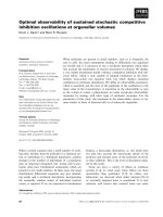
targets are summarized in Table 1 and Fig. 1. Here,
we review the identification and functional character-
ization of such small-molecule modulators of zymogen
activation in the fibrinolytic and coagulation systems.
Plasminogen modulator
The plasminogen
⁄
plasmin system
The plasminogen ⁄ plasmin system plays a central role
in blood clot lysis [2,9]. This system is also important
in other pathophysiological events in which localized
proteolysis is involved [10–12]. The circulating form
of plasminogen (Glu-plasminogen) is a single-chain
zymogen consisting of 791 amino acids, which form
the following domain structures: a plasminogen ⁄
apple ⁄ nematode (PAN) domain, five kringle domains
and a serine protease domain (Fig. 2) [13,14]. It is
proteolytically activated to plasmin by plasminogen
activators (PAs) through specific cleavage at Arg561–
Val562. Urokinase-type and tissue-type PAs (u-PA and
t-PA, respectively) are major physiological activators
Table 1. Small-molecule zymogen modulators identified in this labo-
ratory. PHBP, plasma hyaluronan-binding protein; scu-PA, single-
chain urokinase-type plasminogen activator; SMTP, Stachybotrys
microspora triprenyl phenol.
Target Compound Reference
Plasminogen Complestatin and chloropeptin I [43,44,52]
Staplabin and SMTPs [45–53]
Thioplabins [52,54]
Stachybotrydial [55]
scu-PA ⁄ plasminogen
reciprocal activation
Surfactins and iturins [68,69]
Glucosyldiacylglycerol [70]
Prothrombin Plactins [84–86]
Pro-PHBP Polyamines and carminic acid [104]
OH
H
N
O
N
H
N
HOOC
OH
O
N
H
H
N
O
O
O
N
H
O
O
OH
ClCl Cl
Cl
Cl
OH
HN
O
OH
Cl
HOOC
N
H
H
N
N
H
H
N
O
S
NH
NH
S
NH
S
NH
O
NH
R
NH
OH
CH
2
O
CH
2
O
CH
2
O
CH
2
O
N
N
CH
2
O
CH
2
O
N
O
N
O
N
O
O
N
N
O
R
O
OH
HO
O
CH
3
COOH
OH
OH
HO
HO
O
O
HO
OH
OH
OH
O
HO
O
HN
N
H
NH
NH
NH
H
N
HN
O
O
O
O
O
O
O
O
O
OH
Glu
Leu
D-Leu
Leu
Val
Asp
D-Leu
NH
NH
2
HN
HN
NH
NH
N
H
HN
O
O
O
O
O
D
-
Arg
D-Val
D-Leu
Phe
Leu
HN
HN
NH
NH
H
N
O
O
O
O
O
S
S
D-Cys
D
-
Cys
Val
Ile
D-Leu
O
H
HO
CHO
CHO
O
HO
O
HO
OH
OH
OR
2
OR
1
Complestatin
Thioplabins
Staplabin/SMTPs
Stachybotrydial
Glucosyldiacylglycerol
Surfactin C Plactin D
Malformin A
1
Carminic acid
HO
Fig. 1. Structures of the modulators of zymogen activation. R in the thioplabin structure corresponds to –CH
3
, –CH
2
CH
3
or –CH(CH
3
)
2
for
thioplabin A, B or C, respectively. R in the structure of SMTPs represents one of a variety of substituents, most of which are derived from
amino acids. R
1
and R
2
in the structure of glucosyldiacylglycerol correspond to the oleoyl or palmitoyl group. Malformin A
1
, which enhances
cellular fibrinolytic activity through a mechanism distinct from direct modulation of zymogen activation, is shown to compare its structure
with that of plactin D.
Zymogen activation modulators K. Hasumi et al.
3676 FEBS Journal 277 (2010) 3675–3687 ª 2010 The Authors Journal compilation ª 2010 FEBS
[2]. Glu-plasminogen adopts a closed conformation
because of the intramolecular binding of Lys50 and ⁄ or
Lys62 to the fifth kringle domain (K5) (Fig. 3) [15,16].
The tight conformation renders Glu-plasminogen
less sensitive to activation by PAs [17,18]. The Glu-
plasminogen binding to fibrin or cellular receptors
allows relaxation of Glu-plasminogen conformation,
enabling efficient activation (Fig. 3). This mechanism
facilitates localized activation of plasminogen and
extracellular proteolysis [19,20]. Growing evidence sup-
ports the idea that cellular plasminogen binding plays
roles in physiological processes such as macrophage
recruiting, leukocyte migration and liver regeneration
[21–24].
During the course of fibrinolysis, Lys-plasminogen
(Fig. 2), a truncated form of Glu-plasminogen, pre-
dominantly with an N-terminal Lys78, appears
through autoproteolysis by the resulting plasmin
[25,26]. The molecule no longer has the PAN domain
and therefore adopts a relaxed conformation [27]
because of the lack of intermolecular binding between
the PAN domain and K5 (Fig. 3). Lys-plasminogen is
highly sensitive to activation even in the absence of
fibrin or cells.
PAN–K5 binding is mediated via the aminohexyl
(AH) site in K5 [28,29] (Fig. 3). Unlike lysine-binding
sites in K1, K2 and K4 of plasminogen, the AH site
can bind an internal lysine residue in addition to the
C-terminal lysine [30], which is a preferred ligand for
the lysine-binding site in K1, K2 and K4. Thus, amin-
ohexyl or lysine analogs can interfere with PAN–K5
binding and relax Glu-plasminogen conformation, ren-
dering Glu-plasminogen susceptible to activation by
PAs [31,32]. Some proteins, as well as cell-surface plas-
minogen receptors, interact with kringles and modulate
plasminogen activation [33,34]. It is postulated that the
fibrinolytic process proceeds as follows [35]: at an ini-
tial phase, the K5–fibrin interaction accumulates
Glu-plasminogen on fibrin. Subsequently, partially
degraded fibrin, which has C-terminal lysines, plays a
role in fibrinolytic propagation by serving as more effi-
cient binding sites for plasminogen. The high-affinity
lysine-binding sites in K1 and K4 may be involved in
this propagation process. Thus, plasminogen binding is
essential for the fibrinolytic process. The kringle
ligands, however, inhibit plasminogen binding to fibrin
and cellular receptors and, therefore, suppress fibrino-
lysis. This is the basis of the antihemorrhagic action of
lysine analogs such as tranexamic acid and 6-amino-
hexanoic acid [36].
Competition in the binding between plasminogen
and fibrin or cellular receptors can occur via physio-
logic molecules. Lipoprotein(a) [Lp(a)], a risk factor
for coronary heart disease [37,38], is a plasma lipopro-
tein related to low-density lipoprotein. In the Lp(a)
molecule, apolipoprotein B-100, which is a sole protein
component of low-density lipoprotein, is covalently
modified with apolipoprotein(a), a protein closely
related to plasminogen, consisting of multiple (11–50
repeats) K4-like domains, a K5-like domain and a
pseudo-protease domain [39,40]. Lp(a) can compete
with plasminogen for binding to fibrin or cell-surface
receptors and attenuate localized plasminogen activa-
tion [41] and conversion of Glu-plasminogen to Lys-
plasminogen [42]. These findings suggest a possible link
between thrombosis and atherosclerosis.
Our laboratory has screened up to 10 000 microbial
cultures for their ability to enhance Glu-plasminogen
binding to fibrin or culture cells. This attempt is based
on the hypothesis that relieving the plasminogen bind-
ing competition might enhance fibrinolytic activity.
The active compounds identified include complestatin
K2
P
Gla K1
P
RI
T
285
321320
EK
P
K
E
K
P
IS
1
A
1
F
1
F
1
S
1
159
158
K4K1 K2 K3 K5
P
E
1
K
78
K
78
K4K1 K2 K3 K5
P
R
K4K1 K2 K3 K5
P
V
562
E1 E2
E3
K
P
R
E1
E2 K
P
E3
I
291290
PAN
Prothrombin
α-Thrombin
Glu-
plasminogen
Pro-PHBP
PHBP
Lys-
plasminogen
Plasmin
scu-PA
tcu-PA
561
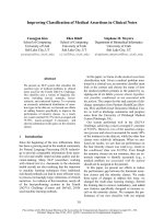
Fig. 2. Structures of key zymogens and their active forms. Each
molecule is shown schematically with each domain in a colored
circle. The disulfide bond that connects the A- and B-chains of the
mature protease is shown in green. Red lines in the PHBP
molecule represent the N-terminal region (NTR). PAN, PAN domain;
K, kringle domain; P, protease domain; Gla, c-carboxyglutamic acid
domain; E, EGF domain.
K. Hasumi et al. Zymogen activation modulators
FEBS Journal 277 (2010) 3675–3687 ª 2010 The Authors Journal compilation ª 2010 FEBS 3677
[43] and its isomer chloropeptin I [44], staplabin [45]
and its congener Stachybotrys microspora triprenyl
phenols (SMTPs) [46–53], thioplabins [54] and stachyb-
otrydial [55].
Complestatin and chloropeptin I
Tachikawa et al. [43] isolated complestatin, a chlorine-
containing peptide metabolite from Streptomyces sp.,
as an active principle that enhanced Glu-plasminogen
binding to cultured U937 monocytoid cells. Complest-
atin enhances the binding by several fold at 1–10 lm.
The enhancement is observed in either the absence or
presence of Lp(a). The compound is also effective in
elevating Glu-plasminogen–fibrin binding. The actions
of complestatin are canceled by a lysine analog. Thus,
the target of complestatin is Glu-plasminogen, and the
agent promotes its binding to fibrin and cells via the
AH site or the lysine-binding sites. However, the initial
characterization does not clarify the exact mechanism
of the complestatin actions. The current understanding
is that the action of complestatin modulates Glu-plas-
minogen conformation [52], but verification of this has
awaited the characterization of staplabin and SMTPs.
However, the concept that small molecule-mediated
augmentation of plasminogen binding results in
increased plasmin formation on cell surfaces has been
confirmed by experiments using complestatin [43].
Chloropeptin I, an isomer of complestatin, has a
similar but slightly different effect on plasminogen
binding compared with complestatin [44]. Chloropep-
tin I is 3–10 times more active than complestatin in
enhancing cellular binding of Glu-plasminogen,
cell-surface plasmin formation, and whole-blood
fibrinolysis assessed by thromboelastography, although
the agent is less active in promoting fibrin binding of
Glu-plasminogen. Although the structural difference
between complestatin and chloropeptin I is only at the
position of the C–C bond that connects the indole ring
of the tryptophan residue and the aromatic ring of the
3,4-dihydroxyphenylglycine residue, a large difference
in conformation between the two compounds is
evident from the nuclear Overhauser effect in NMR
spectroscopy. It is speculated that this difference
may account for the distinct activities of the two
compounds [44].
Staplabin and SMTP
Shinohara et al. [45] discovered a novel triprenyl phe-
nol compound, named staplabin, from a culture of the
fungus Stachybotrys microspora, as an active principle
that enhanced Glu-plasminogen binding to fibrin.
Later, several new staplabin congeners, SMTPs, were
isolated by Kohyama et al. [46], Hasumi et al. [48,53],
and Hu et al. [49,51]. These are named after Stachybot-
rys microspora triprenyl phenol. The staplabin ⁄ SMTP
molecule consists of a tricyclic c-lactam moiety, an iso-
prene side chain and an N-linked side chain (Fig. 1).
Most of the congeners differ in the N-linked side chain
moiety, which is essential for their activity. The
N-linked side chain can be derived from an amine
present in the culture medium, and this finding enabled
selective, efficient production of SMTP congeners
through an amine-feeding cultivation of S. microspora
[50,51,53].
Staplabin enhances Glu-plasminogen binding to
both fibrin and cultured U937 cells [45]. These
K4
K2
K5
P
Fibrin or cellular receptor
PA
K
78
K
78
K4
K1
P
R
561
V
562
Glu-plasminogen
(Closed conformation)
Plasmin
(Open conformation)
K4
K2
K5
P
K1
Lys-plasminogen
(Open conformation)
E
1
E
1
P
K3
K2
K1
K5
K4
PAN
K
50
PAN
Plasmin
PA
D55
D57
W62
W64
F36
Y72
I35
L71
H33
H31
K5 structure
K3
K2
K3
K5
K1
K3
Fig. 3. A model of the conformational regulation of plasminogen activation. The plasminogen and plasmin molecules are shown as in Fig. 2.
The schematic conformation shown is speculative, because detailed experimental data are not yet available. Key disulfide bonds are shown
in green. The molecular structure K5 is shown in the right-hand box. The electrostatic surface image, in which areas of neutral, negativeand
positive potential are depicted in white, red and blue, respectively, was constructed using the coordinate deposited in the RCSB Protein Data
Bank with the code number 2KNF. The labeled amino acids are those implicated in the binding of the lysine analog tranexamic acid [29].
Zymogen activation modulators K. Hasumi et al.
3678 FEBS Journal 277 (2010) 3675–3687 ª 2010 The Authors Journal compilation ª 2010 FEBS
bindings are mediated by the lysine-binding site or the
AH site, because a lysine analog inhibits the bindings.
These activities are quite similar to those of complesta-
tin and chloropeptin I. Takayasu et al. [47] show that
staplabin promotes PA-dependent Glu-plasminogen
activation. The two staplabin activities, the elevation
of Glu-plasminogen-fibrin binding and the promotion
of Glu-plasminogen activation, are observed at the
same range of staplabin concentrations. Thus, a com-
mon mechanism may govern the two effects. The fact
that the activation of Glu-plasminogen is a conforma-
tionally regulated process is the key to understanding
this mechanism. The effect of staplabin on the activa-
tion of Lys-plasminogen, which adopts an open con-
formation, is less prominent than its effect on Glu-
plasminogen. Similarly, smaller effects of staplabin on
Glu-plasminogen activation are observed in the pres-
ence of the lysine analog 6-aminohexanoic acid or
fibrinogen fragments, both of which relax Glu-plasmin-
ogen conformation. The molecular elution time of
both Glu-plasminogen and Lys-plasminogen is slightly
but significantly shortened in the presence of staplabin.
These results support the idea that the staplabin effects
are related to the conformational status of plasmino-
gen, and Takayasu et al. [47] have concluded that
staplabin modulates plasminogen conformation,
rendering the molecule susceptible to proteolytic acti-
vation and to binding to cells and fibrin. The reason
why the effects of staplabin on Glu-plasminogen is lar-
ger than its effect on Lys-plasminogen can be
explained by the fact that the impact of the conforma-
tional effect depends on the initial conformational
status of plasminogen. With respect to the conforma-
tional modulation that leads to an elevated Glu-
plasminogen activation, the staplabin effect is similar
to that of lysine analogs, whereas the effects of each
compound on plasminogen binding are quite different.
This implies that staplabin acts as a plasminogen
modulator that works through a mechanism distinct
from the lysine-binding site (or AH site) occupancy.
Staplabin congener SMTPs may act on plasminogen
activation and binding similarly to staplabin, whereas
some congeners, such as SMTP-7 and -8, are far more
potent than staplabin [49]. The action of stapla-
bin ⁄ SMTP is shown schematically in Fig. 4. Pharma-
cological evaluations of SMTP-7 suggest that the
compound is a promising candidate drug for treating
thrombotic and embolic complications. Detailed inves-
tigations are now under way.
Plasmin formation in the presence of SMTP is tran-
sient in an incubation of Glu-plasminogen with u-PA
[52]. A decrease in plasmin activity follows a rapid
increase in plasmin formation. This is because of auto-
proteolytic degradations of the catalytic domain of the
plasmin molecule. Ohyama et al. speculate that the
conformational change brought about by SMTP affects
susceptibility to autoproteolytic cleavage. 6-Aminohexa-
noic acid does not lead to autoproteolysis, but enhances
plasminogen activation. Thus, the difference between
conformational modulation by SMTP and that by the
lysine analog is also evident from these results. Similar
promotion of plasmin autoproteolysis is observed with
complestatin [52]. This effect is obtained with a concen-
tration range identical to that effective in enhancing
Glu-plasminogen binding, suggesting complestatin’s
action as a plasminogen modulator. As expected, com-
plestatin-mediated enhancement of Glu-plasminogen
activation has been confirmed.
Thioplabins
Ohyama et al. [54] identified thioplabins (or antibiotic
A10255) (Fig. 1), a family of thiopeptide metabolites
from Streptomyces sp., as modulators of plasminogen
binding. Thioplabin B enhances the binding of both
Glu-plasminogen and Lys-plasminogen to fibrin. It
also promotes PA-dependent activation of Glu- and
Lys-plasminogen. Like staplabin, the effect of thiopla-
bin B is smaller on conformationally relaxed Lys-plas-
minogen. Thioplabin B alters patterns of proteolytic
degradation of Glu- and Lys-plasminogen upon
E
1
P
PAN
K4
E
1
Fibrin or cellular receptor
Glu-plasminogen
(Conformational change)
Modulator
PAN
P
K2
K1
K5
K4
K3
K
50
K2
K3
K1
K
78
K4
K1
P
K5
R
561
V
562
Plasmin
PA
K2
K3
E
1
PAN
P
K3
K2
K1
K5
K4
K
50
K5
Fig. 4. A model of the action of the plasminogen modulator stapla-
bin ⁄ SMTP. Staplabin and SMTP alter plasminogen conformation
and enhance both PA-dependent plasminogen activation and bind-
ing to fibrin or cellular receptors. Although these compounds can
also modulate the functions of Lys-plasminogen, the magnitude of
the effects is small, possibly because Lys-plasminogen adopts a
more relaxed conformation compared with Glu-plasminogen. The
schematic model shows only the modulation of Glu-plasminogen
function.
K. Hasumi et al. Zymogen activation modulators
FEBS Journal 277 (2010) 3675–3687 ª 2010 The Authors Journal compilation ª 2010 FEBS 3679
incubation with elastase. The agent also increases auto-
proteolytic degradation of the plasmin catalytic
domain [52]. These features conform to the concept of
the nonlysine-analog plasminogen modulator estab-
lished by staplabin ⁄ SMTP. Thioplabin analogs that
lack the terminal carboxyl group are inactive in
plasminogen modulation [54].
Stachybotrydial
Stachybotrydial (Fig. 1) is a tripreny phenol metabolite
from Stachybotrys sp., structurally distinct from staple-
bin ⁄ SMTPs. Sasaoka et al. [55] show that stachybotry-
dial has a specific effect on Glu-plasminogen. It
enhances the activation and fibrin binding of Glu-plas-
minogen, but not Lys-plasminogen. Unlike the modu-
lation of plasminogen function by other small
molecules described above, the action of stachybotry-
dial involves covalent modification of Glu-plasmino-
gen. Because covalent stachybotrydial modification is
observed even with Lys-plasminogen, the selective
effects on Glu-plasminogen are related to its confor-
mational status. Thus, stachbotrydial represents
another class of plasminogen modulators in addition
to complestatin ⁄ chloropeptin I, staplabin ⁄ SMTP and
thiplabins, which reversibly modulate the function of
Glu- and Lys-plasminogen.
Modulator of the reciprocal activation
of single-chain u-PA and plasminogen
Reciprocal activation of single-chain u-PA and
plasminogen
Of the two major physiological plasminogen activators,
t-PA is postulated to play a role in fibrin dissolution in
the circulation, whereas u-PA is involved in pericellular
proteolysis [2]. t-PA has a significant affinity for fibrin
and exhibits activity more than two orders of magnitude
higher in the presence of fibrin through the formation of
a cyclic ternary complex with plasminogen and fibrin
[56,57]. u-PA, which consists of an epidermal growth fac-
tor (EGF) domain, a kringle domain (which does not
contain a lysine-binding site) and a protease domain
(Fig. 2), has little affinity to fibrin and utilizes a distinct
mechanism to regulate localized proteolysis. u-PA is syn-
thesized as a single-chain zymogen (scu-PA), and binds
to the cell-surface receptor u-PAR via its EGF domain
[58,59] in an autocline manner to facilitate the activation
of cell-bound plasminogen for pericellular proteolysis
involved in a variety of conditions including tissue
remodeling, macrophage function, ovulation and tumor
invasion [60,61]. scu-PA is specifically cleaved at Lys158–
Ile159 by plasmin, affording a two-chain derivative
(Fig. 2) that has a full protease activity [62]. Thus,
the reciprocal activation of scu-PA and plasminogen
constitutes a mechanism of localized initiation and
propagation of pericellular proteolysis [63–66] and fibri-
nolysis on platelets [67]. The mechanism of the initiation
of the reaction (how the two zymogens activate each
other in the initial phase), however, remains to be fully
elucidated.
A screen of natural sources, including microbial cul-
tures and plant extracts, for a modulator of the recipro-
cal activation system has led to the identification of
surfactins [68], iturins Cs [69] and glucosyldiacylglycerol
[70].
Surfactins and iturins
Surfactin C (Fig. 1), a cyclic heptapeptide with a fatty
acid ester in the cyclic structure, is a metabolite from
Bacillus sp. Kikuchi and Hasumi [68] show that surfac-
tin C modulates the reciprocal activation of Glu-
plasminogen and scu-PA. Upon incubation with
Glu-plasminogen and scu-PA, the spontaneous activa-
tion of both Glu-plasminogen and scu-PA proceeds
slowly. Surfactin C markedly enhances the concomi-
tant formation of tcu-PA and plasmin. The surfactin C
action involves modulation of Glu-plasminogen activa-
tion through relaxing the conformation of the protein.
Surfactin C increases the intrinsic fluorescence of
Glu-plasminogen, shortens molecular elution time in
size-exclusion chromatography, and enhances fibrin-
binding and activation of Glu-plasminogen catalyzed
by tcu-PA or t-PA [68]. Thus, surfactin C can be a
plasminogen modulator, but the mechanism by which
the agent promotes the initiation of the reciprocal acti-
vation is not fully understood. A possible explanation
would be that the modulation of Glu-plasminogen con-
formation allows cleavage by scu-PA, which has very
low intrinsic activity toward native Glu-plasminogen
(< 0.4% of tcu-PA [71]). Surfactin C, in combination
with scu-PA, significantly enhances thrombolysis in a
rat pulmonary embolism model [68].
Surfactin C belongs to a large family of lipopeptides
that includes surfactins (heptapeptides consisting of
two acidic and five aliphatic amino acids), iturin As
(heptapeptide consisting of six polar amino acids and
a proline) and iturin Cs (heptapeptide consisting of
five polar and an acidic amino acids as well as a
proline). All the surfactins and iturin As tested are
effective in enhancing the reciprocal activation and
tcu-PA-catalyzed Glu-plasminogen activation, whereas
iturin Cs, which contain no carboxyl group, are inac-
tive [69].
Zymogen activation modulators K. Hasumi et al.
3680 FEBS Journal 277 (2010) 3675–3687 ª 2010 The Authors Journal compilation ª 2010 FEBS
Glucosyldiacylglycerol
Wu et al. [70] identified glucosyldiacylglycerol (Fig. 1),
a cellular constituent of the seaweed Sargassum fulvel-
lum, as a stimulator of the reciprocal activation of scu-
PA and Glu-plasminogen. The apparent action of
glucosyldiacylglycerol is similar to that of surfactin C
in that it leads to the mutual activation of scu-PA and
Glu-plasminogen. Unlike surfactin C, however, gluco-
syldiacylglycerol minimally affects Glu-plasminogen
activation catalyzed by tcu-PA and t-PA. Thus, the
agent likely represents a different class of zymogen
modulator than the plasminogen modulators. Gluco-
syldiacylglycerol markedly enhances scu-PA-mediated
Glu-plasminogen activation in the absence of the con-
version of scu-PA to tcu-PA. Upon incubation with
glucosyldiacylglycerol, the intrinsic fluorescence of
scu-PA, but not tcu-PA or Glu-plasminogen, increases
significantly. Thus, glucosyldiacylglycerol may act on
scu-PA to draw intrinsic PA activity in the zymogen.
The agent enhances fibrin dissolution mediated by
scu-PA and Glu-plasminogen.
Modulator of the activation of
prothrombin
Prothrombin activation
The formation of thrombin from its zymogen prothrom-
bin is the central event in the coagulation cascade. In
addition to the conversion of fibrinogen to fibrin,
thrombin orchestrates the coagulation and fibrinolytic
processes through activation of factors V, VIII, XI and
XIII [1], as well as protein C [72] and thrombin-activat-
able fibrinolysis inhibitor [73] after binding to thrombo-
modulin. In addition, thrombin triggers a variety of
cellular responses by binding to and specifically cleaving
the extracellular domain of the family of G-protein-cou-
pled receptors, protease-activated receptors [74]. Pro-
thrombin, a 579-amino-acid glycoprotein, consists of a
c-carboxyglutamic acid domain, two kringle domains
and a protease domain (Fig. 2) [75]. Prothrombin acti-
vation is due to proteolytic cleavage at Arg320–Ile321,
followed by cleavage at Arg271–Thr272 [76–78]. An
additional cleavage at Arg284–Thr285 by thrombin
itself affords mature a-thrombin. At the site of vascular
injury, prothrombin is rapidly activated to thrombin by
the coagulation factor Xa which is Ca
2+
-dependently
assembled with factor Va on acidic phospholipid mem-
branes of damaged vascular endothelium or activated
platelet aggregates [76,79–82]. Activation of prothrom-
bin by the complex (prothrombinase complex) is
more than 10
5
times faster than that by free Xa [83].
Therefore, physiological coagulation is essentially cata-
lyzed by the prothrombinase complex.
Dual modulation of prothrombin activation by
plactin
Inoue et al. [84] discovered a family of novel cyclic
pentapeptides after screening microbial cultures for
agents that enhanced cellular fibrinolytic activity in an
incubation of U937 cells with plasma. The cyclopenta-
peptides, designated plactins, consist of an aromatic
(Phe or Tyr), a basic (d-Arg) and three bulky aliphatic
amino acids (d-Val, Leu and d-Leu or d-allo-Ile) (see
Fig. 1 for the structure of plactin D). The structure–
activity relationships of 50 synthetic plactins demon-
strate that a sterically restricted arrangement of four
hydrophobic amino acids and a basic amino acid is
essential for their activity [85]. Plactin increases U937
cell-mediated fibrin degradation, which depends on the
presence of plasma. The profibrinolytic action of one
of the promising compounds, plactin D, has been
demonstrated in animal experiments [85].
The action of plactin involves an increase in cellular
u-PA activity [85]. In this mechanism, the presence of
plasma is an absolute requirement. The plasma depen-
dency is characterized in detail by Harada et al. [86],
because plasminogen alone cannot substitute for
plasma, and the presence of a cofactor for the plactin
action is suggested. On cultured U937 cells, most of
the u-PA molecules exist in the zymogen form, scu-
PA. Upon treatment with plactin in the presence of
plasma, scu-PA converts to the two-chain form. Thus,
plactin, in combination with a plasma cofactor, aids
proteolytic activation of cellular scu-PA. It is interest-
ing that malformin A
1
(Fig. 1), which belongs to
another family of cyclopentapeptides, has plasma-
dependent activity to promote cellular fibrinolytic
activity [87]. Although the structural features of mal-
formin partially resemble that of plactin, the mecha-
nisms involved in their actions are quite different. The
malformin action does not involve the increase in
cellular u-PA activity [87].
Using plactin-affinity gels, Harada et al. [86] identi-
fied prothrombin as a candidate for a cofactor from
plasma. The actions of plactin and prothrombin that
lead to the activation of scu-PA are explained as
follows: (a) plactin binds to prothrombin, alters its
conformational status and therefore modulates
prothrombin activation by factor Xa; (b) the conse-
quence of the modulation under the conditions of the
assay for cellular fibrinolytic activity is the promotion
of prothrombin activation; (c) the resulting thrombin
can cleave scu-PA at Arg156–Phe157 (two residues
K. Hasumi et al. Zymogen activation modulators
FEBS Journal 277 (2010) 3675–3687 ª 2010 The Authors Journal compilation ª 2010 FEBS 3681
proximal to the activation cleavage site, Lys158–
Ile159) to form an inactive two-chain u-PA species;
and (d) the tcu-PA derivative, in turn, is processed
to active tcu-PA by dipeptidyl peptidase I-like activity
of U937 cells, possibly through the removal of
Phe157–Lys158 from the newly formed N-terminus
(Fig. 5).
The plactin-modulation of prothrombin activation
leads to different outcomes depending on the form of
the catalyst factor Xa [86]. Plactin inhibits prothrom-
bin activation by factor Xa associated with acidic
phospholipid membranes, especially by the prothrom-
binase complex, which accounts for the physiological
coagulation reaction. By contrast, plactin enhances
prothrombin activation by membrane-free Xa, result-
ing in increased formation of a-thrombin. The activa-
tion of prothrombin is conformationally regulated.
The specificity of prothrombinase for prothrombin is
mediated by exosites, which are physically separated
from the catalytic site. It has been postulated that sub-
strate recognition by prothrombinase involves a two-
step mechanism with an initial docking of prothrombin
to the exosites, followed by a conformational change
to engage the Xa catalytic site [88]. The plactin-medi-
ated dual modulation of prothrombin activation may
be related to the alteration of prothrombin conforma-
tion induced by the agent.
Modulator of the activation of plasma
hyaluronan-binding protein
Plasma hyaluronan-binding protein activation
Plasma hyaluronan-binding protein (PHBP; alterna-
tively designated factor VII activating protease) is a
protease that is implicated in both the coagulation and
fibrinolytic systems, because the enzyme catalyzes the
activation of factor VII and scu-PA [89,90]. It is sug-
gested that PHBP plays a role in the regulation of
inflammation [91], vascular function [92], neointima
formation [93], liver fibrosis [94,95] and atherosclerosis
[92,96]. PHBP exists in plasma as a single-chain zymo-
gen form (pro-PHBP) at a concentration of 170 nm.
Pro-PHBP consists of 537 amino acids which form the
following domains: an N-terminal region, three EGF
domains, a kringle domain and a protease domain
(Fig. 2) [97]. The pro-PHBP activation occurs via
cleavage at Arg290–Ile291. No physiologically relevant
protease that can activate pro-PHBP has been found.
Alternatively, pro-PHBP can be activated autoproteo-
lytically. Negatively charged molecules such as heparin
and RNA accelerate pro-PHBP autoactivation [98–
101], possibly by serving as a scaffold for the accumu-
lation of pro-PHBP, whereas pro-PHBP activation is
observed only after hepatic injury, partial hepatectomy
[102] or inflammation [103]. Thus, pro-PHBP activa-
tion may be a highly regulated process and a particular
mechanism should be involved in pathophysiological
pro-PHBP activation.
Polyamines: promotion of pro-PHBP
autoactivation complex formation
Yamamichi et al. [104] searched for inflammation-asso-
ciated factors that promote pro-PHBP activation and
identified polyamines as potential candidates. The
polyamines, for example, putrescine, spermidine and
spermine, are cationic small molecules that accumulate
in cells undergoing rapid growth and play a role in the
regulation of proliferation, differentiation and pro-
grammed cell death [105,106]. Spermidine markedly
enhances intermolecular association of pro-PHBP to
form the ‘autoactivation complex’ [104]. The impor-
tance of the complex formation is supported by the
result that a ‘pro-PHBP decoy’, with its active site
Ser486 replaced by Ala (S486A), efficiently inhibits the
autoactivation. The experiments aided by a series of
domain-deletion mutants prepared based on the S486A
mutant show that: (a) the mutant lacking the third
EGF domain (DE3) cannot form the autoactivation
complex; (b) heparin, which binds the third EGF
domain, inhibits the complex formation; (c) N-terminal
region binds to the mutant lacking N-terminal region
(DN) and this binding is inhibited by heparin; and (d)
spermidine binds to pro-PHBP but not to the DN
mutant. Thus, the N-terminal region participates in the
formation of the pro-PHBP autoactivation complex,
and this function is regulated by spermidine (Fig. 6)
[104].
R
E
K
S
1
156
P
E
K
S
1
scu-PA (inactive)
Thrombin-cleaved
tcu-PA derivative
(inactive)
tcu-PA (active)
α-Thrombin
F
K
I
F
K
I
Plactin
Prothrombin
Xa
DPP-I-like peptidase
P
R
E
K
S
1
159
156
R
P
156
157
I
Fig. 5. Prothrombin-mediated pathway to cellular scu-PA activation
promoted by plactin. The scu-PA molecule is shown schematically
as in Fig. 2. The amino acids involved in proteolytic cleavages are
given in white circles. DPP-I, dipeptidyl peptidase I.
Zymogen activation modulators K. Hasumi et al.
3682 FEBS Journal 277 (2010) 3675–3687 ª 2010 The Authors Journal compilation ª 2010 FEBS
Carminic acid: specific inhibition of
polyamine-mediated pro-PHBP autoactivation
On the basis of the specific effects of polyamines,
Nishimura et al. screened natural sources for an inhi-
bitor of spermidine-induced pro-PHBP activation [107]
and identified several small molecules including carmi-
nic acid [104], an anthraquinone derived from the
cochineal insect. Carminic acid inhibits spermidine-
promoted pro-PHBP autoactivation selectively, and
does not affect the autoactivation in the absence of
spermidine or that induced by negatively charged mol-
ecules such as heparin or RNA. It also has no effect
on the catalytic activity of the active form of PHBP.
This specific effect is due to the inhibition of autoacti-
vation complex formation (Fig. 6). Carminic acid may
modulate the polyamine-dependent N-terminal region
function, because the agent inhibits binding between
the N-terminal region and DN only in combination
with spermidine [104]. The features of carminic acid,
as well as of polyamines, conform to the idea of the
zymogen modulator.
Conclusions and perspectives
The activation of zymogens, particularly those in the
coagulation and fibrinolytic systems, are regulated by
fine mechanisms programmed in their molecules. The
small molecules described here act by utilizing or mod-
ulating such embedded mechanisms and do not affect
the catalytic activity of the mature enzymes. These fea-
tures discriminate zymogen modulators from inhibitors
or activators that simply act on mature enzymes.
Zymogen activation is an allosteric process, and the
zymogen modulators are allosteric effectors acting dur-
ing zymogen activation. Selective inhibitors or antago-
nists ⁄ agonists have been used as a pharmacologically
powerful means to treat a variety of diseases. Zymogen
modulators will contribute to the development of novel
classes of drugs, as their actions are an ideal on-
demand system, which operates where and when the
physiological system is prompted to work, depending
on the physiological supply of the enzyme that acti-
vates the zymogen.
Acknowledgements
We would like to thank the laboratory staff for their
contribution to the studies of zymogen activation mod-
ulators, Eriko Suzuki for critical reading of the manu-
script and Takashi Tonozuka for construction of the
image of kringle 5.
References
1 Mann KG, Jenny RJ & Krishnaswamy S (1988)
Cofactor proteins in the assembly and expression of
blood clotting enzyme complexes. Annu Rev Biochem 57,
915–965.
2 Rijken DC & Lijnen HR (2009) New insights into the
molecular mechanisms of the fibrinolytic system.
J Thromb Haemost 7, 4–13.
3 Khan AR & James MN (1998) Molecular mechanisms
for the conversion of zymogens to active proteolytic
enzymes. Protein Sci 7, 815–836.
4 Hedstrom L (2002) Serine protease mechanism and
specificity. Chem Rev 102, 4501–4524.
5 Tharp AC, Laha M, Panizzi P, Thompson MW,
Fuentes-Prior P & Bock PE (2009) Plasminogen
substrate recognition by the streptokinase–plasmino-
gen catalytic complex is facilitated by Arg
253
, Lys
256
,
and Lys
257
in the streptokinase beta-domain and
kringle 5 of the substrate. J Biol Chem 284, 19511–
19521.
6 Aneja R, Datt M, Singh B, Kumar S & Sahni G (2009)
Identification of a new exosite involved in catalytic
turnover by the streptokinase–plasmin activator
complex during human plasminogen activation. J Biol
Chem 284, 32642–32650.
7 Panizzi P, Friedrich R, Fuentes-Prior P, Kroh HK,
Briggs J, Tans G, Bode W & Bock PE (2006) Novel
fluorescent prothrombin analogs as probes of
staphylocoagulase–prothrombin interactions. J Biol
Chem 281, 1169–1178.
8 Kroh HK, Panizzi P & Bock PE (2009) Von
Willebrand factor-binding protein is a hysteretic
conformational activator of prothrombin. Proc Natl
Acad Sci USA 106, 7786–7791.
9 Kolev K & Machovich R (2003) Molecular and cellular
modulation of fibrinolysis. Thromb Haemost 89,
610–621.
10 Lund LR, Green KA, Stoop AA, Ploug M, Almholt
K, Lilla J, Nielsen BS, Christensen IJ, Craik CS, Werb
Z et al. (2006) Plasminogen activation independent of
uPA and tPA maintains wound healing in gene-
deficient mice. EMBO J 25, 2686–2697.
Autoactivation
complex
E1
E2
P
E1
E2
P
E1
E2
E3
P
E1
E2
E3
P
E1
E2
E3
P
E1
E2
E3
P
Carminic
acid
PHBP
Polyamine
Pro-PHBP
E3E3
KK
KK
KK
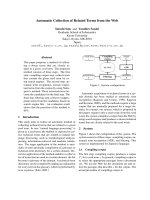
Fig. 6. A model of the modulation of pro-PHBP autoactivation by
polyamine and carminic acid. The pro-PHBP molecule is shown as
in Fig. 2. The schematic conformation shown is speculative.
K. Hasumi et al. Zymogen activation modulators
FEBS Journal 277 (2010) 3675–3687 ª 2010 The Authors Journal compilation ª 2010 FEBS 3683
11 Danø K, Behrendt N, Høyer-Hansen G, Johnsen M,
Lund LR, Ploug M & Romer J (2005) Plasminogen
activation and cancer. Thromb Haemost 93, 676–681.
12 Castellino FJ & Ploplis VA (2005) Structure and
function of the plasminogen ⁄ plasmin system. Thromb
Haemost 93, 647–654.
13 Forsgren M, Raden B, Israelsson M, Larsson K &
Heden L-O (1987) Molecular cloning and
characterization of a full-length cDNA clone for
human plasminogen. FEBS Lett 213, 254–260.
14 Tordai H, Banyai L & Patthy L (1999) The PAN
module: the N-terminal domains of plasminogen and
hepatocyte growth factor are homologous with the
apple domains of the prekallikrein family and with a
novel domain found in numerous nematode proteins.
FEBS Lett 461, 63–67.
15 Cockell CS, Marshall JM, Dawson KM, Cederholm-
Williams SA & Ponting CP (1998) Evidence that the
conformation of unliganded human plasminogen is
maintained via an intramolecular interaction between
the lysine-binding site of kringle 5 and the N-terminal
peptide. Biochem J 333, 99–105.
16 An SS, Carren
˜
o C, Marti DN, Schaller J, Alberico F &
Llinas M (1998) Lysine-50 is a likely site for anchoring
the plasminogen N-terminal peptide to lysine-binding
kringles. Protein Sci 7, 1960–1969.
17 Violand BN, Sodetz JM & Castellino FJ (1975) The
effect of epsilon-aminocaproic acid on the gross
conformation of plasminogen and plasmin. Arch
Biochem Biophys 170 , 300–305.
18 Christensen U & Molgaard L (1991) Stopped-flow
fluorescence kinetic studies of Glu-plasminogen.
Conformational changes triggered by AH-site ligand
binding. FEBS Lett 278, 204–206.
19 Nesheim ME, Fredenburgh JC & Larsen GR (1990)
The dissociation constants and stoichiometries of the
interactions of Lys-plasminogen and chloromethyl
ketone derivatives of tissue plasminogen activator and
the variant DFEIX with intact fibrin. J Biol Chem 265,
21541–21548.
20 Hajjar KA & Nacman L (1988) Endothelial cell-
mediated conversion of Glu-plasminogen to Lys-
plasminogen. Further evidence for assembly of the
fibrinolytic system on the endothelial cell surface.
J Clin Invest 82, 1769–1778.
21 Wygrecka M, Marsh LM, Morty RE, Henneke I,
Guenther A, Lohmeyer J, Markart P & Preissner KT
(2009) Enolase-1 promotes plasminogen-mediated
recruitment of monocytes to the acutely inflamed lung.
Blood 113, 5588–5598.
22 Kawao N, Nagai N, Ishida C, Okada K, Okumoto K,
Suzuki Y, Umemura K, Ueshima S & Matsuo O
(2010) Plasminogen is essential for granulation tissue
formation during the recovery process after liver injury
in mice. J Thromb Haemost 8, 1555–1566.
23 Andronicos NM, Chen EI, Baik N, Bai H, Parmer CM,
Kiosses WB, Kamps MP, Yates JR III, Parmer RJ &
Miles LA (2010) Proteomics-based discovery of a novel,
structurally unique, and developmentally regulated plas-
minogen receptor, Plg-R
KT
, a major regulator of cell
surface plasminogen activation. Blood 115, 1319–1330.
24 O’Connell PA, Surette AP, Liwski RS, Svenningsson P
& Waisman DM (2010) S100A10 regulates
plasminogen-dependent macrophage invasion. Blood,
doi: 10.1182/blood-2010-01-264754.
25 Fredenburgh JC & Nesheim ME (1992) Lys-
plasminogen is a significant intermediate in the
activation of Glu-plasminogen during fibrinolysis
in vitro. J Biol Chem 267, 26150–26156.
26 Violand BN & Castellino FJ (1976) Mechanism of the
urokinase-catalyzed activation of human plasminogen.
J Biol Chem 251, 3906–3912.
27 Marshall JM, Brown AJ & Ponting CP (1994)
Conformational studies of human plasminogen and
plasminogen fragments: evidence for a novel third
conformation of plasminogen. Biochemistry 33,
3599–3606.
28 Chang Y, Mochalkin I, McCance SG, Cheng B,
Tulinsky A & Castellino FJ (1998) Structure and ligand
binding determinants of the recombinant kringle 5
domain of human plasminogen. Biochemistry 37 ,
3258–3271.
29 Battistel MD, Grishaev A, An SS, Castellino FJ &
Llinas M (2009) Solution structure and functional
characterization of human plasminogen kringle 5.
Biochemistry 48, 10208–10219.
30 Christensen U (1984) The AH-site of plasminogen and
two C-terminal fragments. A weak lysine-binding site
preferring ligands not carrying a free carboxylate
function. Biochem J 223, 413–421.
31 Urano T, Chibber BA & Castellino FJ (1987) The
reciprocal effects of epsilon-aminohexanoic acid and
chloride ion on the activation of human [Glu1]plasmi-
nogen by human urokinase. Proc Natl Acad Sci USA
84, 4031–4034.
32 Urano T, Sator de Serrano V, Chibber BA &
Castellino FJ (1987) The control of the urokinase-
catalyzed activation of human glutamic acid
1-plasminogen by positive and negative effectors. J Biol
Chem 262, 15959–15964.
33 Wiles KG, Panizzi P, Kroh HK & Bock PE (2010)
Skizzle is a novel plasminogen and plasmin binding
protein from Streptococcus agalactiae that targets
proteins of human fibrinolysis to promote plasmin
generation. J Biol Chem 285, 21153–21164.
34 Michaeli A, Finci-Yeheskel Z, Dishon S, Linke RP,
Levin M & Urieli-Shoval S (2008) Serum amyloid A
enhances plasminogen activation: implication for a role
in colon cancer. Biochem Biophys Res Commun 368,
368–373.
Zymogen activation modulators K. Hasumi et al.
3684 FEBS Journal 277 (2010) 3675–3687 ª 2010 The Authors Journal compilation ª 2010 FEBS
35 Thorsen S (1992) The mechanism of plasminogen
activation and the variability of the fibrin effector
during tissue-type plasminogen activator-mediated
fibrinolysis. Ann NY Acad Sci 667, 52–63.
36 Krishnamurti C, Vukelja SJ & Alving BM (1994)
Inhibitory effects of lysine analogues on t-PA induced
whole blood clot lysis. Thromb Res 73, 419–430.
37 Utermann G (1989) The mysteries of lipoprotein(a).
Science 246, 904–910.
38 Kraft HG, Lingenhel A, Ko
¨
chl S, Hoppichler F,
Kronenberg F, Abe A, Mu
¨
hlberger V, Scho
¨
nitzer D &
Utermann G (1996) Apolipoprotein(a) kringle IV
repeat number predicts risk for coronary heart disease.
Arterioscler Thromb Vasc Biol 16, 713–719.
39 McLean JW, Tomlinson JE, Kuang W, Eaton DL,
Chen EY, Fless GM, Scanu AM & Lawn RM (1987)
cDNA sequence of human apolipoprotein(a) is
homologous to plasminogen. Nature 300, 132–137.
40 Lackner C, Cohen JC & Hobbs HH (1993) Molecular
definition of the extreme size polymorphism in
apolipoprotein(a). Hum Mol Genet 2, 933–940.
41 Hajjar KA, Gavish D, Breslow JL & Nachman RL
(1989) Lipoprotein(a) modulation of endothelial cell
surface fibrinolysis and its potential role in atheroscle-
rosis. Nature 339, 303–305.
42 Feric NT, Boffa MB, Johnston SM & Koschinsky ML
(2008) Apolipoprotein(a) inhibits the conversion of
Glu-plasminogen to Lys-plasminogen: a novel
mechanism for lipoprotein(a)-mediated inhibition of
plasminogen activation. J Thromb Haemost 6,
2113–2120.
43 Tachikawa K, Hasumi K & Endo A (1997) Enhance-
ment of plasminogen binding to U937 cells and fibrin
by complestatin. Thromb Haemost 77, 137–142.
44 Tachikawa K, Hasumi K & Endo A (1997)
Enhancement of plasminogen binding and fibrinolysis
by chloropeptin I. Thromb Res 87, 571–576.
45 Shinohara C, Hasumi K, Hatumi W & Endo A (1996)
Staplabin, a novel fungal triprenyl phenol which
stimulates the binding of plasminogen to fibrin and
U937 cells. J Antibiot (Tokyo) 49, 961–966.
46 Kohyama T, Hasumi K, Hamanaka A & Endo A
(1997) SMTP-1 and -2, novel analogs of staplabin
produced by Stachybotrys microspora IFO30018.
J Antibiot (Tokyo) 50, 172–174.
47 Takayasu R, Hasumi K, Shinohara C & Endo A
(1997) Enhancement of fibrin binding and activation of
plasminogen by staplabin through induction of a
conformational change in plasminogen. FEBS Lett 418,
58–62.
48 Hasumi K, Ohyama S, Kohyama T, Ohsaki Y,
Takayasu R & Endo A (1998) Isolation of SMTP-3, -4,
-5 and -6, novel analogs of staplabin, and their effects
on plasminogen activation and fibrinolysis. J Antibiot
(Tokyo) 51, 1059–1068.
49 Hu W, Ohyama S & Hasumi K (2000) Activation of
fibrinolysis by SMTP-7 and -8, novel staplabin analogs
with a pseudosymmetric structure. J Antibiot (Tokyo)
53, 241–247.
50 Hu W, Narasaki R, Ohyama S & Hasumi K (2001)
Selective production of staplabin and SMTPs in
cultures of Stachybotrys microspora fed with precursor
amines. J Antibiot (Tokyo) 54, 962–966.
51 Hu W, Kitano Y & Hasumi K (2003) SMTP-4D, -5D,
-6D, -7D and -8D, a new series of the non-lysine-
analog plasminogen modulators with a d-amino acid
moiety. J Antibiot (Tokyo) 56, 832–837.
52 Ohyama S, Harada T, Chikanishi T, Miura Y &
Hasumi K (2004) Nonlysine-analog plasminogen
modulators promote autocatalytic generation of
plasmin(ogen) fragments with angiostatin-like activity.
Eur J Biochem 271, 809–820.
53 Hasumi K, Hasegawa K & Kitano Y (2007) Isolation
and absolute configuration of SMTP-0, a simplest
congener of the SMTP family nonlysine-analog plas-
minogen modulators. J Antibiot (Tokyo) 60, 463–468.
54 Ohyama S, Wada Y & Hasumi K (2002) Antibiotic
A10255 (thioplabin) enhances fibrin binding and activa-
tion of plasminogen. J Antibiot (Tokyo) 55, 83–91.
55 Sasaoka M, Wada Y & Hasumi K (2007)
Stachybotrydial selectively enhances fibrin binding
and activation of Glu-plasminogen. J Antibiot
(Tokyo) 60, 674–681.
56 Stewart RJ, Fredenburgh JC, Leslie BA, Keyt BA,
Rischke JA & Weitz JI (2000) Identification of the
mechanism responsible for the increased fibrin
specificity of TNK-tissue plasminogen activator relative
to tissue plasminogen activator. J Biol Chem 275,
10112–10120.
57 Hoylaerts M, Rijken DC, Lijnen HR & Collen D
(1982) Kinetics of the activation of plasminogen by
human tissue plasminogen activator. Role of fibrin.
J Biol Chem 257, 2912–2919.
58 Ploug M (2003) Structure–function relationships in the
interaction between the urokinase-type plasminogen
activator and its receptor. Curr Pharm Des 9, 1499–
1528.
59 Blasi F & Sidenius N (2010) The urokinase receptor:
focused cell surface proteolysis, cell adhesion and
signaling. FEBS Lett 584, 1923–1930.
60 Blasi F (1993) Urokinase and urokinase receptor:
a paracrine ⁄ autocrine system regulating cell migration
and invasiveness. Bioessays 15, 105–111.
61 Vincenza Carriero M, Franco P, Vocca I, Alfano D,
Longanesi-Cattani I, Bifulco K, Mancini A, Caputi M
& Stoppelli MP (2009) Structure, function and
antagonists of urokinase-type plasminogen activator.
Front Biosci 14, 3782–3794.
62 Danø K, Andreasen PA, Grondahl-Hansen J,
Kristensen P, Nielsen LS & Skriver L (1985)
K. Hasumi et al. Zymogen activation modulators
FEBS Journal 277 (2010) 3675–3687 ª 2010 The Authors Journal compilation ª 2010 FEBS 3685
Plasminogen activators, tissue degradation, and cancer.
Adv Cancer Res 44, 139–266.
63 Stephens RW, Po
¨
lla
¨
nen J, Tapiovaara H, Leung KC,
Sim PS, Salonen EM, Rønne E, Behrendt N, Danø K
& Vaheri A (1989) Activation of pro-urokinase and
plasminogen on human sarcoma cells: a proteolytic
system with surface-bound reactants. J Cell Biol 108,
1987–1995.
64 Ellis V & Danø K (1993) Potentiation of plasminogen
activation by an anti-urokinase monoclonal antibody
due to ternary complex formation. A mechanistic
model for receptor-mediated plasminogen activation.
J Biol Chem 268, 4806–4813.
65 Lijnen HR, Zamarron C, Blaber M, Winkler ME &
Collen D (1986) Activation of plasminogen by
pro-urokinase. I. Mechanism. J Biol Chem 261,
1253–1258.
66 Lijnen HR, Van Hoef B, Nelles L & Collen D (1990)
Plasminogen activation with single-chain urokinase-
type plasminogen activator (scu-PA). Studies with
active site mutagenized plasminogen (Ser
740
fi Ala)
and plasmin-resistant scu-PA (Lys
158
fi Glu). J Biol
Chem 265, 5232–5236.
67 Baeten KM, Richard MC, Kanse SM, Mutch NJ,
Degen JL & Booth NA (2010) Activation of single-
chain urokinase by platelet-associated plasminogen:
a mechanism for stimulation of fibrinolysis by platelets.
J Thromb Haemost 8, 1313–1322.
68 Kikuchi T & Hasumi K (2002) Enhancement of
plasminogen activation by surfactin C: augmentation of
fibrinolysis in vitro and in vivo. Biochim Biophys Acta
1596, 234–245.
69 Kikuchi T & Hasumi K (2003) Enhancement of
reciprocal activation of prourokinase and plasminogen
by the bacterial lipopeptide surfactins and iturin Cs.
J Antibiot (Tokyo) 56, 34–37.
70 Wu W, Narasaki R, Maeda F & Hasumi K (2004)
Glucosyldiacylglycerol enhances reciprocal activation of
prourokinase and plasminogen. Biosci Biotechnol
Biochem 68, 1549–1556.
71 Petersen LC, Lund LR, Nielsen LS, Danø K & Skriver
L (1988) One-chain urokinase-type plasminogen
activator from human sarcoma cells is a proenzyme
with little or no intrinsic activity. J Biol Chem 263,
11189–11195.
72 Esmon CT (2003) The protein C pathway. Chest 124,
26S–32S.
73 Nesheim M & Bajzar L (2005) The discovery of TAFI.
J Thromb Haemost 3, 2139–2146.
74 Coughlin SR & Camerer E (2003) PARticipation in
inflammation. J Clin Invest 111, 25–27.
75 Degen SJ, MacGillivray RT & Davie EW (1983)
Characterization of the complementary deoxyribonu-
cleic acid and gene coding for human prothrombin.
Biochemistry 22, 2087–2097.
76 Krishnaswamy S, Church WR, Nesheim ME & Mann
KG (1987) Activation of human prothrombin by
human prothrombinase. Influence of factor Va on the
reaction mechanism. J Biol Chem 262, 3291–3299.
77 Hacisalihoglu A, Panizzi P, Bock PE, Camire RM &
Krishnaswamy S (2007) Restricted active site docking
by enzyme-bound substrate enforces the ordered
cleavage of prothrombin by prothrombinase. J Biol
Chem 282, 32974–32982.
78 Bradford HN, Micucci JA & Krishnaswamy S (2010)
Regulated cleavage of prothrombin by prothrombinase:
repositioning a cleavage site reveals the unique kinetic
behavior of the action of prothrombinase on its
compound substrate. J Biol Chem 285, 328–338.
79 Krishnaswamy S, Mann KG & Nesheim ME (1986)
The prothrombinase-catalyzed activation of
prothrombin proceeds through the intermediate
meizothrombin in an ordered, sequential reaction.
J Biol Chem 261, 8977–8984.
80 Mann KG, Nesheim ME, Church WR, Haley P &
Krishnaswamy S (1990) Surface-dependent reactions of
the vitamin K-dependent enzyme complexes. Blood 76,
1–16.
81 Bukys MA, Orban T, Kim PY, Nesheim ME &
Kalafatis M (2008) The interaction of fragment 1 of
prothrombin with the membrane surface is a
prerequisite for optimum expression of factor Va
cofactor activity within prothrombinase. Thromb
Haemost 99, 511–522.
82 Qureshi SH, Yang L, Manithody C & Rezaie AR
(2009) Membrane-dependent interaction of factor Xa
and prothrombin with factor Va in the prothrombinase
complex. Biochemistry 48, 5034–5041.
83 Rosing J, Tans G, Govers-Riemslag JW, Zwaal RF &
Hemker HC (1980) The role of phospholipids and
factor Va in the prothrombinase complex. J Biol Chem
255, 274–283.
84 Inoue T, Hasumi K, Kuniyasu T & Endo A (1996)
Isolation of plactins A, B, C and D, novel cyclic
pentapeptides that stimulate cellular fibrinolytic
activity. J Antibiot (Tokyo) 49, 45–49.
85 Inoue T, Hasumi K, Sugimoto M & Endo A (1998)
Enhancement of fibrinolysis by plactins: structure–
activity relationship and effects in human U937 cells
and in mice. Thromb Haemost 79, 591–596.
86 Harada T, Tsuruta T, Yamagata K, Inoue T & Hasumi
K (2009) Dual modulation of prothrombin activation
by the cyclopentapeptide plactin. FEBS J 276, 2516–
2528.
87 Koizumi Y & Hasumi K (2002) Enhancement of
fibrinolytic activity of U937 cells by malformin A
1
.
J Antibiot (Tokyo) 55, 78–82.
88 Bock PE, Panizzi P & Verhamme IM (2007) Exosites
in the substrate specificity of blood coagulation
reactions. J Thromb Haemost 5(Suppl 1), 81–94.
Zymogen activation modulators K. Hasumi et al.
3686 FEBS Journal 277 (2010) 3675–3687 ª 2010 The Authors Journal compilation ª 2010 FEBS
89 Ro
¨
misch J, Vermohlen S, Feussner A & Sto
¨
hr HA
(1999) The FVII activating protease cleaves single-chain
plasminogen activators. Haemostasis 29, 292–299.
90 Ro
¨
misch J, Feussner A, Vermohlen S & Sto
¨
hr HA
(1999) A protease isolated from human plasma
activating factor VII independent of tissue factor. Blood
Coagul Fibrinolysis 10, 471–479.
91 Etscheid M, Kress J, Seitz R & Dodt J (2008) The
hyaluronic acid-binding protease: a novel vascular and
inflammatory mediator? Int Immunopharmacol 8,
166–170.
92 Kanse SM, Parahuleva M, Muhl L, Kemkes-Matthes
B, Sedding D & Preissner KT (2008) Factor VII-
activating protease (FSAP): vascular functions and
role in atherosclerosis. Thromb Haemost 99, 286–
289.
93 Sedding D, Daniel JM, Muhl L, Hersemeyer K,
Brunsch H, Kemkes-Matthes B, Braun-Dullaeus RC,
Tillmanns H, Weimer T, Preissner KT et al. (2006) The
G534E polymorphism of the gene encoding the
factor VII-activating protease is associated with
cardiovascular risk due to increased neointima
formation. J Exp Med 13, 2801–2807.
94 Wasmuth HE, Tag CG, Van de Leur E, Hellerbrand
C, Mueller T, Berg T, Puhl G, Neuhaus P, Samuel D,
Trautwein C et al. (2009) The Marburg I variant
(G534E) of the factor VII-activating protease deter-
mines liver fibrosis in hepatitis C infection by reduced
proteolysis of platelet-derived growth factor BB.
Hepatology 3, 775–780.
95 Roderfeld M, Weiskirchen R, Atanasova S, Gressner
AM, Preissner KT, Roeb E & Kanse SM (2009)
Altered factor VII activating protease expression in
murine hepatic fibrosis and its influence on hepatic
stellate cells. Liver Int 29, 686–691.
96 Parahuleva MS, Kanse SM, Parviz B, Barth A,
Tillmanns H, Bohle RM, Sedding DG &
Holschermann H (2008) Factor Seven Activating
Protease (FSAP) expression in human monocytes and
accumulation in unstable coronary atherosclerotic
plaques. Atherosclerosis 196, 164–171.
97 Choi-Miura NH, Tobe T, Sumiya J, Nakano Y,
Sano Y, Mazda T & Tomita M (1996) Purification
and characterization of a novel hyaluronan-binding
protein (PHBP) from human plasma: it has three
EGF, a kringle and a serine protease domain, similar
to hepatocyte growth factor activator. J Biochem 119,
1157–1165.
98 Etscheid M, Hunfeld A, Konig H, Seitz R & Dodt J
(2000) Activation of proPHBSP, the zymogen of a
plasma hyaluronan binding serine protease, by an
intermolecular autocatalytic mechanism. Biol Chem
381, 1223–1231.
99 Choi-Miura NH, Saito K, Takahashi K, Yoda M &
Tomita M (2001) Regulation mechanism of the serine
protease activity of plasma hyaluronan binding protein.
Biol Pharm Bull 24, 221–225.
100 Nakazawa F, Kannemeier C, Shibamiya A, Song Y,
Tzima E, Schubert U, Koyama T, Niepmann M,
Trusheim H, Engelmann B et al. (2005) Extracellular
RNA is a natural cofactor for the (auto-) activation of
Factor VII-activating protease (FSAP). Biochem J 385,
831–838.
101 Kannemeier C, Feussner A, Sto
¨
hr HA, Weisse J,
Preissner KT & Ro
¨
misch J (2001) Factor VII and
single-chain plasminogen activator-activating protease:
activation and autoactivation of the proenzyme. Eur J
Biochem 268, 3789–3796.
102 Choi-Miura NH, Otsuyama K, Sano Y, Saito K,
Takahashi K & Tomita M (2001) Hepatic injury-
specific conversion of mouse plasma hyaluronan
binding protein to the active hetero-dimer form. Biol
Pharm Bull 24 , 892–896.
103 Wygrecka M, Morty RE, Markart P, Kanse SM,
Andreasen PA, Wind T, Guenther A & Preissner KT
(2007) Plasminogen activator inhibitor-1 is an inhibitor
of factor VII-activating protease in patients with acute
respiratory distress syndrome. J Biol Chem 282, 21671–
21682.
104 Yamamichi S, Nishitani M, Nishimura N, Matsushita
Y & Hasumi K (2010) Polyamine-promoted autoactiva-
tion of plasma hyaluronan-binding protein. J Thromb
Haemost 8, 559–566.
105 Caldarera CM, Barbiroli B & Moruzzi G (1965)
Polyamines and nucleic acids during development of
the chick embryo. Biochem J 97, 84–88.
106 Tabor CW & Tabor H (1984) Polyamines. Annu Rev
Biochem 53, 749–790.
107 Nishimura N, Takai M, Yamamoto E & Hasumi K
(2010) Purpurin as a specific inhibitor of spermidine-
induced autoactivation of the protease plasma hyaluro-
nan-binding protein. Biol Pharm Bull 33, 1430–1433.
K. Hasumi et al. Zymogen activation modulators
FEBS Journal 277 (2010) 3675–3687 ª 2010 The Authors Journal compilation ª 2010 FEBS 3687