Báo cáo khoa học: A single EF-hand isolated from STIM1 forms dimer in the absence and presence of Ca2+ ppt
Bạn đang xem bản rút gọn của tài liệu. Xem và tải ngay bản đầy đủ của tài liệu tại đây (783.58 KB, 9 trang )
A single EF-hand isolated from STIM1 forms dimer in the
absence and presence of Ca
2+
Yun Huang, Yubin Zhou, Hing-Cheung Wong, Yanyi Chen, Yan Chen, Siming Wang,
Adriana Castiblanco, Aimin Liu and Jenny J. Yang
Department of Chemistry, Center for Drug Design and Advanced Biotechnology, Georgia State University, Atlanta, GA, USA
Introduction
Stromal interaction molecule 1 (STIM1), recently iden-
tified by RNA interference (RNAi) screens in Drosoph-
ila S2 cells and HeLa cells by two independent groups
[1,2], is regarded as an endoplasmic reticulum (ER)
luminal Ca
2+
sensor and functions as an essential
component of store-operated Ca
2+
entry. It is a key
linkage between ER Ca
2+
store emptying, Ca
2+
influx
and internal Ca
2+
store refilling in mammalian cells.
On ER Ca
2+
store depletion, STIM1 undergoes oligo-
merization, translocates from the ER membrane to
form ‘punctae’ near the plasma membrane [1,3,4] and
activates the Ca
2+
release-activated Ca
2+
(CRAC)
channel through direct interaction with the pore-form-
ing subunit Orai1 [5]. STIM1 is a single transmem-
brane-spanning protein with 685 amino acids which
contains a canonical EF-hand motif and a sterile
a-motif (SAM) domain in the ER lumen. Previous
studies have strongly indicated that the EF-hand
Keywords
affinity; Ca
2+
; EF-hand; oligomerization;
STIM1
Correspondence
J. J. Yang, Department of Chemistry,
Georgia State University, Atlanta, GA 30303,
USA
Fax: +1 404 413 5551
Tel: +1 404 413 5520
E-mail:
(Received 21 March 2009, revised 26 June
2009, accepted 27 July 2009)
doi:10.1111/j.1742-4658.2009.07240.x
Stromal interaction molecule 1 (STIM1) is responsible for activating the
Ca
2+
release-activated Ca
2+
(CRAC) channel by first sensing the changes
in Ca
2+
concentration in the endoplasmic reticulum ([Ca
2+
]
ER
) via its
luminal canonical EF-hand motif and subsequently oligomerizing to inter-
act with the CRAC channel pore-forming subunit Orai1. In this work, we
applied a grafting approach to obtain the intrinsic metal-binding affinity of
the isolated EF-hand of STIM1, and further investigated its oligomeric
state using pulsed-field gradient NMR and size-exclusion chromatography.
The canonical EF-hand bound Ca
2+
with a dissociation constant at a level
comparable with [Ca
2+
]
ER
(512 ± 15 lm). The binding of Ca
2+
resulted
in a more compact conformation of the engineered protein. Our results
also showed that D to A mutations at Ca
2+
-coordinating loop positions 1
and 3 of the EF-hand from STIM1 led to a 15-fold decrease in the metal-
binding affinity, which explains why this mutant was insensitive to changes
in Ca
2+
concentration in the endoplasmic reticulum ([Ca
2+
]
ER
) and
resulted in constitutive punctae formation and Ca
2+
influx. In addition,
the grafted single EF-hand motif formed a dimer regardless of the presence
of Ca
2+
, which conforms to the EF-hand paring paradigm. These data
indicate that the STIM1 canonical EF-hand motif tends to dimerize for
functionality in solution and is responsible for sensing changes in
[Ca
2+
]
ER
.
Abbreviations
[Ca
2+
]
ER
,Ca
2+
concentration in the endoplasmic reticulum; CaM, calmodulin; CRAC, Ca
2+
release-activated Ca
2+;
ER, endoplasmic reticulum;
GST, glutathione transferase; HSQC, heteronuclear single-quantum correlation; RNAi, RNA interference; SAM, sterile a-motif; STIM1,
stromal interaction molecule 1.
FEBS Journal 276 (2009) 5589–5597 ª 2009 The Authors Journal compilation ª 2009 FEBS 5589
region is responsible for the sensing by STIM1 of the
changes in [Ca
2+
]
ER
. Mutations on the predicted EF-
hand reduce the affinity for Ca
2+
, thus mimicking the
store-depleted state and subsequently triggering STIM1
redistribution to the plasma membrane and activation
of the CRAC channel even without Ca
2+
store deple-
tion [4,6]. However, the site-specific metal-binding
property and the oligomeric state of the canonical
EF-hand of STIM1 alone have not been characterized
thus far.
The EF-hand motif with a characteristic helix–loop–
helix fold was first discovered by Moews and Kretsing-
er [7] in the crystal structure of parvalbumin. To date,
more than 66 members of EF-hand proteins have been
classified [8]. EF-hand proteins often occur in pairs
with the two Ca
2+
-binding loops coupled via a short
antiparallel b-sheet. Ca
2+
is coordinated by the main-
chain carbonyl and side-chain carboxyl oxygens at the
12- or 14-residue loop. One pair of EF-hands usually
forms a globular domain to allow for cooperative
Ca
2+
binding, responding to a narrow range of free
Ca
2+
concentration change. To examine the key deter-
minants for Ca
2+
binding and Ca
2+
-induced confor-
mational change, peptides or fragments encompassing
the helix–loop–helix motif have been produced by
either synthesis or cleavage. Shaw et al. [9] first
reported that an isolated EF-hand III from skeletal
troponin C dimerizes in the presence of Ca
2+
. EF-
hands from parvalbumin and calbindin D9K have also
been shown to exhibit Ca
2+
-dependent dimerization
[10–12]. Wojcik et al. [13] have shown that the isolated
12-residue peptide from calmodulin (CaM) EF-hand
motif III does not dimerize in the presence of Ca
2+
,
but dimerizes to form a native-like structure in the
presence of Ln
3+
, which has a similar ionic radius and
coordination properties to Ca
2+
. They concluded that
local interactions between the EF-hand Ca
2+
-binding
loops alone could be responsible for the observed
cooperativity of Ca
2+
binding to EF-hand protein
domains. Our laboratory has developed a grafting
approach to probe the site-specific Ca
2+
-binding affini-
ties and metal-binding properties of CaM [14] and
other EF-hand proteins, such as the nonstructural pro-
tease domain of rubella virus [15]. We have shown that
an isolated EF-hand loop without flanking helices
grafted in CD2 remains as a monomer instead of a
dimer, as observed in the peptide fragments [16],
implying that additional factors that reside outside of
EF-loop III may contribute to the pairing of the EF-
hand motifs of CaM. Figure 1A shows that most
hydrophobic residues in the flanking helices and loop
are conserved compared with EF-hand III in CaM and
the STIM1 EF-hand, such as position 8 in the loop,
)8, )5, )1 in the E helix and +4, +5 in the F helix,
which leads us to speculate that the EF-hand motif of
STIM1 has the potential to form a dimer. In this
work, we applied a grafting approach [14] to obtain
the site-specific intrinsic metal-binding affinity and to
probe the oligomeric state of the EF-hand of STIM1
using size-exclusion chromatography and pulsed-field
diffusion NMR. We found that mutations on loop
positions 1 and 3 of the EF-hand from STIM1
decreased the binding affinity by more than 10-fold.
Interestingly, the isolated EF-hand motif of STIM1
undergoes Ca
2+
-induced conformational changes and
remains as a dimer in the absence and presence of
Ca
2+
.
Results and Discussion
The isolated EF-hand motif from STIM1 retains
its helical structure
The helix–loop–helix EF-hand motif from STIM1 was
grafted into CD2 with each side flanked by three Gly
residues to render sufficient flexibility (Fig. 1A). Previ-
ous studies in our laboratory have shown that the loop
position in domain 1 of CD2 at 52 between the
b-strands C† and D tolerates the insertion of foreign
EF-hand motifs from CaM whilst retaining its own
structural integrity [15,17]. In Fig. 1B, the modelled
structure of the engineered protein CD2.STIM1.EF is
shown. The structural integrity of the host protein was
then examined by two-dimensional NMR. As shown
in Fig. 1C, the dispersed region of the (
1
H,
15
N)-het-
eronuclear single-quantum correlation (HSQC) NMR
spectrum of CD2.STIM1.EF was very similar to that
of CD2 with grafted EF-loop III of CaM
(CD2.CaM.loopIII) [16], suggesting that the conforma-
tion of the host protein CD2 is largely unchanged.
Additional resonances appearing between 8.2 and
8.8 p.p.m. were caused by the addition of flanking
helices to the grafted EF-hand motif.
To confirm that the grafted EF-hand motif retains
its helical structure, CD spectra of the host protein
CD2 domain 1 (CD2.D1) and CD2.STIM1.EF were
analysed by DICHROWEB, an online server for
protein secondary structure analyses [18]. Figure 1D, E
shows the far-UV CD spectra and the calculated sec-
ondary structure contents of both proteins. The host
protein CD2.D1 contained 3% a-helix and 35%
b-strand, which is in good agreement with the second-
ary structure contents determined by X-ray crystallog-
raphy [19]. Following the insertion of the EF-hand
motif from STIM1, the helical content increased by
7%, which corresponds to approximately 10 residues
Isolated dimeric EF-hand from STIM1 binds to Ca
2+
Y. Huang et al.
5590 FEBS Journal 276 (2009) 5589–5597 ª 2009 The Authors Journal compilation ª 2009 FEBS
in the helical conformation, whereas the b-strand
content largely remained similar to CD2.D1 (Fig. 1E).
The isolated EF-hand binds to Ca
2+
and
lanthanide ions
One of the most important steps to fully understand
the mechanism underlying the Ca
2+
-modulated func-
tions of STIM1 is to investigate the site-specific Ca
2+
-
binding properties of the EF-hand of STIM1. In this
study, we adopted a grafting approach to address this
question. As shown in Fig. 1B, the distance between
the two termini of the inserted Ca
2+
-binding sites in
the model structure of the EF-hand of STIM1 is within
15 A
˚
. Accordingly, a total of six glycine linkers is suffi-
cient to enable the grafted motifs to retain the native
metal conformation. Trp32 and Tyr76 in the host
proteins are approximately 15 A
˚
away from the grafted
sites, which enables aromatic-sensitized energy transfer
to the Tb
3+
bound to the sites, providing a sensitive
spectroscopic method to monitor the metal-binding
process. As shown in Fig. 2A, the addition of Tb
3+
to
the engineered proteins, or vice versa, resulted in large
increases in Tb
3+
fluorescence at 545 nm caused by
energy transfer, which was not observed for wild-type
CD2.D1 [15,20]. The addition of excessive amounts of
Ca
2+
to the Tb
3+
–protein mixture led to a significant
decrease in Tb
3+
luminescence signal as a result of
metal competition (Fig. 2A, inset). The Tb
3+
- and
Ca
2+
-binding affinities could thus be derived from the
Tb
3+
titration and metal competition curves. For the
engineered protein CD2.STIM1.EF, the Tb
3+
- and
Ca
2+
-binding dissociation constants (K
d
) were 170 ± 6
and 512 ± 15 lm, respectively. In contrast, a mutant
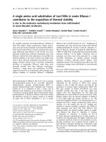
Fig. 1. Grafting the helix–loop–helix EF-hand motif into CD2. (A) The sequence alignment results of calmodulin EF-hand III and the canonical
EF-hand motif in STIM and its mutant. The sequence from S64 to L96 in STIM1 was grafted into CD2.D1. A mutant containing Asp to Ala
substitutions at Ca
2+
-coordinating loop positions 1 and 3 was introduced to perturb the Ca
2+
-binding ability of the grafted EF-hand of STIM1.
(B) Modelled structure of the engineered protein with the grafted EF-hand Ca
2+
-binding motif (magenta) from STIM1. W32 and Y76 in the
host protein are about 15 A
˚
away from the grafted Ca
2+
-binding sites. Ca
2+
is shown as a dark sphere. (C) Overlay of the (
1
H,
15
N)-HSQC
spectrum of CD2.STIM1.EF (red) with that of CD2-loop3 (EF-loop III from calmodulin, cyan) in the absence of Ca
2+
. (D, E) Far-UV CD spectra
of CD2 and CD2.STIM1.EF and the calculated secondary structural contents.
Y. Huang et al. Isolated dimeric EF-hand from STIM1 binds to Ca
2+
FEBS Journal 276 (2009) 5589–5597 ª 2009 The Authors Journal compilation ª 2009 FEBS 5591
with the metal-coordinating residue Asp at positions 1
and 3 in the EF-loop substituted with Ala (denoted as
CD2.STIM1mut) resulted in at least a 12-fold decrease
in the Tb
3+
-binding affinity (K
d
> 2.1 mm, Fig. 2B),
suggesting that these key residues are essential for
metal binding. The direct binding of metal ions to the
grafted sequences was further supported by two-dimen-
sional HSQC NMR studies. As shown in Fig. 2C, the
addition of increasing amounts of La
3+
, a commonly
used trivalent Ca
2+
analogue, led to gradual chemical
shift changes in residues from the grafted sequences.
However, residues from the host protein CD2.D1, such
as T97 ad G107, remained unchanged.
The isolated EF-hand from STIM1 forms dimer in
solution
Next, we examined the oligomeric state of the grafted
EF-hand motif using three independent techniques:
pulsed-field gradient NMR, size-exclusion chromatog-
A
B
C
K
K
K
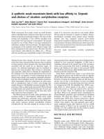
Fig. 2. Metal-binding properties of CD2.STIM1.EF. (A) The enhancement of Tb
3+
luminescence at 545 nm plotted as a function of total
added [Tb
3+
]. The inset shows the Ca
2+
competition curve. (B) The enhancement of fluorescence at 545 nm of the CD2.STIM1.EF mutant
(Asp to Ala substitutions at loop positions 1 and 3) as a function of titrated Tb
3+
. (C) Enlarged areas of (
1
H,
15
N)-HSQC spectrum of CD2.STI-
M1.EF. La
3+
induced chemical shift changes (indicated by arrows) in two residues from the grafted sequences. In contrast, the chemical
shifts of residues from the host protein CD2.D1 (i.e. G107 and T97) remained unchanged.
Isolated dimeric EF-hand from STIM1 binds to Ca
2+
Y. Huang et al.
5592 FEBS Journal 276 (2009) 5589–5597 ª 2009 The Authors Journal compilation ª 2009 FEBS
raphy and chemical cross-linking. Pulsed-field gradient
NMR has been widely used to study the molecular
motion, effective dimensions and oligomeric states of
proteins in solution [21]. With this technique, the size
of proteins can be estimated by measuring diffusion
constants, as the relationship between the translational
motion of spherical molecules in solution and the
hydrodynamic radius is governed by the equation,
D = K
B
T ⁄ 6pag, where g is the solvent viscosity and a
is the radius of the molecules. The diffusion constant
of a dimer is ideally expected to be approximately
79% of the value of a monomer [21].
The diffusion constants of engineered protein
CD2.STIM1.EF were measured under Ca
2+
-depleted
and Ca
2+
-saturated conditions to determine whether
the isolated EF-hand motif from STIM1 undergoes
dimerization on metal binding. Figure 3A shows the
NMR signal decay when the field strength was
increased from 0.2 to 31 GÆcm
)1
. The calculated
hydrodynamic radius of the CD2 monomer was
19.4 ± 0.4 A
˚
, which was close to the previously
reported value of 19.6 A
˚
[16]. The calculated hydrody-
namic radii of the engineered protein CD2.STIM1.
EF were 24.0 ± 0.3 A
˚
with 10 mm EGTA and 24.9 ±
0.2 A
˚
with 10 mm Ca
2+
. According to calculations
using the spherical shape of macromolecules, the
hydrodynamic radius of the protein will increase by
27% on formation of the dimer [22]. The increase in
size for CD2.STIM1.EF is very close to this theoretical
value, indicating that it exists as a dimer in solution,
regardless of the presence of Ca
2+
.
Size-exclusion chromatography was also used to
estimate the size of the engineered protein under
Ca
2+
-saturated and Ca
2+
-free conditions. As shown
in Fig. 3B, the elution profiles of 10 mm Ca
2+
-loaded
and Ca
2+
-depleted CD2.STIM1.EF exhibited a major
peak, with estimated molecular masses of 28 and
32 kDa, respectively, which is close to twice the theo-
retical molecular mass of CD2.STIM1.EF. However,
the Ca
2+
-loaded CD2.STIM1.EF was eluted slightly
later than the Ca
2+
-depleted form. This shift in peak
position suggests that Ca
2+
-loaded CD2.STIM1.EF
has a smaller size than Ca
2+
-depleted CD2.STIM1.EF.
It seems that Ca
2+
induced conformational changes in
the engineered protein and resulted in a more compact
shape of the protein.
One additional method, glutaraldehyde cross-linking,
was applied to study the oligomerization patterns of
the engineered protein at low micromolar concentra-
tion. Figure 3B (inset) shows SDS-PAGE of glutaral-
dehyde-mediated cross-linking of CD2.STIM1.EF
(20 lm) in the presence of 5 mm Ca
2+
or 5 mm
EGTA. Regardless of the presence of Ca
2+
, bands
corresponding to both monomeric and dimeric CD2.
STIM1.EF were observed on SDS-PAGE. In sum-
mary, our data suggest that the grafted EF-hand motif
from STIM1 tends to dimerize in solution.
Implications for Ca
2+
-binding properties of STIM1
Previous studies have demonstrated that STIM1 plays
an important role in store-operated Ca
2+
entry [3]. On
store depletion, STIM1 is redistributed from the ER
membrane to form ‘punctae’ and aggregates near the
plasma membrane [1,6]. The N-terminal region of
STIM1 contains a canonical EF-hand motif and a pre-
dicted SAM domain. Stathopulos et al. [23,24] isolated
the EF-SAM region from STIM1 and studied the
structural and biophysical properties on this domain
after refolding. Their excellent work indicated that the
A
B
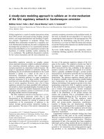
Fig. 3. The oligomeric state of CD2.STIM1.EF. (A) The NMR signal
decay of CD2 (grey circles) and CD2.STIM1.EF with Ca
2+
(crosses)
or EGTA (filled circles) as a function of field strength. The calculated
hydrodynamic radii of the protein samples are indicated. (B) Size-
exclusion chromatography elution profiles of CD2 (thin lines) and
CD2.STIM1.EF (bold lines) in the presence of 10 m
M Ca
2+
or EGTA.
The protein molecular mass standards are indicated by arrows.
Inset: SDS-PAGE of cross-linked CD2.STIM1.EF in the presence of
5m
M EGTA or Ca
2+
.
Y. Huang et al. Isolated dimeric EF-hand from STIM1 binds to Ca
2+
FEBS Journal 276 (2009) 5589–5597 ª 2009 The Authors Journal compilation ª 2009 FEBS 5593
ER Ca
2+
depletion-induced oligomerization of STIM1
occurs via the EF-SAM region. However, the refolding
process may not guarantee the natural conformation
of the EF-SAM region. Furthermore, as both the
EF-hand motif and the SAM region have the potential
to facilitate oligomerization, it is challenging to differ-
entiate which region contributes to the oligomerization
process.
To overcome the limitations of investigating the
Ca
2+
-binding sites in native Ca
2+
-binding proteins, we
established a grafting approach to dissect their site-
specific properties. This approach has been used in the
investigation of single EF-hand motifs in CaM and a
single EF-hand from rubella virus nonstructural prote-
ase [14,15]. CD2 has been shown to be a suitable host
system, as it retains its native structure after the inser-
tion of foreign sequences and in the presence and
absence of Ca
2+
ions, so that the influence from the
host protein to the inserted sites is minimized [14]. Our
NMR spectra shown in Fig. 2A clearly demonstrate
that the conformation of CD2 is unchanged. After the
insertion of the helix–loop–helix EF-hand domain
from STIM1, the helical content of the engineered
protein CD2.STIM1.EF increased, indicating that the
inserted EF-hand motif at least partially maintains the
natural helical structure after grafting. The Ca
2+
dis-
sociation constant of CD2.STIM1.EF (512 lm)isin
good agreement with the previously reported value
(200–600 lm) [25] and is comparable with [Ca
2+
]
ER
(250–600 lm) [15,26]. Such dissociation constants
would ensure that at least one-half of the population
of the EF-hand motif in STIM1 is occupied by Ca
2+
.
Removing the proposed Ca
2+
-coordinating residues in
positions 1 and 3 of the EF-hand motif significantly
compromised the metal-binding capability of the engi-
neered protein, indicating that the metal binding of
CD2.STIM1.EF is through the EF-hand motif from
STIM1. Two-dimensional HSQC NMR studies further
corroborated this view, as only residues from the
grafted sequences underwent chemical shift changes,
whereas residues from the host protein remained
unchanged. The impaired metal-binding ability caused
by Asp to Ala mutations at positions 1 and 3 echoed a
previous observation that these mutations in the intact
STIM1 molecule led to constitutive activation of
CRAC channels even without store depletion [4].
The canonical EF-hand in STIM1 has been regarded
previously to function alone to sense Ca
2+
changes.
The recently determined structure of the EF-SAM
region of STIM1 unveiled a surprising finding [24].
Immediately next to the single canonical EF-hand,
there is a ‘hidden’, atypical, non-Ca
2+
-binding
EF-hand motif that stabilizes the intramolecular inter-
action between the canonical EF-hand and the SAM
domain. This hidden EF-hand pairs with the upstream
canonical EF-hand through hydrogen bonding between
residues at corresponding loop position 8 (V83 and
I115). Indeed, our results suggest that the isolated
canonical EF-hand alone has an intrinsic tendency to
form a dimer, which is in agreement with the EF-hand
pairing paradigm. Clearly, the canonical EF-hand
motif alone is able to sense the ER Ca
2+
concentra-
tion changes. Previous studies have indicated that the
Ca
2+
depletion-induced conformational change of the
EF-SAM region promotes a monomer to oligomer
transition [25]. Our data also suggest that the EF-hand
alone has a tendency to form dimers in solution and
undergoes Ca
2+
-induced conformational changes by
forming a more compact shape. Thus, the [Ca
2+
]
changes in the ER lumen are sensed by the canonical
EF-hand motif and cause conformational changes in
this motif. The Ca
2+
signal change and the accompa-
nying conformational change in the canonical EF-hand
are probably relayed to the SAM domain via the
paired ‘hidden’ EF-hand, resulting in the oligomeriza-
tion of STIM1 on store depletion.
To date, more than 3000 EF-hand proteins have been
reported in various organisms, including prokaryotic
and eukaryotic systems [27]. For example, in bacteria,
about 500 EF-hand motifs were predicted using devel-
oped bioinformatics tools [27]. Many of the predicted
EF-hand proteins are membrane proteins like STIM1.
The determined Ca
2+
-binding affinity and dimerization
properties of STIM1 in this study suggest that our devel-
oped grafting approach can be widely applied to probe
site-specific metal binding and oligomerization proper-
ties of other predicted EF-hand proteins, overcoming
the limitation associated with membrane proteins and
the difficulties encountered in crystallography. In addi-
tion, such information is useful to further develop
predicative tools for predicting the role of Ca
2+
and
Ca
2+
-binding proteins in biological systems.
Materials and methods
Molecular cloning and modelling of engineered
CD2.STIM1.EF
The single EF-hand motif in STIM1 (SFEAVRNIH-
KLMDDDANGDVDVEESDEFLREDL, proposed Ca
2+
-
coordinating ligands in italic) was inserted into the host pro-
tein CD2 domain 1 between residues S52 and G53 with three
Gly at the N-terminus and two at the C-terminus (denoted
as CD2.STIM1.EF) following previous protocols [14].
Site-directed mutagenesis at STIM1 was performed using a
standard PCR method. All sequences were verified by
Isolated dimeric EF-hand from STIM1 binds to Ca
2+
Y. Huang et al.
5594 FEBS Journal 276 (2009) 5589–5597 ª 2009 The Authors Journal compilation ª 2009 FEBS
automated sequencing on an ABI PRISM-377 DNA sequen-
cer (Applied Biosystems, Foster City, CA, USA) in the
Advanced Biotechnology Core Facilities of Georgia State
University. Structural modelling of CD2.STIM1.EF was
performed using modeller9v2 [28] based on the crystal
structures of CD2 domain 1 (pdb entry: 1hng) [29] and the
EF-hand from the EF-SAM region of STIM1 (pdb entry:
2k60) [24].
Protein expression and purification
The engineered protein CD2.STIM1.EF was expressed as a
glutathione transferase (GST) fusion protein in Escherichia
coli BL21 (DE3) cells in Luria–Bertani medium with
100 mgÆL
)1
of ampicillin at 37 °C. For
15
N isotopic labelling,
15
NH
4
Cl was supplemented as the sole source for nitrogen in
the minimal medium. The expression of protein was induced
for 3–4 h by adding 100 lm of isopropyl thio-b-d-galactoside
(IPTG) when the absorbance at 600 nm (A
600
) reached 0.6.
The cells were collected by centrifugation at 5000 g for
30 min. The purification procedures followed the protocols
for GST fusion protein purification using glutathione Sepha-
rose 4B beads, as described previously [14,15,20]. The GST
tag of the proteins was removed from the beads by thrombin.
The eluted proteins were further purified using gel filtration
(Superdex 75) and cation-exchange (Hitrap SP columns, GE
Healthcare, Piscataway, NJ, USA) chromatography. The
protein concentrations were determined using e
280
=
11 700 m
)1
Æcm
)1
[30].
CD spectroscopy
Far-UV CD spectra (190–260 nm) were acquired using a
Jasco-810 spectropolarimeter (JASCO, Easton, MD, USA)
at ambient temperature. A 20 lm sample was placed in a
1 mm path length quartz cell in 10 mm Tris ⁄ HCl at pH 7.4.
All spectra were the average of at least 10 scans with a scan
rate of 50 nmÆmin
)1
. The spectra were converted to the
mean residue molar ellipticity (degÆcm
2
Ædmol
)1
Æper residue)
after subtracting the spectrum of buffer as the blank. The
calculation of secondary structure elements was performed
using DICHROWEB, an online server for protein second-
ary structure analyses [18].
Fluorescence spectroscopy
Steady-state fluorescence was recorded using a PTI fluorime-
ter at 25 °C with a 1 cm path length cell. Intrinsic Trp emis-
sion spectra were recorded using 1.5–3.0 l m protein samples
in 50 mm Tris–100 mm KCl at pH 7.4. The Trp fluorescence
spectra were recorded from 300 to 400 nm with an excitation
wavelength of 282 nm. The slit widths were set at 4 and
8 nm for excitation and emission, respectively. For Tyr ⁄ Trp-
sensitized Tb
3+
luminescence energy transfer experiments,
emission spectra were collected from 500 to 600 nm with
excitation at 282 nm, and the slit widths were set at 8 and
12 nm for excitation and emission, respectively. To circum-
vent secondary Raleigh scattering, a glass filter with a cut-
off of 320 nm was used. The Tb
3+
titration experiments
were performed by gradually adding 5–10 lL aliquots of
Tb
3+
stock solutions (1 mm) to the protein samples (2.5 lm)
in 20 mm Pipes, 100 mm KCl at pH 6.8 to prevent precipita-
tion. For the Ca
2+
competition studies, the solution contain-
ing 30 lm of Tb
3+
and 1.5 lm of protein was set as the
starting point. The stock solution of 10–100 mm CaCl
2
with
the same concentration of Tb
3+
and protein was gradually
added to the initial mixture. The fluorescence intensity was
normalized by subtracting the contribution of the baseline
slope using logarithmic fitting. The Tb
3+
-binding affinity of
the protein was obtained by fitting normalized fluorescence
intensity data using the equation:
f ¼
ð½P
T
þ½M
T
þ K
d
ÞÀ
ffiffiffiffiffiffiffiffiffiffiffiffiffiffiffiffiffiffiffiffiffiffiffiffiffiffiffiffiffiffiffiffiffiffiffiffiffiffiffiffiffiffiffiffiffiffiffiffiffiffiffiffiffiffiffiffiffiffiffiffiffiffiffiffiffiffiffi
ð½P
T
þ½M
T
þ K
d
Þ
2
À 4½P
T
½M
T
q
2½P
T
ð1Þ
where f is the fractional change, K
d
is the dissociation
constant for Tb
3+
, and [P]
T
and [M]
T
are the total concen-
trations of protein and Tb
3+
, respectively. The Ca
2+
competition data were first analysed to derive the apparent
dissociation constant by Eqn (1). By assuming that the
sample is saturated with Tb
3+
at the starting point of the
competition, the Ca
2+
-binding affinity is further obtained
using the equation:
K
d; Ca
¼ K
app
Â
K
d; Tb
K
d; Tb
þ½Tb
ð2Þ
where K
d
,
Ca
and K
d
,
Tb
are the dissociation constants of
Ca
2+
and Tb
3+
, respectively. K
app
is the apparent dissocia-
tion constant.
Size-exclusion chromatography
Size-exclusion chromatography was performed on a
HiLoad Superdex 75 (26 ⁄ 65) column using an AKTA
FPLC System (GE Healthcare) with a flow rate of
2.5 mLÆmin
)1
at 4 °C. The EF-hand samples or molecular
standards (Sigma MW-GF-70; Sigma, St Louis, MO,
USA) were eluted in 20 mm Tris (pH 7.4), 50 mm NaCl
with either 10 mm EGTA or 10 mm CaCl
2
.
NMR spectroscopy
NMR spectra were collected on a Varian 600 MHz NMR
spectrometer (Varian, Palo Alto, CA, USA). Two-dimen-
sional (
1
H,
15
N)-HSQC spectra were collected with 4096
complex data points at the
1
H dimension and 128
Y. Huang et al. Isolated dimeric EF-hand from STIM1 binds to Ca
2+
FEBS Journal 276 (2009) 5589–5597 ª 2009 The Authors Journal compilation ª 2009 FEBS 5595
increments at the
15
N dimension. Samples contained
0.5 mm of the protein in 10 mm Tris–100 mm KCl,
0–1 mm LaCl
3
, 10% D
2
O at pH 7.4. Pulsed-field gradient
NMR diffusion experiments were performed as described
previously [16]. In brief, 0.3 mm protein samples were pre-
pared in a buffer consisting of 10 mm Tris, 100 mm KCl
at pH 7.4 with either 10 mm CaCl
2
or 10 mm EGTA. The
spectra were collected using a modified pulse gradient
stimulated echo longitudinal encode–decode pulse sequence
[21] with 8000 complex data points for each free induction
decay. The diffusion constants were obtained by fitting the
corresponding integrated area of the resonances of the
arrayed spectrum with the following equation:
I ¼ I
0
exp½ÀðcdG
2
ÞðD À d=3ÞDð3Þ
where c is the gyromagnetic ratio of the proton, d is the
pulsed-field gradient duration time (5 ms) and D is the dura-
tion between two pulsed-field gradient pulses (112.5 ms). The
gradient strength (G) was arrayed from 0.2 to approximately
31 GÆcm
)1
using 40 steps. The diffusion constant D was
obtained by fitting the data using a zero-order polynomial
function with R
2
> 0.999. NMR diffusion data for lysozyme
in identical buffer conditions were collected, with a hydrody-
namic radius of 20.1 A
˚
used as standard [16]. All the NMR
data were processed using felix (Accelrys, San Diego, CA,
USA) on a Silicon Graphics computer.
Protein cross-linking with glutaraldehyde
The reaction mixture contained 100 lg protein, 20 mm
Hepes buffer (pH 7.5) and 0.2% (w ⁄ v) glutaraldehyde
(Sigma). The mixtures were reacted at 37 °C for 10 min
and stopped by SDS-PAGE loading buffer, which contains
50 mm Tris ⁄ HCl, followed by boiling for 10 min. Cross-
linked proteins were then resolved by 15% SDS-PAGE.
Acknowledgements
We would like to thank Dan Adams and Michael Kir-
berger for critical review of the manuscript and helpful
discussions, Drs Hsiau-wei Lee and Wei Yang for their
help in the NMR diffusion study and Rong Fu for her
help in the size-exclusion study. This work was sup-
ported in part by the following sponsors: NIH
EB007268 to JJY, Brain and Behavior Predoctoral
Fellowship to YH and Molecular Basis of Disease
Predoctoral Fellowship to YZ.
References
1 Liou J, Kim ML, Heo WD, Jones JT, Myers JW,
Ferrell JE Jr & Meyer T (2005) STIM is a Ca
2+
sensor
essential for Ca
2+
-store-depletion-triggered Ca
2+
influx.
Curr Biol 15, 1235–1241.
2 Roos J, DiGregorio PJ, Yeromin AV, Ohlsen K,
Lioudyno M, Zhang S, Safrina O, Kozak JA, Wagner
SL, Cahalan MD et al. (2005) STIM1, an essential and
conserved component of store-operated Ca
2+
channel
function. J Cell Biol 169, 435–445.
3 Hauser CT & Tsien RY (2007) A hexahistidine-Zn
2+
-
dye label reveals STIM1 surface exposure. Proc Natl
Acad Sci USA 104, 3693–3697.
4 Zhang SL, Yu Y, Roos J, Kozak JA, Deerinck TJ,
Ellisman MH, Stauderman KA & Cahalan MD (2005)
STIM1 is a Ca
2+
sensor that activates CRAC channels
and migrates from the Ca
2+
store to the plasma mem-
brane. Nature 437, 902–905.
5 Yuan JP, Zeng W, Dorwart MR, Choi YJ, Worley PF
& Muallem S (2009) SOAR and the polybasic STIM1
domains gate and regulate Orai channels. Nat Cell Biol
11, 337–343.
6 Spassova MA, Soboloff J, He LP, Xu W, Dziadek MA
& Gill DL (2006) STIM1 has a plasma membrane role
in the activation of store-operated Ca(2+) channels.
Proc Natl Acad Sci USA 103, 4040–4045.
7 Moews PC & Kretsinger RH (1975) Refinement of the
structure of carp muscle calcium-binding parvalbumin
by model building and difference Fourier analysis.
J Mol Biol 91, 201–225.
8 Kawasaki H, Nakayama S & Kretsinger RH (1998)
Classification and evolution of EF-hand proteins.
Biometals 11, 277–295.
9 Shaw GS, Hodges RS & Sykes BD (1992) Determina-
tion of the solution structure of a synthetic two-site cal-
cium-binding homodimeric protein domain by NMR
spectroscopy. Biochemistry 31, 9572–9580.
10 Franchini PL & Reid RE (1999) Investigating site-spe-
cific effects of the -X glutamate in a parvalbumin CD
site model peptide. Arch Biochem Biophys 372, 80–88.
11 Franchini PL & Reid RE (1999) A model for circular
dichroism monitored dimerization and calcium binding
in an EF-hand synthetic peptide. J Theor Biol 199, 199–
211.
12 Julenius K, Robblee J, Thulin E, Finn BE, Fairman R
& Linse S (2002) Coupling of ligand binding and dimer-
ization of helix–loop–helix peptides: spectroscopic and
sedimentation analyses of calbindin D9k EF-hands.
Proteins 47, 323–333.
13 Wojcik J, Goral J, Pawlowski K & Bierzynski A (1997)
Isolated calcium-binding loops of EF-hand proteins can
dimerize to form a native-like structure. Biochemistry
36, 680–687.
14 Ye Y, Lee HW, Yang W, Shealy S & Yang JJ (2005)
Probing site-specific calmodulin calcium and lanthanide
affinity by grafting. J Am Chem Soc 127, 3743–3750.
15 Zhou Y, Tzeng WP, Yang W, Zhou Y, Ye Y, Lee HW,
Frey TK & Yang J (2007) Identification of a Ca
2+
-
binding domain in the rubella virus nonstructural prote-
ase. J Virol 81, 7517–7528.
Isolated dimeric EF-hand from STIM1 binds to Ca
2+
Y. Huang et al.
5596 FEBS Journal 276 (2009) 5589–5597 ª 2009 The Authors Journal compilation ª 2009 FEBS
16 Lee HW, Yang W, Ye Y, Liu ZR, Glushka J & Yang
JJ (2002) Isolated EF-loop III of calmodulin in a scaf-
fold protein remains unpaired in solution using pulsed-
field-gradient NMR spectroscopy. Biochim Biophys Acta
1598, 80–87.
17 Ye Y, Lee HW, Yang W, Shealy SJ, Wilkins AL, Liu
ZR, Torshin I, Harrison R, Wohlhueter R & Yang JJ
(2001) Metal binding affinity and structural properties
of an isolated EF-loop in a scaffold protein. Protein
Eng 14, 1001–1013.
18 Whitmore L & Wallace BA (2004) DICHROWEB, an
online server for protein secondary structure analyses
from circular dichroism spectroscopic data. Nucleic
Acids Res 32, W668–W673.
19 Bodian DL, Jones EY, Harlos K, Stuart DI & Davis SJ
(1994) Crystal structure of the extracellular region of
the human cell adhesion molecule CD2 at 2.5 A
˚
resolu-
tion. Structure 2, 755–766.
20 Huang Y, Zhou Y, Yang W, Butters R, Lee HW, Li S,
Castiblanco A, Brown EM & Yang JJ (2007) Identifica-
tion and dissection of Ca(2+)-binding sites in the extra-
cellular domain of Ca(2+)-sensing receptor. J Biol
Chem 282, 19000–19010.
21 Wilkins DK, Grimshaw SB, Receveur V, Dobson CM,
Jones JA & Smith LJ (1999) Hydrodynamic radii of native
and denatured proteins measured by pulse field gradient
NMR techniques. Biochemistry 38, 16424–16431.
22 Altieri AS & Byrd RA (1995) Randomization approach
to water suppression in multidimensional NMR using
pulsed field gradients. J Magn Reson B 107, 260–266.
23 Stathopulos PB, Zheng L & Ikura M (2009) Stromal
interaction molecule (STIM) 1 and STIM2 calcium
sensing regions exhibit distinct unfolding and oligomeri-
zation kinetics. J Biol Chem 284, 728–732.
24 Stathopulos PB, Zheng L, Li GY, Plevin MJ & Ikura
M (2008) Structural and mechanistic insights into
STIM1-mediated initiation of store-operated calcium
entry. Cell 135, 110–122.
25 Stathopulos PB, Li GY, Plevin MJ, Ames JB & Ikura
M (2006) Stored Ca
2+
depletion-induced oligomeriza-
tion of stromal interaction molecule 1 (STIM1) via the
EF-SAM region: an initiation mechanism for capacitive
Ca
2+
entry. J Biol Chem 281 , 35855–35862.
26 Demaurex N & Frieden M (2003) Measurements of the
free luminal ER Ca(2+) concentration with targeted
‘cameleon’ fluorescent proteins. Cell Calcium 34, 109–
119.
27 Zhou Y, Yang W, Michael K, Lee H-W, Ayalasomayaj-
ula G & Yang J (2006) Prediction of EF-hand calcium
binding proteins and analysis of bacterial EF-hand
proteins. Proteins 65, 643–655.
28 Marti-Renom MA, Stuart AC, Fiser A, Sanchez R,
Melo F & Sali A (2000) Comparative protein structure
modeling of genes and genomes. Annu Rev Biophys
Biomol Struct 29, 291–325.
29 Jones EY, Davis SJ, Williams AF, Harlos K & Stuart
DI (1992) Crystal structure at 2.8 A
˚
resolution of a sol-
uble form of the cell adhesion molecule CD2. Nature
360, 232–239.
30 Driscoll PC, Cyster JG, Somoza C, Crawford DA,
Howe P, Harvey TS, Kieffer B, Campbell ID &
Williams AF (1993) Structure–function studies of CD2
by n.m.r. and mutagenesis. Biochem Soc Trans 21,
947–952.
Y. Huang et al. Isolated dimeric EF-hand from STIM1 binds to Ca
2+
FEBS Journal 276 (2009) 5589–5597 ª 2009 The Authors Journal compilation ª 2009 FEBS 5597