Báo cáo khoa học: Evolutionary changes to transthyretin: structure and function of a transthyretin-like ancestral protein doc
Bạn đang xem bản rút gọn của tài liệu. Xem và tải ngay bản đầy đủ của tài liệu tại đây (821.87 KB, 13 trang )
MINIREVIEW
Evolutionary changes to transthyretin: structure and
function of a transthyretin-like ancestral protein
Sarah C. Hennebry
Department of Biochemistry and Molecular Biology, Bio21 Institute, The University of Melbourne, Victoria, Australia
Introduction
The evolution of the structure and the function of the
thyroid hormone (TH) distributor, transthyretin, has
been well researched. The primary, secondary, tertiary
and quaternary structures of this vertebrate protein are
highly conserved. It was therefore hypothesized that
the transthyretin gene may have evolved in a nonverte-
brate organism. Searches for a transthyretin progenitor
led to the identification of a transthyretin homolog,
which was found initially in nonvertebrate genomes
and subsequently in all major kingdoms. The evolution
of the structure and function of the transthyretin
homolog [referred to as transthyretin-like protein
(TLP)] has been the focus of recent studies by several
research groups. TLPs from various organisms have
been demonstrated to share remarkable structural
similarities to vertebrate transthyretins. Despite this
Keywords
evolution; purines; structure; transthyretin;
transthyretin-like protein
Correspondence
S. C. Hennebry, Human Neurotransmitters
Laboratory, Baker IDI Heart and Diabetes
Institute, P.O. Box 6492, St Kilda Road
Central Melbourne, Victoria 3008, Australia
Fax: +61 3 8532 1100
Tel: +61 3 8532 1734
E-mail:
(Received 2 February 2009, revised 8 June
2009, accepted 8 July 2009)
doi:10.1111/j.1742-4658.2009.07246.x
The structure of the thyroid hormone distributor protein, transthyretin, has
been highly conserved during the evolution of vertebrates. Over the last
decade, studies into the evolution of transthyretin have revealed the exis-
tence of a transthyretin homolog, transthyretin-like protein, in all king-
doms. Phylogenetic studies have suggested that the transthyretin gene in
fact arose as a result of a duplication of the transthyretin-like protein gene
in early protochordate evolution. Structural studies of transthyretin-like
proteins from various organisms have revealed the remarkable conservation
of the transthyretin-like protein ⁄ transthyretin fold. The only significant
differences between the structures of transthyretin-like protein and
transthyretin were localized to the dimer–dimer interface and indicated that
thyroid hormones could not be bound by transthyretin-like protein. All
transthyretin-like proteins studied to date have been demonstrated to
function in purine metabolism by hydrolysing the oxidative product of uric
acid, 5-hydroxyisourate. The residues characterizing the catalytic site in
transthyretin-like proteins are 100% conserved in all transthyretin-like
protein sequences but are absent in transthyretins. Therefore, it was
proposed that following duplication of the transthyretin-like protein gene,
loss of these catalytic residues resulted in the formation of a deep,
negatively charged channel that runs through the centre of the transthy-
retin tetramer. The results thus demonstrate the remarkable evolution of
the transthyretin-like protein ⁄ transthyretin protein from a hydrolytic
enzyme to a thyroid hormone distributor protein.
Abbreviations
5-HIU, 5-hydroxyisourate; COG, cluster of orthologous groups; OHCU, hydroxy-4-carboxy-5-ureidoimidazoline; PTS2, type-two peroxisomal
sequence; RNAi, RNA interference; TH, thyroid hormone; TLP, transthyretin-like protein.
FEBS Journal 276 (2009) 5367–5379 ª 2009 The Authors Journal compilation ª 2009 FEBS 5367
structural similarity, TLP and transthyretin have dif-
ferent functions. TLP is an enzyme functioning in the
purine catabolism pathway, where it hydrolyses 5-hy-
droxyisourate (5-HIU), the oxidation product of uric
acid. Phylogenetic analyses have revealed that it is
likely that the transthyretin gene arose as a result of a
duplication of the TLP gene in early vertebrate evolu-
tion. Thus, the evolution of TLP and transthyretin rep-
resents a remarkable case of the divergent evolution
from an enzyme to a hormone distributor.
This minireview will present and discuss recent find-
ings regarding the identification and distribution of
TLP genes in nature, the structural and functional
characterization of the TLP from various organisms,
and the evolution of TLP and transthyretin.
The identification of TLPs and
transthyretins in nature
The evolution of transthyretin and its distribution in
nature have been well researched [1,2]. Several studies
have demonstrated that all vertebrates synthesize
transthyretin at some stage during their development
[2–4] and this synthesis is primarily localized to the
liver, choroid plexus and retinal pigment epithelium.
The expression of the transthyretin gene in vertebrates
occurs independently in these tissues [5,6]. There is
considerable sequence identity and similarity between
the amino acid sequences of transthyretin from various
vertebrate organisms. The most divergent transthyretin
sequences (for example, human and sea bream trans-
thyretin) still retain 67% similarity (48% identity).
This consensus of primary structure is also reflected in
the highly conserved secondary, tertiary and quater-
nary structures of transthyretin from vertebrates.
Together, these data suggest that the transthyretin
gene may have evolved before the divergence of verte-
brates from invertebrates.
The genomics era has been characterized by the
increasingly rapid sequencing of multiple genomes
alongside the development of sophisticated pairwise
sequence-alignment search tools. These were the cata-
lysts enabling the search for a transthyretin homolog
among nonvertebrate organisms. The first evidence
for such a homolog was published in 2000 [7], when
blast searches [8] revealed the existence of ORFs with
the potential to encode a protein of similar length
and sequence composition to transthyretin. These
ORFs were initially identified in the enteric bacteria
Escherichia coli and Salmonella dublin, in the yeast
Schizosaccharomyces pombe and in the nematode
Caenorhabditis elegans [7]. The predicted protein homo-
log of transthyretin was termed TLP because the name
transthyretin implied a role in the transport of thyroid
hormones and retinol binding protein [9]. Such a func-
tion could not be assumed for the nonvertebrate trans-
thyretin homolog.
Sequence characteristics of TLP and
transthyretin
With the availability of an increasing number of
genomes to mine, Eneqvist et al. [10] used bla st searches
to identify a further 49 putative TLP sequences in the
genomes of bacteria, plants and invertebrate animals.
The TLP genes they identified typically encoded a pro-
tein of 114 amino acid residues compared with, on
average, 125 residues in transthyretin (the number of
residues was species dependent). Furthermore, Eneq-
vist et al. [10] observed that all TLP sequences
possessed a consensus C-terminal tetrapeptide: Tyr-
Arg-Gly-Ser. Alignment of TLP and transthyretin
sequences revealed that the regions of greatest similar-
ity between the two families of proteins were in the
N-terminal and C-terminal regions [11]. In order to
distinguish between the two protein families in greater
detail, a comparative analysis of TLP and transthy-
retin sequences was performed [11]. In this study, a set
of bacterial TLP and vertebrate transthyretin
sequences was probed for motifs that might be con-
served in each group. The study revealed that the
transthyretin sequences in this set were so similar that
a single motif spanned the entire length of each protein
sequence. However, in the set of TLP sequences, five
specific motifs were identified, namely motifs A–E
(with motif A being the most highly conserved). The
motifs in the TLP sequences were found in the follow-
ing arrangement (from N-terminal to C-terminal):
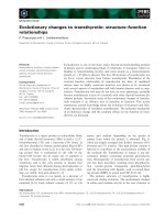
(E)-B-D-C-A (see Fig. 1A). Motif E was only found in
TLPs from plant species and from two alphaproteo-
bacteria: Bradyrhizobium japonicum and Magnetospiril-
lum magnetotacticum. Motif E is homologous to
the proteins of cluster of orthologous groups (COG)
3195, a group of bacterial proteins where the entire
protein is made up of this single domain. Motif E
has been subsequently identified as a unique protein,
2-oxo-4-hydroxy-4-carboxy-5-ureidoimadolazine (OHCU)
decarboxylase, whose function relative to TLP will be
discussed later in this review.
A combined set of TLP and transthyretin sequences
was also probed for motifs to determine whether there
were any motifs in common between the two protein
families. Three motifs (A’–C’), which highlighted
regions of similarity between TLP and transthyretin
sequences, were identified and found in the arrange-
ment B’-C’-A’ (see Fig. 1B). These motifs were shown
The evolution of the transthyretin-like protein S. C. Hennebry
5368 FEBS Journal 276 (2009) 5367–5379 ª 2009 The Authors Journal compilation ª 2009 FEBS
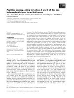
to correspond to regions of structural significance in
the transthyretin molecule (see Fig. 1C). Motif A’ cor-
responds to residues that line the hydrophobic core of
the transthyretin tetramer. Motif B’ corresponds to
residues forming the dimer–dimer interface and resi-
dues in motif C’ are involved in monomer–monomer
interactions (see Fig. 1C). Based on these observations,
it was hypothesized that TLP probably has a tertiary
structure similar to that of transthyretin [11]. The
motifs identified in this study also provided a more
accurate means of differentiating between TLP and
transthyretin sequences and for the identification of
novel TLP ⁄ transthyretin sequences through Hidden
Markov searches in protein databases [11].
Interestingly, whilst motifs A’–C’ represent the
regions of greatest sequence similarity between TLP
and transthyretin, they also contain specific amino acid
substitutions that enabled the distinction of one group
from the other. For instance, at their C-termini (motif
A’ region), nearly all TLP sequences possess a Tyr-
Arg-Gly-Ser tetrapeptide. Specifically, the tyrosine and
glycine residues were found to be 100% conserved
among TLP sequences. Upon sequence alignment with
TLP, the residues at the same positions in transthyretin
are threonine and valine, respectively. At the N-termini
of TLP sequences (the motif B’ region) a conserved his-
tidine residue was found. The equivalent residue in
transthyretin sequences is lysine (also 100% conserved).
Interestingly, the residues involved in TH binding in
transthyretin are not conserved in TLP sequences.
Rather, it appears that residues involved in the struc-
tural integrity of the TLP ⁄ transthyretin molecule have
been conserved. The alignment of representative trans-
thyretin and TLP sequences in Fig. 2 demonstrates the
distribution of residues that are 100% conserved in
both TLP and transthyretin sequences as well as those
that are 100% conserved solely within the set of TLP
sequences.
Distribution of TLPs and transthyretins
in nature
The distribution of TLP in nature and its evolutionary
relationship to transthyretin have been studied exten-
sively in recent years [10,11]. To date, TLP genes have
been identified in over 200 organisms across all king-
A
B
C
Motif A′
A′
ACDB
C′
~ 127 amino acids
~ 114 amino acids
B′
Motif B′
Motif C′
Fig. 1. Motifs common between TLP and
transthyretin indicate conservation of the
TLP ⁄ TTR structure through evolution. Motifs
identified in (A) TLP sequences and (B)
transthyretin+TLP sequences. (A) In the set
of TLP sequences, four motifs were identi-
fied (A–D). The motifs are found in the order
B-D-C-A, with A being the most highly con-
served. (B) In the set of transthyretin+TLP
sequences, three motifs were identified,
A’–C’. Motif A’ is equivalent to motif A from
the TLP motif set. Motif B’ is similar but
extended in the N-terminal and C-terminal
regions to motif B. Motif C’ is shorter than
motif C and its location is shifted towards
the N-terminus. Motif D is specific to the
TLP set of proteins. (C) Motifs A’–C’ were
superimposed on the tertiary structure of
sea bream transthyretin. Motif A’ lines the
hydrophobic core. Motif B’ forms the
dimer–dimer interface and the opening of
the central channel of the TTR molecule.
Residues in motif C’ are involved in mono-
mer–monomer interactions. (Modified from
[11]).
S. C. Hennebry The evolution of the transthyretin-like protein
FEBS Journal 276 (2009) 5367–5379 ª 2009 The Authors Journal compilation ª 2009 FEBS 5369
doms. By contrast, the transthyretin gene is only found
in vertebrates. Whilst the TLP gene is widely distributed
in nature, there are some notable absences or apparent
‘losses’ of the TLP gene. For instance, no protozoans to
date have been found to have a TLP gene, even though
related organisms such as the slime mold Dictyosteli-
um discoideum and the jakobite Jakoba bahemiensis
both express the TLP gene. A TLP gene is absent from
the cnidarian and ascidian phyla, despite the fact that
organisms before and after these branch points in evo-
lution express the TLP gene. This evidence suggests that
whilst TLP might have been conserved throughout evo-
lution because they have an important functional role,
it is by no means essential to all organisms.
Subcellular localization of TLP in
bacteria
In most instances, the TLP gene is present as a single
copy in the organisms in which it has been identified.
The gene typically encodes a cytoplasmic protein and,
in the case of bacteria, is typically located in purine
metabolism operons, neighbouring the gene which
encodes OHCU decarboxylase [11]. This is consistent
with the recently determined role of TLP in this meta-
bolic pathway (to be discussed later). A notable excep-
tion to this is the case of the enterobacterial TLP
genes and a handful of TLP genes from other Gram-
negative bacteria. The TLPs from these bacteria have
been found to possess an N-terminal extension, namely
a periplasmic localization sequence [11]. Interestingly,
these TLP genes are not found to be associated with
purine metabolism operons [11], and it is therefore
tempting to speculate that their primary function is
not purine metabolism.
Some organisms have multiple copies of a TLP gene
(see Table 1) [11]. In these cases, one gene encodes a
cytoplasmic TLP and the ‘additional’ TLP gene
encodes a periplasmic protein that, similarly to entero-
bacterial TLP genes, is not associated on the bacterial
chromosome with genes encoding proteins involved in
purine metabolism. Indeed, phylogenetic analyses of
all periplasmic TLP sequences (S.C. Hennebry, unpub-
lished results) suggests that the genes encoding these
TLPs were probably obtained through horizontal gene
transfer from an enterobacterial ancestor.
Subcellular localization of TLPs in
eukaryotes
In most nonfungal eukaryotic TLP sequences exam-
ined to date, an N-terminal extension has also been
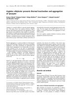
Fig. 2. Alignment of representative transthyretin and TLP sequences. Mature amino acid sequences for transthyretin and TLP from selected
organisms are shown (i.e. with signal peptides removed). The shared secondary structure characteristics of transthyretins and TLPs are indi-
cated above the alignment: motifs A’–C’ are indicated with straight lines and are labelled; b-strands are indicated with arrows and are
labelled A–H. A single a-helix is indicated with a rectangle. The residues that are strongly conserved between transthyretins and TLPs are
indicated with an asterisk (*). Residues 100% conserved among all TLP sequences are indicated with a hash (#). Numbering for human
transthyretin is shown directly beneath the alignment.
The evolution of the transthyretin-like protein S. C. Hennebry
5370 FEBS Journal 276 (2009) 5367–5379 ª 2009 The Authors Journal compilation ª 2009 FEBS
identified. This N-terminal extension contains a nona-
peptide, which is predicted to encode a type-two
peroxisomal sequence (PTS2) [11,12]. Recently, a
proteomic analysis of leaf peroxisomes confirmed the
peroxisomal localization of the Arabidopsis thaliana
TLP [13]. Found in all eukaryotic cells, peroxisomes
are specialized organelles in which oxidative reactions,
such as those associated with purine metabolism, are
compartmentalized. The co-localization of purine-
metabolism enzymes (e.g. uricase) with TLP in peroxi-
somes is therefore in keeping with the function of the
A. thaliana TLP hydrolysis of the purine 5-HIU (S. C.
Hennebry, unpublished results). These observations
contradict those made by Nam and Li [14], where the
A. thaliana TLP was reported to be localized only in
the cytosol and was unlikely to have a function in pur-
ine metabolism. In this study, the authors failed to
take into account that the A. thaliana gene At5g58220
encoded two distinct proteins: OHCU decarboxylase
and TLP. Therefore, conclusions drawn from yeast
two-hybrid studies were based on interactions of the
N-terminal region of OHCU decarboxylase with the
receptor kinase brassinosteroid-insenitive-1, rather than
interactions made by TLP. Furthermore, their conclu-
sion that the TLP could not be peroxisomal was
largely based on the observation that the TLP did not
possess a C-terminal peroxisomal targeting sequence.
Splice variants have been detected for most eukary-
otic TLP genes and some of these variants result in the
truncation of the TLP at the N-terminus. This trunca-
tion has no effect on amino acid residues known to be
involved in the function of the protein, but result in
the deletion of the PTS2 nona-peptide. In the case of
Mus musculus, transcript data available at RIKEN
Mouse Encyclopedia (genome.gsc.riken.go.jp) suggest
that over 90% of TLP gene transcripts possess the
region encoding the PTS2 and were isolated from
hepatocytes. A small proportion of TLP transcripts
(< 10%) do not encode the PTS2 and appear not to
be under tissue-specific regulation. Splice variations
resulting in deletion of the PTS2 have also been
described for plant TLPs [11].
All TLP sequences identified in the Viridiplantae
kingdom are encoded by multiple exons [11]. For
example, the TLP gene from A. thaliana is encoded by
four exons, the last of which encodes the TLP. As pre-
viously mentioned, exons 1–3 (motif E) encode a pro-
tein from COG 3195, which was recently identified as
the enzyme OHCU decarboxylase [12,15]. The
functional relationship between TLP and OHCU
decarboxylase will be discussed below.
Evidence for gene duplication
The most primitive organisms found to have a trans-
thyretin sequence are the lampreys Petromyzon marinus
and Lampetra appendix [16]. By contrast, TLP genes
have been identified in all kingdoms. Given their high
degree of sequence similarity, it has been hypothesized
that the transthyretin gene arose as a result of a dupli-
cation of the TLP gene at some stage in early verte-
brate evolution [11]. Initial phylogenetic analyses of
TLP and transthyretin sequences showed a branching
of transthyretin slightly before the separation of the
chordates [17]. Subsequent analyses using the recently
determined transthyretin sequences from lamprey and
recent additions to echinoderm expressed sequence tag
(EST) databases, suggest that the TLP gene duplica-
tion probably occurred just after the separation of
echinoderms (S. C. Hennebry, unpublished results).
Table 1. Bacteria with multiple copies of TLP genes.
Organism Taxonomy (phylum, class)
Genes encoding
cytoplasmic TLP
Genes encoding
periplasmic TLP
Rhodococcus Actinobacteria, Actinobacteria 2 0
Bradyrhizobium sp. Proteobacteria, Alphaproteobacteria 2 0
Sinorhizobium meliloti Proteobacteria, Alphaproteobacteria 2 0
Dinoroseobacter shibae DFL 12 Proteobacteria, Alphaproteobacteria 2 0
Loktanella vestfoldensis SKA53 Proteobacteria, Alphaproteobacteria 2 0
Roseovarius sp. HTCC2601 Proteobacteria, Alphaproteobacteria 2 0
Ralstonia eutropha H16 Proteobacteria, Betaproteobacteria 2 1
Comamonas testeroni KF-1 Proteobacteria, Betaproteobacteria 2 1
Klebsiella pneumoniae Kp342 Proteobacteria, Gammaproteobacteria 1 1
Salmonella enterica ssp. I choloraesuis Proteobacteria, Gammaproteobacteria 0 2
Chromohalobacter salexigens DSM3034 Proteobacteria, Gammaproteobacteria 1 1
Acinetobacter sp. (strain ADP1) Proteobacteria, Gammaproteobacteria 1 1
Pseudomonas fluorescens Pf5 ATCC BAA-477 Proteobacteria, Gammaproteobacteria 1 2
S. C. Hennebry The evolution of the transthyretin-like protein
FEBS Journal 276 (2009) 5367–5379 ª 2009 The Authors Journal compilation ª 2009 FEBS 5371
Following the gene-duplication event, profound
modifications to the duplicated TLP occurred, leading
to the development of a deep channel into which the
THs 3¢,3,5-triiodo-L-thyronine (T3) and 3¢,5¢,3,5-tetra-
iodo-L-thyronine (thyroxine, T4) could bind. The
nature of this structural modification will be discussed
below.
The function of TLP in purine
metabolism
To date, three studies have been performed examining
the role of TLP in vivo. In a study of the A. thaliana
TLP, no phenotype was observed when an insertional
mutation was introduced into the TLP gene [14]. How-
ever, the lack of phenotype observed may be attributed
to the presence of an additional 5-HIU hydrolase in
plants (see later discussion regarding TLP functional
redundancy). In 2003, Eneqvist et al. [10] performed
RNA interference (RNAi) studies in C. elegans to
determine a loss-of-function phenotype for R09H10.3
and ZK697.8 TLP genes. RNAi-treated worms were
scored for embryonic lethality and for postembryonic
phenotypes (sterility, aberrant morphology, uncoordi-
nated movements, egg-laying defects or slow growth).
No obvious phenotype was detected upon examination
of the gross phenotype of the worms using a dissecting
microscope [10]. However, more in-depth examination
into a possible phenotype was not performed. For
example, the worms were not subjected to any type of
environmental stress. In addition, RNAi was per-
formed using dsRNA for a single TLP gene at a time.
As such, the RNAi studies in C. elegans may have
been more informative had double-knockdown studies
been performed.
A role for TLP in purine metabolism was first pro-
posed in 2001. In an effort to develop a greater under-
standing of purine metabolism in the Gram-positive
bacterium, Bacillus subtilis, Schultz et al. [18] generated
a series of insertion mutants. One of these mutations
was made in the TLP gene (pucM), which is located
immediately downstream of the gene encoding uricase.
The bacteria harbouring this mutation were character-
ized as having a reduced rate of proliferation (com-
pared with wild-type bacteria) on media containing
uric acid as the principal source of nitrogen [18].
Purines are major components of nucleic acids and
nucleotides. Subsequently, de novo and salvage path-
ways for purine biosynthesis are important compo-
nents in the metabolism of all organisms. The ability
to degrade purine compounds, either aerobically or
anaerobically, has been identified in all kingdoms [19].
The aerobic degradation of purines is dependent on
the oxidation of hypoxanthine and xanthine to uric
acid via xanthine dehydrogenase ⁄ oxidase (E.C.
1.1.1.204 ⁄ E.C. 1.1.3.22). In humans, anthropoid apes,
birds, uricotelic reptiles and most insects, uric acid is
the end product of purine metabolism and is thus
excreted [20,21]. Most mammals and gastropods fur-
ther degrade uric acid to allantoin [20,22], fish and
amphibians completely degrade purines to urea,
ammonia and carbon dioxide [20,23,24], whilst most
plants degrade purines to carbon dioxide and ammonia
[25].
Purine oxidation, in particular that of uric acid, is
the major route of ureide biogenesis in nature. Conse-
quently, the enzymes involved in the various stages of
purine metabolism have been the focus of much inves-
tigation. Recently, however, the degradation of uric
acid to allantoin has been shown to be more complex
than originally thought. Previously, it had been
assumed that uricase (EC 1.7.3.3) was the sole enzyme
responsible for the oxidation of uric acid to allantoin.
However, Tipton’s group [26] showed that the oxida-
tion of uric acid by uricase in fact yields the metastable
compound, 5-HIU. They observed the spontaneous
decomposition of 5-HIU to OHCU within 20 min at
neutral pH, followed by the spontaneous decarboxyl-
ation of OHCU to racemic allantoin. The spontaneous
decomposition of 5-HIU results in the generation of
numerous free-radical species, which ultimately con-
tribute to lipid oxidation [27]. Given this fact and the
observation that only (S)-allantoin is found in nature,
Kahn and Tipton [26] proposed the existence of addi-
tional enzymes in the uric acid degradation pathway –
first to hydrolyse 5-HIU and second to decarboxylate
OHCU to (S)-allantoin.
As previously discussed, bacterial TLP genes are fre-
quently found in close proximity to the uricase gene and
to another gene encoding proteins belonging to COG
3195. In 2005, Lee et al. [28] revealed the ability of
recombinant TLP from B. subtilis and E. coli to specifi-
cally hydrolyse 5-HIU. Importantly, they demonstrated
the inability of human transthyretin to hydrolyse the
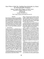
same compound. Ramazzina et al. [12] subsequently
showed that mouse TLP hydrolysed 5-HIU and that the
COG 3195 proteins were responsible for the decarboxyl-
ation of OHCU to (S)-allantoin. Thus, the pathway of
the conversion of uric acid to (S)-allantoin via the three
enzymes uricase, TLP (5-HIUase) and OHCU decar-
boxylase was revealed (see Fig. 3). Whether the three
proteins are able to form a multi-enzyme complex
remains to be determined. One could speculate that the
ability to do so would be favourable given the rapid
kinetics of spontaneous decomposition of both 5-HIU
and OHCU.
The evolution of the transthyretin-like protein S. C. Hennebry
5372 FEBS Journal 276 (2009) 5367–5379 ª 2009 The Authors Journal compilation ª 2009 FEBS
To date, the TLP from three bacteria [28–30], one
plant (A. thaliana; S. C. Hennebry, unpublished
results) and two vertebrate species [12,17], have been
analysed for 5-HIU hydrolytic activity and have all
been shown to be 5-HIU hydrolases. Thus, a role for
TLP in this purine degradation pathway is evident
throughout evolution. In addition, the expression of
the TLP gene in some organisms may be uric acid-
dependent. For example, in the Gram-positive bacte-
rium Deinococcus radiodurans, both the uricase and
TLP genes are regulated by a novel uric acid-respon-
sive transcriptional regulator of the MarR family [31].
Given the similarities in the structures of purine
metabolism operons among Gram-positive bacteria, it
is likely that both uricase and TLP genes are similarly
regulated in other bacteria.
Interestingly, periplasmic TLPs (those from the
Enterobacteria) have also been demonstrated to have
5-HIU hydrolase activity [28,30]. Given that in bacteria,
purine metabolism is localized in the cytosol, it is pos-
sible that the TLP from these organisms acts indepen-
dently of the classical purine catabolism pathway. In
addition, no enterobacteria have been found to possess
homologs of OHCU decarboxylase or uricase genes.
Therefore, the question arises as to the in vivo role of
periplasmic TLP and whether it is capable of hydroly-
sing compounds other than 5-HIU.
The fact that TLP has been demonstrated to hydro-
lyse 5-HIU results in its inclusion in the superfamily of
cyclic amidohydrolases (E.C. 3.5.2). Other cyclic
amidohydrolases include hydantoinase, allantoinase
and dihydrooratase [32]. Cyclic amidohydrolases share
a number of physicochemical characteristics. These
characteristics include quaternary, tertiary, secondary
and primary structure as well as the reliance on a diva-
lent metal cofactor via a conserved metal-binding
motif [33]. Studies have also shown the inhibitory
action of some divalent cations on cyclic amidohydro-
lase activity as well as the ability of many enzymes
within this group to bind a variety of cyclic amides
with varying affinities [32]. TLP does not appear to
share the classic sequence characteristics of cyclic
amidohydrolases (S. C. Hennebry, unpublished
results). Whilst the E. coli TLP was crystallized in the
presence of Zn
2+
, it has been shown that TLP is not a
zinc-dependent hydrolase [17].
Structural comparison of Transthyretin
and TLP
The 3D structures of transthyretin from various organ-
isms have been well characterized. The first transthyre-
tin crystal structure to be solved (that of human) was
published in 1978 [34]. The Protein Database (http://
www.pdb.org) contains multiple crystal structure coor-
dinates for human transthyretin (including multiple
amyloidogenic forms and with various ligands bound).
The crystal structures of transthyretin from rat [35],
chicken [36] and sea bream [37,38] have also been
solved. All of these structures demonstrate the remark-
able conservation of the prealbumin-like fold (as
described by SCOP, ),
which consists of an eight-stranded b-sandwich (strands
A-H) with each sheet adopting a greek-key topology. A
two-turn a-helix usually (with the exception of chicken
transthyretin) exists between strands E and F in trans-
thyretin. The two transthyretin dimers associate, via
nonpolar interactions, between the loops joining stands
G and H with the loops joining strands A and B, mak-
ing the transthyretin tetramer a ‘dimer of dimers.’
Recently, the first crystal structures of TLP from
various organisms were solved. Within a short period
of 3 months, the crystal structures for the TLP from
S. dublin (pdb: 2GPZ; [30]), E. coli (pdb: 2G2N; [39]),
B. subtilis (pdb: 2H0E; [29]) and Danio rerio (zebrafish;
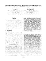
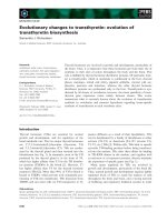
pdb: 2H6U; [17]) were solved. Remarkably, the struc-
tures of these proteins, all tetrameric, showed signifi-
cant similarity to the published structures of
transthyretin. By way of example, a comparison of the
structure of S. dublin TLP with the structures of trans-
thyretin from various organisms is shown in Figure 4.
Generally, the structural deviation between TLPs and
transthyretins from various organisms is of the same
order of magnitude to that within the set of transthyre-
Fig. 3. Schematic of the oxidation of uric acid. Uric acid is oxidized by uricase to 5-HIU, which is subsequently hydrolysed by TLP (5-HIU
hydrolase) to OHCU. The enzyme OHCU decarboxylase generates (S)-allantoin. (Adapted from [26]).
S. C. Hennebry The evolution of the transthyretin-like protein
FEBS Journal 276 (2009) 5367–5379 ª 2009 The Authors Journal compilation ª 2009 FEBS 5373
tin. For instance, the rmsd between equivalent Ca
atoms in the structures of TLPs and human transthy-
retin are 1.0 A
˚
and 1.2 A
˚
for the monomer and dimer
respectively [17]. The rmsd between equivalent Ca
atoms in the structures of transthyretin from various
vertebrates is between 0.34 A
˚
and 1.59 A
˚
[30].
The main differences between the structures of TLP
and transthyretins are found in the loop connecting
b-strands B and C, which is highly exposed to the
solvent in TLP [17]. Interruptions in the b-strands A, G
and H are also observed in TLP structures as a result
of alterations to the formation of hydrogen bonds
between strands. The carbonyls of residues V104 and
P105 (zebrafish TLP numbering), in the middle of
b-strand G, do not form hydrogen bonds with the nitro-
gen atoms of H12 and Y116 of b-strand H in TLP.
The P105 residue, mainly responsible for the b-strand
irregularities, is invariant in TLP sequences, suggesting
a crucial role for the particular conformation observed
in b-strands A, G and H [17].
Structural nature of the TLP and
transthyretin active sites
One of the striking features of transthyretin is the cen-
tral channel of the protein into which the THs bind.
This central channel traverses the entire tetramer. It
has previously been postulated [40] and demonstrated
[7,41] that the characteristics of the N-termini of trans-
thyretin from different organisms account for differ-
ences in the affinity of the two main THs (T3 and T4)
to the channel by hindering or allowing greater accessi-
bility.
The central channel is also present in TLP, albeit
with quite different structural properties. Previously, it
was demonstrated that the regions of greatest similar-
ity between TLP and transthyretin were those forming
this central channel, namely motifs A’ and B’ (see
Fig. 1C). Interestingly, differences between TLP and
transthyretin within these motifs also account for sig-
nificant physicochemical alterations to the central
channel of the protein and provide a structural basis
for the differing function compared with transthyretin.
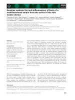
The presence of a conserved, bulky tyrosine residue at
the C-termini of TLP (part of the Tyr-Arg-Gly-Ser tet-
rapeptide) causes the central channel to become
blocked (see Fig. 5A). As a result, the dimer–dimer
interface of TLP is characterized by two ‘grooves’ on
either side of the protein rather than a central channel
[30].
Other key residues at the dimer–dimer interface of
TLP include H14, R49 and H106 (B. subtilis TLP
numbering) [29]. An examination of the active site of
B. subtilis TLP with the uric acid analogue 8-azaxan-
thine bound, reveals that these residues form impor-
tant interactions with the ligand (see Fig. 5B). Indeed,
site-directed mutagenesis studies targeting these resi-
dues show that substitution at these sites has profound
consequences for the 5-HIU hydrolase activity of the
TLP [17,29,30] (and see Table 2).
Mutagenesis of H14 and R49 showed that these resi-
dues are the most sensitive to mutation, with H14A,
H14N and R49E substitutions abolishing enzyme
activity (B. subtilis numbering) [29,30]. However, the
conservative substitution at residue 49 from arginine
to lysine had no effect on activity. This suggests the
need for a positively charged residue at this site. Sub-
stitution of H105 and Y118 also significantly reduced
enzyme activity, by approximately 90% [30]. Deletion
of the C-terminus tetrapeptide Tyr-Arg-Gly-Ser signifi-
cantly affected enzyme activity, but it has been sug-
gested that S121 does not influence the reaction [29].
Fig. 4. Comparison of the tertiary structure
of TLP with transthyretin. Stereo diagram
showing a superimposition of tetramers of
Salmonella dublin TLP (magenta) with trans-
thyretin from human (1F41, cyan), rat (1KGI,
yellow), chicken (1TFP, orange) and sea
bream (1SNO, green). Tetramers were
superimposed using the A chain only.
(Adapted from [30]).
The evolution of the transthyretin-like protein S. C. Hennebry
5374 FEBS Journal 276 (2009) 5367–5379 ª 2009 The Authors Journal compilation ª 2009 FEBS
Interestingly, those residues playing a role in enzyme
activity are 100% conserved in all TLP sequences and
100% substituted in transthyretin (see Table 2). How-
ever, substitutions at position 121 (to threonine or glu-
tamate) have been observed. None of the mutations
affected the tetrameric assembly of the TLP molecule
[30]. Furthermore, the surface charge of the TLP active
site is considerably different from the equivalent region
in transthyretin [29,30]. An electrostatically positive
groove in TLP contrasts the negatively charged
TH-binding site in transthyretin (see Fig. 5C).
In summary, a comparison of the catalytic cavity of
TLP with the equivalent region of transthyretin (the
TH-binding channel) revealed that the TLP cavity is
significantly shallower and ‘groove-like’ compared with
the deep, hollow channel of transthyretin [30]. In par-
ticular, the substitution of the C-terminal tyrosine
(118) with the much less bulky threonine residue fol-
lowing duplication, had a profound effect on the shape
of the channel. Loss of the tyrosine residue opened up
A
B
C
iii
iii
iii
Fig. 5. The active site of TLP. (A) Compari-
son of the ligand-binding cleft at the dimer–
dimer interface in (i) human transthyretin
with (ii) Salmonella dublin TLP. Residues
that contribute to the active site are shown.
Hydrogen bonds are shown as broken cyan
lines. Thyroxine is shown in stick represen-
tation in yellow. For clarity, some elements
of secondary structure are not shown. Resi-
dues His6, His95 and Y108 (S. dublin TLP
numbering) are equivalent, upon structural
alignment, to Lys15, Thr106 and Thr119 of
human TTR. (Adapted from [30].) (B) The
active site of B. subtilis TLP with (i) the uric
acid analog, 8-azaxanthine bound and (ii)
showing interacting residues (from [29]).
Note that the active site of the B. subtilis
TLP is depicted at 90° to those depicted for
transthyretin and TLP in part A. (C) (i) Elec-
trostatic surface potential of human trans-
thyretin with thyroxine bound inside the
negatively charged and deep channel at the
dimer–dimer interface of the protein. (ii) The
equivalent region in TLP is shallow and
positively charged. (Adapted from [30]).
Table 2. Site-directed mutagenesis of conserved residues in TLP.
Transthyretin
residue
Equivalent
residue in TLP
(S. dublin TLP
numbering)
Effect of mutation
on TLP activity Publication
Lys15 His6 Abolishes [30]
Ser52 Asp42 Reduces by 50% [17]
Glu53 Arg44 Abolishes [29]
Thr106 His95 Reduces by 90% [30]
Thr119 Tyr108 Reduces by 90% [30]
Val122 Ser111 No effect [29]
S. C. Hennebry The evolution of the transthyretin-like protein
FEBS Journal 276 (2009) 5367–5379 ª 2009 The Authors Journal compilation ª 2009 FEBS 5375
the central channel of the transthyretin molecule,
allowing for the binding of bulkier ligands such as
THs. Superimposition of the dimer–dimer interface of
TLP with that of transthyretin illustrates the evolu-
tionary changes that resulted in the functional transi-
tion of the enzyme into a transport protein.
Comparison of structures of TLPs from
various organisms
A comparison of the TLP from three species of bacte-
ria with a vertebrate TLP (zebrafish) shows little struc-
tural divergence. Major differences between the
S. dublin TLP and zebrafish TLP are found in the flex-
ible portions of strands B and C that protrude towards
the solvent and in the conformation of the long loop
connecting strands D and E [17]. Greater differences
are observed between the structures of B. subtilis and
zebrafish TLPs: loop B-C is significantly shorter in
B. subtilis TLP whilst the loop connecting the short
a-helix to strand F is extended.
The active sites of TLPs from prokaryotes and
eukaryotes are nearly identical. The location and ori-
entation of the residues present in the catalytic pockets
are well maintained, including the putative main cata-
lytic residues H12 and R52 (zebrafish TLP numbering).
The only significant difference is found in the C-termi-
nal serine residue, which assumed different orientations
in the three structures. However, the role of this resi-
due in catalysis has been shown to be negligible [29].
Evolution of TLP function in the
context of urate metabolism
Ramazzina et al. [12] eloquently demonstrated the
co-evolution of the three proteins [uricase, TLP
(5-HIUase) and OHCU decarboxylase] involved in the
oxidation of uric acid to allantoin. Certainly, the
co-localization of these proteins in the peroxisomes of
metazoan and plant species, and the co-regulation of
TLP genes in some bacteria, suggests a concerted
effort in the rapid generation of allantoin. The co-dis-
tribution of uricase, TLP and OHCU decarboxylase
genes in nature reveals that whenever an organism is
found to have a uricase gene, it always has both TLP
and OHCU decarboxylase genes [12]. In vertebrates,
the loss of these three genes through evolution is mir-
rored. For instance, hominoids lost their ability to
degrade uric acid as the result of the inactivation of
the uricase gene in a primate ancestor, some 15 Ma
[42]. In humans, the TLP gene has several inactivating
mutations and the OHCU decarboxylase gene does
not appear to be expressed [12].
Uric acid is a potent antioxidant in biological sys-
tems. Despite uric acid being the end point of purine
metabolism in humans and birds, high levels of allan-
toin have been detected in their plasma [43,44]. Uric
acid chelates transition metal ions (minimizing metal-
catalysed oxidation), scavenges hypochlorous acid, is a
potent quencher of peroxynitrite and reduces haemo-
globin oxidation by nitrite (for a review, see [45]). It
has been suggested that in humans and birds, the
allantoin generated in these organisms could be a mea-
sure of the levels of oxidative stress [44].
The nonenzymatic oxidation of uric acid generates
5-HIU, just as in the uricase reaction. As previously
discussed, 5-HIU is a highly reactive compound,
which, if left to spontaneously decompose, is capable
of forming numerous free-radical species, which ulti-
mately contribute to lipid peroxidation [27]. Therefore,
the rapid elimination of 5-HIU would be advantageous
to the organism. Whilst birds have lost functional uri-
case and OHCU decarboxylase gene products, TLP
transcripts have been detected. It is tempting to specu-
late that the role of TLP in birds might be to rapidly
‘mop-up’ 5-HIU generated through the nonenzymatic
oxidation of uric acid, thereby reducing the potential
free-radicals generated when 5-HIU is left to spontane-
ously decompose. The role of TLP in scavenging
5-HIU clearly warrants further investigation.
Purine metabolism occurs in the cytosol of bacteria
(for a review, see [46]). The fact that most bacteria
possess a cytosolic TLP is consistent with this. How-
ever, it is not clear what the functional role of a TLP
localized to the periplasm might be. It is possible that
the source of 5-HIU to the periplasm could be from
the external environment. Interestingly, all bacteria
which possess a periplasmic TLP are found to colonize
various animals. Uric acid is secreted on the surface of
mucosal epithelial tissues of all animals as part of the
innate immune system [47] and is also thought to act
as a microbicidal agent in these instances. Because uric
acid can easily permeate the outer membrane of these
bacteria, it might be that the TLP located in the peri-
plasm acts as a primary defence for the bacterium
against oxidized uric acid. Alternatively, it could be
that 5-HIU is generated in small quantities by the non-
specific oxidation of the uric acid by other periplasmic
enzymes, such as cytochrome c or peroxidase [48,49].
TLP: an enzyme with functional
redundancy?
TLP was not the first protein to be identified as having
5-HIU hydrolytic activity. Having hypothesized the
need for additional enzymes to contribute to the oxida-
The evolution of the transthyretin-like protein S. C. Hennebry
5376 FEBS Journal 276 (2009) 5367–5379 ª 2009 The Authors Journal compilation ª 2009 FEBS
tion of uric acid [26], a 5-HIU hydrolase from soybean
(Glycine max) root nodules was purified [50,51]. This
5-HIU hydrolase showed greatest homology to
b-glucosidases (3.2.1.21) (members of the family of
retaining glycosidases) and has quite a different cata-
lytic mechanism to TLP in order to hydrolyse its sub-
strate. The fact that two structurally distinct proteins
have been identified as sharing the same function is
not uncommon in nature [52]. Legumes, such as
soybean, require sophisticated machinery for nitrogen
fixation. Therefore, it is perhaps not surprising that
they have evolved to possess two structurally unrelated
proteins involved in ureide synthesis. The question
remains as to whether this functional redundancy
exists in other plants, or indeed other bacteria, fungi
and metazoan organisms.
Conclusion
The evolution of TLP and transthyretin represents an
intriguing example of divergent evolution. The conser-
vation of catalytic residues at the TLP dimer–dimer
interface, demonstrated to be essential for enzymatic
activity, indicates that it is likely that all TLPs share
5-HIU hydrolytic activity. Following duplication of the
TLP gene in early vertebrate evolution, substitution of
a small number of residues in the active site of TLP
appears to have been sufficient for the acquisition of
new functional properties of the protein whilst its over-
all structure was unchanged. Furthermore, the distri-
bution of TLPs in all kingdoms, but the representation
of transthyretins in vertebrates alone, clearly suggests
that the transthyretin-like fold originally functioned in
purine metabolism. Thus, the evolution of TLP repre-
sents a remarkable example of the divergent evolution
from a hydrolytic enzyme (TLP) to a TH distributor
(transthyretin).
References
1 Power DM, Elias NP, Richardson SJ, Mendes J, Soares
CM & Santos CRA (2000) Evolution of the thyroid
hormone-binding protein, transthyretin. Gen Comp
Endocrinol 119, 241–255.
2 Richardson SJ (2007) Cell and molecular biology of
transthyretin and thyroid hormones. Int Rev Cytol 258,
137–193.
3 Richardson SJ (2002) The evolution of transthyretin
synthesis in vertebrate liver, in primitive eukaryotes and
in bacteria. Clin Chem Lab Med 40, 1191–1199.
4 Richardson SJ, Monk JA, Shepherdley CA, Ebbesson
LO, Sin F, Power DM, Frappell PB, Kohrle J &
Renfree MB (2005) Developmentally regulated thyroid
hormone distributor proteins in marsupials, a reptile
and fish. Am J Physiol Regul Integr Comp Physiol 288,
R1264–R1272.
5 Costa RH, Lai E & Darnell JE (1986) Transcriptional
control of the mouse prealbumin (transthyretin) gene:
both promoter sequences and a distinct enhancer are
cell specific. Mol Cell Biol 6, 4697–4708.
6 Dickson PW, Aldred AR, Marley PD, Bannister D &
Schreiber G (1985) Rat choroid plexus specialises in the
synthesis and the secretion of transthyretin (prealbu-
min). J Biol Chem 261, 3475–3478.
7 Prapunpoj P, Yamauchi K, Nishiyama N, Richardson
SJ & Schreiber G (2000) Evolution of structure, ontog-
eny of gene expression, and function of Xenopus laevis
transthyretin. Am J Physiol Regul Integr Comp Physiol
279, R2026–R2041.
8 Altschul SF, Madden TL, Scha
¨
ffer AA, Zhang Z,
Miller W & Lipman AJ (1997) Gapped BLAST and
PSI-BLAST: a new generation of protein database pro-
grams. Nucleic Acids Res 25, 3389–3402.
9 Nomenclature committee of IUB (NC-IUB) IUB-IU-
PAC joint commission on biochemical nomenclature
(JCBN) (1981) J Biol Chem 256, 12–14.
10 Eneqvist T, Lundberg E, Nilsson L, Abagyan R &
Sauer-Eriksson AE (2003) The transthyretin-related
protein family. Eur J Biochem 270, 518–532.
11 Hennebry SC, Wright HM, Likic V & Richardson SJ
(2006a) Structural and functional evolution of transthy-
retin and transthyretin-like proteins. Proteins: Struct
Funct and Bioinf 64, 1024–1045.
12 Ramazzina I, Folli C, Secchi A, Berni R & Percudani R
(2006) Completing the uric acid degradation pathway
through phylogenetic comparison of whole genomes.
Nat Chem Biol 3, 144–148.
13 Reumann S, Babujee L, Ma C, Wienkoop S, Siemsen
T, Antonicelli GE, Rasche N, Lu
¨
der F, Weckworth W
& Jahn O (2007) Proteome analysis of Arabidopsis leaf
peroxisomes reveals novel targeting peptides, metabolic
pathways and defense mechanisms. Plant Cell 19, 3170–
3193.
14 Nam KH & Li J (2004) The Arabidopsis transthyretin-
like protein is a potential substrate of BRASSINOS-
TEROID-INSENSITIVE 1. Plant Cell 16, 2406–2417.
15 Kim K, Park J & Rhee S (2007) Structural and func-
tional basis for (s)-allantoin formation in the ureide
pathway. J Biol Chem 282, 23457–23464.
16 Manzon RG, Neuls TM & Manzon LA (2007) Molecu-
lar cloning, tissue distribution, and developmental
expression of lamprey transthyretins. Gen Comp Endo-
crinol 151, 55–65.
17 Zanotti G, Cendron L, Ramazzina I, Folli C, Percudani
R & Rodolfo B (2006) Structure of zebrafish HIUase:
insights into evolution of an enzyme to a hormone
transporter. J Mol Biol 363, 1–9.
S. C. Hennebry The evolution of the transthyretin-like protein
FEBS Journal 276 (2009) 5367–5379 ª 2009 The Authors Journal compilation ª 2009 FEBS 5377
18 Schultz AC, Nygaard P & Saxild HH (2001) Functional
analysis of 14 genes that constitute the purine catabolic
pathway in Bacillus subtilis and evidence for a novel
regulon controlled by the PucR Transcription activator.
J Bacteriol 183, 3293–3302.
19 Vogels GD & van der Drift C (1976) Degradation of
purines and pyrimidines in microorganisms. Bacteriol
Rev 40, 403–469.
20 Keilin J (1959) The biological significance of uric acid
and guanine excretion. Biol Rev 34, 265–296.
21 Wu X, Lee CC, Muzny DM & Caskey CT (1989) Urate
oxidase: primary structure and evolutionary implica-
tions. Proc Natl Acad Sci USA 86, 9412–9416.
22 Fujiwara S & Noguchi T (1995) Degradation of purines:
only ureidoglycolate lyase out of four allantoin-degrading
enzymes is present in mammals. Biochem J 312, 315–318.
23 Hayashi S, Jain S, Chu R, Alvares K, Xu B, Erfurth F,
Usada N, Rao MS, Reddy SK & Noguchi T (1994)
Amphibian allantoinase: molecular cloning, tissue distri-
bution and functional expression. J Biol Chem 269,
12269–12276.
24 Mommsen TP & Walsh PJ (1992) Biochemical and
environmental perspectives on nitrogen metabolism in
fishes. Experientia 48, 583–593.
25 Ashihara H & Crozier A (2000) Biosynthesis and
metabolism of caffeine and related purine alkaloids in
plants. Adv Bot Res 30, 117–205.
26 Kahn K, Serfozo P & Tipton P (1997) Identification of
the true product of the urate oxidase reaction. JAm
Chem Soc 119, 5435–5442.
27 Santos CXC, Anjos EI & Augusto O (1999) Uric acid
oxidation by peroxynitrite: multiple reactions, free radi-
cal formation, and amplification of lipid oxidation. Arch
Biochem Biophys 372, 285–294.
28 Lee Y, Lee DH, Kho CW, Lee AY, Jang M, Cho S,
Lee CH, Lee JS, Myung PK, Park BC et al. (2005)
Transthyretin-related proteins function to facilitate the
hydrolysis of 5-hydroxyisourate, the end product of the
uricase reaction. FEBS Lett 579, 4769–4774.
29 Jung D-K, Lee Y, Park SG, Park BC, Kim G-H &
Rhee S (2006) Structural and functional analysis of
PucM, a hydrolase in the ureide pathway and a member
of the transthyretin-related protein family. Proc Nat
Acad Sci USA 103, 9790–9795.
30 Hennebry SC, Law RHP, Richardson SJ, Buckle AM
& Whisstock JC (2006b) The crystal structure of the
transthyretin-like protein from Salmonella dublin, a pro-
karyote 5-hydroxyisourate hydrolase. J Mol Biol 359,
1389–1399.
31 Wilkinson SP & Grove A (2004) HucR, a novel
uric-acid responsive member of the MarR family of
transcriptional regulators from Deinococcus radiodurans.
J Biol Chem 279, 51442–51450.
32 Kim GJ, Lee DE & Kim H-S (2000) Functional
expression and characterisation of the two cyclic
amidohydrolase enzymes, allantoinase and a novel
phenylhydantoinase, from Escherichia coli. J Bacteriol
182, 7021–7028.
33 Kim G-J & Kim H-S (1998) Identification of the
structural similarity in the functionally related
amidohydrolases acting on the cyclic amide ring.
Biochem J 330
, 295–302.
34 Blake CCF, Geisow MJ & Oately SJ (1978) Structure
of prealbumin: secondary, tertiary and quaternary inter-
actions determined by Fourier refinement at 1.8 A
˚
.
J Mol Biol 121, 339–356.
35 Wojtczak A (1997) Crystal structure of rat transthyretin
at 2.5 A
˚
resolution: first report on a unique tetrameric
structure. Acta Biochim Pol 44, 505–517.
36 Sunde M, Richardson SJ, Chang L, Pettersson TM,
Schreiber G & Blake CC (1996) The crystal structure of
transthyretin from chicken. Eur J Biochem 236, 491–499.
37 Folli C, Pasquato N, Ramazzina I, Battistutta R,
Zanotti G & Berni R (2003) Distinctive binding and
structural properties of piscine transthyretin. FEBS
Lett 555, 279–284.
38 Eneqvist T, Lundberg E, Karlsson A, Huang S, Santos
CRA, Power DM & Sauer-Eriksson AE (2004) High-
resolution crystal structures of piscine transthyretin
reveal different binding modes for triiodothyronine and
thyroxine. J Biol Chem 270, 5218–5532.
39 Lundberg E, Backstrom S, Sauer UH & Sauer-Eriksson
E (2006) The transthyretin-related protein: structural
investigation of a novel protein family. J Struct Biol
155, 445–457.
40 Chang L, Munro SLA, Richardson SJ & Schreiber G
(1999) Evolution of thyroid hormone binding by trans-
thyretins in birds and mammals. Eur J Biochem 259,
534–542.
41 Prapunpoj P, Leelawatwatana L, Schreiber G &
Richardson SJ (2006) Change in the structure of the
N-terminal region of transthyretin produces change in
affinity of transthyretin to T4 and T3. FEBS J 273,
4013–4023.
42 Oda M, Satta Y, Takenaka O & Takahata N (2002)
Loss of urate oxidase activity in hominoids and its
evolutionary implications. Mol Biol Evol 19, 640–653.
43 Kaur H & Halliwell B (1990) Action of biologically
relevant oxidising species upon uric acid. Identification
of uric acid oxidation products. Chem-Biol Interact 73,
235–247.
44 Simoyi MF, Falkenstein E, Van Dyke K, Blemings KP
& Klandorf H (2003) Allantoin, the oxidation product
of uric acid is present in chicken and turkey plasma.
Comp Biochem Physiol Pt B 135, 325–335.
45 Ames BN, Cathcart R, Schwiers E & Hochstein P
(1981) Uric acid provides an antioxidant defense in
humans against oxidant- and radical-causing aging and
cancer: a hypothesis. Proc Natl Acad Sci USA 78,
6858–6862.
The evolution of the transthyretin-like protein S. C. Hennebry
5378 FEBS Journal 276 (2009) 5367–5379 ª 2009 The Authors Journal compilation ª 2009 FEBS
46 Reitzer L (2003) Nitrogen assimilation and global regula-
tion in Escherichia coli. Annu Rev Microbiol 57, 155–176.
47 Vorbach C, Harrison R & Capecchi MR (2003)
Xanthine oxidoreductase is central to the evolution and
function of the innate immune system. Trends Immunol
24, 512–517.
48 Howell RR & Wyngaarden JB (1960) On the mecha-
nism of peroxidation of uric acids by hemoproteins.
J Biol Chem 235, 3544–3550.
49 Volk KJ, Yost RA & Brajter-Toth A (1989) On-line
mass spectrometric investigation of the peroxidase-catal-
ysed oxidation of uric acid. Anal Chem 61, 1709–1717.
50 Raychaudhuri A & Tipton P (2002) Cloning and
expression of the gene for soybean hydroxyisourate
hydrolase. Localisation and implications for function
and mechanism. Plant Physiol 130, 2061–2068.
51 Sarma AD, Serfozo P, Kahn K & Tipton P (1999)
Identification and purification of hydroxyisourate
hydrolkasem a novel ureide-metabolising enzyme.
J Biol Chem 274, 33863–33865.
52 Galperin MY, Walker DR & Koonin EV (1998)
Analogous enzymes: independent inventions in enzyme
evolution. Genome Res 8, 779–790.
S. C. Hennebry The evolution of the transthyretin-like protein
FEBS Journal 276 (2009) 5367–5379 ª 2009 The Authors Journal compilation ª 2009 FEBS 5379