Báo cáo khoa học: The nucleosome-binding protein HMGN2 modulates global genome repair potx
Bạn đang xem bản rút gọn của tài liệu. Xem và tải ngay bản đầy đủ của tài liệu tại đây (440.81 KB, 12 trang )
The nucleosome-binding protein HMGN2 modulates global
genome repair
Mangalam Subramanian
1
, Rhiannon W. Gonzalez
1
, Hemangi Patil
1
, Takahiro Ueda
2,
*,
Jae-Hwan Lim
3,
, Kenneth H. Kraemer
2
, Michael Bustin
3
and Michael Bergel
1,3
1 Department of Biology, Texas Woman’s University, Denton, TX, USA
2 Basic Research Laboratory, Center for Cancer Research, National Cancer Institute, National Institutes of Health, Bethesda, MD, USA
3 Protein Section, Laboratory of Metabolism, National Cancer Institute, National Institutes of Health, Bethesda, MD, USA
Introduction
The timely repair of DNA lesions caused by UV irra-
diation is essential for the survival of cells and for the
prevention of cancer. DNA products resulting from
UV irradiation, such as cyclobutane pyrimidine dimers
(CPDs) and pyrimidine(6–4)pyrimidone photoprod-
ucts, are removed from the DNA by a multistep pro-
cess known as the nucleotide excision repair (NER)
pathway. Mutations in the genes coding for compo-
nents of the NER pathway result in severe genetic dis-
orders, such as xeroderma pigmentosum, Cockayne
syndrome, and trichothiodystrophy [1,2]. In the
nucleus of eukaryotic cells, the DNA is packaged into
chromatin, and repair of DNA lesions therefore occurs
within the context of chromatin. The DNA repair rate
in chromatin is slower than that of deproteinized
DNA [3]. The NER process has been recently linked
Keywords
apoptosis; cell cycle; chromatin; DNA repair;
UV irradiation
Correspondence
M. Bergel, Department of Biology, Texas
Woman’s University, PO Box 425799,
Denton, TX 76204-5799, USA
Fax: +1 940 898 2382
Tel: +1 940 898 2471
E-mail:
Present address
*Pharmaceuticals and Medical Devices
Agency, Tokyo, Japan
Department of Biological Science, Andong
National University, Andong 760-749,
Korea
(Received 12 April 2009, revised 17
August 2009, accepted 14 September
2009)
doi:10.1111/j.1742-4658.2009.07375.x
The HMGN family comprises nuclear proteins that bind to nucleosomes
and alter the structure of chromatin. Here, we report that DT40 chicken
cells lacking either HMGN2 or HMGN1a, or lacking both HMGN1a and
HMGN2, are hypersensitive to killing by UV irradiation. Loss of both
HMGN1a and HMGN2 or only HMGN2 increases the extent of UV-
induced G
2
–M checkpoint arrest and the rate of apoptosis. HMGN null
mutant cells showed slower removal of UV-induced DNA lesions from
native chromatin, but the nucleotide excision repair remained intact, as
measured by host cell reactivation assays. These results identify HMGN2
as a component of the global genome repair subpathway of the nucleotide
excision repair pathway, and may indicate that HMGN2 facilitates the
ability of the DNA repair proteins to access and repair UV-induced DNA
lesions in chromatin. Our finding that HMGNs play a role in global DNA
repair expands the role of these proteins in the maintenance of genome
integrity.
Abbreviations
BrdU, bromodeoxyuridine; CPD, cyclobutane pyrimidine dimer; FACS, fluorescence-activated cell sorter; FITC, fluorescein isothiocyanate;
GGR, global genome repair; HAT, histone acetyltransferase; NER, nucleotide excision repair; PI, propidium iodide; TCR, transcription-coupled
repair.
6646 FEBS Journal 276 (2009) 6646–6657 ª 2009 The Authors Journal compilation ª 2009 FEBS
to various factors that remodel and change chromatin
structure. The histone acetyltransferase (HAT) Gcn5
was found to be involved in DNA repair as part of the
STAGA complex [4] and as part of the FCTC com-
plex, which also contains SAP130, a protein homolo-
gous to DDB1 (UV-damaged DNA-binding factor) [5].
Likewise, the HAT CBP ⁄ p300 [6] was also linked to
the NER process. In addition, the ATP-dependent
chromatin-remodeling complexes, such as ACF [7] and
SWI ⁄ SNF [8,9], have been associated with DNA nucle-
otide excision repair. The emerging picture reveals that
modifying the chromatin at the DNA damage site is
an essential step in providing accessibility for the
repair complexes [10–12].
The nucleotide repair pathway is subdivided into
two subpathways, the transcription-coupled repair
(TCR) subpathway, and the global genome repair
(GGR) subpathway [1,2,13]. The TCR subpathway is
involved in repair of the UV irradiation-induced DNA
lesions on the transcribed DNA strands, whereas the
GGR subpathway repairs the damage in the entire
genome. HMGN1, an architectural protein that
remodels chromatin in a nonenzymatic manner, was
found to be involved in the TCR subpathway [14]. The
HMGN1-mediated enhancement of DNA repair in
chromatin was linked to the ability of HMGN1 to
bind to the nucleosomes and unfold chromatin [14].
However, the involvement of HMGNs in the GGR
subpathway has never previously been shown. Further-
more, most of the published data relate to HMGNs as
a family of proteins involved in transcription regula-
tion [15]. The HMGN family comprises structural
proteins that specifically recognize the generic structure
of the 147 bp nucleosome core particle [16,17]. This
family contains several proteins; however, in most spe-
cies, HMGN1 and HMGN2 are the most abundant
family members. Although the overall domain struc-
tures of HMGN1 and HMGN2 are very similar, their
primary sequences differ by almost 40% [17]. In vitro
studies have demonstrated that both proteins bind to
nucleosomes, reduce the compaction of the higher-
order chromatin fibers, and enhance the transcription
potential of chromatin templates [15–17]. HMGNs
may affect the structure and function of chromatin
through several mechanisms. These include competi-
tion with H1 for nucleosomal binding sites [18,19],
facilitating changes in the levels of histone modifica-
tions [20,21], and induction of conformational changes
in the nucleosome itself [22].
Here, we investigated whether the involvement of
HMGNs in NER is only in TCR or also in GGR,
which may indicate that HMGNs’ chromatin-unfold-
ing function in NER is transcription-independent.
Furthermore, we investigated whether, in addition to
HMGN1, other members of the HMGN family play a
role in the repair of UV irradiation-induced DNA
damage. For this purpose, we used wild-type and
gene-targeted chicken lymphoblastoid cells (DT40),
which, like other chicken tissues, contain three HMGN
proteins: HMGN1a, HMGN1b, and HMGN2 [23,24].
HMGN1a has been detected only in chickens, and has
a sequence that is partially homologous to the consen-
sus sequence of vertebrate HMGN1 and HMGN2.
HMGN2 is homologous to the other vertebrate
HMGN2s, whereas the sequence of the chicken
HMGN1b is homologous to the ubiquitous vertebrate
HMGN1. In this study, we focused on HMGN2 and
used cells that lack HMGN2 and null cells for both
HMGN1a and HMGN2 (as HMGN1a is partially
homologous to HMGN2, and therefore it could, in
theory, complement it). As previously described
[25,26], the null DT40 cells lack either HMGN1a or
HMGN2 or both HMGN1a and HMGN2, but they
still contain HMGN1b, a relatively minor component
in most chicken cells. The protein profiles of these cells
differed slightly; however, all lines had normal prolifer-
ation and differentiation rates [25,26].
Although the cells appeared to be normal, it is pos-
sible that their stress response was impaired. There-
fore, chicken cells disrupted for HMGN2 or disrupted
for both HMGN1a and HMGN2 provide a good
model with which to test for functional redundancy
among HMGN variants and the possible role of the
major HMGN proteins in the cellular response to UV
damage.
Here, we show that loss of both HMGN1a and
HMGN2, or of only HMGN2, impairs the rate of UV-
induced GGR. Loss of HMGNs leads to an increase in
both the UV-induced rate of apoptosis and in the level
of checkpoint arrest. In GGR, HMGN2 and
HMGN1a proteins were, for the most part, not redun-
dant in their function, although some additive effects
on cell survival, apoptosis and, mainly, checkpoint
arrest indicated that there is some level of redundancy.
Host cell reactivation assays indicated that HMGNs
do not affect the integrity of the cellular NER machin-
ery. Thus, HMGNs affect the repair of UV-damaged
DNA by altering chromatin.
Results
HMGN null mutants are hypersensitive to UV
irradiation
To investigate the involvement of HMGN variants in
the UV response of DT40 cells, we used cells lacking
M. Subramanian et al. Loss of HMGN impairs DNA repair rate in chromatin
FEBS Journal 276 (2009) 6646–6657 ª 2009 The Authors Journal compilation ª 2009 FEBS 6647
either HMGN2 (clone D108-1), or HMGN1a (clone
8 ⁄ bsr8), or cells lacking both HMGN2 and HMGN1a
(clones Nh43, Bp39, Nh52, and Bp5). The Bp lines
were derived by first deleting the HMGN2 gene, and
the Nh lines were derived by first targeting the
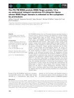
HMGN1a gene [25,26]. We used western analysis to
verify that the targeted genes were indeed disrupted
(Fig. 1). Interestingly, loss of HMGN1a increased the
amounts of HMGN2 (Fig. 1A). In addition, all cells
contained HMGN1b, which is a minor HMGN vari-
ant in chicken cells [23].
Wild-type and mutant DT40 cells were irradiated
with UV doses ranging from 3 to 12 JÆm
)2
. Seventy-
two hours after the irradiation, the viability of cells
was measured by a Trypan blue exclusion assay. As
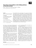
shown in Fig. 2 and Table 1, all of the HMGN null
mutants were significantly more UV-sensitive than
wild-type DT40 control cells. The LD
50
(UV dose
resulting in 50% survival) for the wild-type cells was
9.4 ± 2.33 JÆm
)2
, whereas the LD
50
for the cell vari-
ants lacking HMGN was in the range of 2.63 ± 0.51
to 3.69 ± 0.83 JÆm
)2
. These differences in the LD
50
values between the wild-type cells and all of the
HMGN null cells were found to be significant (non-
parametric Mann–Whitney U-tests, all P-values
< 0.01). The level of UV sensitivity of HMGN2
) ⁄ )
cells (D108-1) (3.69 ± 0.83 JÆm
)2
) was somewhat
lower than that of the other null cells, but a statistical
analysis showed that D108-1 cells were statistically
similar to the other HMGN null cells (all P-values
> 0.127). No major additive or synergistic effect was
observed in the sensitivity of the doubly disrupted cell
lines. These results may indicate that, for the most
part, HMGN2 and HMGN1a function in the same
pathway in conferring UV resistance to cells, as
disrupting each of them alone was sufficient to reduce
the UV tolerance to almost the same level as disrupt-
ing both genes. However, these results cannot rule out
a partial redundancy between HMGN2 and HMGN1a
that contributes to the minor additive effect.
HMGN null mutants have a higher rate of
UV-induced apoptosis
The increased rate of mortality in UV-irradiated cells
is linked to the activation of the apoptotic pathway
[27,28]. To test whether the UV-hypersensitive HMGN
null cells have a higher apoptosis rate, we UV-irradi-
ated the various cell lines with 6 JÆm
)2
, and, 48 h
following UV irradiation, stained control and UV-irra-
diated cells with annexin V and propidium iodide (PI).
Fluorescein isothiocyanate (FITC)-conjugated annex-
in V detects translocation of phosphatidylserine across
membranes, an early apoptotic event, and PI is used to
detect the permeabilization of the plasma membrane,
an event that occurs late in apoptosis. Fluorescence-
activated cell sorter (FACS) analysis of these cells pro-
vided a quantitative measure of the apoptotic events in
the cells. The quadrant analysis of the FACS results
demonstrated that, after UV irradiation, the late and
total apoptosis rates were higher in both HMGN2
) ⁄ )
cells (D108-1) and in the HMGN1a
) ⁄ )
⁄ HMGN2
) ⁄ )
double-knockout clones (Nh43 and Bp5), as compared
with the wild-type DT40 cells (Fig. 3 and Table 2)
(independent group t-test, P < 0.05). In contrast to
this, the early apoptotic rates were lower in the
HMGN null cell lines than in the wild-type DT40 cells
(independent group t-test, P < 0.05). The sum totals
of both early and late apoptotic cells were as follows:
33.7% for the wild-type DT40 cells, 41.7% for the
D108-1 cells, 47.9% for the Nh43 cells, and 58.6% for
DT40
Wild-type
8/bsr8
HMGN1a
–/–
Bp5
Nh43
D108-1
HMGN2
–/–
Nh52
HMGN1a
–/–
;N2
–/–
HMGN1a
HMGN1b
H3
H2A
H2B
H4
HMGN2
DT40
Wild-type
8/bsr8
HMGN1a
–/–
Bp5
Bp39
Nh43
D108-1
HMGN2
–/–
Nh52
HMGN1a
–/–
;N2
–/–
H3
H2A
H2B
H4
A
B
Fig. 1. Loss of HMGN variant expression in DT40 clones with dis-
rupted HMGN genes. (A) Western blot analysis of HMGN2 in
whole cell lysates fractionated by 15% SDS ⁄ PAGE. The cell lines
tested are identified at the top of each lane, and the location of the
HMGN2 protein is indicated by an arrow. The bottom panel shows
Coomassie blue staining of a similar gel to demonstrate equal load-
ing of cell lysates (shown from top are the core histones H3, H2B,
H2A, and H4). (B) Western blot analysis of HMGN1a and HMGN1b
in whole cell lysates fractionated by 18% SDS ⁄ PAGE. The cell lines
tested are identified at the top of each lane, and the locations of
the HMGN1a and HMGN1b proteins are indicated by arrows. The
bottom panel shows Coomassie blue staining of a similar gel to
demonstrate equal loading of cell lysates (shown from the top are
the core histones H3, H2B, H2A and H4).
Loss of HMGN impairs DNA repair rate in chromatin M. Subramanian et al.
6648 FEBS Journal 276 (2009) 6646–6657 ª 2009 The Authors Journal compilation ª 2009 FEBS
the Bp5 cells. These results, taken together, indicate
that cells lacking HMGN proteins had a higher apop-
tosis rate following UV irradiation but also activated
the apoptotic pathway faster, and therefore moved fas-
ter from early to late apoptosis.
Loss of HMGN increases the rate of G
2
–M
checkpoint arrest following UV irradiation
The cellular response to UV radiation is known to
involve not only apoptosis but also cell cycle arrest,
mainly in the G
1
–S or the G
2
–M checkpoints. To test
whether the UV hypersensitivity of cells lacking
HMGNs involves increased activation of one of these
checkpoints, cells were UV-irradiated (12 JÆm
)2
), and,
48 h after irradiation, their cell cycle distribution was
measured as follows. The cells were pulsed for 30 min
with the thymidine analog bromodeoxyuridine (BrdU),
fixed in 70% ethanol, and double-stained with anti-
bodies against BrdU and PI. In FACS analysis, a plot
of BrdU levels against PI levels produces a typical
‘horseshoe’ shape (Fig. 4), in which the G
1
and G
0
cells are represented in the lower left corner of the
plot, and the G
2
–M cells in the right side of the plot.
The cells in S-phase are between these two groups, in
the arch of the ‘horseshoe’, which is high in BrdU.
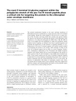
The results reveal that UV irradiation of cells lacking
HMGN2 (D108-1 cells), or lacking both HMGN1a and
HMGN2 (Nh43 cells), decreases the relative number of
cells in S-phase as compared with the more moderate
decrease in the wild-type DT40 cells. The S-phase pop-
ulation significantly decreased in D108-1 cells, from
40.7% to 21.1%, and in Nh43 cells from 48.2% to
14.0% (P < 0.05 by paired t-test), as opposed to an
insignificant decrease in the number of wild-type cells,
from 38.7% to 30.3% (Fig. 4). The decrease in the
number of cells in S-phase was associated with a con-
comitant increase in the population of G
2
–M cells for
the null mutants after UV irradiation (paired t-test,
P < 0.05), and a significant increase in the G
2
–M pop-
ulation of irradiated Nh43 cells in comparison with
irradiated wild-type cells (independent t-test,
P < 0.05). This increase in the G
2
–M population indi-
cates activation of either the G
2
–M checkpoint arrest
or one of the mitotic checkpoints. In order to distin-
guish between these two possibilities, we conducted a
western blot analysis with two antibodies, which served
as specific markers. We used antibody against phos-
phorylated Chk1 Ser345 as a marker for activation of
G
2
–M checkpoint arrest [29]. Antibody against his-
tone H3 phosphorylated on Ser10 was used as a mar-
ker for mitotic cells [30]. The western blot analysis
(Fig. 4C) indicated that the cells lacking HMGN2 and
also the double-null cells lacking HMGN1a and
HMGN2 had a longer arrest time in the G
2
–M check-
point. In the wild-type DT40 cells, there was a sharp
drop in the phosphorylation of Chk1 Ser345 10 h after
UV irradiation. In contrast, in the D108-1 cell line, the
high levels of Chk1 phosphorylation continued to the
24 h point, and in the double-null Nh43 cells, the high
level of phosphorylation remained even 48 h after UV
irradiation. The number of mitotic cells among the
double-null cells was also higher after UV irradiation,
indicating activation of mitotic checkpoints. These
results explain the significantly higher levels of G
2
and
M cells among the HMGN1a
) ⁄ )
⁄ N2
) ⁄ )
cells (Nh43)
that were observed with PI and BrdU double staining
(Fig. 4B). It is important to note that the differences
between the null cell lines D108-1 and Nh43 and the
wild-type DT40 cells cannot be attributed to random
mutations that may have accumulated in these cells,
but only to the lack of HMGN proteins. The reason
for excluding this possibility is that the two null cell
0
20
40
60
80
100
120
140
0 3 6 9
12
UV dose to cells (J·m
–2
)
Cell survival (%)
DT 40
Bp 5 (N1a
–/–
; N2
–/–
)
Nh 43 (N1a
–/–
; N2
–/–
)
D108-1 (N2
–/–
)
8bsr8 (N1a
–/–
)
Fig. 2. Loss of HMGN variants leads to UV hypersensitivity in
DT40 cells. Shown are survival curves of wild-type and mutant
HMGN DT40 cells 72 h after irradiation with various doses of UV.
Each data point represents the mean of three independent
measurements (±standard deviation).
Table 1. LD
50
values of UV-irradiated wild-type DT40 cells and
DT40-derived null HMGN cell lines. The LD
50
values [in
JÆm
)2
± standard deviation] of the wild-type DT40 cells and the
derived null HMGN2 cells (D108-1), null HMGN1a cells (8bsr8) and
HMGN1
) ⁄ )
⁄ N2
) ⁄ )
double-null cells (Bp5 and Nh43) were calcu-
lated on the basis of the experiments presented in Fig. 2 (n ‡ 3).
DT40 D108-1 8bsr8 Bp5 Nh43
9.40 ± 2.33 3.69 ± 0.83
a
2.83 ± 0.35
a
3.03 ± 0.80
a
2.63 ± 0.51
a
a
Significant difference of HMGN null cells from the wild-type cells
as determined by nonparametric Mann–Whitney U-tests (P < 0.01).
M. Subramanian et al. Loss of HMGN impairs DNA repair rate in chromatin
FEBS Journal 276 (2009) 6646–6657 ª 2009 The Authors Journal compilation ª 2009 FEBS 6649
lines were independently derived from DT40 cells;
Nh43 cells were first disrupted for HMGN1a alleles
and then for the two HMGN2 alleles, so they were not
derived from the D108-1 (HMGN2
) ⁄ )
) cells [25,26].
A decreased rate of CPD removal in the context
of chromatin from cells lacking HMGN proteins
The increased rates of apoptosis and checkpoint arrest
in cells lacking HMGNs could be due to an impaired
ability to repair the damaged DNA. To test this
possibility directly, we analyzed the kinetics of CPD
removal in HMGN2
) ⁄ )
cells, in HMGN2
) ⁄ )
⁄ HMGN1a
) ⁄ )
cells, and in wild-type cells, following
UV irradiation. DNA purified immediately after irradi-
ation (time 0), and 7 and 20 h after irradiation, was
slot blotted onto a nylon membrane, probed with anti-
bodies directed against CPDs, and stained with ethidi-
um bromide (Fig. 5A–C). The negative control was
DNA from nonirradiated cells. The CPDs and DNA
were measured within the linear range, based on a
standard curve (data not shown). Following irradia-
tion, there was a gradual decrease in the CPD content
of the DNA of all the cells, an indication of active
repair of the damaged DNA. However, the removal of
CPDs from the chromatin of the HMGN2
) ⁄ )
and
HMGN2
) ⁄ )
⁄ HMGN1a
) ⁄ )
cells was significantly slower
than from the chromatin of wild-type cells (P < 0.05
Table 2. Apoptosis levels following UV irradiation of cells lacking HMGNs and wild-type DT40 cells. The various cell lines were irradiated at
6JÆm
)2
, and 48 h later they were double-labeled with PI and annexin V (see explanations in legend to Fig. 3 and Experimental procedures).
After labeling, the cells were subjected to FACS quadrant analysis. Early apoptotic cells were positively stained with annexin V–FITC, and
late apoptotic cells were positive for annexin V–FITC as well as PI (see more details in Results).
Apoptosis before UV irradiation (%) Apoptosis 48 h after UV irradiation (%)
Cell line Early Late Total Early Late Total
DT-40 2.9 ± 0.7 2.2 ± 0.4 5.1 ± 1.2 10.4 ± 0.9
a
23.3 ± 3.3
a
33.7 ± 3.6
a
D108-1 1.3 ± 0.1 3.3 ± 0.5 4.5 ± 0.5 5.2 ± 1.5
a,b
36.5 ± 1.1
a,b
41.7 ± 2.4
a,b
Nh43 2.1 ± 0.5 6.5 ± 3.4 8.6 ± 3.9 5.4 ± 0.7
a,b
42.6 ± 2.5
a,b
47.9 ± 3.1
a,b
Bp5 1.3 ± 0.3 2.5 ± 0.6 3.9 ± 0.9 6.7 ± 0.4
a,b
51.9 ± 2.0
a,b
58.6 ± 2.0
a,b
a
Early and late phases in each cell line showed a significant difference when compared before and after UV irradiation. This difference was
tested using a paired t-test, and was shown to be significant (P < 0.05).
b
There were significant differences in early, late and total apoptosis
levels after UV irradiation between null HMGN cell lines and the wild-type DT40 cells. These differences were tested using independent
group t-tests, and shown to be significant (P < 0.05).
10 000
10
100
1000
Propidium iodide
Propidium iodide
UV–
UV+
Bp5
(HMGN1a
–/–
/N2
–/–
)
D108-1
(HMGN2
–/–
)
DT40
(WT)
Annexin V–FITC
Nh43
(HMGN1a
–/–
/N2
–/–
)
1
1
10 100 1000 10 000
1
10
100
1000
10 000
Fig. 3. Higher UV-induced apoptosis rate in HMGN2
) ⁄ )
and HMGN1a
) ⁄ )
⁄ HMGN2
) ⁄ )
cells. Early and late apoptosis rates were measured
by annexin V and PI double staining. Wild-type DT40 cells, knockout HMGN2
) ⁄ )
(clone D108-1) cells and double-knockout
HMGN1a
) ⁄ )
⁄ HMGN2
) ⁄ )
(clones Nh43 and Bp5) cells were irradiated with UV at 6 JÆm
)2
. The apoptosis rate was measured 48 h after irradi-
ation. Shown is a dot plot of the cell population as detected by FACS and analyzed by quadrant statistics. The bottom left rectangle repre-
sents live and nonapoptotic cells, which are negative for both annexin V and PI; the bottom right rectangle represents early apoptotic
cells, which are annexin-positive, but PI-negative. The top right rectangle represents late apoptotic and dead cells (annexin V-positive and
PI-positive); the top left rectangle includes dead cells (only PI-positive). This experiment was repeated three times, and the averages are
summarized in Table 1.
Loss of HMGN impairs DNA repair rate in chromatin M. Subramanian et al.
6650 FEBS Journal 276 (2009) 6646–6657 ª 2009 The Authors Journal compilation ª 2009 FEBS
by nonparametric Kruskal–Wallis test). Thus, 7 h after
irradiation, 60% of the CPDs were removed from
wild-type DT40 cells, but less than 20% were removed
from cells lacking HMGN variants (Fig. 5B). After
20 h, the amount of CPDs present in wild-type cells
was 10% of the initial content, whereas in the
HMGN null cells, 40% of the original damage still
remained in the DNA. These results, however, could
also have been obtained if the HMGN null cells had
an initially higher susceptibility to UV irradiation. This
would result in a higher number of CPDs immediately
after UV irradiation, and therefore a slower repair
process, by virtue of the cells having more CPD sites
to repair. For example, lack of the chromatin architec-
tural factor HMGB1 has been previously found to
increase the number of CPDs after UV irradiation
[31]. To test this possibility, we analyzed the
CPD ⁄ DNA ratio at time 0 after UV irradiation in the
wild-type DT40 cells and in the null HMGN cells,
without standardizing these values to 100% (Fig. 5C).
The data indicate that although there are small differ-
ences between the wild-type DT40 cells and the null
cells, these differences are not consistent between the
null cell lines, and they are not statistically significant,
either between the HMGN null cells and DT40 cells,
or between the null D108-1 cells and Nh43 cells (non-
parametric Kruskal–Wallis test, all P-values > 0.275).
These results therefore suggest that HMGNs affect the
rate of repair of DNA damage induced by UV irradia-
tion and not the initial number of CPDs formed by
UV. The repair kinetics in cells lacking only HMGN2
DT-40
Nh43
D108-1
DT-40
Nh43
D108-1
No UV
30 min
4 h
10 h
24 h
48 h
72 h
H3
DT-40
Nh43
D108-1
H2B
H4
H2A
Phospho-Chk1 Ser 345
Phospho-H3 Ser 10
Coomassie
0
10
20
30
40
50
60
70
DT40 DT40
+ UV
D108-1 D108-1
+ UV
Nh43 Nh43
+ UV
% cells
G
1
-G
0
S
G
2
-M
a
a
b
b
c
d
d
e
f
f
c
e
DT
-40
Nh
43
Propidium iodide
600 400 200
1
600 400 200
0
600 400 200 0 0
BrdU
UV+
UV–
27.3
53.6
11.3
27.5
57.5
22.7
21.1
32.6
31.0 33.9
16.8
37.7
10 000
1000
100
10
36.8
57.9
G
1
-G
0
S
D
108-1
1
10 000
1000
100
10
1
G
2
-M
14.6
45.6
22.4
18.9
A
B
C
Fig. 4. UV-induced G
2
–M arrest in HMGN2
) ⁄ )
cells and G
2
–M and
mitotic arrest in HMGN1a
) ⁄ )
⁄ HMGN2
) ⁄ )
cells. Cells (1 · 10
6
cellsÆmL
)1
) were irradiated with 12 JÆm
)2
. Forty-eight hours later,
the cells were labeled with BrdU, fixed, incubated with FITC-conju-
gated antibody against BrdU, stained with PI, and analyzed by
FACS. The results indicate that cells lacking HMGN2 and, to an
even greater extent, cells lacking both HMGN1a and HMGN2 have
a lower rate of transition to S-phase after UV irradiation, and conse-
quently show greater accumulation at G
2
–M. The bar graph (B) rep-
resents the averages of three experiments such as the one
depicted in the dot plots of Fig. 5A. The letters a–d indicate the col-
umns with significant statistical differences as determined by
paired t-test (one-tailed, P < 0.05). The letters e and f indicate the
columns with significant statistical differences as determined by
independent t-test (one-tailed, P < 0.05). (C) The wild-type cell line
DT40, HMGN2
) ⁄ )
cells (D108-1) and HMGN1a
) ⁄ )
⁄ N2
) ⁄ )
cells
(Nh43) were analyzed for the levels and kinetics of G
2
–M check-
point and mitotic checkpoint activation after UV irradiation. The
cells were lysed at various time intervals after UV irradiation at
12 JÆm
)2
. Whole cell extracts were resolved by SDS ⁄ PAGE and
analyzed by western blot. The antibody used to detect the levels of
cells arrested in the G
2
–M checkpoint was antibody against phos-
phor-Chk1 Ser345. The antibody used to detect accumulation of
cells in mitosis was antibody against phospho-H3 Ser10. Equal
loading of proteins was demonstrated by Coomassie staining of a
similar gel.
M. Subramanian et al. Loss of HMGN impairs DNA repair rate in chromatin
FEBS Journal 276 (2009) 6646–6657 ª 2009 The Authors Journal compilation ª 2009 FEBS 6651
were similar to those in cells lacking both HMGN2
and HMGN1a.
Host cell reactivation reveals the integrity of the
NER machinery
Most of the UV-induced damage in DNA is removed
by NER, an evolutionarily conserved pathway that
repairs the damage in the context of cellular chroma-
tin. To determine whether loss of HMGN affected the
activity of proteins in this pathway, we used the host
cell reactivation assay [32,33] (Fig. 6). This assay mea-
sures the repair of a UV-irradiated plasmid containing
the reporter gene for luciferase that was transiently
transfected into various cells. The level of the repair of
the episomal DNA can be estimated from the levels of
luciferase activity in the cellular extracts prepared 48 h
after transfection [32,33]. In this assay, the levels of
luciferase activity recovered from wild-type DT40 cells
were the same as those recovered from DT40 variants
lacking both HMGNs, an indication that the UV-irra-
diated plasmids were repaired at the same rate in these
cell types (Fig. 6B,C). Thus, the UV hypersensitivity in
the DT40 cells lacking HMGNs is not due to a lack of
function in the NER components, but probably to the
direct unfolding activity of HMGNs at the damage
sites, a finding consistent with previous results
obtained with mouse cells lacking HMGN1 [14]. It is
important to note that the chromatin structure of the
transfected plasmid DNA is different from that of the
cellular chromatin [34,35]; therefore, it is conceivable
that the effect of HMGN proteins on the repair of the
transfected plasmid is different from their effect on the
cellular chromatin. However, in the cell line D108-1,
which lacks HMGN2, there was even higher DNA
damage repair activity than in the wild-type control
(Fig. 6A). One possible explanation for this observa-
tion is that D108-1 cells might have an increased
expression level of one or more of the genes involved
in TCR, which is the major NER subpathway detected
by the host cell reactivation.
Discussion
Our main finding is that the nucleosome-binding pro-
tein HMGN2 plays a role in the NER GGR subpath-
way. We found that, in DT40 cells, loss of HMGN2 or
HMGN2 and HMGN1a reduces the rate of CPD
removal from chromatin. Taken together with our
previous finding, that loss of HMGN1 from mouse
embryonic fibroblasts reduces the rate of transcription-
coupled UV repair [14], our present findings indicate
that both HMGN1 and HMGN2 play more general
D108-1DT40 Nh43
No UV
UVC 12 J·m
–2
0
20 h
7 h
Lesions
(Antibody
against CPD)
A
B
C
Fig. 5. Decreased CPD removal rate in cells lacking HMGN vari-
ants. (A) Shown is a southwestern analysis of the CPD removal
rates in the DT40 cells lacking HMGN2 (D108-1) or both HMGN2
and HMGN1a (Nh43) as compared with that of wild-type DT40
cells. DNA was extracted from cells that were not irradiated and
from cells immediately after UV irradiation, and 7 and 20 h after
irradiation with a dose of 12 JÆm
)2
. One microgram of DNA was
loaded per slot in a slot blot system, and transferred to a Hybond-
N+ membrane. The membrane was incubated with monoclonal
antibody against CPD. The CPD levels were normalized against the
DNA levels by staining the membranes with ethidium bromide.
Note the absence of signal in DNA samples that were not exposed
to UV. The CPD ⁄ DNA ratio was determined using densitometry of
the CPD blot. (B) A bar graph representing the averages (±standard
error) of three repetitions of the experiment described in (A). The
CPD ⁄ DNA averages are presented as percentage of the initial level
of CPD ⁄ DNA detected at time 0 (immediately after UV irradiation).
DT40 cells have a significantly more efficient CPD-removal rate, 7 h
and 20 h after irradiation, than D108-1 and Nh43 null cells (P < 0.05
by nonparametric Kruskal–Wallis test). (C) A bar graph presenting
the averages (±standard error) of the CPD ⁄ DNA ratio at time 0
after UV irradiation of three repetitions of the experiment described
in (A) and (B). A nonparametric Kruskal–Wallis test showed that the
wild-type DT0 cells and the null D108-1 and Nh43 cells were not
statistically different from each other (all P > 0.275).
Loss of HMGN impairs DNA repair rate in chromatin M. Subramanian et al.
6652 FEBS Journal 276 (2009) 6646–6657 ª 2009 The Authors Journal compilation ª 2009 FEBS
roles in repair of UV-induced DNA damage in the
context of chromatin. Our previous studies with mouse
embryonic fibroblasts lacking HMGN1 [14] did not
provide information on GGR, as this repair is not effi-
cient in murine cells. Our present studies reveal that
loss of HMGNs reduced the rate of CPD removal not
only from transcriptionally active genes (as was shown
in mice), but also at the global genomic level. Thus,
HMGN proteins affect UV-induced DNA damage
removal, both in TCR and in GGR.
Both the higher apoptosis rate and increased check-
point arrest of HMGN2
) ⁄ )
and HMGN1a
) ⁄ )
⁄ HMGN2
) ⁄ )
cells can be attributed to the lower rate
of removal of CPDs from chromatin. It is well estab-
lished that cells do arrest in various cell cycle check-
points in response to induced DNA damage. Cells
have various mechanisms in place for sensing DNA
damage [10] and switching between alternative
response pathways [36]. After their arrest at the cell
cycle checkpoints, the cells respond either by repairing
the DNA damage or, if the damage is beyond repair,
by activating an apoptotic pathway [37,38]. HMGNs
affect the rate of CPD removal, and in their absence
the removal of CPDs is slower. The lower repair rate
means that a higher CPD ⁄ DNA content will persist
in the cell, resulting in more robust cell cycle arrest
and a higher rate of activation of the apoptotic
pathway following UV irradiation. Interestingly, the
HMGN1a
) ⁄ )
⁄ N2
) ⁄ )
cells (Nh43) were not only
arresting in the G
2
–M checkpoint, but also signifi-
cantly accumulated during mitosis. In contrast, the
null HMGN2
) ⁄ )
cells (D108-1) showed mainly G
2
–M
arrest. A possible explanation might be that in the
HMGN1a
) ⁄ )
⁄ N2
) ⁄ )
cells, there is a leakage of cells
with DNA damage through the G
2
–M checkpoint to
the mitosis phase, and their arrest in the mitotic check-
points. This leakage is indicative of a possible role of
HMGN1a in activation of the G
2
–M checkpoint.
Involvement of HMGN1 in activation of the G
2
–M
checkpoint has also been suggested to occur in mouse
cells. In Hmgn1
) ⁄ )
mouse embryonic fibroblasts there
was no decrease in the level of mitotic cells following
c-irradiation, as opposed to wild-type mouse fibro-
blasts, which showed a drop of 70% in the number of
mitotic cells [39].
In considering the possible molecular mechanisms
whereby HMGNs affect the rate of CPD removal from
damaged DNA, we note that the DT40 cells lacking
HMGN2 and HMGN1a, as well as the murine cells
lacking HMGN1 [14], repair irradiated plasmids with
the same efficiency as wild-type cells. Thus, the host
cell reactivation assays suggest that the known NER
factors are functional and normally expressed in the
cells lacking HMGNs. Further support for this conclu-
sion comes from microarray analysis, which could not
detect changes in the transcription levels of NER-
related genes between Hmgn1
+ ⁄ +
and Hmgn1
) ⁄ )
mouse cells [14]. These results suggest that the
impaired UV repair is not due to a significant change
in one of the components of the NER repair complex,
and that the loss of HMGN does not have significant
effects on the transcription levels of genes coding for
these components. Most likely, the effects of HMGNs
are related to their ability to induce structural changes
ABC
Fig. 6. Intact NER of a luciferase reporter plasmid in DT40 cells lacking HMGN variants. Shown are host cell reactivation assays of wild-type
DT40 cells and cells lacking HMGN variants. (A) Null D108-1 cells (HMGN2
) ⁄ )
) in comparison with wild-type DT40 cells. (B) The null cell line
Nh43 (HMGN1a
) ⁄ )
⁄ HMGN2
) ⁄ )
) in comparison with wild-type DT40 cells. (C) The null cell line Bp5 (HMGN1a
) ⁄ )
⁄ HMGN2
) ⁄ )
) in comparison
with wild-type DT40 cells. Luciferase expression plasmids were irradiated with various doses of UV and then used for transfection of cells.
Cell extracts prepared 48 h after transfection were examined for luciferase activity, an indicator of DNA repair potential of the cells [32].
Each point of irradiation was checked independently in triplicate.
M. Subramanian et al. Loss of HMGN impairs DNA repair rate in chromatin
FEBS Journal 276 (2009) 6646–6657 ª 2009 The Authors Journal compilation ª 2009 FEBS 6653
in chromatin, the substrate of the NER factors. The
chromatin structure of transiently transfected plasmids
is different from that of ‘native’ cellular chromatin
[34,35]. Therefore, the NER machinery could effi-
ciently repair the damage to the transfected plasmids
but not that to the cellular chromatin. Interestingly,
D108-1 cells lacking HMGN2 were even more efficient
in repairing the DNA damage than the wild-type
DT40 cells in the host cell reactivation assay. To
explain why the same cells had an impaired DNA
repair rate in the southwestern analysis (Fig. 5), we
need to stress that the host cell reactivation assay mea-
sures predominantly TCR, with a small contribution
from GGR, whereas the southwestern assay quantifies
mainly GGR. The simplest explanation could therefore
be that the HMGN2 null cells (D108-1) have higher
expression of a TCR-specific protein or proteins, which
therefore do not contribute to the repair demonstrated
in the CPD-removal southwestern assay, which mainly
detects GGR. A recent study has shown that, during
TCR, HMGN1 is recruited to the damage site by asso-
ciation with Cockayne syndrome A protein, which also
interacts with the UV-stalled hyperphosphorylated
RNA polymerase II [40]. As the recruitment of
HMGN1 takes place after the incision complex is
assembled, this work suggests that, in TCR, HMGN1
may be involved in establishing epigenetic conforma-
tion post-repair, or additional remodeling beyond that
needed for preincision complex activation. This role of
HMGN in TCR may differ from that in early chromatin
unfolding, which we presume HMGNs to be involved in
during the NER pathway. These possibly two different
modes of action of HMGNs, which may specify their
different modes of involvement in TCR and GGR, may
explain the conflicting results obtained with the host cell
reactivation assays and the southwestern whole genome
analysis. We suggest that, in GGR, HMGNs affect the
ability of the NER proteins to access and repair the
damaged site in cellular chromatin.
Our findings demonstrate that the UV sensitivity of
HMGN2
) ⁄ )
cells is very similar to that of cells lacking
HMGN1a and even to the double-knockout
HMGN1a
) ⁄ )
⁄ HMGN2
) ⁄ )
cells. The similarity in the
response level is also demonstrated in the rate of CPD
removal. Despite partial compensation of HMGN levels
in HMGN1a
) ⁄ )
cells by over-expression of HMGN2
protein, which could be detected by western blotting
(Fig. 1), we could not observe an increase in UV resis-
tance relative to the double-disrupted HMGN1a
) ⁄ )
⁄
HMGN2
) ⁄ )
cells, suggesting a lack of redundancy. On
the other hand D108-1 cells were somewhat less sensitive
to UV in the survival curve assay, they demonstrated a
lower level of apoptosis relative to the double null cells,
and also had weaker cell cycle arrest at G
2
–M. Although
the differences in the cell survival curve and the apopo-
tosis assay did not reach statistical significance, the over-
all implication from these three assays is that HMGN2
and HMG1a also have a level of redundancy. Thus, the
results indicate that HMGN1a and HMGN2 could be
active in the same pathway in GGR, probably
consecutively, but that they may also be capable of
partially compensating for each other.
HMGN2 and HMGN1, which are nonhistone chro-
matin architectural proteins, form part of a growing list
of chromatin modifiers found in recent years to be
involved in DNA repair [10,12,41,42]. HMGA1 was
reported to inhibit the removal of CPDs [43,44], and
HMGB proteins inhibited cisplatin-induced DNA inter-
strand cross-link removal by NER [44,45] but enhanced
the removal of UV-induced DNA adducts in vivo [46].
HMGB1 was also found to be involved in mammalian
base excision repair [47] and in enhancing the initial thy-
mine dimer levels after UV irradiation [31]. Some of the
modifiers are suggested to function as chromatin accessi-
bility factors that remodel or unfold the damaged site
and make it accessible to the repair complex. These
modifiers include the following: ATP-dependent chro-
matin-remodeling factors such as ACF [7,48]; HATs
such as p300 [6], Tip60 [49], and the TFTC complex [5];
and other proteins such as Gadd45 [50] and p53 [51].
Our studies establish a role for all HMGN variants in
the repair of UV-induced DNA damage.
Experimental procedures
Cells
The DT40 cell line was obtained from the American Type
Culture Collection (Manassas, VA, USA). The DT40-
derived cell lines, null for HMGN1a, HMGN2, or both
HMGN1a and HMGN2, were generated by targeted gene
disruption in the laboratory of J. B. Dodgson, who gave
them to us as a gift [25,26]. Cells were cultured in suspen-
sion at 37 °C under 7.5% CO
2
in DMEM (Gibco BRL;
catalog number 11960-044), supplemented with 10% fetal
bovine serum, 5% chicken serum, 10 lgÆmL
)1
gentamicin,
0.5 lgÆmL
)1
amphotericin B (Fungizone), 4 mml-gluta-
mine, and 50 lm 2-mercaptoethanol.
Western blotting
Whole cell lysates were resolved on 15% SDS ⁄ PAGE (for
HMGN2) or on 18% SDS ⁄ PAGE (for HMGN1a and
HMGN1b). Western blotting was performed using a
Bio-Rad semidry transfer cell. The proteins were transferred
to poly(vinylidene difluoride) membranes and detected with
Loss of HMGN impairs DNA repair rate in chromatin M. Subramanian et al.
6654 FEBS Journal 276 (2009) 6646–6657 ª 2009 The Authors Journal compilation ª 2009 FEBS
antibody against hHMGN2 (0.25 lgÆ mL
)1
) for HMGN2,
and with antibody against hHMGN1 (0.1 lgÆmL
)1
) for
HMGN1a and HMGN1b. The bound antibodies were
detected with secondary antibodies and an ECL kit from
Amersham.
Survival after UV irradiation
Cells (1 · 10
6
) were washed once with NaCl ⁄ P
i
and plated
at 0.5 · 10
6
cellsÆmL
)1
in 60 mm tissue culture dishes. The
cells were irradiated with UVC from a 254 nm germicidal
lamp while being gently agitated, at doses of 3–12 JÆm
)2
,
resuspended in DMEM, and cultured at 37 °C under 7.5%
CO
2
for 72 h; their survival was determined by a Trypan
blue exclusion assay (0.2%). All experiments were per-
formed in triplicate.
Apoptosis
Cells irradiated with a UVC dose of 6 JÆm
)2
were labeled
using the annexin V–FITC Apoptosis Detection Kit I (BD
Pharmingen), according to the manufacturer’s recommenda-
tions, with slight modifications. Briefly, 48 h following irradi-
ation, the cells were rinsed twice with chilled 1 · NaCl ⁄ P
i
and centrifuged at 196 g at room temperature for 8 min; the
pellet was then resuspended in 500 lL(1· 10
6
cellÆmL
)1
)of
1 · annexin V Binding Buffer. Nonirradiated cells were used
as controls. Then, cells (200 lL) were stained with 10 lLof
PI and 5 lL of annexin V–FITC, and incubated for 15 min
at room temperature in the dark. The samples were brought
to a volume of 800 lL with Binding Buffer, and run on a
FACS Calibur (BD Biosciences, San Jose, CA, USA). A min-
imum of 10 000 cells was acquired for each sample. cell
quest pro software (BD Biosciences) was used for both
acquisition and analysis. The experiments were performed in
triplicate, and the averages were statistically analyzed by
paired or independent group t-tests as indicated in Table 1.
Cell cycle analysis
Cells grown at 5 · 10
6
cells in 90 mm Petri dishes were
UV-irradiated at 12 JÆm
)2
. Forty-eight hours after irradia-
tion, the cells were pulsed with 20 lm BrdU for 30 min.
Cells were washed with NaCl ⁄ P
i
, resuspended, fixed with
chilled 70% ethanol, and stored at )20 °C. For analysis,
the cells were incubated with 3 mL of 2 m HCl for 30 min,
and 6 mL of 0.1 m sodium borate (pH 8.5) was then added.
The cells were washed twice with NaCl ⁄ P
i
containing 0.5%
Tween and 0.5% BSA (NaCl ⁄ P
i
⁄ T ⁄ B). The cells were
stained with FITC-conjugated antibody against BrdU for
60 min at room temperature. This was followed by a
20 min treatment with 200 lgÆmL
)1
RNase A, and over-
night incubation with 20 lgÆmL
)1
PI. Samples were run on
a FACS Calibur (BD Biosciences). A minimum of 20 000
cells was acquired for each sample. cellquest software
was used for both acquisition and analysis.
Western blot assays for cell cycle analysis
Cell lysates were prepared from chicken cells at various
time intervals after irradiation with 12 JÆm
)2
. The time
intervals were as follows: 30 min, 4, 10, 24, 48, and 72 h; a
nonirradiated control was included. The extracts were
resolved on SDS ⁄ PAGE and subjected to western blotting
against phospho-Chk1 Ser345 (Cell Signaling; 0.1 lgÆlL
)1
)
and phospho-H3 Ser10 (Upstate Biotech; 0.04 lgÆlL
)1
) (the
gels used were 12% and 15%, respectively). The bound
antibodies were detected with secondary antibodies and an
ECL kit from Amersham. Standardization of protein
loading was performed by Coomassie Blue staining. All
experiments were performed in triplicate.
Southwestern analysis of photoproduct levels
DNA was extracted from cells at various times after UV irra-
diation at a dose of 12 JÆm
)2
and slot-blotted onto Hybond-
N+ membranes (GE Lifesciences, Pittsburgh, PA, USA).
The DNA was cross-linked by a 15 min incubation in an
80 °C vacuum-oven. The relative levels of CPD dimers were
assessed using mouse monoclonal antibody against CPD
(TDM-2; gift from T. Tadokoro, Department of Dermatol-
ogy, Osaka University, Japan). The relative level of DNA
loaded on each blot was determined by staining with
0.5 lgÆmL
)1
ethidium bromide in 1 · Tris ⁄ acetate ⁄ EDTA
(40 mm Tris ⁄ acetate; 2 mm disodium EDTA). The
CPD ⁄ DNA ratio was determined using densitometry of the
CPD blot and imagequant software (Molecular Dynamics).
The tests were performed in a linear range according to the
calibration curve.
Host cell reactivation
The host cell reactivation assay was performed as previously
described [32,33]. Briefly, plasmids containing the reporter
gene for luciferase were irradiated with UV, at energy levels
of 250, 500, 1000 and 2000 JÆm
)2
. The wild type and various
null mutant cells were transfected with 2 lg of either control
or irradiated plasmid, using DMRIE-C (Invitrogen). Forty-
eight hours later, the transfected cells were harvested.
Extracts were examined for luciferase activity levels. Each
dose was assessed as independent triplicates.
Acknowledgements
This work was supported by Texas Woman’s Univer-
sity (TWU) Research Enhancement Program grants
for the years 2004 and 2005 (to M. Bergel), TWU
M. Subramanian et al. Loss of HMGN impairs DNA repair rate in chromatin
FEBS Journal 276 (2009) 6646–6657 ª 2009 The Authors Journal compilation ª 2009 FEBS 6655
Chancellor’s Research Fellowship for the year 2005–
2006 and 2006–2007 (awarded to M. Bergel), and an
MBRS program funded by NIH GM 55380. This
research was supported in part by the Intramural
Research Program of the NIH, National Cancer Insti-
tute, Center for Cancer Research. We thank J. B.
Dodgson, who gave us the DT40-derived HMGN null
cell lines. We thank T. Tadokoro for the gift of the
monoclonal antibody TDM-2. We thank B. J. Taylor
from the NCI, NIH, for technical assistance with the
FACS analysis. We thank R. M. Paulson from TWU
for help with the statistical analysis. We thank S. Dha-
nireddy from TWU for his technical assistance. We
thank D. Hynds and M. Bergel for critical reading,
editing and proofreading of the manuscript.
References
1 van Steeg H & Kraemer KH (1999) Xeroderma pigmen-
tosum and the role of UV-induced DNA damage in
skin cancer. Mol Med Today 5, 86–94.
2 Friedberg EC (2001) How nucleotide excision repair
protects against cancer. Nat Rev Cancer 1, 22–33.
3 Wilkins RJ & Hart RW (1974) Preferential DNA repair
in human cells. Nature 247, 35–36.
4 Martinez E, Palhan VB, Tjernberg A, Lymar ES, Gam-
per AM, Kundu TK, Chait BT & Roeder RG (2001)
Human STAGA complex is a chromatin-acetylating
transcription coactivator that interacts with pre-mRNA
splicing and DNA damage-binding factors in vivo. Mol
Cell Biol 21, 6782–6795.
5 Brand M, Moggs JG, Oulad-Abdelghani M, Lejeune F,
Dilworth FJ, Stevenin J, Almouzni G & Tora L (2001)
UV-damaged DNA-binding protein in the TFTC
complex links DNA damage recognition to nucleosome
acetylation. EMBO J 20, 3187–3196.
6 Datta A, Bagchi S, Nag A, Shiyanov P, Adami GR,
Yoon T & Raychaudhuri P (2001) The p48 subunit of
the damaged-DNA binding protein DDB associates
with the CBP ⁄ p300 family of histone acetyltransferase.
Mutat Res 486, 89–97.
7 Ura K, Araki M, Saeki H, Masutani C, Ito T, Iwai S,
Mizukoshi T, Kaneda Y & Hanaoka F (2001) ATP-
dependent chromatin remodeling facilitates nucleotide
excision repair of UV-induced DNA lesions in synthetic
dinucleosomes. EMBO J 20, 2004–2014.
8 Hara R & Sancar A (2002) The SWI ⁄ SNF chromatin-
remodeling factor stimulates repair by human excision
nuclease in the mononucleosome core particle. Mol Cell
Biol 22, 6779–6787.
9 Hara R & Sancar A (2003) Effect of damage type on
stimulation of human excision nuclease by SWI ⁄ SNF
chromatin remodeling factor. Mol Cell Biol 23,
4121–4125.
10 Peterson CL & Cote J (2004) Cellular machineries for
chromosomal DNA repair. Genes Dev 18, 602–616.
11 Green CM & Almouzni G (2002) When repair meets
chromatin. First in series on chromatin dynamics.
EMBO Rep 3, 28–33.
12 Ura K & Hayes JJ (2002) Nucleotide excision repair
and chromatin remodeling. Eur J Biochem 269,
2288–2293.
13 Park CJ & Choi BS (2006) The protein shuffle. Sequen-
tial interactions among components of the human
nucleotide excision repair pathway. FEBS J 273,
1600–1608.
14 Birger Y, West KL, Postnikov YV, Lim JH, Furusawa
T, Wagner JP, Laufer CS, Kraemer KH & Bustin M
(2003) Chromosomal protein HMGN1 enhances the
rate of DNA repair in chromatin. EMBO J 22 ,
1665–1675.
15 Bustin M (2001) Chromatin unfolding and activation
by HMGN(*) chromosomal proteins. Trends Biochem
Sci 26, 431–437.
16 Bustin M (1999) Regulation of DNA-dependent activi-
ties by the functional motifs of the high-mobility-group
chromosomal proteins. Mol Cell Biol 19, 5237–5246.
17 Bustin M & Reeves R (1996) High-mobility-group chro-
mosomal proteins: architectural components that facili-
tate chromatin function. Prog Nucleic Acid Res Mol
Biol
54, 35–100.
18 Catez F, Yang H, Tracey KJ, Reeves R, Misteli T &
Bustin M (2004) Network of dynamic interactions
between histone H1 and high-mobility-group proteins in
chromatin. Mol Cell Biol 24, 4321–4328.
19 Bustin M, Catez F & Lim JH (2005) The dynamics of his-
tone H1 function in chromatin. Mol Cell 17, 617–620.
20 Lim JH, West KL, Rubinstein Y, Bergel M, Postnikov
YV & Bustin M (2005) Chromosomal protein HMGN1
enhances the acetylation of lysine 14 in histone H3.
EMBO J 24, 3038–3048.
21 Lim JH, Catez F, Birger Y, West KL, Prymakowska-
Bosak M, Postnikov YV & Bustin M (2004) Chromo-
somal protein HMGN1 modulates histone H3 phos-
phorylation. Mol Cell 15, 573–584.
22 Postnikov YV, Shick VV, Belyavsky AV, Khrapko KR,
Brodolin KL, Nikolskaya TA & Mirzabekov AD (1991)
Distribution of high mobility group proteins 1 ⁄ 2, E and
14 ⁄ 17 and linker histones H1 and H5 on transcribed
and non-transcribed regions of chicken erythrocyte
chromatin. Nucleic Acids Res 19, 717–725.
23 Srikantha T, Landsman D & Bustin M (1990) A single
copy gene for chicken chromosomal protein HMG-14b
has evolutionarily conserved features, has lost one of its
introns and codes for a rapidly evolving protein. J Mol
Biol 211, 49–61.
24 Srikantha T, Landsman D & Bustin M (1988) Cloning
of the chicken chromosomal protein HMG-14 cDNA
Loss of HMGN impairs DNA repair rate in chromatin M. Subramanian et al.
6656 FEBS Journal 276 (2009) 6646–6657 ª 2009 The Authors Journal compilation ª 2009 FEBS
reveals a unique protein with a conserved DNA binding
domain. J Biol Chem 263, 13500–13503.
25 Li Y, Strahler JR & Dodgson JB (1997) Neither HMG-
14a nor HMG-17 gene function is required for growth
of chicken DT40 cells or maintenance of DNaseI-hyper-
sensitive sites. Nucleic Acids Res 25, 283–288.
26 Li Y & Dodgson JB (1995) The chicken HMG-17 gene
is dispensable for cell growth in vitro. Mol Cell Biol 15,
5516–5523.
27 Martin SJ & Cotter TG (1991) Ultraviolet B irradiation
of human leukaemia HL-60 cells in vitro induces
apoptosis. Int J Radiat Biol 59, 1001–1016.
28 Assefa Z, Van Laethem A, Garmyn M & Agostinis P
(2005) Ultraviolet radiation-induced apoptosis in
keratinocytes: on the role of cytosolic factors. Biochim
Biophys Acta 1755, 90–106.
29 Liu Q, Guntuku S, Cui XS, Matsuoka S, Cortez D,
Tamai K, Luo G, Carattini-Rivera S, DeMayo F,
Bradley A et al. (2000) Chk1 is an essential kinase that
is regulated by Atr and required for the G(2) ⁄ M DNA
damage checkpoint. Genes Dev 14, 1448–1459.
30 Hendzel MJ, Wei Y, Mancini MA, Van Hooser A,
Ranalli T, Brinkley BR, Bazett-Jones DP & Allis CD
(1997) Mitosis-specific phosphorylation of histone H3
initiates primarily within pericentromeric heterochroma-
tin during G2 and spreads in an ordered fashion coinci-
dent with mitotic chromosome condensation.
Chromosoma 106, 348–360.
31 Giavara S, Kosmidou E, Hande MP, Bianchi ME,
Morgan A, d’Adda di Fagagna F & Jackson SP (2005)
Yeast Nhp6A ⁄ B and mammalian Hmgb1 facilitate the
maintenance of genome stability. Curr Biol 15, 68–72.
32 Protic-Sabljic M & Kraemer KH (1985) One pyrimidine
dimer inactivates expression of a transfected gene in
xeroderma pigmentosum cells. Proc Natl Acad Sci USA
82, 6622–6626.
33 Gozukara EM, Khan SG, Metin A, Emmert S, Busch
DB, Shahlavi T, Coleman DM, Miller M, Chinsom-
boon N, Stefanini M et al. (2001) A stop codon in xero-
derma pigmentosum group C families in Turkey and
Italy: molecular genetic evidence for a common
ancestor. J Invest Dermatol 117, 197–204.
34 Archer TK, Lefebvre P, Wolford RG & Hager GL
(1992) Transcription factor loading on the MMTV pro-
moter: a bimodal mechanism for promoter activation.
Science 255, 1573–1576.
35 Horrocks P & Lanzer M (1999) Differences in nucleo-
some organization over episomally located plasmids
coincides with aberrant promoter activity in P. falcipa-
rum. Parasitol Int 48, 55–61.
36 Levine AJ (1997) p53, the cellular gatekeeper for
growth and division. Cell 88, 323–331.
37 Bender K, Blattner C, Knebel A, Iordanov M, Herrlich
P & Rahmsdorf HJ (1997) UV-induced signal transduc-
tion. J Photochem Photobiol B 37, 1–17.
38 Shackelford RE, Kaufmann WK & Paules RS (1999)
Cell cycle control, checkpoint mechanisms, and
genotoxic stress. Environ Health Perspect 107(Suppl
1), 5–24.
39 Birger Y, Catez F, Furusawa T, Lim JH, Pry-
makowska-Bosak M, West KL, Postnikov YV, Haines
DC & Bustin M (2005) Increased tumorigenicity and
sensitivity to ionizing radiation upon loss of chromo-
somal protein HMGN1. Cancer Res 65, 6711–6718.
40 Fousteri M, Vermeulen W, van Zeeland AA &
Mullenders LH (2006) Cockayne syndrome A and B
proteins differentially regulate recruitment of chromatin
remodeling and repair factors to stalled RNA
polymerase II in vivo. Mol Cell
23, 471–482.
41 Gong F, Kwon Y & Smerdon MJ (2005) Nucleotide
excision repair in chromatin and the right of entry.
DNA Repair (Amst) 4 , 884–896.
42 Morrison AJ & Shen X (2005) DNA repair in the
context of chromatin. Cell Cycle 4, 568–571.
43 Adair JE, Kwon Y, Dement GA, Smerdon MJ &
Reeves R (2005) Inhibition of nucleotide excision repair
by high mobility group protein HMGA1. J Biol Chem
280, 32184–32192.
44 Reeves R & Adair JE (2005) Role of high mobility
group (HMG) chromatin proteins in DNA repair. DNA
Repair (Amst) 4, 926–938.
45 Huang JC, Zamble DB, Reardon JT, Lippard SJ &
Sancar A (1994) HMG-domain proteins specifically
inhibit the repair of the major DNA adduct of the
anticancer drug cisplatin by human excision nuclease.
Proc Natl Acad Sci USA 91, 10394–10398.
46 Lange SS, Mitchell DL & Vasquez KM (2008) High
mobility group protein B1 enhances DNA repair and
chromatin modification after DNA damage. Proc Natl
Acad Sci USA 105, 10320–10325.
47 Prasad R, Liu Y, Deterding LJ, Poltoratsky VP, Kedar
PS, Horton JK, Kanno S, Asagoshi K, Hou EW,
Khodyreva SN et al. (2007) HMGB1 is a cofactor in
mammalian base excision repair. Mol Cell 27, 829–841.
48 Kosmoski JV, Ackerman EJ & Smerdon MJ (2001)
DNA repair of a single UV photoproduct in a designed
nucleosome. Proc Natl Acad Sci USA 98, 10113–10118.
49 Ikura T, Ogryzko VV, Grigoriev M, Groisman R,
Wang J, Horikoshi M, Scully R, Qin J & Nakatani
Y (2000) Involvement of the TIP60 histone acetylase
complex in DNA repair and apoptosis. Cell 102,
463–473.
50 Carrier F, Georgel PT, Pourquier P, Blake M, Kontny
HU, Antinore MJ, Gariboldi M, Myers TG, Weinstein
JN, Pommier Y et al. (1999) Gadd45, a p53-responsive
stress protein, modifies DNA accessibility on damaged
chromatin. Mol Cell Biol 19, 1673–1685.
51 Rubbi CP & Milner J (2003) p53 is a chromatin
accessibility factor for nucleotide excision repair of
DNA damage. EMBO J 22, 975–986.
M. Subramanian et al. Loss of HMGN impairs DNA repair rate in chromatin
FEBS Journal 276 (2009) 6646–6657 ª 2009 The Authors Journal compilation ª 2009 FEBS 6657