Báo cáo khoa học: Cell biology, regulation and inhibition of b-secretase (BACE-1) potx
Bạn đang xem bản rút gọn của tài liệu. Xem và tải ngay bản đầy đủ của tài liệu tại đây (739.36 KB, 15 trang )
REVIEW ARTICLE
Cell biology, regulation and inhibition of b-secretase
(BACE-1)
Clare E. Hunt and Anthony J. Turner
Proteolysis Research Group, Institute of Molecular and Cellular Biology, Faculty of Biological Sciences, University of Leeds, UK
The proteinase originally termed ‘b-secretase’, cataly-
ses the initial step in the amyloidogenic metabolism of
the large transmembrane amyloid precursor protein
(APP), releasing a soluble APPb (sAPPb) ectodomain
and simultaneously generating a membrane-bound,
C-terminal fragment consisting of 99 amino acids
(CTF99) [1]. The latter is then further processed by
the c-secretase enzyme complex which, in turn, gener-
ates the APP intracellular domain and releases the
39–42-amino-acid amyloid b-peptide (Ab) [2]. An
alternative and protective (‘non-amyloidogenic’) path-
way of APP metabolism is initiated by the metallo-
proteinase, a-secretase pathway, which predominates
in most cell types (Fig. 1). The identification of the
Ab peptide as the main constituent of the extracellular
plaques which characterize Alzheimer’s disease (AD)
[3,4] led to the formulation of the ‘amyloid cascade’
hypothesis of AD [5]. Interruption of this metabolic
cascade at one of several sites could potentially reduce
the amyloid burden, and slow or even reverse the
devastating consequences of the disease. Hence, the
identification of b-secretase and the formulation of
potent and selective inhibitors of the enzyme that can
cross the blood–brain barrier have been the primary
targets of pharmaceutical development for almost two
decades. b-Secretase is particularly attractive in this
context, as it catalyses the first and rate-limiting step
in the pathway. Its deletion in mice has minimal
Keywords
Alzheimer’s disease; amyloid; amyloid
precursor protein; aspartic proteinase;
BACE; inhibitors; memapsin;
neurodegeneration; protease; secretase
Correspondence
A. J. Turner, Institute of Molecular and
Cellular Biology, Faculty of Biological
Sciences, University of Leeds, Leeds LS2
9JT, UK
Fax: 44 113 343 3157
Tel: 44 113 343 3131
E-mail:
(Received 1 December 2008, revised 16
January 2009, accepted 23 January 2009)
doi:10.1111/j.1742-4658.2009.06929.x
Since the discovery of the b-secretase responsible for initiating the
Alzheimer’s amyloid cascade as a novel membrane-bound aspartic protein-
ase, termed ‘b-site amyloid precursor protein cleaving enzyme’, ‘aspartyl
protease-2’ or ‘membrane-anchored aspartic proteinase of the pepsin
family-2’, huge efforts have been devoted to an understanding of its biol-
ogy and structure in the subsequent decade. This has paid off in many
respects, as it has been cloned, its structure solved, novel physiological sub-
strates of the enzyme discovered, and numerous inhibitors of its activity
developed in a relatively short space of time. The inhibition of b-secretase
activity in vivo remains one of the most viable strategies for the treatment
of Alzheimer’s disease, although progress in getting inhibitors to the clinic
has been slow, partly as a consequence of its aspartic proteinase character,
which poses considerable problems for the production of potent, selective
and brain-accessible compounds. This review reflects on the development
of b-secretase biology and chemistry to date, highlighting the diverse and
innovative strategies applied to the modulation of its activity at the molec-
ular and cellular levels.
Abbreviations
AD, Alzheimer’s disease; ADAM, a disintegrin and metalloprotease; APP, amyloid precursor protein; Asp-2, aspartyl protease-2; Ab, amyloid
b-peptide; BACE, b-site APP cleaving enzyme; CTF, C-terminal fragment; eIF, eukaryotic initiation factor; ER, endoplasmic reticulum; EST,
expressed sequence tag; HEK, human embryonic kidney; memapsin-2, membrane-anchored aspartic proteinase of the pepsin family-2.
FEBS Journal 276 (2009) 1845–1859 ª 2009 The Authors Journal compilation ª 2009 FEBS 1845
phenotypic and behavioural consequences [6],
although more recent data have suggested subtle phe-
notypic changes in b-secretase-deficient mice [7], and
the enzyme appears to play a role in both peripheral
and central myelination. This review article provides
current progress in this context, and also highlights
alternative strategies to the modulation of b-secretase
activity and expression independent of targeting its
active site directly (Table 1).
Identification of the b-secretase
The protein responsible for the activity of b-secretase
was reported almost simultaneously by a number of
independent groups using quite distinct methodologies.
It is unique in being a transmembrane aspartic
protease of type I topology, in which the N-terminus
and catalytic site reside on the lumenal or extracellular
side of the membrane. It has variously been named by
B
A
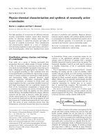
Fig. 1. Processing of APP to form Ab peptides. (A) Schematic diagram of the alternative processing pathways of APP. The transmembrane
APP undergoes two alternative and competing pathways of metabolism. The major and non-amyloidogenic, or a-secretase, pathway
precludes the formation of Alzheimer’s Ab peptide. The amyloidogenic, or b-secretase, pathway initiates the formation of Ab, which is
completed by the action of the c-secretase. a-Secretase has been identified as a zinc metalloproteinase of the ADAMs family, whereas both
b- and c-secretases are membrane-bound aspartic proteinases (see text for full details). (B) Sites of cleavage of APP by b- and c-secretases
to form Ab peptides. The sites of the juxtamembrane and intramembrane cleavages of transmembrane APP by b- and c-secretases, respec-
tively, are indicated by arrows. The c-secretase cleavages are heterogeneous, mainly producing Ab peptides of 40 and 42 amino acids. The
amino acid sequences of Ab and around the scissile bonds are indicated by the one letter code for amino acids. The sequence shown is the
wild-type sequence. The ‘Swedish mutant’ APP sequence around the b-secretase cleavage site is . NL ⁄ DAEF. rather than . KM ⁄ DAEF
The development of many BACE-1 inhibitors has used the sequence around the scissile bond in the Swedish mutant as the lead for
synthetic chemistry to produce potent and selective compounds.
Biology and chemistry of b-secretase (BACE-1) C. E. Hunt and A. J. Turner
1846 FEBS Journal 276 (2009) 1845–1859 ª 2009 The Authors Journal compilation ª 2009 FEBS
different groups as ‘b-site APP cleaving enzyme’
(BACE), ‘aspartyl protease-2’ (Asp-2) or ‘membrane-
anchored aspartic proteinase of the pepsin family-2’
(memapsin-2) [8–12]. Vassar et al. [8] originally used
an expression cloning strategy to identify genes that
altered Ab production in human embryonic kidney
(HEK) cells overexpressing APP containing the
amyloidogenic Swedish mutation. This cell line was
known to express both the b- and c-secretases. They
isolated a sequence from a clone that produced ele-
vated levels of Ab and that encoded a novel aspartic
protease, which they termed ‘BACE’ (subsequently
BACE-1). A classical biochemical strategy involving
affinity chromatographic isolation of the enzyme activ-
ity and its subsequent cloning also proved to be highly
effective [9]. In another approach, b-secretase was
independently identified using expressed sequence tag
(EST) databases. Hussain et al. [10] screened a proprie-
tary EST database, from which they identified a
sequence of interest which they termed Asp-2. Subse-
quently, they cloned the cDNA, transfected it into
HEK cells and observed an increase in the b-cleavage
of APP. In an alternative strategy, Yan et al. [11] visu-
ally inspected the b-cleavage sites within APP, and
concluded that the cleavage may be carried out by an
aspartic protease. They subsequently searched the
database of the newly emerging Caenorhabditis elegans
genome using the characteristic active site motif for
aspartic proteases, D(S ⁄ T)G. Using these isolated
sequences, they next searched human EST databases,
which identified four novel aspartic proteases that they
named Asp-1–4. Accordingly, they transfected two of
these sequences into HEK cells, and those containing
the Asp-2 construct were found to possess b-secretase
activity. From the human EST database at the time,
Lin et al. [12] identified, and subsequently cloned and
expressed, two novel human aspartic proteinases which
they named memapsin-1 and memapsin-2. All groups
succeeded in identifying the same protein as the
putative b-secretase (BACE-1, Asp-2, memapsin-2),
together with a close homologue (BACE-2, Asp-1,
memapsin-1). The localization, specificity and other
enzymological properties of BACE-1 most closely fitted
the profile of b-secretase. Although BACE-2 is interest-
ing in comparative terms, its precise physiological roles
are unclear, and there is no compelling evidence that it
plays a direct role in the b-secretase processing of APP.
The rest of this article focuses exclusively on BACE-1,
although inhibitor development studies must clearly
consider compound discrimination between the two
activities (and other relevant protease activities).
Molecular cell biology of BACE-1
BACE-1 is synthesized as a proprotein in the
endoplasmic reticulum (ER) before it is transported to
the trans-Golgi network, where it undergoes matura-
tion [13]. The efficient exit of the enzyme from the ER
is determined by the prodomain [13], which is subse-
quently removed by the proprotein convertase, furin or
a furin-like protease [13–15]. This process is not
required for its activation as pro-BACE can still cleave
APP [14]; however, removal of its prodomain increases
BACE-1 activity by approximately twofold [16].
Molecular dynamics simulation studies have suggested
that the partial catalytic activity of the zymogen could
be explained by the high mobility of the prosegment in
comparison with that of other zymogens, resulting in
the occasional exposure of the catalytic site for access
by its substrate, APP [17]. During maturation, BACE-1
also undergoes a number of post-translational modifica-
tions during its transport through the cell. The catalytic
domain contains four potential N-linked glycosylation
sites at asparagines 153, 172, 223 and 354, all of which
appear to be occupied with some degree of heterogene-
ity between the bound carbohydrates [18]. The simple
carbohydrates added in the ER produce an immature
BACE-1 protein of approximately 65 kDa [14]. These
sugars are further processed to an endoglycosidase
H-resistant, complex form producing the mature
75 kDa species [14,19]. These modifications appear to
be important for the maximal catalytic activity of the
enzyme, as site-directed mutagenesis of these aspara-
gine residues significantly reduces the proteolytic activ-
ity [20]. BACE-1 also contains three disulphide bonds
in the catalytic domain between cysteines 216–420,
278–443 and 330–380 [18], which are important for the
correct folding, and hence proteolytic activity, of the
enzyme [21]. Within the membrane, BACE-1 probably
functions as a dimer, as may the APP molecule [22,23].
The dimerization of BACE-1 could facilitate the bind-
ing and cleavage of physiological substrates, as the
Table 1. Potential strategies to inhibit b-secretase processing of
APP by BACE-1.
Active site-directed (competitive) inhibition of enzyme activity.
Transition state, small-molecule inhibitors; peptidic or non-peptidic
Non-competitive or allosteric inhibition, e.g. targeting protein
processing, conformational changes (‘flap movement’), distant
subsites from scissile bond
Modulation of oligomeric state and hence activity of the enzyme
Modulation of protein–protein interactions affecting localization
and ⁄ or activity
Modulation of lipid environment of the enzyme
Immunization with BACE-1
Modulation of miRNA regulation of BACE-1
C. E. Hunt and A. J. Turner Biology and chemistry of b-secretase (BACE-1)
FEBS Journal 276 (2009) 1845–1859 ª 2009 The Authors Journal compilation ª 2009 FEBS 1847
purified native BACE-1 dimer revealed a higher affin-
ity and turnover rate in comparison with the soluble
BACE-1 ectodomain, which exists as a monomer
[22,23]. Understanding the oligomeric states and nature
of the molecular interactions between the secretases
and their protein substrates could allow the develop-
ment of secretase inhibitors which specifically bind to
the contact sites of dimers and hence inhibit Ab
formation. In addition, serine 498 is phosphorylated
by casein kinase 1, which appears to determine its
subsequent subcellular location [24]. Both the wild-
type, phosphorylated BACE-1 and an unphosphorylat-
able mutant localize to early endosomes, but only the
phosphorylated form is recycled back to the membrane
[24]. Adjacent to serine 498 within the extreme C-ter-
minus of BACE-1, there is also a dileucine motif. This
sequence has been shown previously in a variety of
proteins to determine their trafficking from the cell
surface to the endosomal and lysosomal compartments
[25]. Mutation of the dileucine motif [26] resulted in
increased levels of BACE-1 at the cell surface,
consistent with decreased internalization to endosomes.
The cytoplasmic domain also contains several cysteine
residues which are subject to palmitoylation [13]. This
modification may function to anchor the protein to the
membrane, as mutation of these cysteine residues
increases the release of the BACE-1 ectodomain into
the medium [13]. The stability and turnover of
BACE-1, like that of the low-density lipoprotein
receptor, is regulated by reversible acetylation of seven
lysine residues in its lumenal (N-terminal) domain, this
event occurring in the ER and serving as a ‘quality
control’ step in protein maturation [27,28]. Acetylated
BACE-1 can then traffic to the Golgi, where deacetyla-
tion of the mature protein can occur. Non-acetylated,
immature BACE-1 is degraded in a non-proteasomal,
post-ER compartment [27]. The proprotein convertase
PCSK9 appears to be involved in the disposal of non-
acetylated BACE-1 [28].
BACE-1 is shed from cells through cleavage at its
membrane anchor between alanine 429 and valine 430
[29] to generate a soluble BACE-1 ectodomain [13] by
an as yet unidentified proteinase activity. Metallopro-
teinase inhibitors block BACE-1 shedding from cells
overexpressing BACE-1 [29,30], from which it was con-
cluded that the BACE-1 ‘sheddase’ is likely to be a
member of the ‘a disintegrin and metalloprotease’
(ADAM) family of proteins [31]. Shedding is a process
by which many integral membrane proteins, such as
angiotensin-converting enzyme and tumour necrosis
factor-a, are cleaved to release a large soluble ectodo-
main by a protease referred to as a ‘sheddase’ or ‘sec-
retase’ [31,32]. The physiological role of soluble
BACE-1, if any, and its potential to modulate the
amyloidogenic processing of APP still remain conten-
tious. Hussain et al. [30] showed that the inhibition of
BACE-1 shedding using metalloprotease inhibitors had
no effect on the b-cleavage of APP. In contrast, the
activation of protein kinase C, which is known to
upregulate the shedding of BACE-1 [30], has been
shown by a number of groups to decrease Ab produc-
tion in cell lines [33,34], primary cells [34] and mouse
brain [35]. However, this decrease may largely reflect
the upregulation of the competing a-secretase pathway.
Soluble BACE-1 is still able to process APP, as
Benjannet et al. [14] clearly showed that the overex-
pression of soluble BACE-1 resulted in a dramatic
increase in the production of Ab, and so membrane
anchorage in the vicinity of its substrate is not
essential.
Expression and localization of BACE
BACE-1 mRNA [8,9,11] and enzyme activity [9] levels
are highest in the brain, with lower expression in
peripheral tissues, consistent with its role as an APP
b-secretase. Surprisingly, significant BACE-1 mRNA
has also been detected in the pancreas [8,9,11],
although the enzyme activity is very low in this tissue
[9]. In the brain, BACE-1 is largely expressed by
neurons, with seemingly little produced by glial cells
[8,10,36–38]. However, in animal models of chronic
gliosis and in brains of AD patients, BACE-1
expression can be detected in reactive astrocytes,
suggesting that astrocyte activation may play a role in
the development of AD (for a review, [39]).
Hence, targeting astrocyte activation could be a viable
strategy in the treatment of AD for this and other
reasons.
Evidence that BACE-1 is the sole b-secretase activity
in the brain (at least in transgenic mouse models) was
provided by the observations that BACE-1 knockout
mice completely lacked both b-secretase enzyme
activity and the product of b-cleavage, CTF99 [6,40].
In addition, cultured primary neurons from these
animals do not secrete detectable levels of Ab
[6,7,40,41]. In support of this view, a commercial
BACE-1 inhibitor administered to wild-type mice was
shown to decrease the levels of endogenous Ab
compared with those in control animals [42]. Increased
levels of BACE-1 activity have been reported in the
brains of patients with sporadic AD [36,43,44], and a
truncated, soluble form of BACE-1 can be detected
by activity assay in cerebrospinal fluid, which may
provide a useful biomarker in AD and a source for
monitoring the efficacy of drug candidates [45].
Biology and chemistry of b-secretase (BACE-1) C. E. Hunt and A. J. Turner
1848 FEBS Journal 276 (2009) 1845–1859 ª 2009 The Authors Journal compilation ª 2009 FEBS
Elevated BACE-1 levels have been reported in the cere-
brospinal fluid of patients with mild cognitive impair-
ment [46]. Nevertheless, some studies have shown that
other proteases could contribute to the b-secretase
activity in brain against the wild-type b-secretase APP
site, e.g. cathepsins B and D, and that cathepsin inhibi-
tors may be therapeutically useful in AD [47,48].
A recent study of the effect of glutaminyl cyclase inhi-
bition on AD-like pathology in mouse and Drosophila
disease models also indirectly suggests the occurrence
of a very low abundant but pathologically relevant
b-secretase activity distinct from BACE-1 [49].
The precise subcellular location(s) at which BACE-1
cleaves APP is still controversial. BACE-1 undergoes
recycling and is transported to the cell surface from
where it is internalized. The enzyme has been found,
through co-localization studies, to be associated with
the Golgi apparatus [8,14,19,24] and endosomal
compartments [8,14,50,51] from where the Ab product
may be routed to multivesicular bodies and then
secreted via exosomes [52]. Specialized membrane
domains, referred to as lipid rafts, have also been
proposed as the location for b-cleavage [53–55]. The
direct targeting of BACE-1 to lipid rafts by the addi-
tion of a glycosyl-phosphatidylinositol anchor has been
shown to upregulate both sAPPb and Ab production
in SH-SY5Y cells [56]. In addition, the disruption of
lipid rafts by the depletion of cellular cholesterol levels
has been shown to decrease Ab production in both
cells [56–58] and in vivo [58,59], whilst animals fed a
diet high in cholesterol showed enhanced accumulation
of Ab [59]. Interestingly, data presented in [53] suggest
that these differing concepts regarding the location of
b-cleavage of APP can be reconciled. Using antibody
co-patching, evidence was provided to suggest that
BACE-1 and APP in lipid rafts come together during
endocytosis into endosomes where b-cleavage occurs.
Not all studies are consistent with the elevation of
cellular cholesterol enhancing amyloid peptide forma-
tion, and an optimal level of neuronal membrane
cholesterol may be critical as, under some conditions,
loss of membrane cholesterol can potentiate amyloid
peptide synthesis [60]. Palmitoylation-deficient mutants
of BACE-1, which are not raft-localized, can still
cleave APP, suggesting that b-site processing can take
place in both raft and non-raft microdomains [61].
Chronic treatment with statins as inhibitors of choles-
terol biosynthesis (and hence lipid raft stability) has, in
some studies, been reported to reduce the risk of
developing AD, although the literature is conflicting
(for example, [62]). Indeed, any effect of statins on
amyloid production may relate to the inhibition of
protein isoprenylation, rather than any direct effect on
cholesterol levels [63]. A specific inhibitor of choles-
terol biosynthesis, BM15.766, does however reduce the
expression of b-secretase, and consequently the
production of amyloid-b, at least in vitro [64].
BACE-1 activity itself is highly sensitive to its lipid
environment and is stimulated by glycosphingolipids,
glycerophospholipids and sterols [65]. Glycosaminogly-
cans may also act as allosteric modulators of BACE-1
activity, as heparan sulphate specifically inhibits the
BACE-1 cleavage of APP, but not that by a-secretase
[66]. Heparin itself has a complex mode of action by
activating the partially active BACE-1 zymogen at low
concentrations, but promoting autocatalytic cleavage
and hence inhibition of the protease domain at higher
concentrations [67,68]. Hence, in total, these studies
suggest that modulation of the subcellular site(s) of
APP processing may represent a potential therapeutic
strategy in the treatment of AD [69]. In this context,
APP may normally be segregated from BACE-1 in
distinct membrane domains through its interaction
with X11 ⁄ Munc18 [70] proteins, but neuronal activity,
coupled with the phosphorylation of Munc18, appears
to influence the movement of APP into BACE-1-con-
taining membrane domains, a process referred to as
‘membrane microdomain switching’ [71]. A variety of
BACE-interacting proteins have been reported that
might influence enzyme localization and ⁄ or activity,
for example reticulon ⁄ NOGO proteins, which can inhi-
bit the access of BACE-1 to its substrate APP [72,73].
A conserved C-terminal QID sequence among reticu-
lon family members is involved in the interaction with
the BACE-1 cytoplasmic domain [74]. The cellular
form of the prion protein also negatively regulates
b-secretase cleavage of APP, probably through its raft
interaction with glycosaminoglycans [75]. Hence, the
cellular form of the prion protein may normally sup-
press Ab formation through its inhibition of BACE-1
[76]. Small-molecule mimics of such modulating
interactions could provide novel BACE-1-inhibiting
therapeutics.
Regulation of BACE-1 expression
A variety of physiological stressors and signalling
pathways have been found to regulate BACE-1 and
may be a factor in the reported increased BACE-1
protein levels and enzyme activity in AD brains
[36,43,44], although BACE-1 transcript levels
generally appear unchanged in AD brains [77,78].
Hypoxia and ischaemia are important risk factors
for AD, and chronic hypoxia in the neuroblastoma
line SH-SY5Y promotes amyloidogenic processing of
APP [79]. Hypoxia-inducible factor-1a binds to the
C. E. Hunt and A. J. Turner Biology and chemistry of b-secretase (BACE-1)
FEBS Journal 276 (2009) 1845–1859 ª 2009 The Authors Journal compilation ª 2009 FEBS 1849
BACE-1 promoter, and several studies have reported
the upregulation of BACE-1 mRNA both in vitro and
in vivo following hypoxia [80–82]. Oxidative stress can
stimulate BACE-1 expression in cells through the
c-jun N-terminal kinase pathway in a mechanism
which requires the presence of presenilin [83]. The
lipid peroxidation product 4-hydroxynonenal also
upregulates BACE-1 expression through the stress-
activated protein kinase pathway [84]. The activation
of cyclin-dependent kinase 5 also leads to increased
levels of BACE-1 mRNA and protein in vivo and
in vitro, and the BACE-1 promoter contains a cyclin-
dependent kinase 5-responsive region [85]. Other
stressors that can cause the activation of BACE-1
expression include traumatic brain injury, a strong
risk factor for AD [86], and infection of neuronal
cells with herpes simplex virus 1 [87]. Herpes simplex
virus 1 is also a risk factor for AD, particularly when
in association with the e4 allele of the apolipoprotein
E4 gene [88], and the viral DNA is localized within
amyloid plaques in AD brains [89].
Post-transcriptional mechanisms have a major
influence on BACE-1 levels, and BACE-1 translation
is regulated at multiple stages, consistent with the
presence of a long and highly conserved transcript
leader [90,91]. In particular, the 5¢-UTR represses the
rate of BACE-1 translation [92], and alternative splic-
ing of the transcript leader can influence the rate of
translation in a tissue-dependent manner [90]. A
detailed mutagenesis analysis suggested that the
GC-rich region of the 5¢-UTR acts as a ‘translation
barrier’ [92]. The presence of several upstream ATGs
also strongly reduces the translation of the main open
reading frame, which implies that BACE-1 translation
might increase in conditions that favour phosphoryla-
tion of the translation eukaryotic initiation factor-2a
(eIF2a) [90]. More recent studies have shown that
cellular energy deprivation (glucose deprivation in cell
culture) produces a post-transcriptional increase in
BACE-1 levels, which is indeed mediated through
increased eIF2a phosphorylation [92]. These observa-
tions in vitro correlated with in vivo studies in AD
transgenic (Tg2576) mice, in which chronic energy
inhibition with 2-deoxyglucose or 3-nitropropionic
acid was shown to increase eIF2a phosphorylation,
BACE-1 levels and amyloidogenesis [93]. Thus, a
common mechanism by which stress (e.g.
hypoxia ⁄ ischaemia, viral infection, etc.) can influence
BACE-1 levels may be through the regulation of
translation initiation at the level of eIF2a. BACE-1
protein stability can also be influenced by the
lysosomal and proteasomal pathways [94] and
through its lysine acetylation status [27,28].
Substrates of BACE-1
Like most proteases, BACE-1 is not uniquely specific
to one substrate, and APP may not even be the
primary substrate of the enzyme, except where muta-
tions in the enzyme or in APP render it far more effec-
tive in this reaction. Hence, in addition to APP,
BACE-1 is also involved in the proteolytic processing
of a number of other proteins. The amyloid precursor-
like proteins 1 and 2, which are closely related to and
structurally similar to APP, are also processed by
BACE-1 [95], as are the APPe product (the e-secretase-
derived N-terminal product of APP) [96] and Ab itself,
which is cleaved at the 34 ⁄ 35 site [97]. Additional sub-
strates include the sialyltransferase ST6Gal I [98], the
cell adhesion protein P-selectin glycoprotein ligand-1
[99], the low-density lipoprotein receptor-related pro-
tein [100] and the b-subunits of voltage-gated sodium
channels [101]. Recently, using BACE-1 knockout
mice, Willem et al. [102] have suggested a role for
BACE-1 in the myelination of peripheral nerves
through the processing of type III neuregulin 1, and
the enzyme also appears to modulate myelination in
the central nervous system [103]. However, inhibition
of BACE-1 in vivo in adult mice expressing human
wild-type APP lowered brain Ab levels and increased
sAPPa, but did not affect neuregulin processing [104].
Given the diversity of the BACE-1 substrates so far
identified, there are probably considerably more to dis-
cover. In order to validate BACE-1 as a realistic thera-
peutic target, it is important that the manifestations of
inhibiting these alternative activities are understood,
particularly in the adult and aging animal.
Inhibitors of aspartic proteinases
Aspartic proteinases are endopeptidases which use two
aspartic acid residues to catalyse the hydrolysis of a
peptide bond. These aspartic acid residues in the active
site bind and activate a water molecule, which, in turn,
acts as a nucleophile to attack the scissile bond at the
cleavage site of its substrate. Of the various clans of
aspartic proteases, BACE-1 belongs to the same clan
as pepsin, although it is only very weakly inhibited
(IC
50
= 0.3 mm) by the statine-based transition state
inhibitor of pepsin, pepstatin. The statine moiety of
pepstatin represents a tetrahedral, hydroxymethylene
isostere of the scissile peptide bond, and hence mimics
the putative transition state intermediate of the
catalytic reaction. This mode of inhibition has been
generally applied to the development of BACE-1 inhib-
itors (see below). Members of the pepsin family are
only found in eukaryotes and are most active at an
Biology and chemistry of b-secretase (BACE-1) C. E. Hunt and A. J. Turner
1850 FEBS Journal 276 (2009) 1845–1859 ª 2009 The Authors Journal compilation ª 2009 FEBS
acidic pH (approximately pH 4 for BACE-1, although
it rapidly and irreversibly loses activity at pH 3.5 or
lower). Most are synthesized as proproteins with a
signal domain targeting them to the secretory pathway.
The crystal structures of several of the members of this
family have revealed a bilobed structure, in which each
lobe contributes one of the aspartic acid residues
which makes up the catalytic pair at the active site (for
a review, [105]). The two lobes are structurally similar
and appear to have evolved from a gene duplication
event [106].
Towards the development of BACE-1
inhibitors
Ever since the elucidation of the metabolic pathway
leading to the formation of Ab (Fig. 1), the b-secretase
has been a primary target for inhibitor design in AD
therapy. Considerable efforts have been directed
towards the identification of low-molecular-mass,
specific and stable non-peptide analogues as BACE-1
inhibitors that can lead to the development of a suc-
cessful therapeutic. Such compounds must be of high
potency, stable to hydrolysis, deliver low toxicity and
be able to cross the blood–brain barrier. Approaches to
the discovery of novel BACE-1 inhibitors have involved
understanding the substrate specificity of the enzyme,
coupled with structure-based design and high-through-
put screening in vitro and in silico. To date, the screen-
ing of extensive libraries for non-peptide-based BACE-
1 inhibitors has resulted in the discovery of relatively
few, generally low-affinity, compounds, indicating that
this is not an easy protein target to inhibit effectively
in vivo. This is partly because of the extended sub-
strate-binding site requirements [107], a problem also
seen with other aspartic proteinase targets. The crystal
structure of the protease domain of BACE-1 complexed
to an eight-residue, peptide-based inhibitor (OM99-2)
was determined shortly after the enzyme was identified
[108]. The design strategy for OM99-2 was based on
comparisons of the amino acid sequences around the
scissile bond in the wild-type APP (–EVKM ⁄ DAEF–),
which is a relatively poor substrate for BACE-1, and
the very efficiently hydrolysed, Swedish mutant APP (–
EVNL ⁄ DAEF–), with a 60-fold higher k
cat
⁄ K
m
relative
to the wild-type. The residues of the inhibitor in the
S
1
–S
4
subsites were unchanged from the Swedish
mutant sequence (EVNL), but those at the S
1
¢–S
2
¢ sub-
sites were changed from Asp–Ala to Ala–Val, as the
key specificity of BACE-1 appeared to reside mainly at
the S
1
¢ site, where small side-chains, such as alanine,
are highly preferred over aspartic acid. The aim was
also to reduce the polarity and increase the lipophilicity
of the inhibitor to aid penetration across the blood–
brain barrier. This peptide backbone was used to gener-
ate a typical aspartic proteinase inhibitor by converting
the P
1
–P
1
¢ peptide bond to a hydroxyethylene transition
state isostere, leading to the compound OM99-2 (EVN-
L*AAEF, where * indicates the isostere), which is
shown in Fig. 2.
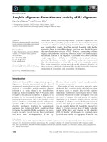
The structural solution of BACE-1 [108] revealed a
bilobed structure with the same general folding pattern
as other known aspartic proteases, such as pepsin,
including high conservation of the hydrogen-bonding
structure around the active site (Fig. 3). However,
there are important structural differences between
BACE-1 and pepsin. The most significant differences
are four insertions, which considerably increase the
molecular boundary of BACE compared with pepsin,
and a 35-residue C-terminal extension in the C-lobe
which contains two of the disulphide bonds unique to
BACE-1. The large, active site cleft which contains the
two catalytic aspartate residues is located between the
two lobes and appears to be more open and accessible
than that of pepsin.
GSK 188909
OM99-2
P
3
Val
P
1
Leu
P
2
' Ala
P
1
' Ala
P
3
' Glu
P
2
Asn
P
4
Glu
P
4
' Phe
O
O
SF
HN
HN N
H
N
O
OH
F
O
OO
O
O
OH
OO
O
O
O
OH
OH
OH
N
H
N
H
N
H
N
H
H
2
N
H
2
N
H
N
H
N
F
F
Fig. 2. BACE-1: from peptide-based to non-peptidic BACE-1
inhibitors. Examples of two BACE-1 inhibitors: the first reported
compound OM99-2 (reproduced from [108] with permission of the
American Association for the Advancement of Science) and a
recently described orally active, non-peptidic inhibitor GSK 188909
(reproduced from [117] with permission of the International Society
for Neurochemistry). In OM99-2, the constituent amino acids and
their subsite designations are indicated. The hydroxyethylene
transition state isostere is between P
1
-Leu and P
1
¢-Ala. Figure
reproduced from Hussain et al. [117] by kind permission.
C. E. Hunt and A. J. Turner Biology and chemistry of b-secretase (BACE-1)
FEBS Journal 276 (2009) 1845–1859 ª 2009 The Authors Journal compilation ª 2009 FEBS 1851
More extensive studies of the specificity requirements
of BACE-1 have subsequently been carried out, estab-
lishing that the enzyme has a relatively loose substrate
specificity, which has been defined in detail by Turner
et al. [109]. A peptide containing the sequence of the
eight most favoured residues around the scissile bond
[–EIDLMVLD–] is the most efficient known substrate
of the enzyme [109]. A variety of other short peptides
have typically been used in BACE-1 assays, usually as
fluorogenic substrates incorporating a fluorophore and
a quencher, mimicking the sequence around the b-secre-
tase cleavage site in APP or in the Swedish mutated
form (for example, [110]). However, caution should
always be used in interpreting data from small peptide
substrates as they lack many of the subsite and other
interactions of the genuine protein substrate. Neverthe-
less, such studies have led to the development of novel
BACE-1 inhibitors, usually transition state analogues
incorporating the hydroxyethylene transition state
isostere, or statine, residue typical of many aspartic
proteinase inhibitors. Refinement of OM99-2 [111] led
to the development of OM00-3 (Glu-Leu-Asp-Leu*
Ala-Val-Glu-Phe), the most potent inhibitor known to
date with a K
i
value of 0.3 nm. The cell permeability
and blood–brain barrier penetrance of such compounds
are, however, often a problem compounded by active
P-glycoprotein-mediated efflux, leading to poor inhibi-
tion constants in vivo. Ideally, such compounds should
be < 500 Da for passive barrier penetration. An
alternative is to permit facilitated penetration. The cell
permeability problem has been overcome, in one suc-
cessful example, by the incorporation of a penetratin
sequence to the inhibitor, considerably enhancing the
cell potency [112]. The inhibitor itself [JMV1195;
EVN(statine)AEF-NH
2
] represents one of the statine-
based peptidomimetic BACE-1 inhibitors [109], again
modelled on the Swedish mutant peptide sequence. In
another approach, a series of isonicotinamides derived
from traditional aspartic proteinase transition state iso-
stere inhibitors has been optimized to yield low-nanom-
olar inhibitors with sufficient penetration across the
blood–brain barrier to demonstrate b-amyloid reduc-
tion in a murine model [113]. Hence, structure-based
approaches to inhibitor design against BACE-1 are now
beginning to yield potential therapeutic compounds.
Recent disclosures of the crystal structures of BACE-1
with lower M
r
inhibitors have provided further insights
into active site interactions, producing more potent and
selective, cell-permeable compounds, including both
peptidomimetic and non-peptidic compounds [114–
116]. For example, using a rational drug design
approach, Hussain et al. [117] identified GSK188909
(Fig. 2) as a small-molecule (M
r
600), potent and
selective non-peptidic inhibitor able to block Ab
formation in transgenic mice when co-administered
with a P-glycoprotein inhibitor.
BACE-1 inhibition cannot be considered in isolation
from that of BACE-2. Detailed studies on BACE-2
AB
Fig. 3. The crystal structure of BACE-1 complexed to the peptide-based inhibitor OM99-2. (A) Stereoview of the polypeptide backbone of
BACE-1 is shown as a ribbon diagram. The N-lobe and C-lobe of the bilobed aspartic proteinase structure are shown in blue and yellow,
respectively. The inhibitor bound between the lobes is shown in red. (B) The chain tracings of human BACE-1 (dark blue) and human pepsin
(grey) are compared. The light blue balls represent identical residues which are topologically equivalent. The disulphide bonds are shown in
red for BACE-1 and orange for pepsin. The C-terminal extension in BACE-1 is shown in green and the active site aspartic acid residues are
shown in yellow. Reproduced from [108] with permission of the American Association for the Advancement of Science. Figure reproduced
from Hong et al. [108] by kind permission.
Biology and chemistry of b-secretase (BACE-1) C. E. Hunt and A. J. Turner
1852 FEBS Journal 276 (2009) 1845–1859 ª 2009 The Authors Journal compilation ª 2009 FEBS
specificity [118] indicate that it is broad-based and, not
surprisingly, rather similar to BACE-1, consistent with
several BACE-1 inhibitors also inhibiting BACE-2.
Nevertheless, in a separate study, inhibitors with
isophthalamide derivatives as the P
2
–P
3
ligands showed
good selectivity between BACE-1 and BACE-2,
nanomolar potency in vitro and in cell-based studies,
and a significant reduction in Ab40 levels in vivo in
transgenic mice after intraperitoneal administration
[119]. Relatively few detailed kinetic and mechanistic
studies have been carried out on BACE-1 inhibition.
A notable exception is provided by Marcinkeviciene
et al. [120], in which steady state and stopped flow
kinetics of BACE-1 inhibition by a statine-based inhi-
bitor [Ac-KTEEISEVN(statine)VAEF-COOH] were
carried out. These studies revealed a two-step mecha-
nism involving an initial low-affinity binding, followed
by a tightening up of the binding, induced either by a
conformational change (‘flap movement’) or displace-
ment of a catalytic water molecule. The scene is now
set for the refinement of existing molecules and the
exploration of their efficacy further in animal models.
The ability of an orally administered BACE-1 inhibitor
to reduce cerebrospinal fluid and plasma Ab levels in a
non-human primate (rhesus monkey) has recently been
reported [121] and, at long last, clinical trials of
BACE-1 inhibitor drug candidates are being initiated
almost a decade on from the original cloning of the
enzyme. This has largely been because of the problems
inherent in the design of potent and selective aspartic
proteinase inhibitors sufficiently small to penetrate the
blood–brain barrier. The BACE-1 inhibitor CTS-2166
has entered a phase I study in healthy volunteers, and
the drug was reported to be well tolerated and effective
in lowering plasma Ab levels [122], and further trials
are ongoing.
Future approaches and therapeutic
potential of b-secretase inhibition
The development of clinically successful BACE-1
inhibitors has been hampered by a number of factors,
including effective inhibitor design, selectivity and
stability, brain access and potential toxicity. Combina-
tion therapies employing BACE inhibition with other
strategies may provide a more versatile treatment in
AD, and other novel strategies are also currently being
explored. An innovative and promising recent experi-
mental approach has been to attempt to immunize
transgenic AD mice with BACE-1, which resulted in a
significant reduction in brain Ab levels and an
improvement in cognitive function, without any
reported evidence of inflammatory responses [123]. The
rationale for this study was that immunization with
BACE-1 could produce a proportion of brain-pene-
trant antibodies, which, in turn, bind to neuronal cell
surface BACE-1 molecules. Internalization of the
BACE-1 antibody complexes then results in inhibition
of the enzyme activity within endosomes, and hence of
Ab production. Recent studies have also shown that
microRNAs (miRNAs) can bind to the 3¢-UTR of
BACE-1 mRNA, and hence regulate BACE-1 levels.
Loss of specific miRNAs (e.g. miR-107, 298, 328 and
the cluster miR-29a ⁄ b-1) during AD progression could
contribute to increases in BACE-1 and Ab levels
[124–126], but exploiting miRNAs therapeutically is
currently very challenging. Only time will tell which of
these diverse approaches to the modulation of
b-secretase activity of BACE-1, directly or indirectly, is
likely to have the potential to reach the clinic.
Conclusions
Almost 10 years since BACE-1 was unequivocally
identified, it still remains a promising, indeed probably
the most viable, target for therapy in AD, although
some have urged caution in adopting this approach
[7]. Although much has been learned about the struc-
ture and action of the enzyme, there are still many
unanswered questions relating to its true physiological
roles, its locations and the physiological consequences
of its inhibition in vivo. Targeting aspartic proteinases
is not a trivial exercise and there remains considerable
scope for innovative design and application of BACE-1
inhibitors, but their efficacy and safety still remain to
be demonstrated, particularly in the chronic treatment
regimes that would be required. Alternative
strategies that seek to manipulate the location, lipid
environment, antigenicity, transcriptional regulation or
processing of the enzyme may also be strategically
useful, as described above. The importance of the
problem demands both an imaginative and thorough
approach to rational drug design and application.
Acknowledgements
CEH was supported by a UK Biotechnology and
Biological Sciences Research Council PhD studentship,
and we also acknowledge the financial support of the
UK Medical Research Council.
References
1 Sinha S & Lieberburg I (1999) Cellular mechanisms of
b-amyloid production and secretion. Proc Natl Acad
Sci USA 96, 11049–11053.
C. E. Hunt and A. J. Turner Biology and chemistry of b-secretase (BACE-1)
FEBS Journal 276 (2009) 1845–1859 ª 2009 The Authors Journal compilation ª 2009 FEBS 1853
2 Wolfe MS (2007) When loss is gain: reduced presenilin
proteolytic function leads to increased Ab42 ⁄ Ab40.
Talking point on the role of presenilin mutations in
Alzheimer disease. EMBO Rep 8, 136–140.
3 Glenner GG & Wong CW (1984) Alzheimer’s disease:
initial report of the purification and characterization of
a novel cerebrovascular amyloid protein. Biochem
Biophys Res Commun 120, 885–890.
4 Masters CL, Simms G, Weinman NA, Multhaup G,
McDonald BL & Beyreuther K (1985) Amyloid plaque
core protein in Alzheimer disease and Down
syndrome. Proc Natl Acad Sci USA 82, 4245–4249.
5 Hardy JA & Higgins GA (1992) Alzheimer’s disease:
the amyloid cascade hypothesis. Science 256, 184–185.
6 Roberds SL, Anderson J, Basi G, Bienkowski MJ,
Branstetter DG, Chen KS, Freedman SB, Frigon NL,
Games D, Hu K et al. (2001) BACE knockout mice
are healthy despite lacking the primary b-secretase
activity in brain: implications for Alzheimer’s disease
therapeutics. Hum Mol Genet 10, 1317–1324.
7 Dominguez D, Tournoy J, Hartmann D, Huth T,
Cryns K, Deforce S, Serneels L, Camacho IE, Marjaux
E, Craessaerts K et al. (2005) Phenotypic and
biochemical analyses of BACE1- and BACE2-deficient
mice. J Biol Chem 280, 30797–30806.
8 Vassar R, Bennett BD, Babu-Khan S, Kahn S,
Mendiaz EA, Denis P, Teplow DB, Ross S, Amarante
P, Loeloff R et al. (1999) b-Secretase cleavage of
Alzheimer’s amyloid precursor protein by the
transmembrane aspartic protease BACE. Science 286,
735–741.
9 Sinha S, Anderson JP, Barbour R, Basi GS,
Caccavello R, Davis D, Doan M, Dovey HF, Frigon
N, Hong J et al. (1999) Purification and cloning of
amyloid precursor protein b-secretase from human
brain. Nature 402, 537–540.
10 Hussain I, Powell D, Howlett DR, Tew DG, Meek
TD, Chapman C, Gloger IS, Murphy KE, Southan
CD, Ryan DM et al. (1999) Identification of a novel
aspartic protease (Asp2) as b-secretase. Mol Cell
Neurosci 14, 419–427.
11 Yan R, Bienkowski MJ, Shuck ME, Miao H, Tory
MC, Pauley AM, Brashier JR, Stratman NC, Mathews
WR, Buhl AE et al. (1999) Membrane-anchored
aspartyl protease with Alzheimer’s disease b-secretase
activity. Nature 402, 533–537.
12 Lin X, Koelsch G, Wu S, Downs D, Dashti A & Tang
J (2000) Human aspartic protease memapsin 2 cleaves
the b-secretase site of b-amyloid precursor protein.
Proc Natl Acad Sci USA
97, 1456–1460.
13 Benjannet S, Elagoz A, Wickham L, Mamarbachi M,
Munzer JS, Basak A, Lazure C, Cromlish JA, Sisodia
S, Checler F et al. (2001) Post-translational processing
of b-secretase (b-amyloid-converting enzyme) and its
ectodomain shedding. J Biol Chem 276, 10879–10887.
14 Creemers JW, Ines Dominguez D, Plets E, Serneels L,
Taylor NA, Multhaup G, Craessaerts K, Annaert W
& De Strooper B (2001) Processing of b-secretase by
furin and other members of the proprotein convertase
family. J Biol Chem 276, 4211–4217.
15 Bennett BD, Denis P, Haniu M, Teplow DB, Kahn S,
Louis JC, Citron M & Vassar R (2000) A furin-like
convertase mediates propeptide cleavage of BACE, the
Alzheimer’s b-secretase. J Biol Chem 275, 37712–
37717.
16 Shi XP, Chen E, Yin KC, Na S, Garsky VM, Lai MT,
Li YM, Platchek M, Register RB, Sardana MK et al.
(2001) The pro domain of b-secretase does not confer
strict zymogen-like properties but does assist proper
folding of the protease domain. J Biol Chem 276,
10366–10373.
17 Zuo Z, Gang C, Zou H, Mok PC, Zhu W, Chen K &
Jiang H (2007) Why does b-secretase zymogen possess
catalytic activity? Molecular modelling and molecular
dynamics simulation studies. Comput Biol Chem 31,
186–195.
18 Haniu M, Denis P, Young Y, Mendiaz EA, Fuller J,
Hui JO, Bennett BD, Kahn S, Ross S, Burgess T et al.
(2000) Characterisation of Alzheimer’s b-secretase
protein BACE. J Biol Chem 275, 21099–21106.
19 Capell A, Steiner H, Willem M, Kaiser H, Meyer C,
Walter J, Lammich S, Multhaup G & Haass C (2000)
Maturation and pro-peptide cleavage of b-secretase.
J Biol Chem 275, 30849–30854.
20 Charlwood J, Dingwall C, Matico R, Hussain I,
Johanson K, Moore S, Powell DJ, Skehel JM, Ratc-
liffe S, Clarke B et al. (2001) Characterisation of the
glycosylation profiles of Alzheimer’s b-secretase protein
Asp-2 expressed in a variety of cell lines. J Biol Chem
276, 16739–16748.
21 Fischer F, Molinari M, Bodendorf U & Paganetti P
(2002) The disulphide bonds in the catalytic domain of
BACE are critical but not essential for amyloid
precursor protein processing activity. J Neurochem 80,
1079–1088.
22 Westmeyer GG, Willem M, Lichtenthaler SF, Lurman
G, Multhaup G, Assfalg-Machleidt I, Reiss K, Saftig
P & Haass C (2004) Dimerization of b-site b-amyloid
precursor protein-cleaving enzyme. J Biol Chem 279,
53205–53212.
23 Multhaup G (2006) Amyloid precursor protein and
BACE function as oligomers. Neurodegener Dis 3,
270–274.
24 Walter J, Fluhrer R, Hartung B, Willem M, Kaether
C, Capell A, Lammich S, Multhaup G & Haass C
(2001) Phosphorylation regulates intracellular traffick-
ing of b-secretase. J Biol Chem 276, 14634–14641.
25 Sandoval IV & Bakke O (1994) Targeting of
membrane proteins to endosomes and lysosomes.
Trends Cell Biol 4, 292–297.
Biology and chemistry of b-secretase (BACE-1) C. E. Hunt and A. J. Turner
1854 FEBS Journal 276 (2009) 1845–1859 ª 2009 The Authors Journal compilation ª 2009 FEBS
26 Pastorino L, Ikin AF, Nairn AC, Pursnani A & Bux-
baum JD (2002) The carboxy-terminus of BACE con-
tains a sorting signal that regulates BACE trafficking
but not the formation of total Ab. Mol Cell Neurosci
19, 175–185.
27 Costantini C, Ko MH, Jonas MC & Puglielli L (2007)
A reversible form of lysine acetylation in the ER and
Golgi lumen controls the molecular stabilization of
BACE1. Biochem J 407 , 383–395.
28 Jonas MC, Costantini C & Puglielli L (2008) PCSK9 is
required for the disposal of non-acetylated intermedi-
ates of the nascent membrane protein BACE1. EMBO
Rep 9, 916–922.
29 Murayama KS, Kametani F & Araki W (2005) Extra-
cellular release of BACE1 holoproteins from human
neuronal cells. Biochem Biophys Res Commun 338,
800–807.
30 Hussain I, Hawkins J, Shikotra A, Riddell DR, Faller
A & Dingwall C (2003) Characterisation of the
ectodomain shedding of the b-site amyloid precursor
protein-cleaving enzyme 1 (BACE 1). J Biol Chem 278,
36264–36268.
31 Huovila AP, Turner AJ, Pelto-Huikko M, Karkkainen
I & Ortiz RM (2005) Shedding light on ADAM metal-
loproteinases. Trends Biochem Sci 30, 413–422.
32 Hooper NM, Karran EH & Turner AJ (1997)
Membrane protein secretases. Biochem J 321,
265–279.
33 Skovronsky DM, Moore DB, Milla ME, Doms RW &
Lee VMY (2000) Protein kinase C-dependent a-secre-
tase competes with b-secretase for cleavage of amyloid-
b precursor protein in the trans-Golgi network. J Biol
Chem 275, 2568–2575.
34 Hung AY, Haass C, Nitsch RM, Qiu WQ, Citron M,
Wurtman RJ, Growdon JH & Selkoe DJ (1993)
Activation of protein kinase C inhibits cellular
production of the amyloid b-protein. J Biol Chem 268,
22959–22962.
35 Savage MJ, Trusko SP, Howland DS, Pinsker LR,
Mistretta S, Reaume AG, Greenberg BD, Siman R &
Scott RW (1998) Turnover of amyloid b-protein in
mouse brain and acute reduction of its level by
phorbol ester. J Neurosci 18, 1743–1752.
36 Harada H, Tamaoka A, Ishii K, Shoji S, Kametaka S,
Kametani F, Saito Y & Murayama S (2006) Beta-site
APP cleaving enzyme 1 (BACE1) is increased in
remaining neurons in Alzheimer’s disease brains.
Neurosci Res 54, 24–29.
37 Laird FM, Cai H, Savonenko AV, Farah MH, He
K, Melnikova T, Wen H, Chiang HC, Xu G,
Koliatsos VE et al. (2005) BACE1, a major
determinant of selective vulnerability of the brain to
amyloid-b amyloidogenesis, is essential for cognitive,
emotional, and synaptic functions. J Neurosci 25,
11693–11709.
38 Zhao J, Fu Y, Yasvoina M, Shao P, Hitt B, O’Connor
T, Logan S, Maus E, Citron M, Berry R et al. (2007)
b-Site amyloid precursor protein cleaving enzyme-1
levels become elevated in neurons around amyloid pla-
ques: implications for Alzheimer’s disease pathogene-
sis. J Neurosci 27
, 3639–3649.
39 Rossner S, Lange-Dohna C, Zeitschel U & Perez-Polo
JR (2005) Alzheimer’s disease b-secretase BACE1 is
not a neuron-specific enzyme. J Neurochem 92, 226–
234.
40 Luo Y, Bolon B, Kahn S, Bennett BD, Babu-Khan S,
Denis P, Fan W, Kha H, Zhang J, Gong Y et al.
(2001) Mice deficient in BACE1, the Alzheimer’s
b-secretase, have normal phenotype and abolished
b-amyloid generation. Nat Neurosci 4, 231–232.
41 Cai H, Wang Y, McCarthy D, Wen H, Borchelt DR,
Price DL & Wong PC (2001) BACE1 is the major
b-secretase for generation of Ab peptides by neurons.
Nat Neurosci 4, 233–234.
42 Nishitomi K, Sakaguchi G, Horikoshi Y, Gray AJ,
Maeda M, Hirata-Fukae C, Becker AG, Hosono M,
Sakaguchi I, Minami SS et al. (2006) BACE1
inhibition reduces endogenous Ab and alters APP
processing in wild-type mice. J Neurochem 99, 1555–
1563.
43 Li R, Lindholm K, Yang LB, Yue X, Citron M, Yan
R, Beach T, Sue L, Sabbagh M, Cai H et al. (2004)
Amyloid b peptide load is correlated with increased
b-secretase activity in sporadic Alzheimer’s disease
patients. Proc Natl Acad Sci USA 101, 3632–3637.
44 Stockley JH, Ravid R & O’Neill C (2007) Altered
b-secretase enzyme kinetics and levels of both BACE1
and BACE2 in the Alzheimer’s disease brain. FEBS
Lett 580, 6550–6552.
45 Verheijen JH, Huisman LG, van Lent N, Neumann U,
Paganetti P, Hack CE, Bouwman F, Lindeman J,
Bollen EL & Hanemaaijer R (2006) Detection of a
soluble form of BACE-1 in human cerebrospinal fluid
by a sensitive activity assay. Clin Chem 52, 1168–1174.
46 Zhong Z, Ewers M, Teipel S, Bu
¨
rger K, Wallin A,
Blennow K, He P, McAllister C, Hampel H & Shen Y
(2007) Levels of b-secretase (BACE1) in cerebrospinal
fluid as a predictor of risk in mild cognitive
impairment. Arch Gen Psychiatr 64, 718–726.
47 Hook VY, Kindy M & Hook G (2008) Inhibitors of
cathepsin B improve memory and reduce b-amyloid in
transgenic Alzheimer disease mice expressing the
wild-type, but not the Swedish mutant, b-secretase site
of the amyloid precursor protein. J Biol Chem 283,
7745–7753.
48 Haque A, Banik NL & Ray SK (2008) New insights
into the roles of endolysosomal cathepsins in the
pathogenesis of Alzheimer’s disease: cathepsin
inhibitors as potential therapeutics. CNS Neurol Disord
Drug Targets 7, 270–277.
C. E. Hunt and A. J. Turner Biology and chemistry of b-secretase (BACE-1)
FEBS Journal 276 (2009) 1845–1859 ª 2009 The Authors Journal compilation ª 2009 FEBS 1855
49 Schilling S, Zeitschel U, Hoffmann T, Heiser U,
Francke M, Kehlen A, Holzer M, Hutter-Paier B,
Prokesch M, Windisch M et al. (2008) Glutaminyl
cyclase inhibition attenuates pyroglutamate Ab and
Alzheimer’s disease-like pathology. Nat Med 14, 14.
50 Huse JT, Pijak DS, Leslie GJ, Lee VM & Doms RW
(2000) Maturation and endosomal targeting of b-site
amyloid precursor protein-cleaving enzyme. The
Alzheimer’s disease b-secretase. J Biol Chem 275 ,
33729–33737.
51 He X, Li F, Chang WP & Tang J (2005) GGA
proteins mediate the recycling pathway of memapsin 2
(BACE). J Biol Chem 280, 11696–11703.
52 Rajendran L, Honsho M, Zahn TR, Keller P, Geiger
KD, Verkade P & Simons K (2006) Alzheimer’s
disease b-amyloid peptides are released in association
with exosomes. Proc Natl Acad Sci USA 103, 11172–
11177.
53 Ehehalt R, Keller P, Haass C, Thiele C & Simons K
(2003) Amyloidogenic processing of the Alzheimer
b-amyloid precursor protein depends on lipid rafts.
J Cell Biol 160, 113–123.
54 Wolozin B (2001) A fluid connection: cholesterol and
Ab. Proc Natl Acad Sci USA 98, 5371–5373.
55 Tun H, Marlow L, Pinnix I, Kinsey R & Sambamurti
K (2002) Lipid rafts play an important role in Ab
biogenesis by regulating the b-secretase pathway.
J Mol Neurosci 19, 31–35.
56 Cordy JM, Hussain I, Dingwall C, Hooper NM &
Turner AJ (2003) Exclusively targeting b-secretase to
lipid rafts by GPI-anchor addition up-regulates b-site
processing of the amyloid precursor protein. Proc Natl
Acad Sci USA 100, 11735–11740.
57 Simons M, Keller P, De Strooper B, Beyreuther K,
Dotti CG & Simons K (1998) Cholesterol depletion
inhibits the generation of b-amyloid in hippocampal
neurons. Proc Natl Acad Sci USA 95, 6460–
6464.
58 Fassbender K, Simons M, Bergmann C, Stroick M,
Lutjohann D, Keller P, Runz H, Kuhl S, Bertsch T,
von Bergmann K et al. (2001) Simvastatin strongly
reduces levels of Alzheimer’s disease b-amyloid
peptides Ab 42 and Ab 40 in vitro and in vivo. Proc
Natl Acad Sci USA 98, 5856–5861.
59 Refolo LM, Pappolla MA, LaFrancois J, Malester B,
Schmidt SD, Thomas-Bryant T, Tint GS, Wang R,
Mercken M, Petanceska SS et al. (2001) A cholesterol-
lowering drug reduces
b-amyloid pathology in a
transgenic mouse model of Alzheimer’s disease.
Neurobiol Dis 8, 890–899.
60 Abad-Rodriguez J, Ledesma MD, Craessaerts K,
Perga S, Medina M, Delacourte A, Dingwall C, De
Strooper B & Dotti CG (2004) Neuronal membrane
cholesterol loss enhances amyloid peptide generation.
J Cell Biol 167, 953–960.
61 Vetrivel KS, Meckler X, Chen Y, Nguyen PD, Seidah
NG, Vassar R, Wong PC, Fukata M, Kounnas MZ &
Thinakaran G (2009) Alzheimer disease Ab production
in the absence of S-palmitoylation-dependent targeting
of BACE1 to lipid rafts. J Biol Chem 284, 3793–3803.
62 Rockwood K & Darvesh S (2003) The risk of dementia
in relation to statins and other lipid lowering agents.
Neurol Res 25, 601–604.
63 Ostrowski SM, Wilkinson BL, Golde TE & Landreth
G (2007) Statins reduce amyloid-b production through
inhibition of protein isoprenylation. J Biol Chem 282,
26832–26844.
64 Parsons RB, Subramaniam D & Austen BM (2007) A
specific inhibitor of cholesterol biosynthesis,
BM15.766, reduces the expression of b-secretase and
the production of amyloid-b in vitro. J Neurochem
102, 1276–1291.
65 Kalvodova L, Kahya N, Schwille P, Ehehalt R,
Verkade P, Drechsel D & Simons K (2005) Lipids as
modulators of proteolytic activity of BACE: involve-
ment of cholesterol, glycosphingolipids, and anionic
phospholipids in vitro. J Biol Chem 280, 36815–36823.
66 Scholefield Z, Yates EA, Wayne G, Amour A,
McDowell W & Turnbull JE (2003) Heparan sulfate
regulates amyloid precursor protein processing by
BACE1, the Alzheimer’s b-secretase. J Cell Biol 163,
97–107.
67 Beckman M, Holsinger RM & Small DH (2006)
Heparin activates b-secretase (BACE1) of Alzheimer’s
disease and increases autocatalysis of the enzyme.
Biochemistry 45, 6703–6714.
68 Small DH, Klaver DW & Beckman M (2008) Regula-
tion of proBACE1 by glycosaminoglycans. Neurodegen
Dis 5, 206–208.
69 Cheng H, Vetrivel KS, Gong P, Meckler X, Parent A
& Thinakaran G (2007) Mechanisms of disease: new
therapeutic strategies for Alzheimer’s disease – target-
ing APP processing in lipid rafts. Nat Clin Pract
Neurol 3, 374–382.
70 Saito Y, Sano Y, Vassar R, Gandy S, Nakaya T,
Yamamoto T & Suzuki T (2008) X11 proteins regulate
the translocation of APP into detergent resistant mem-
brane and suppress the amyloidogenic cleavage of APP
by BACE in brain. J Biol Chem 283, 35763–35771.
71 Sakurai T, Kaneko K, Okuno M, Wada K,
Kashiyama T, Shimizu H, Akagi T, Hashikawa T &
Nukina N (2008) Membrane microdomain switching: a
regulatory mechanism of amyloid precursor protein
processing. J Cell Biol 183, 339–352.
72 He W, Lu Y, Qahwash I, Hu XY, Chang A & Yan R
(2004) Reticulon family members modulate BACE1
activity and amyloid-b peptide generation. Nat Med
10, 959–965.
73 Murayama KS, Kametani F, Saito S, Kume H,
Akiyama H & Araki W (2006) Reticulons RTN3 and
Biology and chemistry of b-secretase (BACE-1) C. E. Hunt and A. J. Turner
1856 FEBS Journal 276 (2009) 1845–1859 ª 2009 The Authors Journal compilation ª 2009 FEBS
RTN4-B ⁄ C interact with BACE1 and inhibit its ability
to produce amyloid b-protein. Eur J Neurosci 24,
1237–1244.
74 He W, Hu X, Shi Q, Zhou X, Lu Y, FisherC&Yan
R (2006) Mapping of interaction domains mediating
binding between BACE1 and RTN ⁄ Nogo proteins.
J Mol Biol 363, 625–634.
75 Parkin ET, Watt NT, Hussain I, Eckman EA, Eckman
CB, Manson JC, Baybutt HN, Turner AJ & Hooper
NM (2007) Cellular prion protein regulates b-secretase
cleavage of the Alzheimer’s amyloid precursor protein.
Proc Natl Acad Sci USA 104, 11062–11067.
76 Hooper NM & Turner AJ (2008) A new take on
prions: preventing Alzheimer’s disease. Trends Biochem
Sci 33, 151–155.
77 Preece P, Virley DJ, Costandi M, Coombes R, Moss
SJ, Mudge AW, Jazin E & Cairns NJ (2003)
b-Secretase (BACE) and GSK-3 mRNA levels in
Alzheimer’s disease. Brain Res Mol Brain Res 116,
155–158.
78 Matsui T, Ingelsson M, Fukumoto H, Ramasamy K,
Kowa H, Frosch MP, Irizarry MC & Hyman BT
(2007) Expression of APP pathway mRNAs and
proteins in Alzheimer’s disease. Brain Res 1161,
116–123.
79 Webster NJ, Green KN, Peers C & Vaughan PF
(2002) Altered processing of amyloid precursor protein
in the human neuroblastoma SH-SY5Y by chronic
hypoxia. J Neurochem 83, 1262–1271.
80 Xue S, Jia L & Jia J (2006) Hypoxia and reoxygen-
ation increased BACE1 mRNA and protein levels in
human neuroblastoma SH-SY5Y cells. Neurosci Lett
405, 231–235.
81 Sun X, He G, Qing H, Zhou W, Dobie F, Cai F,
Staufenbiel M, Huang LE & Song W (2006) Hypoxia
facilitates Alzheimer’s disease pathogenesis by up-regu-
lating BACE1 gene expression. Proc Natl Acad Sci
USA 103, 18727–18732.
82 Zhang X, Zhou K, Wang R, Cui J, Lipton SA, Liao
FF, Xu H & Zhang YW (2007) Hypoxia-inducible
factor 1a (HIF-1a)-mediated hypoxia increases BACE1
expression and b-amyloid generation. J Biol Chem 282,
10873–10880.
83 Tamagno E, Guglielmotto M, Aragno M, Borghi R,
Autelli R, Giliberto L, Muraca G, Danni O, Zhu X,
Smith MA et al. (2008) Oxidative stress activates a
positive feedback between the gamma- and
beta-secretase cleavages of the beta-amyloid precursor
protein. J Neurochem 104 , 683–695.
84 Tamagno E, Parola M, Bardini P, Piccini A, Borghi R,
Guglielmotto M, Santoro G, Davit A, Danni O, Smith
MA et al. (2005) b-Site APP cleaving enzyme up-regu-
lation induced by 4-hydroxynonenal is mediated by
stress-activated protein kinase pathways. J Neurochem
92, 628–636.
85 Wen Y, Yu WH, Maloney B, Bailey J, Ma J, Marie
´
I,
Maurin T, Wang L, Figueroa H, Herman M
et al.
(2008) Transcriptional regulation of b-secretase by
p25 ⁄ cdk5 leads to enhanced amyloidogenic processing.
Neuron 57, 680–690.
86 Blasko I, Beer R, Bigl M, Apelt J, Franz G, Rudzki
D, Ransmayr G, Kampfl A & Schliebs R (2004)
Experimental traumatic brain injury in rats stimulates
the expression, production and activity of Alzheimer’s
disease b-secretase (BACE-1). J Neural Transm 111,
523–536.
87 Wozniak MA, Itzhaki RF, Shipley SJ & Dobson CB
(2007) Herpes simplex virus infection causes cellular
b-amyloid accumulation and secretase upregulation.
Neurosci Lett 429, 95–100.
88 Itzhaki RF, Lin WR, Shang D, Wilcock GK, Faragher
B & Jamieson GA (1997) Herpes simplex virus type 1
in brain and risk of Alzheimer’s disease. Lancet 349,
241–244.
89 Wozniak MA, Mee AP & Itzhaki RF (2009) Herpes
simplex virus type 1 DNA is located within Alzhei-
mer’s disease amyloid plaques. J Pathol 217, 131–138.
90 De Pietri Tonelli D, Mihailovich M, Di Cesare A,
Codazzi F, Grohovaz F & Zacchetti D (2004) Transla-
tional regulation of BACE-1 expression in neuronal
and non-neuronal cells. Nucleic Acids Res 32, 1808–
1817.
91 Wong PC (2008) Translational control of BACE1 may
go awry in Alzheimer’s disease. Neuron 60, 941–943.
92 Lammich S, Scho
¨
bel S, Zimmer AK, Lichtenthaler SF
& Haass C (2004) Expression of the Alzheimer prote-
ase BACE1 is suppressed via its 5¢-untranslated region.
EMBO Rep 5, 620–625.
93 O’Connor T, Sadleir KR, Maus E, Velliquette RA,
Zhao J, Cole SL, Eimer WA, Hitt B, Bembinster LA,
Lammich S et al. (2008) Phosphorylation of the
translation initiation factor eIF2a increases BACE1
levels and promotes amyloidogenesis. Neuron 60,
988–1009.
94 Tesco G, Koh YH, Kang EL, Cameron AN, Das S,
Sena-Esteves M, Hiltunen M, Yang SH, Zhong Z,
Shen Y et al. (2007) Depletion of GGA3 stabilizes
BACE and enhances b-secretase activity. Neuron 54 ,
721–737.
95 Li Q & Su
¨
dhof TC (2004) Cleavage of amyloid-b
precursor protein and amyloid-b precursor-like protein
by BACE 1. J Biol Chem 279, 10542–10550.
96 Lefranc-Jullien S, Sunyach C & Checler F (2006)
APPe, the e-secretase-derived N-terminal product of
the b-amyloid precursor protein, behaves as a type I
protein and undergoes a
-, b-, and c-secretase cleavages.
J Neurochem 97, 807–817.
97 Shi XP, Tugusheva K, Bruce JE, Lucka A, Wu GX,
Chen-Dodson E, Price E, Li Y, Xu M, Huang Q et al.
(2003) b-Secretase cleavage at amino acid residue 34 in
C. E. Hunt and A. J. Turner Biology and chemistry of b-secretase (BACE-1)
FEBS Journal 276 (2009) 1845–1859 ª 2009 The Authors Journal compilation ª 2009 FEBS 1857
the amyloid b peptide is dependent upon c-secretase
activity. J Biol Chem 278, 21286–21294.
98 Kitazume S, Tachida Y, Oka R, Shirotani K, Saido
TC & Hashimoto Y (2001) Alzheimer’s b-secretase,
b-site amyloid precursor protein-cleaving enzyme, is
responsible for cleavage secretion of a Golgi-resident
sialyltransferase. Proc Natl Acad Sci USA 98, 13554–
13559.
99 Lichtenthaler SF, Dominguez DI, Westmeyer GG,
Reiss K, Haass C, Saftig P, De Strooper B & Seed B
(2003) The cell adhesion protein P-selectin glycoprotein
ligand-1 is a substrate for the aspartyl protease
BACE1. J Biol Chem 278, 48713–48719.
100 von Arnim CA, Kinoshita A, Peltan ID, Tangredi
MM, Herl L, Lee BM, Spoelgen R, Hshieh TT,
Ranganathan S, Battey FD et al. (2005) The low
density lipoprotein receptor-related protein (LRP) is a
novel b-secretase (BACE1) substrate. J Biol Chem 280,
17777–17785.
101 Wong HK, Sakurai T, Oyama F, Kaneko K, Wada K,
Miyazaki H, Kurosawa M, De Strooper B, Saftig P &
Nukina N (2005) b Subunits of voltage-gated sodium
channels are novel substrates of b-site amyloid
precursor protein-cleaving enzyme (BACE1) and
c-secretase. J Biol Chem 280, 23009–23017.
102 Willem M, Garratt AN, Novak B, Citron M,
Kaufmann S, Rittger A, DeStrooper B, Saftig P,
Birchmeier C & Haass C (2006) Control of peripheral
nerve myelination by the b-secretase BACE1. Science
314, 664–666.
103 Hu X, Hicks CW, He W, Wong P, Macklin WB,
Trapp BD & Yan R (2006) Bace1 modulates myelina-
tion in the central and peripheral nervous system. Nat
Neurosci 9, 1520–1525.
104 Sankaranarayanan S, Price EA, Wu G, Crouthamel
MC, Shi XP, Tugusheva K, Tyler KX, Kahana J,
Ellis J, Jin L et al. (2008) In vivo b-secretase 1
inhibition leads to brain Ab lowering and increased
a-secretase processing of amyloid precursor protein
without effect on neuregulin-1. J Pharmacol Exp
Ther 324, 957–969.
105 Rawlings ND & Barrett AJ (2004) Introduction:
aspartic peptidases and their clans. In Handbook of
Proteolytic Enzymes, 2nd edn (Barrett AJ, Rawlings
ND & Woessner JF, eds), pp. 3–12. Elsevier Academic
Press, London.
106 Tang J, James MN, Hsu IN, Jenkins JA & Blundell
TL (1978) Structural evidence for gene duplication in
the evolution of the acid proteases. Nature 271, 618–
621.
107 Villaverde MC, Gonzalez-Louro L & Sussman F
(2007) The search for drug leads targeted to the
b-secretase: an example of the roles of computer
assisted approaches in drug discovery. Curr Top Med
Chem 7, 980–990.
108 Hong L, Koelsch G, Lin X, Wu S, Terzyan S, Ghosh
AK, Zhang XC & Tang J (2000) Structure of the
protease domain of memapsin 2 (b-secretase)
complexed with inhibitor. Science 290, 150–153.
109 Turner RT, Koelsch G, Hong L, Castanheira P,
Ermolieff J, Ghosh AK & Tang J (2001) Subsite
specificity of memapsin 2 (b-secretase): implications for
inhibitor design. Biochemistry 40, 10001–10006.
110 Andrau D, Dumanchin-Njock C, Ayral E, Vizzavona
J, Farzan M, Boisbrun M, Fulcrand P, Hernandez JF,
Martinez J, Lefranc-Jullien S et al. (2003) BACE1- and
BACE2-expressing human cells: characterization of
b-amyloid precursor protein-derived catabolites, design
of a novel fluorimetric assay, and identification of new
in vitro inhibitors. J Biol Chem 278, 25859–25866.
111 Hong L, Turner RT, Koelsch G, Shin D, Ghosh AK
& Tang J (2002) Crystal structure of memapsin 2
(b-secretase) in complex with an inhibitor OM00-3.
Biochemistry 41, 10963–10967.
112 Lefranc-Jullien S, Lisowski V, Hernandez JF, Martinez
J & Checler F (2005) Design and characterization of a
new cell-permeant inhibitor of the b-secretase BACE1.
Br J Pharmacol 145, 228–235.
113 Stanton MG, Stauffer SR, Gregro AR, Steinbeiser M,
Nantermet P, Sankaranarayanan S, Price EA, Wu G,
Crouthamel MC, Ellis J et al. (2007) Discovery of iso-
nicotinamide derived b-secretase inhibitors: in vivo
reduction of b-amyloid. J Med Chem 50 , 3431–3433.
114 Yang W, Lu W, Lu Y, Zhong M, Sun J, Thomas AE,
Wilkinson JM, Fucini RV, Lam M, Randal M et al.
(2006) Aminoethylenes: a tetrahedral intermediate
isostere yielding potent inhibitors of the aspartyl
protease BACE-1. J Med Chem 49, 839–842.
115 Coburn CA, Stachel SJ, Jones KG, Steele TG, Rush
DM, DiMuzio J, Pietrak BL, Lai MT, Huang Q,
Lineberger J et al. (2006) BACE-1 inhibition by a
series of w[CH
2
NH] reduced amide isosteres. Bioorg
Med Chem Lett 16, 3635–3638.
116 Hanessian S, Yun H, Hou Y, Yang G, Bayrakdarian
M, Therrien E, Moitessier N, Roggo S, Veenstra S,
Tintelnot-Blomley M et al. (2005) Structure-based
design, synthesis, and memapsin 2 (BACE) inhibitory
activity of carbocyclic and heterocyclic peptidomimet-
ics. J Med Chem 48, 5175–5190.
117 Hussain I, Hawkins J, Harrison D, Hille C, Wayne G,
Cutler L, Buck T, Walter D, Demont E, Howes C
et al. (2007) Oral administration of a potent and
selective non-peptidic BACE-1 inhibitor decreases
b-cleavage of amyloid precursor protein and amyloid-b
production in vivo. J Neurochem 100, 802–809.
118 Turner RT III, Loy JA, Nguyen C, Devasamudram T,
Ghosh AK, Koelsch G & Tang J (2002) Specificity of
memapsin 1 and its implications on the design of
memapsin 2 (b-secretase) inhibitor selectivity.
Biochemistry 41, 8742–8746.
Biology and chemistry of b-secretase (BACE-1) C. E. Hunt and A. J. Turner
1858 FEBS Journal 276 (2009) 1845–1859 ª 2009 The Authors Journal compilation ª 2009 FEBS
119 Ghosh AK, Kumaragurubaran N, Hong L, Kulkarni
SS, Xu X, Chang W, Weerasena V, Turner R, Koelsch
G, Bilcer G et al. (2007) Design, synthesis, and X-ray
structure of potent memapsin 2 (b-secretase) inhibitors
with isophthalamide derivatives as the P2–P3-ligands.
J Med Chem 50, 2399–2407.
120 Marcinkeviciene J, Luo Y, Graciani NR, Combs AP &
Copeland RA (2001) Mechanism of inhibition of b-site
amyloid precursor protein-cleaving enzyme (BACE) by
a statine-based peptide. J Biol Chem 276, 23790–23794.
121 Sankaranarayanan S, Holahan MA, Colussi D,
Crouthamel MC, Devanarayan V, Ellis J, Espeseth A,
Gates AT, Graham SL, Gregro AR et al. (2008) First
demonstration of CSF and plasma Ab lowering with
oral administration of a BACE1 inhibitor in nonhuman
primates. J Pharmacol Exp Ther 328, 131–140.
122 Lukiw WJ (2008) Emerging amyloid b peptide
modulators for the treatment of Alzheimer’s disease
(AD). Expert Opin Emerg Drugs 13, 255–271.
123 Chang WP, Downs D, Huang XP, Da H, Fung KM &
Tang J (2007) Amyloid-b reduction by memapsin 2
(b-secretase) immunization. FASEB J 21, 3184–3196.
124 Wang WX, Rajeev BW, Stromberg AJ, Ren N, Tang
G, Huang Q, Rigoutsos I & Nelson PT (2008) The
expression of microRNA miR-107 decreases early in
Alzheimer’s disease and may accelerate disease pro-
gression through regulation of b-site amyloid precursor
protein-cleaving enzyme 1. J Neurosci 28, 1213–1223.
125 He
´
bert SS, Horre
´
K, Nicolaı
¨
L, Papadopoulou AS,
Mandemakers W, Silahtaroglu AN, Kauppinen S,
Delacourte A & De Strooper B (2008) Loss of micro-
RNA cluster miR-29a ⁄ b-1 in sporadic Alzheimer’s
disease correlates with increased BACE1 ⁄ b-secretase
expression. Proc Natl Acad Sci USA 105, 6415–6420.
126 Boissonneault V, Plante I, Rivest S & Provost P (2009)
MicroRNA-298 and microRNA-328 regulate
expression of mouse b-amyloid precursor protein
converting enzyme 1. J Biol Chem 284, 1971–1981.
C. E. Hunt and A. J. Turner Biology and chemistry of b-secretase (BACE-1)
FEBS Journal 276 (2009) 1845–1859 ª 2009 The Authors Journal compilation ª 2009 FEBS 1859