Báo cáo khoa học: Dual modulation of prothrombin activation by the cyclopentapeptide plactin pptx
Bạn đang xem bản rút gọn của tài liệu. Xem và tải ngay bản đầy đủ của tài liệu tại đây (475.16 KB, 13 trang )
Dual modulation of prothrombin activation by the
cyclopentapeptide plactin
Tomotaka Harada*, Tomoko Tsuruta*, Kumi Yamagata, Toshiki Inoue and Keiji Hasumi
Department of Applied Biological Science, Tokyo Noko University, Tokyo, Japan
Plactin is a family of cyclic pentapeptides that enhance
fibrinolytic activity both in vitro and in vivo [1,2].
Structure–activity relationship studies using 50 plactin
congeners revealed that a sterically restricted arrange-
ment of four hydrophobic amino acids and one basic
amino acid is essential for their activity. The plactin-
mediated increase in fibrinolytic activity accompanies
an elevation in cellular urokinase-type plasminogen
activator (u-PA) activity [2]. In this mechanism, the
presence of plasma is an absolute requirement.
u-PA, as well as tissue-type plasminogen activator, is
a physiologically relevant protease that catalyzes the
limited proteolysis of plasminogen to afford the fibri-
nolytic enzyme plasmin [3,4]. u-PA is produced as an
inactive, single-chain proenzyme (scu-PA) that binds to
a cell-surface receptor in an autocrine fashion follow-
ing secretion [5]. Activation of scu-PA is catalyzed by
plasmin [4] and some other proteases, such as cathep-
sin B [6], plasma kallikrein [7] and mast cell tryptase
[8], involves cleavage at Lys158–Ile159 (numbering
Keywords
blood coagulation; fibrinolysis; proteolysis;
prothrombin; urokinase
Correspondence
K. Hasumi, Department of Applied Biological
Science, Tokyo Noko University, 3-5-8
Saiwaicho, Fuchu-shi, Tokyo 183 8509,
Japan
Fax: +81 42 367 5708
Tel: +81 42 367 5710
E-mail:
*These authors contributed equally to this
work
(Received 30 January 2009, revised 17
February 2009, accepted 20 February 2009)
doi:10.1111/j.1742-4658.2009.06976.x
Plactin, a family of cyclopentapeptides, enhances fibrinolytic activity by
elevating the activity of cellular urokinase-type plasminogen activator
(u-PA), a protease involved in a variety of extracellular proteolytic events.
Factor(s) in the blood plasma is an absolute requirement for this plactin
activity. In this study, we found that plactin promoted plasma cofactor-
dependent conversion of inactive single-chain u-PA to active two-chain
u-PA on U937 cells. Using plactin-affinity chromatography, we identified
prothrombin as one of the plasma cofactors. In incubations of U937 cells
with prothrombin and Xa, plactin increased the formation of thrombin,
which cleaved single-chain u-PA to afford the inactive two-chain form.
Thrombin-cleaved two-chain u-PA was alternatively activated by cellular
cystatin-sensitive peptidase activity, yielding fully active two-chain u-PA. In
a purified system, plactin bound to prothrombin, altered its conformation
and dually modulated factor Xa-mediated proteolytic activation of pro-
thrombin to a-thrombin. Plactin inhibited the activation catalyzed by Xa
in complex with Va, Ca
2+
and phospholipids (prothrombinase), whereas
the activations catalyzed by nonmembrane-associated Xa were enhanced
markedly by plactin. Plactin inhibited in vitro plasma coagulation, which
involved prothrombinase formation. Plactin did not cause prothrombin
activation or thrombosis in normal mice at doses that produced a protec-
tive effect in a thrombin-induced pulmonary embolism mouse model.
Therefore, the dual modulation of prothrombin activation by plactin may
be interpreted as leading to anticoagulation under physiological coagulat-
ing conditions.
Abbreviations
DAPA, dansylarginine-N-(3-ethyl-1,5-pentanediyl)amide; DPP-I, dipeptidyl peptidase I; GGA-MCA, glutaryl-Gly-Arg-4-methylcoumarin-7-amide;
PCPS, phospholipid vesicles composed of 75% (w ⁄ w) phosphatidylcholine and 25% (w ⁄ w) phosphatidylserine; scu-PA, single-chain u-PA;
tcu-PA, two-chain u-PA; tcu-PA ⁄ T, thrombin-cleaved two-chain u-PA; u-PA, urokinase-type plasminogen activator.
2516 FEBS Journal 276 (2009) 2516–2528 ª 2009 The Authors Journal compilation ª 2009 FEBS
based on the human scu-PA sequence), and yields an
active two-chain form of the enzyme (tcu-PA). u-PA
establishes a localized cell-surface proteolytic system
through activation of plasminogen and some matrix-
degrading metalloproteinases [9,10].
In this study, we investigated the plasma-dependent
mechanism by which plactin increases cellular u-PA
activity and identified prothrombin as one plasma
component that supported the action of plactin. Pro-
thrombin is a zymogen of the blood coagulation
enzyme thrombin that proteolytically forms fibrin from
fibrinogen [11]. At the site of vascular injury,
prothrombin is rapidly activated to thrombin by
coagulation factor Xa, which is assembled in a Ca
2+
-
dependent manner with factor Va on acidic phospho-
lipid membranes of damaged vascular endothelium or
activated platelet aggregates [12–14]. Activation of pro-
thrombin by the complex (prothrombinase complex) is
>10
5
times faster than activation by free Xa [15].
Therefore, physiological coagulation is eventually cata-
lyzed by the prothrombinase complex. In addition to
promoting fibrin formation, thrombin in complex with
thrombomodulin can activate protein C [16,17] and
thrombin-activated fibrinolysis inhibitor [18], which
modulate coagulation and fibrinolysis. Thus, thrombin
plays multiple roles in hemostatic processes.
In this study, we show that in a cultured cell system,
plactin enhances prothrombin activation to thrombin,
which cleaves cellular scu-PA to afford inactive two-
chain u-PA, which is activated by cystatin-sensitive
peptidase activity to yield fully active tcu-PA. In a
purified system, plactin dually modulates prothrombin
activation, depending on the conditions of catalysis by
Xa. Under conditions where membrane-associated Xa
formation is restricted, plactin enhances the formation
of a-thrombin, whereas plactin inhibits prothrombin
activation by membrane-associated Xa. Plactin is
inhibitory to plasma coagulation in vitro and does not
cause prothrombin activation or thrombosis in vivo.
Thus, we suggest that the dual modulation of pro-
thrombin activation by plactin leads to an antithrom-
botic state under physiological coagulating conditions.
Results and Discussion
Plactin promotes cell-surface activation of scu-PA
Previous experiments have demonstrated that plac-
tin D promotes a plasma-dependent elevation in u-PA
activity in U937 cells. The increase in u-PA activity
was not associated with an increase in the total
amount of u-PA [2]. Therefore, we tested whether plac-
tin D increased the conversion of inactive scu-PA to
active tcu-PA on cell surfaces in the plasma milieu.
First, we determined the levels of total and active
u-PA on U937 cells. Total u-PA activity was obtained
by treating U937 cells with plasmin, which could acti-
vate scu-PA to tcu-PA by cleaving at Lys158–Ile159.
Taking this value as 100%, the level of cellular active
u-PA, obtained without plasmin pretreatment, was as
low as 1% (Fig. 1A). This implied that 99% of
the total u-PA on U937 cells was in the inactive single-
chain form. Treatment of U937 cells with 50 lm plac-
tin D increased the level of active u-PA to 35% of
scu-PA
B-chain
A-chain
66
45
29
(kDa)
20% plasma
Plactin D
+
−
+
−
No plasma
B
0
1
2
3
4
5
A
706050403020100
u-PA activity (fluorescence intensity)
Time of second incubation (min)
1
st
plactin
2
nd
PM
1
st
plactin
2
nd
none
1
st
none
2
nd
none
1
st
none
2
nd
PM
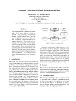
Fig. 1. Promotion of scu-PA activation on U937 cells by plactin. (A)
U937 cells were first incubated with or without plactin D in the
presence of 20% (v ⁄ v) human plasma. After washing, cells were
incubated in the absence or presence of 100 n
M plasmin (PM) for
the indicated time (second incubation). After incubation, cellular
uPA activity was determined using a chromogenic u-PA substrate
in the presence of aprotinin, an inhibitor of plasmin. Line indicates
the average of duplicate determinations. (B) U937 cells were incu-
bated with
125
I-labeled scu-PA in the absence or presence of 50 lM
plactin D and 20% plasma. Aliquots of cell lysates were resolved
on reduced SDS ⁄ PAGE on a 12.5% gel. The positions of molecular
mass standards, as well as scu-PA, A- and B-chains of tcu-PA, are
shown.
T. Harada et al. Dual modulation of prothrombin activation
FEBS Journal 276 (2009) 2516–2528 ª 2009 The Authors Journal compilation ª 2009 FEBS 2517
the total u-PA level (Fig. 1A). The finding that 60%
of u-PA in plactin-treated cells was not activated by
plasmin might be partly explained by the observation
that thrombin-cleaved tcu-PA (see below for the
involvement of thrombin-cleaved tcu-PA) was 500
times less sensitive to activation by plasmin when com-
pared with scu-PA [19]. Next, we determined the con-
version of scu-PA to tcu-PA on the cell surface. In this
experiment, U937 cells equilibrated with
125
I-labeled
scu-PA were treated with plactin D, followed by
SDS ⁄ PAGE of the labeled protein to resolve scu-PA
and tcu-PA. As shown in Fig. 1B, plactin D markedly
promoted conversion of scu-PA to the two-chain form.
The apparent molecular masses of the resulting poly-
peptide chains were comparable with those of the
A- and B-chains of tcu-PA (an A-chain doublet was
caused by differential glycosylation) [20]. The plactin
effect was specific in the presence of plasma (Fig. 1B),
consistent with previous observations [2]. From these
results, we concluded that plactin D promoted cell-
surface activation of scu-PA to tcu-PA, and that the
conversion (specific proteolysis) required a cofactor in
the plasma.
Identification of prothrombin as a plasma factor
participating in plactin activity
To identify the plasma cofactor required for plactin
promotion of scu-PA proteolysis to tcu-PA, we
attempted to develop affinity media to purify plactin-
binding protein. To immobilize plactin onto a gel
matrix, we first looked for plactin derivatives with a
free amino group. One such candidate was plactin-14
(Fig. 2A). Although plactin-14 itself had no activity,
modification of its amino group with a dansyl group
converted the molecule an active form (Fig. 2B). This
0
2.
0
4
.
0
6
.0
8.
0
lortnoC
D nitc
a
l
P
u-PA activity (fluorescence intensity)
-41-nitcalP
e
sor
a
hpeS
-eso
r
ahpeS
B
4
o
N
s
d
a
eb
-
41-
ni
tc
a
l
P
Sephaose
Sepharose-4B
6
6
54
9
2
)a
D
k(
502
611
4.79
Fraction E4A50
RVXXLFGKNA
Prothrombin
T
AV
Q
DANVS
I-A-opA
V
I
-
A
-op
A
Sequence
not obtained
VRDWSSQPDD
0
2.0
4.0
6
.0
8.0
1
2.1
4.1
05040302010
D nitcalP
SND-41-nitcalP
41-nitcalP
Concentration (μ
M
)
u
-PA activity (fluorescence intensity)
HN
HN
2
NH
N
H
H
N
HN
N
H
NH
O
O
O
O
O
N
H
O
HN
e
s
orahpe
S
d
a
eb
esorahpeS-41-n
it
calPSND-41-nitcalP41-nitca
lPD nitcalP
HN
HN
2
N
H
A
C
D
E
B
N
H
HN
HN
N
H
NH
O
O
O
O
O
-
D
g
r
A
-D
laV
ueL
-D ue
L
eh
P
HN
H
N
2
N
H
NH
HN
HN
N
H
N
H
O
O
O
O
O
HN
2
syL
HN
HN
2
N
H
NH
HN
HN
N
H
N
H
O
O
O
O
O
N
H
S
O
O
N
S
N
D
-
s
yL
0
1.0
2.0
3.0
4.0
u-
PA activity (fluorescence intensity)
aX
+ T
Pa
XT
P
lortnoC
D nitcalP
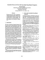
Fig. 2. Identification of prothrombin as a plasma cofactor required for plactin activity. (A) Structures of plactin D and its analogs. DNS,
dansyl. (B) The activities of plactin D, plactin-14 and plactin-14–DNS to enhance cellular u-PA activity were measured by incubating each
compound with U937 cells at the concentrations shown. (C) Human plasma (diluted to 25% v ⁄ v in buffer D) was incubated with or without
Sepharose 4B or plactin-14–Sepharose at 4 °C for 20 min. After centrifugation, the resulting supernatant was assayed for scu-PA activation
on U937 cells at a concentration of 10% (v ⁄ v) of original plasma in the presence or absence of plactin D (50 l
M). (D) Partially purified bovine
plasma fraction E4A50 was subjected to plactin-14–Sepharose chromatography. After flow-through fraction (FT) was collected and the col-
umn was washed with buffer D, elution was carried out successively with buffer D containing 0.5
M NaCl or 6 M guanidine ⁄ HCl (Gnd-HCl).
All fractions were dialyzed against buffer A before the assay. Fractions were resolved on reduced SDS ⁄ PAGE on a 10% gel. Arrowheads
denote specifically enriched proteins. N-terminal sequences of such proteins, and their identifications, are shown. Apo-A, apolipoprotein A.
(E) U937 cells were incubated with the indicated protein(s) at 37 °C for 30 min in the absence or presence of 50 l
M plactin D. The con-
centrations of prothrombin and Xa were 347 n
M and 50 pM, respectively. After washing, cellular u-PA activity was measured. Error bars
represent SD from triplicate determinations. In some data points, error bars are too small to be recognized.
Dual modulation of prothrombin activation T. Harada et al.
2518 FEBS Journal 276 (2009) 2516–2528 ª 2009 The Authors Journal compilation ª 2009 FEBS
result was consistent with the idea that a sterically
restricted arrangement of four hydrophobic amino
acids and a basic amino acid is essential for plactin
activity [2]. Therefore, we speculated that coupling of
plactin-14 to CNBr-activated Sepharose gels via its
amino group should afford an active affinity matrix
(Fig. 2A). Indeed, plactin cofactor activity in human
plasma was successfully adsorbed to plactin-14–Sepha-
rose affinity gel (Fig. 2C). Similar results were
obtained when partially purified bovine plasma (frac-
tion E4A50; see Experimental Procedures) was used
for plactin-14–Sepharose chromatography, and cofac-
tor activity was recovered in fractions eluted with
0.5 m NaCl or 6 m guanidine ⁄ HCl. Some proteins
were specifically enriched in these fractions, although
many protein bands were detected on reduced
SDS ⁄ PAGE (Fig. 2D). No significantly adsorbed pro-
tein was detected when Sepharose 4B alone was used,
suggesting that the nonspecific protein binding in the
plactin-14–Sepharose chromatography was caused by
the hydrophobic surface provided by the coupled
plactin-14. The N-terminal amino acid sequences of
three specifically enriched proteins suggested that these
were prothrombin, apolipoprotein A-IV and apolipo-
protein A-I (Fig. 2D).
We chose prothrombin for further analysis because
prothrombin, but not apolipoproteins, might participate
in the proteolytic cleavage of scu-PA. When prothrom-
bin was used in place of plasma to determine plactin
cofactor activity, it did not support plactin D-dependent
enhancement of u-PA activity in U937 cells (Fig. 2E).
This was not unexpected, as prothrombin itself is an
inactive protease zymogen. Specific proteolysis by the
coagulation factor Xa activates prothrombin to
thrombin. Simultaneous incubation of U937 cells with
prothrombin and factor Xa produced a plactin
D-dependent increase in u-PA activity (Fig. 2E). There-
fore, we suggest that prothrombin is one of the plasma
cofactors participating in plactin D-promoted scu-PA
activation on U937 cells.
Mechanism of prothrombin- and plactin-mediated
enhancement of scu-PA activation
The above results suggested that prothrombin activa-
tion (thrombin formation) was involved in the mecha-
nism of plactin action and that plactin affected this
reaction. Indeed, plactin D increased prothrombin
activation in the U937 cell system (Fig. 3A), and
a-thrombin alone could produce a significant increase
in scu-PA activation on U937 cells (Fig. 3B,C). Plac-
tin D affected a-thrombin-mediated scu-PA activation
only slightly (Fig. 3B). Hirudin, a specific inhibitor of
thrombin, abolished the plactin D effect on scu-PA
activation by prothrombin ⁄ Xa (Fig. 3C). Thus, it
seemed likely that plactin D played a role in increasing
the formation of a-thrombin in prothrombin ⁄ Xa-medi-
ated promotion of scu-PA activation.
a-Thrombin can specifically cleave human scu-PA at
Arg156–Phe157 [7], two residues proximal to the acti-
vation cleavage site (Lys158–Ile159). Thrombin-cleaved
tcu-PA (tcu-PA ⁄ T), however, showed < 1% activity
of tcu-PA (consistent with previous reports) [7,19,21].
Plactin D failed to activate tcu-PA ⁄ T (data not
shown). Nevertheless, incubation of tcu-PA ⁄ T with
U937 cells resulted in the generation of u-PA activity
(Fig. 3D). Thus, there was an additional, cell-associ-
ated mechanism to achieve the generation of fully
active u-PA. One possible candidate is dipeptidyl pep-
tidase I (DPP-I), a thiol protease that could activate
tcu-PA ⁄ T [19], and is expressed at high levels in cyto-
toxic lymphocytes and myeloid cells, including U937
cells. Therefore, we examined the effects of cystatin, an
inhibitor of DPP-I, on tcu-PA ⁄ T activation by U937
cells. Cystatin effectively inhibited tcu-PA activation
by U937 cells (Fig. 3D) and prothrombin ⁄ Xa-mediated
scu-PA activation on U937 cells (Fig. 3E). These
results were consistent with the observation that DPP-I
was able to activate tcu-PA ⁄ T by removing two amino
acids (Phe157–Lys158) from the N-terminus of its
B-chain [19]. The sequential mechanism leading to
enhancement of scu-PA activation is shown in Fig. 3F.
Dual modulation of prothrombin activation
by plactin
The above results suggested that plactin D affected
Xa-catalyzed activation of prothrombin not only in
the U937 system, but also under other conditions. To
characterize the plactin action, prothrombin activation
was assayed using a purified system. Consistent with
the results obtained with the U937 system, prothrom-
bin activation was markedly increased by plactin D
when prothrombin was incubated with Xa (Fig. 4A).
Xa activity, measured using a chromogenic peptide
substrate (Spectrozyme Xa), was minimally affected by
plactin D (Fig. 4A, inset). Thus, it appeared likely that
plactin D altered prothrombin such that it was suscep-
tible to activation by Xa. Under physiological coagula-
tion conditions, prothrombin activation is catalyzed by
the prothrombinase complex (factor Xa in complex
with factor Va, phospholipids and Ca
2+
). When pro-
thrombin activation was assayed using prothrombinase
complex, plactin D inhibited the reaction (Fig. 4B).
Because plactin did not affect the activity of prothrom-
binase toward Spectrozyme Xa (Fig. 4B, inset), it was
T. Harada et al. Dual modulation of prothrombin activation
FEBS Journal 276 (2009) 2516–2528 ª 2009 The Authors Journal compilation ª 2009 FEBS 2519
also likely that plactin D altered prothrombin such
that it became resistant to the activation by Xa that
formed prothrombinase.
To understand the mechanism for these conflicting
effects of plactin D on prothrombin activation, we
tested several combinations of the factors that make
prothrombinase a catalyst (Fig. 4C–H). Prominent
enhancement by plactin D was observed when the
catalyst was Xa ⁄ phospholipids, Xa ⁄ phospholipids ⁄ Va,
Xa ⁄ Va or Xa ⁄ Ca
2+
. However, plactin D led to marked
inhibition when Xa ⁄ phospholipids ⁄ Ca
2+
was used. A
marginal promotive effect of plactin D was observed
when the catalyst was Xa ⁄ Va ⁄ Ca
2+
. Under all these
conditions, plactin D did not affect Xa activity (Fig. 4,
insets) or the activity of isolated a-thrombin (data not
shown). In summary, the data demonstrated that plac-
tin D could promote or inhibit prothrombin activation,
depending on the conditions of activation (Fig. 4I). For
the inhibitory plactin D effect, the presence of both
phospholipids and Ca
2+
was required, whereas the pro-
motive effect was seen in the absence of either phospho-
lipids or Ca
2+
, irrespective of the presence or absence of
factor Va. Phosphatidylserine-containing phospholipid
membranes act as a scaffold for the Ca
2+
-dependent
0
20.0
40.0
60.0
0.08
0.1
A
DE F
B
C
080604020
aX
+
T
P
)nitcalp +(
aX
+ TP
)lortnoc(
lo
rtnoc
ni
t
cal
p +
enola TP
Thrombin activity (Δ A
405
)
)nim( emiT
66
54
92
)aDk(
6
11
4.79
AP-ucs
niahc-B
niahc-A
nibmorhT
D nitc
a
lP
+
−
+
−
aX + nibmorhtorP
−
ni
duriH
−−
−
+++
−
u-PA activity (fluorescence intensity)
α
-
T
h
r
o
m
b
i
n
0
1.0
2.0
3.0
4.0
5.0
6.0
7.0
8.0
P
r
o
t
h
r
o
m
b
i
n
α
-
T
h
r
o
m
b
i
n
+
p
l
a
c
t
i
n
D
0
2
.0
4
.0
6
.
0
8.0
1
2
.1
4.1
6
.
1
u-PA activity (fluorescence intensity)
l
ort
n
o
C
ni
t
at
sy
C
T/AP-uct
+
−
0
20.0
4
0
.
0
6
0
.
0
8
0
.
0
1
.0
21.0
41.
0
u-PA activity (fluorescence intensity)
nitatsyC
+
–
D nitcalP
niduri
H
++
–––+–––+
–+
–
––+
–
–
)
evi
tc
a
n
i(
AP-
ucs
)
e
vi
t
c
a
ni(
T
/
AP
-
uc
t
)
evitc
a
(
A
P
-
u
c
t
α nibmorhT-α nibmorhT-
es
a
et
orP
FGE
R
F
K
I
elgnir
K
es
a
et
orP
FGE
R
F
K
I
elgnir
K
FGE
F
K
I
es
a
e
t
orP
elgnirK
R
FGE
F
K
I
es
a
e
t
orP
elgnirK
R
FGE
elgnirK
R
es
aetorP
I
FGE
elgnirK
R
es
aetorP
I
e
sa
d
i
t
p
e
p
e
k
i
l
-
I
-PPD
e
sa
d
i
t
p
e
p
e
k
i
l
-
I
-PPD
nitc
a
lPnitc
a
lP
nibmorhtorPnibmorhtorP
aXaX
Fig. 3. Mechanism of plactin promotion of prothrombin-mediated scu-PA activation in U937 cells. (A) U937 cells were incubated with human
prothrombin in the presence of 2 m
M CaCl
2
and 0.1 mM Spectrozyme TH to determine thrombin formation. Where indicated, 0.1 nM human
Xa and 25 l
M plactin D were included in the incubation. (B) U937 cells were incubated with the indicated protein in the absence or presence
of 50 l
M plactin D. The concentrations of prothrombin and a-thrombin were 347 and 27 nM, respectively. After washing, cellular u-PA activity
was measured. (C) U937 cells equilibrated with
125
I-labeled scu-PA were incubated with either prothrombin (347 nM) plus factor Xa (3 nM)or
a-thrombin (10 n
M) in the absence or presence of 50 lM plactin D and 30 nM hirudin. After washing, cells were lysed and subjected to
reduced SDS ⁄ PAGE on a 12.5% gel, followed by autoradiography. Positions of scu-PA, A- and B-chains of tcu-PA are shown. (D) U937 cells
(5.0 · 10
6
) were equilibrated with tcu-PA ⁄ T (10 nM)at4°C for 30 min in buffer A. After washing, cells were incubated with GGA-MCA in
the absence or presence of 100 n
M cystatin to determine u-PA activity. (E) U937 cells were treated with prothrombin (347 nM) and factor Xa
(100 p
M) in the absence or presence of 25 lM plactin D. After washing, cells received GGA-MCA to determine u-PA activity in the second
incubation. Where indicated, 30 n
M hirudin or 100 nM cystatin was included both in the first and second incubations. Error bars represent
SD from determinations carried out in triplicate. In some data points, error bars are too small to be recognized. (F) Schematic representation
of prothrombin- and plactin-mediated enhancement of scu-PA activation on U937 cells. The u-PA molecule is shown schematically with each
domain in a colored circle. Amino acid residues involved in proteolytic cleavages are given in white circles. A disulfide bond that connects
A- and B-chains of tcu-PA is shown as red dashed line.
Dual modulation of prothrombin activation T. Harada et al.
2520 FEBS Journal 276 (2009) 2516–2528 ª 2009 The Authors Journal compilation ª 2009 FEBS
assembly of the protein components that form pro-
thrombinase [11]. Therefore, the plactin D action can be
interpreted as follows: plactin D is inhibits prothrombin
activation by membrane-associated Xa, whereas it is
promotive when free Xa is used as a catalyst.
We next examined the possibility that plactin D
induced alternative proteolytic prothrombin cleavages
that resulted in increases or decreases in thrombin
activity. When prothrombin activation was catalyzed
by fully assembled prothrombinase complex, plactin D
inhibited the formation of a-thrombin without produc-
ing proteolytic fragment species other than those
produced during the course of normal prothrombinase
catalysis (Fig. 5A). When free Xa was used to activate
prothrombin, plactin D increased the level of a-throm-
bin (Fig. 5B). Thus, the plactin D effects were increas-
ing or decreasing the formation of a-thrombin without
the accompanying conversion of prothrombin to highly
active or inactive thrombin species.
Interaction between plactin and prothrombin
To investigate the interaction between plactin and
prothrombin, we synthesized a radiolabeled plactin
analog. The analog, [
14
C]plactin-50 [cyclo(-d-Val-l-
[
14
C]Leu-d-Leu-l-Phe-d-Lys-)], had two to three times
the activity of plactin D. Binding of [
14
C]plactin-50 to
prothrombin gave a curve that appeared to become
sigmoidal (Fig. 6A), although maximum binding was
not obtained because of the low solubility of
[
14
C]plactin-50. This observation was consistent with
the promotion and inhibition of prothrombin activa-
tion by plactin, which gave sigmoidal or bell-shaped
dose–response curves (Fig. 4). These properties of the
plactin–prothrombin interaction suggested a change in
the conformation of prothrombin after plactin bind-
ing. We measured the intrinsic fluorescence of
prothrombin to assess any conformational change.
When prothrombin was incubated with plactin D in
the absence of Ca
2+
, the intrinsic fluorescence
increased by 4.6% (P < 0.01) (Fig. 6B). Prothrombin
has several Ca
2+
-binding sites, and Ca
2+
binding
alters its conformation. Accordingly, the intrinsic fluo-
rescence of prothrombin in the presence of Ca
2+
was
significantly lower (6.6%, P < 0.01) than in the
absence of Ca
2+
. Plactin D increased the internal
fluorescence by 7.7% (P < 0.001), even under these
conditions (Fig. 6B). Therefore, it was likely that
plactin–prothrombin binding altered the conformation
of prothrombin and resulted in dual modulation of
prothrombin activation, depending on the conditions
of factor Xa catalysis.
0
05
001
051
002
052
0504
0
3020
1
0
0
002
004
0
0
6
0
0
8
0001
0
02
1
0
5
0
40302010
0
0
2
04
06
08
0
01
0
504
030201
0
Xa activity
0
1
2
3
0
5
0
4
0302
0
10
0
05
0
01
0
51
002
05
2
003
053
05040302010
0
05
0
01
051
0
02
0
52
003
0
5
0
4
0302010
0
001
0
02
003
004
005
05040302010
0
05
001
051
002
052
003
05040302010
0
01
0
2
0
3
04
05
06
050403020
1
0
0
1
2
3
05
04
03
0
2
0
1
0
Xa activity
0
1
2
3
0
5040302
0
10
Xa activity
0
1
2
3
0
504
0
3
0
2
010
Xa activity
0
1
2
3
0
50
4
030
2
010
Xa activity
0
1
2
3
05040302010
Xa activity
0
1
2
3
050
40
30
2
010
Xa activity
0
1
2
3
05040302010
Xa activity
a
X
aX
LP /aX
LP /aX
aC /aX
+2
aC /aX
+
2
a
V
/
aX
a
V
/
a
X
aC / LP /aX
+
2
aC
/ LP
/
aX
+2
aC / LP /aX
+2
aV /
aC /
L
P /aX
+
2
aV /
aV / LP
/a
X
aV
/ LP
/
aX
aC /aX
+2
aV /
aC /aX
+2
aV /
Generation of thrombin activity (Δ A
405
·
h
–2
× 10
3
)
Plactin D (μM)
A
B
C
D
G
EF H
I
Maximal response (%)
001–
05
–
0
0
001
4121%
0002
LP
aC
aV
––––++++
––++++––
–+–+–+–+
Fig. 4. Dual modulation of prothrombin activation by plactin D. (A–H) Factor Xa-catalyzed activation of human prothrombin was determined
by measuring the generation of thrombin using the chromogenic substrate Spectrozyme TH, in the presence of the indicated concentrations
of plactin D. Where indicated, factor Va (4 p
M in panel B and 2 nM in the other panels), PCPS (PL) (50 lM) or CaCl
2
(2 mM) were included.
The concentration of Xa was 1 p
M in (B) and 0.5 nM in the other panels. Inset shows the effect of plactin D on factor Xa activity in each con-
dition. The Xa concentration was 0.5 n
M for all incubations and the Va concentration was 2 nM when added. Ordinate denotes Xa activity as
expressed in A
405
Æmin
)1
· 10
3
, and abscissa plactin D concentration in lM. Each value represents the mean ± SD from determinations
performed in triplicate. (I) Summary of the plactin D effects on prothrombin activation. Maximal response values are plotted.
T. Harada et al. Dual modulation of prothrombin activation
FEBS Journal 276 (2009) 2516–2528 ª 2009 The Authors Journal compilation ª 2009 FEBS 2521
Can plactin be a procoagulant or an anticoagulant?
We asked whether plactin inhibited or stimulated the
blood coagulation system, because plactin dually
modulated prothrombin activation. As shown in
Fig. 7A, plactin D showed anticoagulant activity in
experimental coagulation tests: plactin D significantly
prolonged both activated partial thromboplastin time
(fibrin clot formation time after Ca
2+
addition to
phospholipid-supplemented, contact-phase-activated
plasma) and prothrombin time (clot formation time
after the addition of tissue factor–phospholipids com-
plex and Ca
2+
to plasma). In these measurements,
the enzyme that catalyzed prothrombin activation
was in situ-generated, membrane-associated Xa. How-
ever, thrombin time (clot formation time after the
addition of a-thrombin to plasma), which did not
involve prothrombin activation, was not affected by
plactin D (data not shown). These observations
appeared consistent with results obtained using puri-
fied systems.
Finally, the effect of plactin D on prothrombin
activation was examined in vivo using normal mice.
In one experiment, prothrombin activation was evalu-
ated as the formation of a thrombin ⁄ antithrombin III
complex. When mice were treated with plactin D at
0.1 and 1 mgÆkg
)1
, the level of the complex was not
elevated significantly (Fig. 7B). In another experiment,
the fate of intravenously injected
125
I-labeled pro-
thrombin was determined. Forty minutes after plac-
tin D treatment,
125
I-labeled prothrombin ⁄ thrombin
species in the blood were immunopurified and resolved
by SDS ⁄ PAGE. We did not detect the formation of
thrombin or its complex with antithrombin III in plac-
tin D-treated mice (Fig. 7C). The dose of plactin D
used in these experiments (0.1 or 1 mgÆkg
)1
) was suffi-
cient for plactin D to produce a protective effect in a
thrombin-induced pulmonary embolism model. In this
model, plactin D improved the survival of thrombin-
treated mice. A plactin D dose of 0.1 mgÆkg
)1
increased the survival rate to levels comparable with
that produced by 0.01 UÆkg
)1
of the fibrinolytic
enzyme plasmin (Fig. 7D). Furthermore, plactin D did
not show acute toxicity after intravenous injection at
25 mgÆkg
)1
. These results may exclude the idea that
plactin D is a procoagulant.
It is possible that plactin inhibits physiological
coagulation, which proceeds via membrane-associated
processes [11], and that plactin does not behave as a
procoagulant under normal circulation conditions.
5.00
6
6
5
4
92
)aDk(
611
4
.7
9
54
35.
2
25.
1
15.005435.22
5.11
)
1
Fs
e
d
(T
P
A-2,1F
B
T
P
2,
1
F
A
nim
ni
tcalPl
ort
noC
A
B
noitavitca dezylatac-esanibmorhtorP
nitcalP
α nibm
or
hT
-
dr
a
d
n
a
ts
66
5
4
9
2
)aD
k
(
lo
r
tnoC
noitavitca dezylatac-aX
Fig. 5. Analysis of thrombin species formed
in the presence of plactin D. (A) Human pro-
thrombin was activated by prothrombinase
complex in the absence or presence of
50 l
M plactin D. At the indicated times,
aliquots of the incubation mixtures were
withdrawn to analyze by reduced
SDS ⁄ PAGE on a 10% gel. Proteins were
visualized by Coomassie Brilliant Blue
R-250. The positions of prothrombin (PT),
prothrombin(desF1) [prothrombin without
fragment 1; PT(desF1)] and fragment
1 + 2 + A-chain (F1,2-A), as well as A-chain
and B-chain of a-thrombin B, are shown. (B)
Human prothrombin was activated by free
factor Xa in buffer F containing 2 m
M CaCl
2
in the absence or presence of 25 lM
plactin D. Proteolytically active molecular
species were visualized by casein zymo-
graphy after resolving on nonreduced
SDS ⁄ PAGE on a 10% gel. Human
a-thrombin (0.3 lg) was used as a
standard.
Dual modulation of prothrombin activation T. Harada et al.
2522 FEBS Journal 276 (2009) 2516–2528 ª 2009 The Authors Journal compilation ª 2009 FEBS
Conclusion
Our studies demonstrate plactin-mediated modulation
of prothrombin activation. Plactin binds to prothrom-
bin and dually modulates its activation, depending on
the form of the catalyst, factor Xa. Under physiologi-
cal conditions, the coagulation reaction proceeds via
membrane-associated processes. Plactin inhibits pro-
thrombin activation catalyzed by membrane-associated
Xa. This is consistent with the observation that plactin
inhibits the coagulation of plasma in activated partial
thromboplastin time tests and prothrombin time tests.
However, plactin enhances prothrombin activation
when the catalyst is nonmembrane-bound Xa. This
mechanism may participate in the enhancement of
fibrinolytic activity in the U937 cell system, in which
plactin enhances prothrombin activation and the for-
mation of inactive tcu-PA ⁄ T, which is subsequently
converted to fully active tcu-PA by cellular cystatin-
sensitive, DPP-I-like peptidase.
The specificity of prothrombinase for prothrombin is
mediated by exosites, which are physically separated
from the catalytic site, on the surfaces of the catalytic
domains. It is postulated that substrate recognition by
prothrombinase involves a two-step mechanism with
initial docking of prothrombin to exosites, followed
by a conformational change to engage the Xa catalytic
site [22]. Thus, prothrombin activation is a conforma-
tionally regulated process. This may partly explain
the plactin-mediated dual modulation of prothrombin
activation. The pharmacological application of dual
thrombin modulation would be an intriguing approach
to intervention in thromboembolic diseases.
Experimental procedures
Plactins
Plactin D [cyclo(-d-Val-l-Leu-d-Leu-l-Phe-d-Arg-)] and
plactin-14 [cyclo(-d-Val-l-Lys-d-Leu-l-Phe-d-Arg-)] were
synthesized according to Fmoc chemistry, as described pre-
viously [1,2]. Dansylplactin-14 (plactin-14-DNS) was syn-
thesized by mixing 1 mL of plactin-14 (1 mgÆmL
)1
in
water), 1 mL of dansyl chloride (4 mgÆ mL
)1
in acetone)
and 90 mg of NaCO
3
overnight at ambient temperature.
Plactin-14–Sepharose was prepared by reacting 35 mL of
0.7 mgÆmL
)1
plactin-14 with 1.5 g of CNBr-activated
Sepharose 4B (GE Healthcare Biosciences, Tokyo, Japan)
in 0.1 m sodium bicarbonate, pH 9.0, and 0.5 m NaCl, fol-
lowed by blocking with 1 m ethanolamine. The amount of
plactin-14 immobilized was 7.0 lmolÆmL
)1
of gel. [
14
C]Plac-
tin-50 [cyclo(-d-Val-l-Leu-d-Leu-l-Phe-d-Lys-)] was synthe-
sized using Fmoc-l-Leu (1-
14
C) (American Radiolabeled
Chemicals Inc, St Louis, MO, USA). The specific radioac-
tivity was 1.02 BqÆ pmol
)1
. For assays, plactins dissolved in
dimethylsulfoxide were used at a solvent concentration of
1% (v ⁄ v).
Other materials
Human scu-PA was provided by Mitsubishi Tanabe
Pharma Corporation (Osaka, Japan). Other proteins and
chemicals were from the following sources: human tcu-PA
from JCR Pharmaceutical (Kobe, Japan); human plasmin
and aprotinin from Wako (Osaka, Japan); human pro-
thrombin, human coagulation factor Xa, the thrombin
1.8
1.5
1.2
0.9
0.6
0.3
0
86
4
2
0
[
14
C]Plactin-50 (µM)
[
14
C]Plactin-50 binding to prothrombin
(pmol bound per pmol prothrombin)
*
*
21
20
19
18
22
B
A
Ca
2+
Control
Plactin D (50 µ
M
)
Intrinsic fluorescence
+
−
Fig. 6. Interaction between plactin and prothrombin. (A) The bind-
ing of [
14
C]plactin-50 to human prothrombin was determined in the
presence of the indicated concentrations of [
14
C]plactin-50. Specific
binding data are shown. (B) The intrinsic fluorescence of human
prothrombin was measured in the absence or presence of CaCl
2
(2 mM) and plactin D (50 lM). *P < 0.01 by Student’s t-test, com-
pared with control. Error bars represent SD from determinations
performed in triplicate.
T. Harada et al. Dual modulation of prothrombin activation
FEBS Journal 276 (2009) 2516–2528 ª 2009 The Authors Journal compilation ª 2009 FEBS 2523
inhibitor dansylarginine-N-(3-ethyl-1,5-pentanediyl)amide
(DAPA) and polyclonal anti-(human thrombin) sheep IgG
from Haematologic Technologies (Essex Junction, VT,
USA); human coagulation factor V from Serbio (Paris,
France); human a-thrombin, BSA, cystatin and l-a-phos-
phatidylcholine (egg yolk) from Sigma (St Louis, MO,
USA); l-a-phosphatidylserine (porcine brain) from Avanti
Polar Lipids (Alabaster, AL, USA); glutaryl-Gly-Arg-4-
00.0
02.0
04.0
06.0
08
.
0
00
.
1
nitcalP
*
*
51
/
3
41/31
5
1
/
21
Fraction survived
n
im
s
a
l
Plort
n
oC
TP
)1Fsed(TP
α nibmor
h
T-
66
54
92
)a
D
k
(
4.
7
9
11.0
Coagulating
blood
Plactin D
(mg·kg
–1
)
Control
Plactin D
(mg·kg
–1
)
10.1
Control
Plasma TAT level (ng·mL
–1
)
4
6
40
30
20
10
2
0
31.8
Coagu-
lating
blood
0
35
AB
CD
30
25
20
15
10
5
060504030201
0
Prothrombin
time
Activated partial
thromboplastin time
Plactin D (μ
M)
Percent change
*
**
**
**
**
**
**
**
**
Fig. 7. Effects of plactin D on plasma coagulation in vitro and prothrombin activation in vivo. (A) Activated partial thromboplastin time and
prothrombin time were measured using normal human plasma. Plactin D was added 5 min before the initiation of each reaction. The clotting
times in the absence of plactin D were 26.9 ± 0.2 s for activated partial thromboplastin time and 15.1 ± 0.7 s for prothrombin time. Error bars
represent SD from triplicate determinations. *P < 0.05 and **P < 0.01 by Dunnett’s test, compared with control. (B) Plactin D, at the indicated
dose, was given intravenously to mice (n = 5 for each group), and blood was drawn in a mixture of protease inhibitors, 40 min after the treat-
ment. The level of thrombin ⁄ antithrombin III complex in the resulting plasma was determined by enzyme immunoassay. Serum obtained from
blood drawn without anticoagulants from normal mice (Coagulating blood) was used as a standard. There were no statistical differences among
control, 0.1 mgÆkg
)1
plactin D and 1 mgÆkg
)1
plactin D groups by Dunnett’s test. (C) Plactin D and human
125
I-labeled prothrombin were
successively given intravenously to mice (n = 3 for each group). Blood was drawn in a mixture of protease inhibitors, 40 min after treatment.
Labeled proteins were purified from plasma with anti-(human thrombin) IgG–Sepharose and resolved on nonreduced SDS ⁄ PAGE on a 10% gel.
Serum from control mouse blood was similarly processed as a standard to detect prothrombin activation (Coagulating blood). Data shown are
representative. Essentially the same results were obtained in each group. The positions of prothrombin (PT), and prothrombin(desF1)
[PT(desF1)] and a-thrombin are shown. (D) Effect of plactin D on thrombin-induced pulmonary embolism in mice. Mice received intravenous
injection with saline (Control) plactin D (0.1 mgÆkg
)1
) or plasmin (0.01 UÆkg
)1
). After 15 min, human a-thrombin was injected intravenously to
induce pulmonary thromboembolism. Next day, the number of surviving animals was counted. Numbers above bars denote the number of
survived ⁄ total animals in each group. *P < 0.01 by Fisher’s exact test, compared with control.
Dual modulation of prothrombin activation T. Harada et al.
2524 FEBS Journal 276 (2009) 2516–2528 ª 2009 The Authors Journal compilation ª 2009 FEBS
methylcoumarin-7-amide (GGA-MCA) from Peptide
Institute (Osaka, Japan); Spectrozyme TH (H-d-hexahydro-
tyrosyl-Ala-Arg-p-nitroanilide), Spectrozyme Xa (methoxy-
carbonyl-d-hexahydrotyrosyl-Ala-Arg-p-nitroanilide) and
recombinant hirudin from American Diagnostica (Green-
wich, CT, USA).
Factor V (300 nm) was activated to Va by incubating
with a-thrombin (3 nm)at37°C for 10 min. Thrombin-
cleaved tcu-PA (tcu-PA ⁄ T) was prepared by incubating
scu-PA (1 lm) with a-thrombin (10 nm)at37°C for 22 h,
followed by the addition of 30 nm hirudin to neutralize
thrombin. Phospholipid vesicles (PCPS) composed of 75%
(w ⁄ w) phosphatidylcholine and 25% (w ⁄ w) phosphatidyl-
serine were prepared as described previously [23]. Radio-
iodination of scu-PA and prothrombin was performed by
the IODO-GEN method [24], using carrier-free Na
125
Itoa
specific activity of 2000–3000 cpmÆ ng
)1
of protein.
Buffers used were: buffer A, 20 mm sodium phosphate,
pH 7.4, and 150 mm NaCl; buffer B, 50 mm Tris ⁄ HCl,
pH 7.4, and 100 mm NaCl; buffer C, 50 mm sodium phos-
phate, pH 7.4, and 80 mm NaCl; buffer D, 50 mm sodium
phosphate, pH 7.4; buffer E, 50 mm Tris ⁄ HCl, pH 7.4,
100 mm NaCl and 0.01% (w ⁄ v) Tween 80; buffer F, 20 mm
Tris ⁄ HCl, pH 7.4, 150 mm NaCl and 0.1% (w ⁄ v)
Tween 80; buffer G, 62.5 mm Tris ⁄ HCl, pH 6.8, 2% SDS,
10% glycerol, 5% 2-mercapthoethanol and 0.002% bromo-
phenol blue.
Cell culture
Human monocytoid line U937 cells (obtained from the
Japanese Cancer Research Resources Bank, Tokyo) were
maintained in RPMI-1640 medium supplemented with 10%
fetal bovine serum (JRH Biosciences, Lenexa, KS, USA),
100 UÆmL
)1
penicillin G and 100 lgÆmL
)1
streptomycin.
For assays, cells were seeded at 2 · 10
5
cellsÆmL
)1
in 15 mL
of the medium and grown for 2 days. Prior to use in experi-
ments, exponentially growing cells were harvested, washed
twice and suspended with buffer A.
Assay for cellular scu-PA activation
U937 cells were suspended with buffer A at a density of
5.0 · 10
6
cellsÆmL
)1
. Cells were incubated in the absence or
presence of 20% (v ⁄ v) human plasma and plactin at 37 °C
for 30 min, with shaking. After washing with buffer B, cells
were resuspended in buffer B containing 0.1 mm GGA-
MCA, a chromogenic peptide substrate for u-PA. After
incubation at 22 °C for 1 h, the supernatant was removed
and acetic acid was added to 10% to stop the reaction. The
fluorescence of 7-amino-4-methylcoumarine liberated from
GGA-MCA by the u-PA cleavage was measured (excitation
at 380 nm and emission at 480 nm).
In the experiment shown in Fig. 1A, cells treated in the
first incubation were further incubated with plasmin
(100 nm)at22°C for the indicated time in buffer B con-
taining BSA (10 mgÆmL
)1
). After addition of aprotinin (40
kallikrein inhibitor unitsÆmL
)1
to neutralize plasmin) and
washing, cells were processed to determine u-PA activity as
described above.
In some experiments, scu-PA activation on U937 cells
was also determined as the proteolytic cleavage of
125
I-
labeled scu-PA. In this experiment,
125
I-labeled scu-PA
(5.6 nm, 3000 cpmÆng
)1
) was included in the first incuba-
tion. After washing twice with buffer B, cells were lysed
with buffer G. An aliquot of the lysate was subjected to
SDS ⁄ PAGE on a 12.5% gel. After fixing and drying, the
gel was exposed to an X-ray film at )80 °C for 16 h. In the
experiment shown in Fig. 3C,
125
I-labeled scu-PA was
bound to cell surface at 4 °C for 30 min in RPMI-1640
medium supplemented with 10% fetal bovine serum and
20 mm Hepes, pH 7.4. The labeled cells were used for
incubations, as described in the legend to Fig. 3.
Partial purification of plactin cofactor from
bovine plasma
Citrated bovine platelet-poor plasma (490 mL) was frac-
tionated using the method described by Cohn et al. [25].
Most of the cofactor activity to support plactin-dependent
activation of cellular scu-PA was recovered in the ‘precipi-
tate IV-1¢ fraction. The fraction was subjected to ammo-
nium sulfate fractionation at 4 °C, and precipitates
obtained from 25–50% saturation were dialyzed against
buffer C, yielding 2.3 g of partially purified cofactor
preparation (fraction E4A50). The specific activity of the
preparation was 24 times that of the original plasma.
Plactin-14–Sepharose chromatography
A column containing 0.5 mL of plactin-14–Sepharose was
equilibrated with buffer D at room temperature, and
0.6 mL of fraction E4A50 (11 mg protein) was applied to
the column. After washing with 2.5 mL of buffer D, the
column was developed with 2.5 mL of buffer D containing
0.5 m NaCl, followed by 2.5 mL of buffer D containing
6 m guanidine ⁄ HCl. Each eluate was dialyzed overnight
against buffer A before SDS ⁄ PAGE and assay for plactin-
dependent promotion of scu-PA activation on U937 cells.
Assay for prothrombin activation
The activation of prothrombin was assayed by incubating
human prothrombin (20 nm) and Spectrozyme TH
(0.1 mm) in the presence of factor Xa in buffer F with or
without factor Va (4 pm or 2 nm), PCPS (50 lm) or CaCl
2
(2 mm). The concentration of Xa was 1 pm when the incuba-
tion contained Va (4 pm), PCPS and CaCl
2
to assemble pro-
thrombinase complex. In other assays, the Xa concentration
T. Harada et al. Dual modulation of prothrombin activation
FEBS Journal 276 (2009) 2516–2528 ª 2009 The Authors Journal compilation ª 2009 FEBS 2525
was 0.5 nm, and the concentration of Va was 2 nm when it
was included in the mixture, other than that for prothrom-
binase formation. The reaction was started by adding Xa,
and the change in absorbance at 405 nm was monitored
kinetically at 37 °C. From the slope of the plots of A
405
versus t
2
[26], the initial velocity of thrombin generation
was calculated.
In the SDS ⁄ PAGE assay for the prothrombinase-cata-
lyzed reaction, 1.4 lm prothrombin was incubated with
30 lm PCPS, 2 mm CaCl
2
,3lm DAPA, 1 nm Xa and
5nm Va at 37 °C for the indicated times in buffer F
(DAPA was included to inhibit the feedback proteolysis of
prothrombin by the generated thrombin). The reaction was
stopped by the addition of an equal volume of acetic acid,
and the resulting mixture was dialyzed against 0.2 m acetic
acid. After lyophilization, followed by dissolving in
buffer G, samples were resolved on reduced SDS ⁄ PAGE.
In the zymography assay for the free Xa-catalyzed reac-
tion, a reaction mixture (20 lL) containing 4 lm prothrom-
bin, 2 mm CaCl
2
,3nm Xa and 10 lm DAPA was
incubated at 37 °C for 1 h in buffer F. After incubation,
aliquots of the mixture were subjected to nonreduced
SDS ⁄ PAGE on a 10% gel containing 0.5 mgÆmL
)1
casein.
The gel was washed twice for 30 min with 2.5% (w ⁄ v)
Triton X-100 to remove SDS, followed by overnight incu-
bation at 37 °C with buffer F containing 2 mm CaCl
2
.
After staining with Coomassie Brilliant Blue R-250 the pro-
teolytically active position appeared as a colorless band on
dark blue background.
The activation of prothrombin in the presence of U937
cells was assayed by incubating 1.0 · 10
6
cellsÆmL
)1
in buf-
fer A containing prothrombin (20 nm), 0.1 nm Xa, 2 mm
CaCl
2
and 0.1 mm Spectrozyme TH at 37 °C for the times
indicated in Fig. 3A. After centrifugation, the absorbance
at 405 nm in the supernatant was measured.
[
14
C]Plactin-50 binding to prothrombin
Human prothrombin (0.95 lm) was incubated with
[
14
C]plactin-50 in buffer E at 37 °C for 1 h, followed by
standing on ice for 15 min. Bound and free [
14
C]plactin-50
was separated by spin column chromatography. We applied
20 lL of the reaction mixture to a spin column (prepared
by centrifuging 500 lL of a 15.2% w ⁄ v suspension of
Sephadex G-25 in buffer E at 2000 g for 1 min), and the
column was centrifuged at 2000 g for 1 min. The radioac-
tivity in the eluate was counted for 3 min in a liquid scintil-
lation counter. The amount of bound [
14
C]plactin-50 was
calculated by subtracting the radioactivity obtained in the
absence of prothrombin from that obtained in its presence.
Animal experiments
Animal experiments were performed in accordance with
guidelines for animal experiments at Tokyo Noko Univer-
sity. We took adequate steps to ensure that animals did not
suffer unnecessarily at any stage of an experiment. The
protocol was approved by the Animal Experiment Commit-
tee of Tokyo Noko University.
To determine the level of thrombin ⁄ antithrombin III
complex in plasma, male ICR mice ( 30 g; Japan SLC,
Hamamatsu) were anesthetized with intraperitoneal
urethane ⁄ a-chlorarose (750 and 60 mgÆkg
)1
, respectively).
Plactin D dissolved in saline was given to the mice intrave-
nously from caudal vein. After 40 min, blood collected by
cardiac puncture (720 lL) was immediately mixed with
80 lL of 3.8% (w ⁄ v) sodium citrate containing an inhibitor
cocktail (300 mm benzamidine, 50 lm leupeptin, 10 lm
antipain, 5 mm EDTA, 28 lm E64, 1 lm pepstatin and
0.2 mm FUT-175) to obtain plasma. The level of throm-
bin ⁄ antithrombin III complex was determined by enzyme
immunoassay at SRL (Tokyo, Japan).
To assay for
125
I-labeled prothrombin activation in vivo ,
male ICR mice (anesthetized with urethane ⁄ a-chlorarose)
received intravenous plactin D. Immediately after the plactin
injection, mice received an intravenous injection of human
125
I-labeled prothrombin (3.8 · 10
7
cpmÆkg
)1
). After 40 min,
blood was collected in sodium citrate ⁄ inhibitor cocktail as
described above. The resulting platelet-poor plasma (200 lL)
was incubated with 10 lL of anti-(human thrombin) IgG–
Sepharose 4B at room temperature for 30 min. The Sepha-
rose beads were washed three times with buffer A containing
0.1% (w ⁄ v) Nonidet P-40 and 1 ⁄ 10 volume of inhibitor cock-
tail, followed by boiling for 2 min in 10 lL of 2% SDS, 10%
glycerol and 20 mm sodium phosphate, pH 6.0. Aliquots of
supernatant were resolved on a 7.5% SDS-polyacrylamide
gel and processed for autoradiography.
In the thrombin-induced pulmonary embolism model,
male ddY mice ( 30 g; Japan SLC) were fasted for 5 h
before experiment. Plactin D was injected intravenously
through caudal vein. Fifteen minutes after the plactin injec-
tion, 1 mL of human a-thrombin (10 UÆmL
)1
) was given
intravenously to the mice to induce pulmonary embolism
[27]. Next day, the number of surviving animals was counted.
Other methods
Thrombin activity was determined at 37 °C in buffer F con-
taining 2 mm CaCl
2
using 1.5 nm human a-thrombin and
0.1 mm Spectrozyme TH. Xa activity was measured at
37 °C in buffer F with or without CaCl
2
(2 mm), PCPS
(50 lm)orVa(2nm) using 0.5 nm human Xa and Spectro-
zyme Xa. The intrinsic fluorescence of human prothrombin
was measured in buffer E with or without 2 mm CaCl
2
after incubation of 100 nm prothrombin and 50 lm plac-
tin D for 5 min at room temperature. Excitation and emis-
sion wave lengths were 290 and 340 nm, respectively. The
N-terminal amino acid sequence was determined after
transferring to poly(vinylidene difluoride) membrane using
an Applied Biosystems model 476A protein sequencer.
Dual modulation of prothrombin activation T. Harada et al.
2526 FEBS Journal 276 (2009) 2516–2528 ª 2009 The Authors Journal compilation ª 2009 FEBS
Activated partial thromboplastin time and prothrombin
time were measured using commercial kits (Sysmex Interna-
tional Reagents, Kobe, Japan) according to the manufac-
turer’s instructions.
Acknowledgements
We thank Akira Endo for encouragement and Hiro-
yuki Yoshii and Emiko Iwao for technical assistance.
This work was supported by a grant from the Japan
Society for the Promotion of Science.
References
1 Inoue T, Hasumi K, Kuniyasu T & Endo A (1996)
Isolation of plactins A, B, C and D, novel cyclic
pentapeptides that stimulate cellular fibrinolytic activity.
J Antibiot (Tokyo) 49, 45–49.
2 Inoue T, Hasumi K, Sugimoto M & Endo A (1998)
Enhancement of fibrinolysis by plactins: structure–activ-
ity relationship and effects in human U937 cells and in
mice. Thromb Haemost 79, 591–596.
3 Collen D & Lijnen HR (2005) Thrombolytic agents.
Thromb Haemost 93, 627–630.
4 Danø K, Andreasen PA, Grondahl-Hansen J, Kristen-
sen P, Nielsen LS & Skriver L (1985) Plasminogen acti-
vators, tissue degradation, and cancer. Adv Cancer Res
44, 139–266.
5 Stoppelli MP, Tacchetti C, Cubelis MV, Corti A, Hear-
ing VJ, Cassani G, Appella E & Blasi F (1986) Auto-
crine saturation of pro-urokinase receptors on human
A431 cells. Cell 45, 675–684.
6 Kobayashi H, Moniwa N, Sugimura M, Shinohara H,
Ohi H & Terao T (1993) Effects of membrane-associ-
ated cathepsin B on the activation of receptor-bound
prourokinase and subsequent invasion of reconstituted
basement membranes. Biochim Biophys Acta 1178, 55–
62.
7 Ichinose A, Fujikawa K & Suyama T (1986) The
activation of pro-urokinase by plasma kallikrein and
its inactivation by thrombin. J Biol Chem 261, 3486–
3489.
8 Stack MS & Johnson DA (1994) Human mast cell tryp-
tase activates single-chain urinary-type plasminogen
activator (pro-urokinase). J Biol Chem 269, 9416–9419.
9 Andreasen PA, Kjøller L, Christensen L & Duffy MJ
(1997) The urokinase-type plasminogen activator system
in cancer metastasis: a review. Int J Cancer 72, 1–22.
10 Danø K, Behrendt N, Høyer-Hansen G, Johnsen M,
Lund LR, Ploug M & Romer J (2005) Plasminogen
activation and cancer. Thromb Haemost 93, 676–681.
11 Mann KG (1999) Biochemistry and physiology of blood
coagulation. Thromb Haemost 82, 165–174.
12 Krishnaswamy S, Mann KG & Nesheim ME (1986)
The prothrombinase-catalyzed activation of prothrom-
bin proceeds through the intermediate meizothrombin
in an ordered, sequential reaction. J Biol Chem 261,
8977–8984.
13 Krishnaswamy S, Church WR, Nesheim ME & Mann
KG (1987) Activation of human prothrombin by human
prothrombinase. Influence of factor Va on the reaction
mechanism. J Biol Chem 262, 3291–3299.
14 Mann KG, Nesheim ME, Church WR, Haley P &
Krishnaswamy S (1990) Surface-dependent reactions of
the vitamin K-dependent enzyme complexes. Blood 76,
1–16.
15 Rosing J, Tans G, Govers-Riemslag JW, Zwaal RF &
Hemker HC (1980) The role of phospholipids and fac-
tor Va in the prothrombinase complex. J Biol Chem
255, 274–283.
16 Esmon CT (1989) The roles of protein C and thrombo-
modulin in the regulation of blood coagulation. J Biol
Chem 264, 4743–4746.
17 Esmon CT (1995) Thrombomodulin as a model of
molecular mechanisms that modulate protease
specificity and function at the vessel surface. FASEB J
9, 946–955.
18 Von dem Borne PAK, Bajzar L, Meijers JCM, Nesheim
ME & Bouma BN (1997) Thrombin-mediated activation
of factor XI results in a thrombin-activatable fibrinoly-
sis inhibitor-dependent inhibition of fibrinolysis. J Clin
Invest 99, 2323–2327.
19 Nauland U & Rijken DC (1994) Activation of throm-
bin-inactivated single-chain urokinase-type plasminogen
activator by dipeptidyl peptidase I (cathepsin C). Eur J
Biochem 223, 497–501.
20 Narasaki R, Kuribayashi H, Shimizu K, Imamura D,
Sato T & Hasumi K (2005) Bacillolysin MA, a novel
bacterial metalloproteinase that produces angiostatin-
like fragments from plasminogen and activates protease
zymogens in the coagulation and fibrinolysis systems.
J Biol Chem 280, 14278–14287.
21 Abercrombie DM, Buchinski B, Salvato KA, Vovis GF,
Stump DC & Broeze RJ (1990) Fibrin specific throm-
bolysis by two-chain urokinase-type plasminogen activa-
tor cleaved after arginine 156 by thrombin. Thromb
Haemost 64, 426–432.
22 Bock PE, Panizzi P & Verhamme IM (2007) Exosites in
the substrate specificity of blood coagulation reactions.
J Thromb Haemost 5(Suppl. 1), 81–94.
23 Barenholz Y, Gibbes D, Litman BJ, Goll J, Thompson
TE & Carlson FD (1977) A simple method for the
preparation of homogeneous phospholipid vesicles.
Biochemistry 16, 2806–2810.
24 Salacinski PRP, McLean C, Sykes JEC, Clement-Jones
VV & Lowry PJ (1981) Iodination of proteins,
glycoproteins, and peptides using a solid-phase
oxidizing agent, 1,3,4,6-tetrachloro-3 alpha,6
alpha-diphenyl glycoluril (Iodogen). Anal Biochem 117,
136–146.
T. Harada et al. Dual modulation of prothrombin activation
FEBS Journal 276 (2009) 2516–2528 ª 2009 The Authors Journal compilation ª 2009 FEBS 2527
25 Cohn EJ, Strog LE, Hughes WL, Mulford DJ, Ash-
worth JN, Melin M & Taylor HL (1946) Preparation
and properties of serum and plasma proteins. IV. A
system for the separation into fractions of the protein
and lipoprotein components of biological tissues and
fluid. J Am Chem Soc 68, 459–475.
26 Verheijen JH, Mullaart E, Chang GTG, Kluft C &
Wijngaards J (1982) A simple, sensitive spectrophoto-
metric assay for extrinsic (tissue-type) plasminogen
activator applicable to measurements in plasma.
Thromb Haemost 48, 266–269.
27 Gresele P, Momi S, Berrettini M, Nenci GG, Schwarz
HP, Semeraro N & Colucci M (1998) Activated human
protein C prevents thrombin-induced thromboembolism
in mice. Evidence that activated protein c reduces intra-
vascular fibrin accumulation through the inhibition of
additional thrombin generation. J Clin Invest 101, 667–
676.
Dual modulation of prothrombin activation T. Harada et al.
2528 FEBS Journal 276 (2009) 2516–2528 ª 2009 The Authors Journal compilation ª 2009 FEBS