Báo cáo khoa học: In vivo RNA interference in oyster – vasa silencing inhibits germ cell development pptx
Bạn đang xem bản rút gọn của tài liệu. Xem và tải ngay bản đầy đủ của tài liệu tại đây (641.86 KB, 8 trang )
In vivo RNA interference in oyster – vasa silencing inhibits
germ cell development
Caroline Fabioux
1,2
, Charlotte Corporeau
1,3
, Virgile Quillien
1,3
, Pascal Favrel
1,3
and
Arnaud Huvet
1,3
1 UMR 100 PE2M Ifremer-Universite
´
de Caen, Ifremer centre de Brest, B.P.70, Plouzane
´
, France
2 UMR CNRS 6539, LEMAR, Universite
´
de Bretagne Occidentale, IUEM, Plouzane
´
, France
3 UMR 100 PE2M Ifremer-Universite
´
de Caen, IBFA, IFR 146 ICORE, Caen Cedex, France
The oyster Crassostrea gigas has stimulated a great
deal of biological research, as it represents a major
economic resource for aquaculture (production:
4.2 million metric tons; [1]), it plays a sentinel role in
estuarine and coastal marine habitats [2], and it
belongs to the Lophotrochozoa, a vast and diverse
branch of bilaterian animals that have been little stud-
ied with respect to genomics. The recent emergence of
bivalve genomics, with substantial characterization of
genome-wide expression sequences, especially for
C. gigas [2,3], argues for the rapid development of
methodologies to unravel gene function in these
species.
Classic functional genetic approaches such as muta-
genesis are not yet available for bivalve molluscs. A
powerful alternative method for reverse genetics is
RNA interference (RNAi), which can be a quick and
efficient technique for determining the loss-of-function
phenotype of a gene [4]. The RNAi revolution was
started by evidence that dsRNA could knock down
the expression of specific genes [5]. The 25 nucleo-
tide small interfering RNA fragments generated by
processing long dsRNAs are reported to be the media-
tors of RNAi [6]. Small interfering RNA provides
sequence specificity to the RNA-induced silencing com-
plex, which inhibits the corresponding mRNA, thereby
silencing the targeted gene [7]. RNAi has been widely
used in vitro and in vivo in vertebrate and invertebrate
species [5,8–11]. Conversely, RNAi studies are scarce
in molluscs. RNAi has been used, for example, in
gastropods to explore gene functions in the nervous
system [12], and in the cephalopod Sepia officinalis to
analyse the role of muscle regulatory factor in tentacle
muscle differentiation [13]. In bivalve molluscs, RNAi
remains a technical challenge. To document in vivo
gene silencing by RNAi in the oyster, we injected
dsRNA targeting the oyster vasa-like gene (Oyvlg). In
Drosophila and Caenorhabditis, vasa plays a key role in
Keywords
Crassostrea gigas; germline; marine bivalve;
RNAi; vasa
Correspondence
C. Fabioux, UMR CNRS 6539, LEMAR,
Universite
´
de Bretagne Occidentale, IUEM,
Plouzane
´
, France
Fax: +33 0 2 98 49 8645
Tel: +33 0 2 98 49 8744
E-mail:
(Received 9 December 2008, revised 20
February 2009, accepted 25 February 2009)
doi:10.1111/j.1742-4658.2009.06982.x
This study investigated the potential of RNA interference, which is techni-
cally challenging in bivalve mollusc species, to assess gene function in the
oyster Crassostrea gigas. We designed dsRNA targeting the oyster vasa-like
gene (Oyvlg), specifically expressed in oyster germ cells. In vivo injection of
oyvl-dsRNA into the gonad provokes a knockdown phenotype correspond-
ing to germ cell underproliferation and prematurely arrested meiosis throu-
gout the organ. The most severe phenotype observed is sterile. This
knockdown phenotype is associated with a decrease in Oyvlg mRNA level
of between 39% and 87%, and a strong reduction in OYVLG protein, to
an undetectable level. Therefore, Oyvlg appears to be essential for germ cell
development in Crassostrea gigas, particularly for mitotic proliferation and
early meiosis. Our results demonstrate for the first time that in vivo RNA
interference works efficiently in a bivalve species, opening major perspec-
tives for functional genetic studies.
Abbreviations
DIG, digoxygenin; EFI, elongation factor I; NPY, neuropeptide Y; RNAi, RNA interference.
2566 FEBS Journal 276 (2009) 2566–2573 ª 2009 The Authors Journal compilation ª 2009 FEBS
germ cell differentiation, as clearly demonstrated by
functional analysis of mutation or inactivation of the
gene, which in the most striking cases can lead to total
sterility [14,15]. In the oyster C. gigas, Oyvlg is specifi-
cally expressed in germ cells and was thought to play a
role in germline development [16,17]. In this study, the
oyster vasa-like gene was chosen to develop in vivo
RNAi in the oyster, not only to assess the function of
Oyvlg in germline formation, but also to investigate
the potential of this methodology to serve as a
routine means for gene function assignment in bivalve
molluscs.
Results and Discussion
Validation of OYVLG-specific antibody
As demonstrated by immunodetection on western blot
against total protein extracts from oyster tissues
(mantle, gills, muscle, labial palps, digestive gland, and
gonad), the synthetic polyclonal antibodies (Millegen,
Labege, France) targeting two peptides specific to
OYVLG recognized a unique band of apparent mole-
cular mass of 79 kDa corresponding to the predicted
size for OYVLG (Fig. 1). The distribution of the anti-
genic protein appeared to be restricted to gonadic tissue
in both sexes, with a higher quantity of protein in
female than in male mature gonads, in accordance with
the Oyvlg mRNA expression pattern [17]. As a result,
antibodies (Fab1 + Fab2) were used in this study to
detect and quantify the amount of OYVLG protein.
Design of RNAi experiment in the oyster
The oyster vasa-like gene was chosen for the develop-
ment of an RNAi method in the oyster for several
important reasons: (a) the determination of the role of
Oyvlg in C. gigas is of major interest for our physio-
logical research into oyster reproduction; (b) the
spatiotemporal expression of Oyvlg mRNA has been
clearly characterized in the oyster [17], showing specific
expression in germ cells; (c) inactivation of the vasa
gene has been successful for several species [14,15,18],
leading to a clear phenotypic effect that is easily mea-
surable (i.e. partial or total sterility); and (d) specific
antibodies are now available against OYVLG to mea-
sure the effect of oyvl dsRNA administration at the
protein level, in addition to real-time PCR for the
mRNA level [16].
Because long dsRNAs have been shown to perform
efficient gene silencing in invertebrates [4], we synthe-
sized two long dsRNAs, oyvl4-dsRNA and
oyvl5-dsRNA, by in vitro transcription. Designing two
targets is recommended, and is commonly called a
‘redundancy experiment’ to avoid false positives [19].
Both dsRNAs were designed to contain vasa-specific
domains, and to be outside the sequence amplified by
real-time PCR primers, so as to avoid any bias from
the injected dsRNA when quantifying Oyvlg mRNA.
In our preliminary experiments, no differences were
observed in response to injection of oyvl4-dsRNA
alone, oyvl5-dsRNA alone, or a mixture of both dsR-
NAs (data not shown). All the experiments presented
in this article were therefore performed with a mix of
oyvl4-dsRNA and oyvl5-dsRNA, called ‘oyvl-dsRNA’.
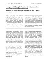
To validate the in vivo dsRNA injection method in
oyster, we used an original technique that consisted of
monitoring, by in situ hybridization, the administration
of digoxygenin-labelled (DIG-labelled) oyvl-dsRNA
into the target organ. The DIG-labelled oyvl-dsRNA
has been observed in a large part of the gonad around
the injection point, showing the efficiency of the
administration of the dsRNA into the gonad (Fig. 2).
Direct injection into the target organ is therefore an
Fig. 1. Western blot probed with antibodies against OYVLG to ana-
lyse the level of OYVLG protein in oyster tissues: mantle (lane 1),
gills (lane 2), muscle (lane 3), labial palps (lane 4), digestive gland
(lane 5), male gonad (lane 6), and female gonad (lane 7). Twelve
micrograms of total protein extract from each tissue was loaded
into the gel. A single band of about 79 kDa was detected in female
and male gonads.
Digestive
gland
Gonad
Mantle
Oo
Fig. 2. In vivo dispersion of DIG-labelled oyvl-dsRNA injected into
oyster gonad. DIG-labelled dsRNA, stained in dark blue, appeared
to have dispersed into a large part of the gonad. Oo, oocyte.
Magnification: · 100. Scale bar: 100 lm.
C. Fabioux et al. In vivo RNA interference in oyster
FEBS Journal 276 (2009) 2566–2573 ª 2009 The Authors Journal compilation ª 2009 FEBS 2567
efficient method for introducing dsRNA into oyster
tissues. The DIG-labelled dsRNA developed in the
present study represents an important technical
advance for examining the first crucial step in success-
fully using in vivo RNAi: the introduction of dsRNA
into animal tissues.
In vivo injection of oyvl-dsRNA provokes
abnormal germ cell development
One month postinjection, 44% of the oysters injected
with 20 lgofoyvl-dsRNA and 80% of the oysters
injected with 100 lgofoyvl-dsRNA presented defects
in germ cell development affecting all of the gonadic
area, in both females and males. Upon histological
examination of gonads injected with 20 lg of dsRNA,
there were fewer germ cells, and development was pre-
maturely curtailed as compared with control gonads
(Fig. 3). Females with the abnormal phenotype halted
their gametogenesis at prophase I of meiosis, before
vitellogenesis, whereas vitellogenic oocytes were
observed in all control females. In males with the
abnormal phenotype, germ cells developed no further
than the spermatocyte stage. Conversely, spermatids
and spermatozoids were observed in all control males
(Fig. 3). Moreover, in oysters showing the abnormal
phenotype, apoptotic germ cells were visible, with a
significant number of haemocytes invading the gonadic
tubules, probably reflecting active resorption of degen-
erating germ cells (Fig. 3).
Defects in gonad development appeared to be even
stronger in females and males injected with 100 lgof
oyvl-dsRNA. The gonadic tubules appeared to be
almost fully regressed throughout the gonadic area.
They contained scarce germ cells, all blocked at early
stages of gametogenesis, whereas the gonads of control
oysters were fully mature (Fig. 3). Haemocyte infiltra-
tion was also observed in the gonadic area of oysters
injected with 100 lgofoyvl-dsRNA. This suggests that
gonadic tubules had stopped developing and started to
degenerate. This most severe defect is clearly similar to
the sterile phenotypes described in mouse and Drosoph-
ila vasa mutants. Tanaka et al. [20] demonstrated that
male mice homozygous for a mutation of vasa exhib-
ited reproductive deficiency. The premeiotic male germ
cells ceased their differentiation before the pachytene
Gt
OI
CT
Gt
og
VO
A
AtO
OI
HH
ApO
og
ApO
g
B
CTCT
H
RGt
og
H
G
og
C
CT
RGt
G
G
F
spc
spgspg
CT
E
spz
Gt
spz
Gt
spc
spd
D
Fig. 3. Effects of in vivo oyvl-dsRNA injection on germ cell development in oysters, 1 month postinjection. (A) Female control, injected with
saline solution. Oocytes are in vitellogenesis. (B) Female injected with 20 lgofoyvl-dsRNA (no. 20.19). Gonadic tubules are composed of
oogonia, oocytes I, and atretic oocytes phagocytized by haemocytes. (C) Female injected with 100 lgofoyvl-dsRNA (no. 100.10). Gonadic
tubules are mostly degenerated. (D) Male control injected with saline solution. Germ cells are in active gametogenesis. (E) Male injected
with 20 lgofoyvl-dsRNA (no. 20.20). Gonadic tubules are filled with a limited number of germ cells, spermatogonia, and spermatocytes. (F)
Male injected with 100 lgofoyvl-dsRNA (no. 100.8). Gonadic tubules are degenerated. Gt, gonadic tubule; CT, conjunctive tissue; H, hae-
mocytes; og, oogonia; OI, oocyte I; VO, vitellogenic oocyte; AtO, atretic oocyte; ApO, apoptotic oocyte; RGt, residual gonadic tubule; Spg,
spermatogonia; Spc, spermatocytes; Spd, Spermatids; Spz, spermatozoı
¨
ds. Magnification: · 400. Scale bars: 100 lm.
In vivo RNA interference in oyster C. Fabioux et al.
2568 FEBS Journal 276 (2009) 2566–2573 ª 2009 The Authors Journal compilation ª 2009 FEBS
spermatocyte stage, and underwent apoptosis. In Dro-
sophila, ovaries of null vasa mutants contained fewer
developing cysts than ovaries of wild-type Drosophila
[21]. No nonspecific defects were observed in gonads of
oysters injected with oyvl-dsRNA, and no oyster mor-
tality was recorded during RNAi experiments, indicat-
ing that dsRNAs were not toxic for oysters.
We demonstrated here that the oyvl-dsRNA injection
into oyster gonads provoked partial or total sterility,
probably associated with Oyvlg gene product deficiency.
The knockdown phenotype was observed throughout
the gonad, although we injected oyvl-dsRNA at only
one point. This pattern confirmed systemic spread of
dsRNA throughout the gonad, as demonstrated in
other species [22]. This systemic spread of dsRNA could
not be followed using DIG-labelled dsRNA, as it was
probably the result of newly synthesized oyvl-dsRNA
issued from the injected oyvl-dsRNA. The severity of
the knockdown phenotypes appeared to be dsRNA
dose-dependent and resulted in complete sterility, repre-
sented by the complete regression of the gonadic
tubules and the degeneration of germ cells at the highest
dose (100 lg). Moreover, the knockdown phenotype
appeared to be more severe 1 month postinjection than
after 9 days, when only 40% of the oysters injected with
100 lgofoyvl-dsRNA displayed a knockdown pheno-
type, probably because it was too soon to visualize
alterations of cellular processes occurring during germ
cell development.
Knockdown of Oyvlg mRNA and protein
expression
A 70% inhibition of mRNA level after dsRNA treat-
ment was considered to be a threshold for effective
RNAi [23]. In our data, a ‡ 70% reduction of Oyvlg
mRNA level as compared with the control was
obtained for three of 21 oysters injected with 20 lgof
dsRNA (14%) and for four of 10 oysters injected with
100 lg of dsRNA (40%) (Fig. 4). Nevertheless, the
knockdown phenotype visible at 1 month postinjection
was already clearly observed, with only 39% inhibition
of Oyvlg mRNA, for four of nine oysters injected with
20 lg of dsRNA (44%) and for four of five oysters
injected with 100 lg of dsRNA (80%) (Fig. 4). The
injection of oyvl-dsRNA clearly triggered an RNAi
mechanism, and a threshold around 40% for mRNA
level reduction appeared to be enough to obtain the
knockdown phenotype. The mRNA level reduction
was greater for oysters injected with 100 lg than with
20 lgofoyvl-dsRNA (Fig. 4), and was correlated with
the most severe knockdown phenotype, confirming the
dose-dependent effect of RNAi discussed previously.
The quantity of 100 lg of dsRNA, corresponding to a
mean concentration of 20 lg of dsRNA per gram of
oyster body weight, is within the range of dsRNA
quantities injected into other adult invertebrates to
obtain RNAi: about 50 lg dsRNA ⁄ g was used in hon-
eybee, and 15 lg dsRNA ⁄ g was used in shrimp [10,24].
The level of 20 lg dsRNA
⁄ g of body weight could be
therefore considered as an optimal quantity of dsRNA
for in vivo RNAi experiments in adult oysters. The
inhibition rates for Oyvlg mRNA levels were similar at
9 days and 1 month postinjection, indicating no
decrease of the RNAi effect during this time. These
results suggest the existence of a dsRNA amplification
process in oyster cells, as was demonstrated in organ-
isms such as Drosophila and Caenorhabditis [25,26].
A
B
Fig. 4. Levels of Oyvlg transcripts relative to EFI transcripts analy-
sed by real-time PCR and expressed as ‘number of copies of Oyvlg
per copy of EFI’ for controls, oysters injected with 20 lgofoyvl-
dsRNA (N = 12 at T9, and N = 9 at T30) (light grey), and oysters
injected with 100 lgofoyvl-dsRNA (N = 5 at T9 and T30) (dark
grey). The control is the mean of Oyvlg mRNA levels of all control
oysters (N = 12 at T9 and T30). The bar represents the confidence
interval at the 5% level. Asterisks (*) indicate oysters showing the
knockdown phenotype. (A) Nine days postinjection. (B) One month
postinjection. The horizontal black line indicates the threshold of
39% inhibition of Oyvlg mRNA level as compared with control at
1 month postinjection. The grey dotted line indicates the threshold
of 70% inhibition of Oyvlg mRNA level as compared with control,
considered as the threshold for effective RNAi [23].
C. Fabioux et al. In vivo RNA interference in oyster
FEBS Journal 276 (2009) 2566–2573 ª 2009 The Authors Journal compilation ª 2009 FEBS 2569
Whereas a significant reduction in Oyvlg mRNA
level was observed as early as 9 days postinjection, no
reduction of mRNA level was observed for two other
gonad-specific genes; the specificity of the dsRNA
effect is therefore clearly shown. Mean relative levels
of og-TGFb mRNA, specifically expressed in auxiliary
cells of the germ cells [27], were 0.54 ± 0.20 for con-
trols, 0.69 ± 0.30 and 0.59 ± 0.17 for oysters injected
with 20 and 100 lgofoyvl-dsRNA, respectively.
Furthermore, the relative levels of a neuropeptide Y
(NPY)-related receptor, specifically expressed in
C. gigas germ cells (Genbank accession number
AM856249, unpublished data), were also statistically
similar in the three tested conditions: 1.98 ± 1.28,
1.81 ± 0.96 and 3.90 ± 2.05 for controls, and oysters
injected with 20 and 100 lgofoyvl-dsRNA, respec-
tively. These assays were not repeated at 1 month
postinjection, because the defects in the gonad were
already so strong that most of the gonad-specific genes
would be affected.
Oysters showing reductions in Oyvlg mRNA levels
after dsRNA treatment also displayed dramatic reduc-
AB
CD
Fig. 5. Levels of both Oyvlg transcripts relative to EFI transcripts measured by real-time PCR (expressed as ‘number of copies of Oyvlg per
copy of EFI’’), and OYVLG protein quantified on western blot (expressed in D ⁄ mm
2
) for oysters injected with 100 lgofoyvl-dsRNA (N =5
at T9 and T30). Bars represent confidence intervals at the 5% level. (A) mRNA levels 9 days postinjection. The inhibition of Oyvlg mRNA
level ranged from 0% to 82%. (B) mRNA levels 1 month postinjection. The inhibition of Oyvlg mRNA level ranged from 0% to 87%. The
control used for mRNA level measurement is the mean of Oyvlg mRNA levels of all control oysters (N = 12 at T9 and T30). (C) OYVLG pro-
tein level 9 days postinjection (D). The values presented on the graph were calculated from the western blot of OYVLG shown below. The
inhibition of OYVLG protein level ranged from 15% to 100%. In the same samples, the protein level of histone H3 (blot under the graph)
was unchanged. (D) OYVLG protein level 1 month postinjection (D). The values presented on the graph were calculated from the western
blot of OYVLG shown below. The inhibition ranged from 0% to 83%. In the same samples, the protein level of histone H3 (blot under the
graph) was unchanged. The control used for protein measurement is a pool of proteins from all control oysters injected with saline solution.
Asterisks indicate oysters showing the knockdown phenotype.
In vivo RNA interference in oyster C. Fabioux et al.
2570 FEBS Journal 276 (2009) 2566–2573 ª 2009 The Authors Journal compilation ª 2009 FEBS
tions in OYVLG protein levels (Fig. 5). Nine days
postinjection, when the mRNA decrease reached 70%,
OYVLG protein was totally absent from gonadic tis-
sue (Fig. 4). One month postinjection, the decrease in
OYVLG protein level had reached 83%, but appeared
to be weaker overall than the mRNA level reduction
(except in one oyster, no. 100.6; Fig. 5). Post-transcrip-
tional gene silencing triggered by RNAi stems from
degradation of target mRNAs. The OYVLG protein
detected probably results from the progressive accumu-
lation of translated ‘residual’ Oyvlg mRNA escaping
from the RNAi machinery. In our data, ‘residual’
Oyvlg mRNA varied from 13% to 48%.
High variability in RNAi response was observed
between individuals (Figs 4 and 5). Variation in the
amount of dsRNA actually penetrating into the germ
cells probably contributed, to a large extent, to the
variability in RNAi response. Direct injection of
dsRNA solution into the circulatory system, through
the adductor muscle or in the pericardic region, would
probably improve the delivery of dsRNA into the cells
of the target organ, as haemolymph efficiently reaches
all the organs of the oyster.
The role of the oyster vasa-like gene in germ cell
development
In previous studies, we demonstrated that Oyvlg is spe-
cifically expressed in germ cells of both male and female
oysters, and we hypothesized that Oyvlg had a role in
germ cell formation [17]. However, the function of Oyvlg
in germline development had never been demonstrated,
as no functional genetic tools were available for the oys-
ter. In this study, in vivo oyvl-dsRNA injection was
achieved in the gonad of oysters at the initiation of
reproduction, when gonadic tubules are filled with germ
stem cells and some gonia at the start of proliferation.
The oyvl-dsRNA injection was clearly associated with
defective germ cell development, which was particularly
visible 1 month later, when control oysters reached
maturity. The number of germ cells was reduced, and
their development was arrested at the first step of meio-
sis. The most severe phenotype showed total sterility, as
represented by the complete degeneration of germ cells
and the regression of gonadic tubules in the whole
gonadic area (Fig. 3). Our results demonstrate that
Oyvlg has an essential role in germ cell (germ stem cells
and gonia) proliferation, and is probably implicated in
oocyte and spermatocyte differentiation. Conversely,
Oyvlg would not be essential in the last step of gameto-
genesis, vitellogenesis, or spermiogenesis, as RNAi
experiments performed according to the same protocol
in maturing oysters did not lead to knock-down pheno-
type (data not shown). In Drosophila, vasa appeared to
have an essential function in female gametogenesis
but not in male gametogenesis. In the mouse, however,
the Mvh gene appeared to be necessary for spermato-
genesis completion but not for oogenesis. In oysters,
we observed defects in both male and female germ
cell development in oyvl-dsRNA-treated gonads. A simi-
lar molecular regulation of early gametogenesis is
suggested to occur in both sexes, probably owing to
the alternative hermaphrodite status of oysters, as
observed in Caenorhabditis [14].
Experimental procedures
Biological material
Oysters were obtained from Marennes-Ole
´
ron (France) cul-
tured stocks, and transferred to the Ifremer Laboratory in
Argenton (France). They were acclimated for 1 week, under
optimal conditions for germ cell maturation [28].
dsRNA synthesis
Two fragments from positions 495 to 1020 (oyvl4) and 29 to
906 (oyvl5)ofOyvlg cDNA (GenBank accession number
AY423380) were amplified by RT-PCR using total RNA
extracted from gonad as template. PCR fragments were
subcloned into the pCR4-TOPO vector (Invitrogen, Paisley,
UK) and sequenced. Recombinant plasmids were purified
by using the Plasmid midi kit (Qiagen, Valencia, CA, USA),
linearized with either NotIorSpeI (Promega, Madison, WI,
USA) enzymes (4 h at 37 °C, using 5 UÆlg
)1
plasmid), phe-
nol ⁄ chloroform-extracted, and finally ethanol-precipitated
and suspended in RNase-free water. The purified plasmids
were transcribed in vitro on both strands, using a T7 and T3
MEGAscript Kit (Ambion, Austin, TX, USA) to produce
oyvl4 and oyvl5 sense and antisense ssRNAs. The ssRNAs
were phenol ⁄ chloroform-extracted, ethanol-precipitated, and
suspended in RNase-free saline solution (10 mm Tris, 10 mm
NaCl) to a final concentration of 0.5 lgÆlL
)1
after quantifi-
cation by spectrophotometry (Nanodrop; Thermo Scientific,
Villebon-sur-Yvette, France). Equimolar amounts of sense
and antisense ssRNA were heated at 100 °C for 1 min, and
left to cool at room temperature for 10 h for annealing. Each
dsRNA (1 lg) was analysed by 1% agarose gel electrophore-
sis to ensure that it existed as a single band of 525 bp (oyvl4)
or 877 bp (oyvl5).
DIG-labelled dsRNA synthesis
Recombinant plasmids (oyvl4 and oyvl5) were synthesized
and linearized as described above. Single-stranded RNAs
were synthesized and DIG-labelled using a T3 or T7 RNA
polymerase (20 UÆlg
)1
plasmid) and DIG RNA-labelling
C. Fabioux et al. In vivo RNA interference in oyster
FEBS Journal 276 (2009) 2566–2573 ª 2009 The Authors Journal compilation ª 2009 FEBS 2571
mix (Roche, Meylan, France). Sense and antisense
DIG-labelled ssRNAs were annealed as described above, and
dsRNAs were stored at )80 °C.
dsRNA administration and sampling
Oysters were anesthetized in MgCl
2
solution (60 : 40 fresh
water ⁄ seawater and 50 gÆL
)1
MgCl
2
) for 3 h. Anesthetized
oysters were injected in the gonad with 100 lL of saline
solution containing dsRNA, or saline solution for the con-
trol. After dsRNA injection, oysters were maintained in
raceways in conditions allowing optimal gonad maturation.
Oysters were injected at T0 (initiation of reproduction),
T7 (7 days) and T14, with 20 lg(N = 24) or 100 lg
(N = 10) of oyvl-dsRNA (a mixture of oyvl4 dsRNA and
oyvl5 dsRNA in equal amounts) or with the same volume
of saline solution (control, N = 24).
At T9 and T30, 12 oysters injected with 20 lgofoyvl-
dsRNA, five oysters injected with 100 lgofoyvl-dsRNA
and 12 control oysters were sampled. Their gonads were
immediately dissected: a large transverse section of all the
gonadic area was taken for histological examination, and
the rest of the gonad was placed in total RNA and protein
extraction solution.
For dsRNA tracking, 10 oysters were injected with 20 lg
of DIG-labelled dsRNA and sampled 9 days after injection
for histological and in situ hybridization examinations.
Histology, in situ hybridization and real-time
RT-PCR analysis
Gonadic development was assayed on histological slides of
a transverse section of all the gonadic area according to
Fabioux et al. [28] for dsRNA-injected and control oysters
at T0, T9, and T30. The DIG-labelled oyvl-dsRNAs
sampled were analysed by in situ hybridization, using Oyvlg
DNA probes according to Fabioux et al. [17].
Total RNA was isolated from the gonads of treated and
control oysters, using Extract All (Invitrogen, Cergy-Pon-
toise, France). Samples were then treated with DNase I
(1 UÆlg
)1
total RNA; Sigma, Saint-Quentin, France) to
prevent DNA contamination. RNA concentrations were
measured as described above, and RNA quality was
checked with a Bioanalyser 2100 (Agilent, Massy, France).
From 2 lg of total RNA, RT-PCR amplifications were car-
ried out as described in Fabioux et al. [16], using specific
primers for the Oyvlg [16], oyster-gonadal-TGFb-like (og-
TGFb) [27] and NPY-related-receptor-like (NPY-receptor)
genes (forward, 5¢-GTGGCTTGTGGGCTTATTGT-3¢;
reverse, 5¢-CTGAAATCCGAATGGACGAC-3¢). The cal-
culation of relative mRNA levels of target genes was based
on the the comparative C
t
method (see [16] for DDC
t
for-
mulae), and was normalized to elongation factor I (EFI), as
no significant differences in C
t
values were observed for
EFI between control and injected oysters (Kruskall–Wallis
test = 3.74; P = 0.15, coefficient of variation = 3.6%).
The relative mRNA levels are expressed as ‘number of
copies of target gene per copy of EFI.
Antibodies and western blot analysis
Polyclonal antibodies (Fab1 and Fab2) against two peptides
[GSKNDGESSGFGGG(126–139) and EEGHFARECPE
PRK(165–178), respectively] encoded in the Oyvlg cDNA
sequence were produced in rabbits by MilleGen.
Total protein extracts were obtained from gonadic tissue
of mature female and mature male mantle, gills, muscle,
labial palps, and digestive glands, according to Corporeau
& Auffret [29]. Before denaturation of protein samples,
total protein extracts were quantified using a DC protein
assay (Bio-Rad, Hercules, CA, USA), and adjusted to a
final concentration of 1 mgÆmL
)1
. Twelve micrograms of
each protein extract was loaded onto SDS ⁄ polyacrylamide
gel to ensure identical amounts of protein between samples.
Western blot was performed as described in Corporeau
& Auffret [29], using the polyclonal antibody against
OYVLG produced in this study (dilution 1 : 5000). Blots
were revealed using an Immun-star AP detection kit (Bio-
Rad). The amount of OYVLG protein was quantified using
multi-analyst software (Bio-Rad), with the background
signal removed. The obtained value is expressed in
OD ⁄ mm
2
, and represents the spot intensity expressed as
mean count per pixel, multiplied by the spot surface. After
visualization and signal quantification, membranes were de-
hybridized for 1 h at room temperature in dehybridizing
buffer (100 mm glycine, 100 mm NaCl, pH 3.2), and rehy-
bridized with an antibody against histone H3 (#9715; Cell
Signaling Technology, Danvers, MA, USA; dilution
1 : 5000) to control for identical amounts of total protein
between samples.
Acknowledgements
The authors are grateful to J. F. Samain and M. Mat-
hieu for their support. The authors are indebted to
V. Boulo, J. P. Cadoret, F. Le Roux and J. S. Joly for
advice, and to J. Y. Daniel for technical assistance.
We thank all the staff of the Argenton experimental
hatchery for conditioning oysters. We thank
H. McCombie for her help with editing the English.
C. Fabioux was funded by Ifremer and a Re
´
gion
Basse-Normandie postdoctoral grant.
References
1 FAO (2004) The State of World Fisheries and Aquacul-
ture. FAO Fisheries Department, part 1, 65.
2 Saavedra C & Bache
`
re E (2006) Bivalve genomics.
Aquaculture 256, 1–14.
In vivo RNA interference in oyster C. Fabioux et al.
2572 FEBS Journal 276 (2009) 2566–2573 ª 2009 The Authors Journal compilation ª 2009 FEBS
3 Jenny MJ, Chapman RW, Mancia A, Chen YA,
McKillen DJ, Trent H, Lang P, Escoubas J-M,
Bachere E, Boulo V et al. (2007) Characterization of
a cDNA microarray for Crassostrea virginica and
C. gigas. Mar Biotechnol 9, 577–591.
4 Elbashir SM, Harborth J, Lendeckel W, Yalcin A,
Weber K & Tuschl T (2001) Duplexes of 21-nucleotide
RNAs mediate RNA interference in cultured mamma-
lian cells. Nature 411, 494–498.
5 Fire A, Xu S, Montgomery MK, Kostas SA, Driver SE
& Mello CC (1998) Potent and specific genetic interfer-
ence by double-stranded RNA in Caenorhabditis
elegans. Nature 391, 806–811.
6 Hannon GJ (2002) RNA interference. Nature 418,
244–251.
7 Meister G & Tuschl T (2004) Mechanisms of gene silenc-
ing by double-stranded RNA. Nature 431, 343–349.
8 Berns K et al. (2004) A large-scale RNAi screen in
human cells identifies new components of the p53 path-
way. Nature 428, 431–437.
9 Xia H, Mao Q, Paulson HL & Davidson BL (2002)
siRNA-mediated gene silencing in vitro and in vivo. Nat
Biotechnol 20, 1006–1010.
10 Robalino J, Browdy CL, Prior S, Metz A, Parnell P,
Gross P & Warr G (2004) Induction of antiviral immu-
nity by double-stranded RNA in a marine invertebrate.
J Virol 78, 10442–10448.
11 Dash PK, Tiwari M, Santhosh SR, Parida M &
Lakshmana Rao PV (2008) RNA interference
mediated inhibition of Chikungunya virus replication in
mammalian cells. Biochem Biophys Res Commun 376,
718–722.
12 Van Diepen MT, Spencer GE, Van Minnen J, Gouwen-
berg Y, Bouwman J, Smit AB & Van Kesteren RE
(2005) The molluscan RING-finger protein L-TRIM is
essential for neuronal outgrowth. Mol Cell Neurosci 29 ,
74–81.
13 Grimaldi A, Tettamanti G, Rinaldi L, Brivio MF, Cas-
tellani D & de Eguileor M (2004) Muscle differentiation
in tentacles of Sepia officinalis (Mollusca) is regulated
by muscle regulatory factors (MRF) related proteins.
Dev Growth Differ 46, 83–95.
14 Kuznicki KA, Smith PA, Leung-Chiu WM, Estevez
AO, Scott HC & Bennett KL (2000) Combinatorial
RNA interference indicates GLH-4 can compensate for
GLH-1; these two P granule components are critical for
fertility in C. elegans . Development 127, 2907–2916.
15 Lasko F & Ashburner M (1988) The product of the
Drosophila gene vasa is very similar to eucaryotic initia-
tion factor-4A. Nature 335, 611–617.
16 Fabioux C, Huvet A, Lelong C, Robert R, Pouvreau S,
Daniel JY, Mingant C & Le Pennec M (2004) Oyster
vasa-like gene as a marker of the germline cell develop-
ment in Crassostrea gigas. Biochem Biophys Res
Commun 320, 592–598.
17 Fabioux C, Pouvreau S, Le Roux F & Huvet A (2004)
The oyster vasa-like gene: a specific marker of the
germline in Crassostrea gigas. Biochem Biophys Res
Commun 315, 897–904.
18 Knaut H, Pelegri F, Bohmann K, Schwarz H & Nuess-
lein-Volhard C (2000) Zebrafish vasa RNA but not its
protein is a component of the germ plasm and segre-
gates asymmetrically before germline specification.
J Cell Biol 149, 875–888.
19 Echeverri CJ et al. (2006) Minimizing the risk of report-
ing false positives in large-scale RNAi screens. Nat
Methods 3, 777–779.
20 Tanaka SS, Toyooka Y, Akasu R, Katoh-Fukui Y,
Nakahara Y, Suzuki R, Yokoyama M & Noce T (2000)
The mouse homolog of Drosophila Vasa is required for
the development of male germ cells. Gene Dev 14, 841–
853.
21 Styhler S, Nakamura A, Swan A, Suter B & Lasko P
(1998) vasa is required for GURKEN accumulation in
the oocyte, and is involved in oocyte differentiation and
germline cyst development. Development 125, 1569–1578.
22 Saleh M-C, van Rij RP, Hekele A, Gillis A, Foley E,
O’Farrell PH & Andino R (2006) The endocytic path-
way mediates cell entry of dsRNA to induce RNAi
silencing. Nat Cell Biol 8, 793–802.
23 Jiang Y, Loker ES & Zhang S-M (2006) In vivo and
in vitro knockdown of FREP2 gene expression in the
snail Biomphalaria glabrata using RNA interference.
Dev Comp Immunol 30, 855–866.
24 Amdam GV, Simoes ZL, Guidugli KR, Norberg K &
Omholt SW (2003) Disruption of vitellogenin gene func-
tion in adult honeybees by intra-abdominal injection of
double-stranded RNA. BMC Biotechnol 3,1,
doi:10.1186/1472-6750-3-1.
25 Sijen T, Fleenor J, Simmer F, Thijssen L, Parrish S,
Timmons L, Plasterk R & Fire A (2001) On the role of
RNA amplification in dsRNA-triggered gene silencing.
Cell 107, 465–476.
26 Agrawal N, Dasaradhi PVN, Mohmmed A, Malhotra
P, Bhatnagar RK & Mukherjee SK (2003) RNA inter-
ference: biology, mechanism, and applications. Micro-
biol Mol Biol Rev 67, 657–685.
27 Fleury E, Fabioux C, Lelong C, Favrel P & Huvet A
(2008) Characterization of a gonad-specific transforming
growth factor-[beta] superfamily member differentially
expressed during the reproductive cycle of the oyster
Crassostrea gigas. Gene 410, 187–196.
28 Fabioux C, Huvet A, Le Souchu P, Le Pennec M &
Pouvreau S (2005) Temperature and photoperiod drive
Crassostrea gigas reproductive internal clock. Aquacul-
ture 250, 458–470.
29 Corporeau C & Auffret M (2003) In situ hybridisation
for flow cytometry: a molecular method for monitoring
stress-gene expression in hemolymph cells of oysters.
Aquat Toxicol 64, 427–435.
C. Fabioux et al. In vivo RNA interference in oyster
FEBS Journal 276 (2009) 2566–2573 ª 2009 The Authors Journal compilation ª 2009 FEBS 2573