Báo cáo khoa học: Typical 2-Cys peroxiredoxins – structures, mechanisms and functions ppt
Bạn đang xem bản rút gọn của tài liệu. Xem và tải ngay bản đầy đủ của tài liệu tại đây (354.81 KB, 9 trang )
REVIEW ARTICLE
Typical 2-Cys peroxiredoxins – structures, mechanisms
and functions
Andrea Hall
1
, P. A. Karplus
1
and Leslie B. Poole
2
1 Department of Biochemistry and Biophysics, Oregon State University, Corvallis, OR, USA
2 Department of Biochemistry, Wake Forest University School of Medicine, Winston-Salem, NC, USA
Peroxiredoxins (Prxs, EC 1.11.1.15) are ubiquitous
antioxidant enzymes found in all organisms, with the
single exception, to our knowledge, of Borrelia burg-
dorferi (and other Borrelia species). The broad distri-
bution of Prxs and the high levels of expression [1]
suggest that they are both an ancient and important
enzyme family. Prxs are assumed to have evolved from
a thioredoxin-like precursor protein, and a model for
the evolutionary path has been presented [2]. The ini-
tial publication defining this family of enzymes intro-
duced the term ‘peroxidoxin’ [3], but, shortly
thereafter, ‘peroxiredoxin’ was suggested [4]. Although
some publications use peroxidoxin, it is the latter name
that has been widely adopted.
Prx research has expanded rapidly in recent years,
with a PubMed search for the term ‘peroxiredoxin’
yielding 2, 43, 227 and 542 papers in 1992–95, 1996–
99, 2000–03 and 2004–07, respectively. Despite this
recent increase in study, Prxs are still not well charac-
terized relative to glutathione peroxidase and catalase.
For comparison, 6523 papers were published on cata-
lase and 4205 on glutaredoxin in 2004–07.
A number of excellent reviews have summarized
much of the work on Prx structure, function and
biology [1,5–8], and on the developing view of hydro-
gen peroxide signaling [5,9–11]. The purpose of this
review is to highlight recent advances in our under-
standing of the chemistry of ‘typical 2-Cys’ Prxs and
Keywords
antioxidants; hydrogen peroxide;
hydroperoxides; mechanisms; NADPH
oxidase; oxidative stress; peroxiredoxins;
redox signaling; signal transduction; sulfenic
acid
Correspondence
L. B. Poole, Department of Biochemistry,
Wake Forest University School of Medicine,
Medical Center Boulevard, Winston-Salem,
NC 27157, USA
Fax: +1 336 716 6711
Tel: +1 336 777 3242
E-mail:
(Received 6 October 2008, revised 3
February 2009, accepted 25 February 2009)
doi:10.1111/j.1742-4658.2009.06985.x
Peroxiredoxins are abundant cellular antioxidant proteins that help to con-
trol intracellular peroxide levels. These proteins may also function, in part,
through an evolved sensitivity of some peroxiredoxins towards peroxide-
mediated inactivation in hydrogen peroxide signaling in eukaryotes. This
review summarizes recent progress in our understanding of the catalytic
and regulatory mechanisms of ‘typical 2-Cys’ peroxiredoxins and of the
biological roles played by these important enzymes in oxidative stress
and nonstress-related cellular signaling. New evidence suggests localized
peroxide buildup plays a role in nonstress-related signaling.
Abbreviations
1-Cys, Prx with C
R
coming from another molecule; 2-Cys, Prx with C
R
coming from a Prx; AhpC, alkyl hydroperoxide reductase C;
C
P
, peroxidatic cysteine; C
R
, resolving cysteine; Cys-C
P
O
2
H, peroxidatic cysteine sulfinic acid; Cys-S
P
OH, peroxidatic cysteine sulfenic acid;
ER, endoplasmic reticulum; NOXs, NADPH oxidases; PDGF, platelet-derived growth factor; PTP1B, protein tyrosine phosphatase 1B;
Srx, sulfiredoxin; SP, sulfur of peroxidatic cysteine; SR, sulfur of resolving cysteine; VEGF, vascular endothelial growth factor.
FEBS Journal 276 (2009) 2469–2477 ª 2009 The Authors Journal compilation ª 2009 FEBS 2469
evidence for the role(s) of the sensitivity to inactivation
by hydrogen peroxide seen in some members of this
group.
Typical 2-Cys Prxs
All Prxs have in common an overall fold and cata-
lytic mechanism involving a conserved, fully folded
active site and an unfolding event [6] (Figs 1A and
2). The enzymatic mechanism relies on a conserved
cysteine residue, the peroxidatic cysteine (C
P
), to
reduce various peroxide substrates with catalytic effi-
ciencies on the order of 10
6
–10
7
m
)1
Æs
)1
[7]. Follow-
ing oxidation of C
P
by the peroxide substrate,
regions of the protein around the active site change
conformation, allowing for subsequent reactivation
steps (Fig. 2A). A second free thiol (C
R
for the
resolving cysteine or thiol group) then forms a disul-
fide with C
P
. 1-Cys and 2-Cys Prxs are differentiated
by whether C
R
comes from another molecule (1-Cys)
or from a Prx (2-Cys).
All of the early characterized Prxs were ‘typical
2-Cys’ Prxs. In this subclass of 2-Cys Prxs, the basic
active unit is a dimer, with the catalytically relevant
disulfide bond being formed between the C
P
on one
chain and the C
R
from near the C-terminus of the
other chain [6] (Fig. 2). The subclass of ‘atypical
2-Cys’ Prxs is differentiated from typical 2-Cys Prxs
in that the catalytic disulfide bond is intramolecular in
most cases, and the resolving cysteine is not at the
‘typical’ conserved position in the C-terminus. Despite
the different positions observed for C
R
, the catalytic
cycle for peroxidase activity in all Prxs can be broken
down into three steps: peroxidation (reaction 1); reso-
lution (reaction 2); and recycling (reaction 3)
(Fig. 1A). Peroxidation occurs in the fully folded
active site, which contains four conserved residues: C
P
,
Arg and Thr residues, presumed to stabilize the thio-
late anion, and Pro, which shields the active site from
water (Fig. 2A). This active site environment lowers
the pK
a
value of C
P
, which has recently been shown to
be in the range of 5–6 for the few Prxs thus studied
[12–14]. The thiolate anion attacks the peroxide sub-
strate to generate water (or alcohol) and a Cys-sulfenic
acid (Cys-S
P
OH) at the active site [7]. Resolution
occurs when C
R
attacks Cys-S
P
OH to release water
and form an intersubunit disulfide bond. The catalytic
cycle is completed when the disulfide bond is recycled,
typically by a thioredoxin-like molecule, regenerating
the free thiol forms of C
P
and C
R
.
For these enzymes, catalysis not only involves chem-
ical transformations, but also requires the protein to
undergo certain conformational gymnastics. The form
of the enzyme carrying out peroxidation is fully folded;
in typical 2-Cys Prxs, the C
R
side-chain is buried and
roughly 14 A
˚
away from C
P
. For C
P
and C
R
to form a
disulfide, both the active site region (known as the C
P
loop) and the C-terminal region must locally unfold
(Fig. 2A). Details of the structural changes occurring
with catalysis for various Prxs have been reviewed
recently by Karplus and Hall [6].
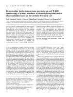
Fig. 1. Mechanisms of catalysis by typical 2-Cys Prxs. (A) The per-
oxidatic catalytic cycle of typical 2-Cys Prxs involves three main
reactions: 1 peroxidation, 2 resolution and 3 recycling. Not shown
is the local unfolding event that occurs in both the C
P
loop and
C-terminus during reaction 2, so that the disulfide bond can form
(see Fig. 2A). The protein is represented as one of two active sites
within a functional dimer, with S
P
and S
R
(red) designating the sul-
fur atoms of the peroxidatic and resolving cysteines, respectively,
from different subunits. ‘2 R¢SH’ in reaction 3 represents a thiore-
doxin-like protein or domain. Overoxidation of C
P
(reaction 4) and
reduction of Cys-S
P
O
2
H by Srx (reaction 5) depict redox regulation
and repair occurring in some eukaryotic typical 2-Cys Prxs. (B)
Mutants of AhpC which suppress the growth defect (dithiothreitol
dependence) of the trxB gor mutant from E. coli have been shown
to catalyze the deglutathionylation of Grx1 (using the C14S mutant)
in vitro [24]. Although the catalytic intermediate is shown with
glutathione attached in a mixed disulfide to Prx, the alternative
mechanism with Grx attached to Prx is also possible. The truncated
cycle shown with dotted lines (reaction 4 in B) illustrates the find-
ing that only the ‘resolving cysteine’ (with the sulfur depicted as
S
R
) is required for this activity.
2-Cys peroxiredoxins A. Hall et al.
2470 FEBS Journal 276 (2009) 2469–2477 ª 2009 The Authors Journal compilation ª 2009 FEBS
An additional mechanistic complexity of these pro-
teins is that, during catalysis, they shift quaternary
structure between a homodimer and a doughnut-
shaped decamer (which consists of a pentamer of
dimers). In addition to decamers, octamers and
dodecamers have also been observed; the role of
oligomerization is thought to be the same in all
three cases [6] (Fig. 2B). Studies with the alkyl
hydroperoxide reductase C (AhpC) Prx from Salmo-
nella typhimurium have suggested that the decameric
form is stabilized in all catalytic states of the
enzyme, except for the disulfide form (between reac-
tions 2 and 3 in Fig. 1A); the decamer falls apart on
disulfide formation because the unfolding of the C
P
loop destabilizes the decamer building (dimer-to-
dimer) interface [15]. Analogous redox-dependent
oligomerization effects are now known to extend to
mammalian and plant-derived typical 2-Cys Prxs [16–
18]. The physiological role of the dimer–decamer
transition remains unclear; however, studies indicat-
ing that decamers are better peroxidases than dimers
[19], but are less amenable to reduction by thiore-
doxins [18], suggest that the quaternary structure
transition aids efficient catalysis. There is also
evidence for some Prxs that the decamers associate
with membranes [20,21], and so it is possible that
cellular localization is influenced by the oligomeric
state.
Recent mechanism-relevant discoveries
for Prxs
Although Prxs have been described as broad-specificity
peroxidases that reduce substrates such as hydrogen per-
oxide, lipid hydroperoxides and peroxynitrite, recent
data suggest that at least some of the typical 2-Cys Prxs
are much more active with hydrogen peroxide than with
bulkier hydroperoxide substrates [22], and that the reac-
tivity with other oxidants and alkylating agents is
remarkably limited [13]. The thermodynamic driving
force for peroxide reduction by thiols (reaction 1 in
Fig. 1A) is highly favorable, and so the relative reactiv-
ity for thiol-based peroxidases is, instead, dominated by
kinetic factors [11]. However, the turnover of Prxs with
reductants (reaction 3 in Fig. 1A) may be subject to
greater influence by their midpoint reduction (redox)
potentials. In this regard, it is perhaps surprising that
the redox potentials for typical 2-Cys Prxs from plant
chloroplasts are in the region of )300 mV, similar to or
even lower than their physiological reductants [23]. As
pointed out by Dietz et al. [23], however, these low
potentials probably reflect the unique regulatory envi-
ronment of this photosynthetic organelle. Recent data
for the bacterial antioxidant AhpC indicate a redox
potential of )178 mV, sufficiently high for AhpC to
remain predominantly reduced even under conditions in
which the cell is oxidatively stressed [22].
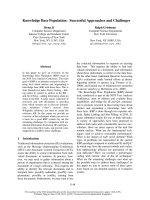
Fig. 2. Structural aspects of Prx sensitivity and resurrection. (A) The active site of a sensitive 2-Cys Prx in the fully folded (FF) and locally
unfolded (LU) conformations. Both chains of the dimer are colored gray. The C-terminal helix containing the YF motif (cyan) and the loop
associated with the GGLG motif (yellow), which are characteristic of sensitive Prxs, can be seen to pack against each other and cover the
active site C
P
loop (pink) in the FF conformation (PDB code 1QMV). Comparing the LU structure (PDB code 1QQ2) with the FF structure
shows how the C
P
loop and the protein C-terminus unfold for disulfide formation, and how the C-terminal helix hinders this required local
unfolding. In robust Prxs, the C-terminal helix containing the YF motif is absent, allowing for more facile unfolding (see fig. 2 in Wood et al.
[27]). In the LU form, a star indicates the presence of the additional disordered C-terminal residues. In both the FF and LU images, the four
residues conserved in all Prxs (C
P
, Arg, Thr and Pro) and C
R
are colored green, with sulfur, oxygen and nitrogen atoms colored dark yellow,
red and blue, respectively. (B) The typical 2-Cys dimer (magenta and dark blue) associates with other dimers (light blue) as part of the normal
catalytic cycle to form higher order oligomers. The overoxidized state is stabilized in this form [20]. Modeling of Srxs (green) on a Prx decam-
er shows that Srx can associate with such a structure without significant changes to the decamer, consistent with the role of Srx in the
reduction of the overoxidized Prx [32].
A. Hall et al. 2-Cys peroxiredoxins
FEBS Journal 276 (2009) 2469–2477 ª 2009 The Authors Journal compilation ª 2009 FEBS 2471
In addition, an alternative catalytic activity that may
be exhibited by a subset of Prxs has recently come to
light using Escherichia coli mutants with thioredoxin
reductase and glutathione reductase gene deletions (the
trxB gor mutant). These E. coli are compromised in
cytoplasmic disulfide reduction, resulting in growth
defects. This condition leads to frequent selection of a
mutation causing the insertion of one amino acid
between residues 37 and 38 of AhpC, converting it
from a peroxidase to a disulfide reductase that acts as a
glutaredoxin deglutathionylating enzyme [24]. Other
single point mutations were also able to confer this
activity without eliminating peroxidase activity. Sur-
prisingly, C
R
(Cys165) rather than C
P
(Cys46) of E. coli
AhpC is critical for the suppression of the growth
defect of the trxB gor mutant, and for the disulfide
reductase activity measured in vitro (Fig. 1B). Although
only speculative at present, it is conceivable that this
activity may be more constitutively present in wild-type
forms of other Prxs in which C
R
is particularly reactive,
or switched on in some Prxs by protein modifications.
Robust and sensitive typical 2-Cys Prxs
The catalytic cycle explained above is all that is needed
to describe the peroxidase activity of Prxs. However,
beginning with a yeast Prx (now known as Tpx1), a
number of Prxs have been shown to be quickly inacti-
vated by submillimolar concentrations of both hydro-
gen peroxide and alkyl hydroperoxides [4,25]. Yang
et al. [26] reported that, for human Prx1 at 100 lm
hydrogen peroxide, the half-life for inactivation during
catalytic cycling with reductant is approximately
2 min. The inactivation is a result of overoxidation of
the C
P
side-chain that occurs when a second peroxide
substrate molecule attacks Cys-S
P
OH, forming a dead-
end sulfinic acid (Cys-S
P
O
2
H) (reaction 4 in Fig. 1A).
This reaction is in competition with the resolution step
(reaction 2 in Fig. 1A) of the normal catalytic cycle.
In contrast with these ‘sensitive’ Prxs, a number of
members of the typical 2-Cys Prx family from bacteria,
such as Salmonella typhimurium AhpC, are robust,
requiring more than 100-fold higher hydrogen peroxide
levels to be inactivated. Wood et al. [27] followed up
this observation to draw two conclusions: first, that
the sensitivity to inactivation by hydrogen peroxide
correlated with two amino-acid sequence motifs – a
GGLG-containing motif in the middle of the protein,
and a YF-containing C-terminal extension (Fig. 2A);
second, that the Prxs that conserved these two motifs
(i.e. were putative sensitive Prxs) were from eukaryotic
organisms. These or similar motifs are largely absent
from bacterial Prx sequences, although potentially
interesting exceptions exist. {Among the exceptions to
the distribution of Prxs with GGLG and YF motifs
within eukaryotes only are some parasitic bacteria
(Helicobacter pylori, Yersinia pestis, Chlamydia pneu-
moniae), which have Prxs with a GGIG motif and a
C-terminal extension containing a YL motif. The sensi-
tivity to overoxidation of Prxs with these and other
variations on the GGLG and YF motifs, found within
both prokaryotes and eukaryotes, has not been fully
characterized, although it is clear that point mutations
within the C-terminus of sensitive Prxs are sufficient to
disrupt packing of the C-terminal helix [28]. The pres-
ence of these potentially sensitive Prxs in parasitic bac-
teria may be a result of horizontal gene transfer [29],
and so does not necessarily break from the expected
limited distribution of sensitive Prxs to eukaryotes.}
As described by Wood et al. [27], the YF motif is
not part of the peroxidatic active site itself, but forms
a helix that packs just above the active site in the fully
folded form of the protein (Fig. 2A). In contrast, this
feature is missing in robust Prxs, so that the peroxidatic
active site region is much more open. The explanation
for sensitivity runs as follows: the YF-containing
C-terminal helix packs above the active site region like
a cork in a bottle, limiting the active site dynamics and
hindering it from unfolding; because of this, the local
unfolding of the active site required for the resolution
reaction (reaction 2 in Fig. 1A) is disfavored, causing
the Cys-S
P
OH-containing active site to be longer lived
and thus more susceptible to attack by a second
molecule of peroxide [27]. That the presence of the
C-terminal helix is responsible for sensitivity has been
confirmed by mutagenesis [30].
The reason for conservation of the GGLG motif is
less clear, but it is speculated to be required for rescu-
ing the overoxidized (sulfinic acid, Cys-S
P
O
2
H) form
of the protein [31]. Although Cys-S
P
O
2
H formation
was originally thought to be biologically irreversible,
sulfiredoxins (Srxs) and possibly sestrins are able to
reduce Cys-S
P
O
2
H to Cys-S
P
OH in an ATP-dependent
reaction [31]. The existence of the ‘resurrection’ activ-
ity supports a physiological role for the overoxidized
form of the protein. Recent structural studies of a
Prx–Srx complex have revealed a surprising C-terminal
tail ‘embrace’ [32] (Fig. 2B). In this structure, the
GGLG motif forms part of the ATP binding site
and thus may play an important role in the reduction
reaction with Srx [32].
That some Prxs are sensitive to overoxidation by
their own substrates, making them worse peroxidases,
raises the question of why a worse peroxidase would
be maintained. In theory, the selective pressure to
maintain sensitivity could be a direct result of the
2-Cys peroxiredoxins A. Hall et al.
2472 FEBS Journal 276 (2009) 2469–2477 ª 2009 The Authors Journal compilation ª 2009 FEBS
importance of sensitivity, or may be a pressure
directed at conserving the C-terminal extension for
another reason with the sensitivity being an unwanted
byproduct. The existence of robust Prxs that otherwise
conserve the active site and mechanistic details proves
that the sensitivity is not an obligatory limitation
related to the enzyme mechanism. Furthermore,
because the sensitivity is not only avoidable, but could
be very easily lost during evolution (simply through
mutation or loss of one or a few C-terminal residues)
[28], its conservation throughout eukarya implies that
there must be a very strong selective pressure to con-
serve it. Wood et al. [27] proposed that the built-in
sensitivity is important for facilitating nonstress-related
hydrogen peroxide signaling in eukaryotes, but this
remains to be proven.
Stress and nonstress-related peroxide
signaling
Much evidence has accumulated that implicates hydro-
gen peroxide as an important and widespread signaling
molecule [5,9–11], both as an indicator of oxidative
stress and as part of normal cellular development.
Although both of these processes involve signaling,
some authors use ‘H
2
O
2
signaling’ to refer to the sec-
ond process only, leading to some confusion. In this
review, hydrogen peroxide signaling includes both, and
will be referred to as either stress (exogenous peroxide
induced) or nonstress (endogenous peroxide induced)
related. It is necessary to differentiate between the two
because the supporting evidence and pathways of the
two processes are quite different.
All of the well-characterized pathways for hydrogen
peroxide signaling describe stress-related signaling, and
the response triggered is generally the protective activa-
tion of a broad antioxidant response involving increased
transcription of antioxidants and repair proteins. Well-
characterized examples of stress-related signaling
include the OxyR and OhrR transcriptional regulators
in prokaryotes, which act as both molecular sensors and
transducers of the H
2
O
2
signal, and the Yap1 ⁄ Gpx3 sys-
tem in Saccharomyces cerevisiae [9,10,33]. In the latter
system, yeast transcriptional responses to elevate protec-
tive antioxidant enzyme levels rely on communication of
the H
2
O
2
signal from a thiol-based peroxidase, the glu-
tathione peroxidase-like Gpx3 (also known as Orp1), to
a transcriptional regulator, Yap1, through condensation
between the two proteins and thiol–disulfide interchange
[34]. There is no controversy about the relevance of
these events.
In contrast, the role of hydrogen peroxide in non-
stress-related signaling associated with endogenously
generated hydrogen peroxide is still controversial.
These mechanisms require that the hydrogen peroxide
signal is generated in a regulated manner without a
global change in the redox state of the cell. Long-
standing evidence for such signaling comes from stud-
ies showing that the exposure of cells to low levels of
hydrogen peroxide stimulates proliferation. More
recent evidence has shown that, at least in mammals,
tightly regulated NADPH oxidases (NOXs) become
activated by hormones and produce superoxide, which
is converted to hydrogen peroxide and, in turn, oxi-
dizes specific cysteine residues in target proteins (such
as protein tyrosine phosphatases) to influence the fate
of the cell. These pathways can be blocked by
increased catalase expression, supporting a role for
hydrogen peroxide as a signaling molecule [9–11].
Despite the growing body of evidence for such non-
stress-related peroxide signaling, there is active debate
about the physiological relevance of these putative sig-
naling pathways. One of the major concerns is that
many of the implicated target proteins, such as Prxs and
phosphatases, appear to require peroxide concentrations
in the 10–300 lm range in order to become (over)oxi-
dized, whereas, in healthy cells, the peroxide levels are
not thought to exceed 700 nm [9]. As summarized by
Stone and Yang [9], alternative hypotheses to explain
the discrepancy include the following: (a) there are as
yet unidentified, much more sensitive peroxide sensor
proteins that transduce the signals; (b) hydrogen perox-
ide itself is not the key signaling molecule, but it may be
superoxide, peroxynitrite or a nitrosothiol; and (c)
hydrogen peroxide build-up is highly localized.
Among these possibilities, new evidence suggests
that subcellular localization [possibility (c)] is indeed a
key component of certain nonstress-related peroxide
signaling pathways. Early evidence supporting localiza-
tion was published by Choi et al. [35], who demon-
strated that Prx2 from mouse embryonic fibroblasts is
recruited to the platelet-derived growth factor (PDGF)
receptor in response to PDGF stimulation. This site-
specific recruitment of Prx2 was associated with the
suppression of protein tyrosine phosphatase inactiva-
tion. Later, Li et al. [36] showed that specific recruit-
ment of Nox2 to the endosome was required for
redox-dependent recruitment of TRAF6 to the active
interleukin-1 receptor complex, ultimately leading to
interleukin-1b-dependent nuclear factor-jB activation.
Similarly, Nox localization has been implicated in
vascular endothelial growth factor (VEGF) signaling
in angiogenesis [37]. Most recently, Chen et al. [38]
showed that, for epidermal growth factor signaling,
activated Nox4 is localized to the endoplasmic
reticulum (ER) and is able to oxidatively inactivate
A. Hall et al. 2-Cys peroxiredoxins
FEBS Journal 276 (2009) 2469–2477 ª 2009 The Authors Journal compilation ª 2009 FEBS 2473
ER-localized protein tyrosine phosphatase 1B
(PTP1B), but not cytosolic PTP1B. Furthermore,
ER-localized antioxidant enzymes were able to block
the signal, whereas untargeted counterparts were not.
This last report provides powerful evidence that locali-
zation allows for levels of reactive oxygen species that
can oxidize a less reactive target, such as PTP1B.
What is the role of Prx sensitivity
in peroxide signaling?
The use of hydrogen peroxide as a signaling molecule
requires very tight regulation because of the damaging
nature of peroxides. The high expression level (0.1–1%
of total soluble protein) and ubiquitous distribution [1]
make Prxs one of the first proteins encountered by a
hydrogen peroxide molecule. This, combined with their
fast reactivity ( 10
7
m
)1
Æs
)1
), implies that, in mamma-
lian cells, 10 000 times more hydrogen peroxide would
react with Prx than with glutathione [11]. In character-
ized systems, the most highly expressed Prxs are sensi-
tive: mammalian mRNA levels suggest that this is
generally Prx1 [39] and, in yeast, Tpx1 [40]. {Of the
five Prxs expressed in S. cerevisiae, three are present in
relatively low abundance (cTpxII, mTpx and nTpx,
each at less than 5000 molecules per cell), whereas
cTpxI and cTpxIII are present at 378 000 and 162 000
molecules per cell, respectively. cTpxI and cTpxII are
sensitive Prxs. Expression levels were estimated using
green fluorescent protein fusion proteins [40]}. The
importance of Prx1 expression for controlled growth in
mammals is demonstrated by the high rate of malig-
nant cancers in the Prx1 knockout mouse [41].
So what is the role of sensitivity? In principle, there
are two possibilities – overoxidation could cause a gain
in function and ⁄ or overoxidation could cause a loss of
function. In either case, Prxs could act as a molecular
switch, influenced by a change in peroxide level,
whether for stress- or nonstress-related signaling. It is
worth noting that phosphorylation, nitrosylation and
C-terminal cleavage can also modulate Prx activity
and sensitivity, and contribute to the regulation of cell
signaling [5]. Three models describing a role for sensi-
tivity in signal transduction have been proposed. Two
models rely on a gain-of-function mechanism: disulfide
exchange with other downstream sensor proteins
(Fig. 3A) and chaperone activity (Fig. 3B). Both of
these models are supported by evidence derived from
stress-related signaling pathways. The only loss-of-
function paradigm is the floodgate model (Fig. 3C),
proposed to be involved in nonstress-related signaling.
Figure 3 summarizes these three models and the role
of sensitivity in each, as described below.
In the disulfide exchange model for peroxide sig-
naling (Fig. 3A), Prxs act as a specific transducer of
the peroxide signal by forming an intermolecular
disulfide bond with a partner protein, much like the
Gpx3 ⁄ Yap1 system mentioned earlier. Although, in
theory, the target protein may itself continue to
transmit the signal through continued disulfide
exchange with downstream proteins, this has not
been seen in the two examples of intermolecular
disulfide bond formation observed in stress-related
signaling. Vivancos et al. [42] showed that, at low
levels of hydrogen peroxide, the Schizosaccharomyces
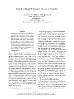
Fig. 3. Three proposed roles for Prxs in peroxide signaling. In each
case, different high levels of hydrogen peroxide cause a shift in
function from peroxidase activity. (A) Disulfide exchange, repre-
sented by an interprotein disulfide bond between Prx and a down-
stream protein (pink). In the one case studied, signaling is not
stress related and does not require sensitivity. (B) The chaperone
model, represented by the formation of higher order oligomers of
overoxidized Prxs. This is involved in stress-related signaling and
requires sensitivity. In (A) and (B), the Prxs are represented as pur-
ple and blue decamers under normal cellular conditions. (C) The
floodgate model is an unproven mechanism. Prxs are represented
as tall barriers made up of gray rectangles – vertical for active, hori-
zontal for overoxidized and inactive. The multiple barriers on the
right reflect the cell-wide Prx distribution; Prxs that are close to the
peroxide generation site (marked by an arrow) are overwhelmed
and inactivated, whereas those at increasing distances away are
not. This creates a steep peroxide gradient and allows for localized
peroxide build-up after endogenous peroxide generation. The level
of hydrogen peroxide is represented by both color gradient and
height. This proposed role may be involved in both stress- and non-
stress-related signaling and requires sensitivity.
2-Cys peroxiredoxins A. Hall et al.
2474 FEBS Journal 276 (2009) 2469–2477 ª 2009 The Authors Journal compilation ª 2009 FEBS
pombe Prx, Tpx1, activates the transcription factor
Pap1 through intermolecular disulfide bond forma-
tion. At higher hydrogen peroxide levels, Pap1 is not
activated, but instead the transcriptional factor Sty1
is activated through an intermolecular disulfide bond
with Tpx1 [25]. Activation of Sty1 leads to the tran-
scription of antioxidant defense proteins and Srx.
Although early evidence suggested that the regulation
of these two pathways required sensitivity, mutants
with C-terminal truncations that rendered Tpx1
insensitive to overoxidation were fully functional in
these two pathways [43].
In the second gain-of-function model, Prxs have
been shown to act as chaperones or cell cycle regu-
lators when the overoxidized enzyme aggregates into
larger assemblies [44,45] (Fig. 3B). Higher order
molecular mass oligomers have been shown to have
chaperone activity in H. pylori, S. cerevisiae and
Homo sapiens [29,46,47], an activity which could pro-
tect cells from oxidation-induced protein unfolding.
Interestingly, chaperone activity appears to be higher
for Ho. sapiens Prx1 than Prx2, because an additional
cysteine present in Prx1 forms a disulfide to stabilize
the higher order aggregates [48]. Consistent with the
proposed requirement for sensitivity, C-terminal trun-
cation mutants of Ho. sapiens Prx2 do not respond to
oxidative stress with increasing levels of chaperone
activity [47]. Although sensitivity is important for Prx
chaperone activity in S. cerevisiae and Ho. sapiens, the
case for H. pylori AhpC is unclear, as studies docu-
menting the sensitivity of H. pylori AhpC have not
been published.
Although direct evidence exists supporting both of
the gain-of-function models for stress-related signaling,
the loss-of-function model for Prxs in nonstress-related
signaling is still largely speculative. This mechanism
requires sensitivity. The ‘floodgate model’ [27] predicts
that, under normal conditions, Prxs act as a barrier, hin-
dering peroxide encounters with sensitive cellular com-
ponents (Fig. 3C). In the presence of a high peroxide
pulse, such as could be produced by hormone-triggered
activation of cellular NOXs, the rapid production of
hydrogen peroxide causes high local peroxide concentra-
tions which would inactivate the proximal floodgate
Prxs, allowing peroxide to locally build up to concentra-
tions that can oxidize specific downstream target pro-
teins. The recent demonstrations of localized NOX
signaling are consistent with this model, but do not
prove that Prxs play this role. As shown in Fig. 3C, the
high Prx concentration throughout the cell implies that
the term ‘floodgate’ is somewhat of a misnomer, as sen-
sitive Prxs will actually not allow peroxide to spread
throughout the cell, but will instead localize the peroxide
build-up. Thus, Prxs are more similar to an adjustable
buffer than a floodgate. The recent evidence that non-
stress-related growth factor signaling involves localized
peroxide build-up of a sufficient concentration to oxi-
dize PTP1B implies that the nearby sensitive Prxs would
also be overoxidized [38]. Operation of a Prx floodgate
in such systems may be challenging to discern because
of the difficulty in detecting a small overoxidized popu-
lation located close to the peroxide source within the
large cellular pool of Prxs [10].
Hormone-triggered apoptosis is, in contrast, a signal-
ing process that appears to involve more global Prx
overoxidation. This was shown years ago for signaling
by tumor necrosis factor [49]. More recently, a report of
apoptotic signaling in dopaminergic neurons (used as an
experimental model of Parkinson’s disease) has shown
that 6-hydroxydopamine activation of p38 mitogen-
activated protein kinase and caspase-3 is associated with
significant overoxidation of Prx1 and other Prxs [50].
The cells were protected against elevated levels of
reactive oxygen species and apoptotic death by over-
expression of Prx1 or the addition of other antioxidants,
and displayed enhanced apoptosis when Prx1 expression
was knocked down. Whether or not these two cases can
truly be considered as ‘nonstress-related redox signaling’
is debatable, because the extensive overoxidation of Prxs
implies that apoptotic signaling is accompanied by
increases in reactive oxygen species that are much
higher than in other types of signaling.
With the increasing attention given to Prxs, we are
learning that they are efficient catalysts and that their
catalytic repertoire is broader than that of a simple per-
oxidase. The high level of expression of sensitive Prxs in
eukaryotes allows them to carry out functions under
conditions of oxidative stress, such as that of a chaper-
one, which depend on their high concentration rather
than on their peroxidase activity. Nevertheless, why the
sensitivity of the highly expressed eukaryotic Prxs is so
strongly conserved and why repair systems have evolved
to recover activity in the overoxidized Prxs are still open
questions. Some answers could come from the study of
a Prx1 knockout mouse with a robust Prx gene knockin
that mimics the expression pattern of the missing sensi-
tive Prxs. The local nature of nonstress-related redox
signaling has made it challenging to determine the mech-
anisms involved and to discern whether the overoxidized
Prxs act primarily as a passive floodgate ⁄ buffer or as an
active positive signal, or both.
Acknowledgements
The authors thank Todd Lowther, Kim Nelson and
Derek Parsonage for their editorial suggestions. This
A. Hall et al. 2-Cys peroxiredoxins
FEBS Journal 276 (2009) 2469–2477 ª 2009 The Authors Journal compilation ª 2009 FEBS 2475
publication was made possible in part by a grant from
the National Institute of General Medical Sciences to
L.B.P. with a subcontract to P.A.K. (RO1 GM50389),
and by a grant from the National Institute of Environ-
mental Health Sciences (P30 ES00210).
References
1 Wood ZA, Schro
¨
der E, Harris JR & Poole LB (2003)
Structure, mechanism and regulation of peroxiredoxins.
Trends Biochem Sci 28, 32–40.
2 Copley SD, Novak WR & Babbitt PC (2004) Divergence
of function in the thioredoxin fold suprafamily: evidence
for evolution of peroxiredoxins from a thioredoxin-like
ancestor. Biochemistry 43, 13981–13995.
3 Chae HZ, Robison K, Poole LB, Church G, Storz G &
Rhee SG (1994) Cloning and sequencing of thiol-spe-
cific antioxidant from mammalian brain: alkyl hydro-
peroxide reductase and thiol-specific antioxidant define
a large family of antioxidant enzymes. Proc Natl Acad
Sci USA 91, 7017–7021.
4 Chae HZ, Chung SJ & Rhee SG (1994) Thioredoxin-
dependent peroxide reductase from yeast. J Biol Chem
269, 27670–27678.
5 Fourquet S, Huang ME, D’Autreaux B & Toledano
MB (2008) The dual functions of thiol-based
peroxidases in H
2
O
2
scavenging and signaling. Antioxid
Redox Signal 10, 1565–1576.
6 Karplus PA & Hall A (2007) Structural survey of the
peroxiredoxins. In Peroxiredoxin Systems (Flohe
´
L&
Harris JR, eds), pp. 41–60. Springer, New York, NY.
7 Poole LB (2007) The catalytic mechanism of peroxi-
redoxins. In Peroxiredoxin Systems (Flohe
´
L & Harris
JR, eds), pp. 61–81. Springer, New York, NY.
8 Rhee SG, Chae HZ & Kim K (2005) Peroxiredoxins: a
historical overview and speculative preview of novel
mechanisms and emerging concepts in cell signaling.
Free Radic Biol Med 38, 1543–1552.
9 Stone JR & Yang S (2006) Hydrogen peroxide: a
signaling messenger. Antioxid Redox Signal 8 , 243–270.
10 Veal EA, Day AM & Morgan BA (2007) Hydrogen
peroxide sensing and signaling. Mol Cell 26, 1–14.
11 Winterbourn CC (2008) Reconciling the chemistry and
biology of reactive oxygen species. Nat Chem Biol 4,
278–286.
12 Ogusucu R, Rettori D, Munhoz DC, Soares Netto LE
& Augusto O (2007) Reactions of yeast thioredoxin
peroxidases I and II with hydrogen peroxide and perox-
ynitrite: rate constants by competitive kinetics. Free
Radic Biol Med 42, 326–334.
13 Peskin AV, Low FM, Paton LN, Maghzal GJ, Hamp-
ton MB & Winterbourn CC (2007) The high reactivity
of peroxiredoxin 2 with H
2
O
2
is not reflected in its reac-
tion with other oxidants and thiol reagents. J Biol Chem
282, 11885–11892.
14 Nelson KJ, Parsonage D, Hall A, Karplus PA & Poole
LB (2008) Cysteine pK
a
values for the bacterial perox-
iredoxin AhpC. Biochemistry 47 , 12860–12868.
15 Wood ZA, Poole LB, Hantgan RR & Karplus PA
(2002) Dimers to doughnuts: redox-sensitive oligomeri-
zation of 2-cysteine peroxiredoxins. Biochemistry 41,
5493–5504.
16 Barranco-Medina S, Kakorin S, Lazaro JJ & Dietz KJ
(2008) Thermodynamics of the dimer–decamer transi-
tion of reduced human and plant 2-cys peroxiredoxin.
Biochemistry 47, 7196–7204.
17 Cao Z, Bhella D & Lindsay JG (2007) Reconstitution of
the mitochondrial PrxIII antioxidant defence pathway:
general properties and factors affecting PrxIII activity
and oligomeric state. J Mol Biol 372, 1022–1033.
18 Matsumura T, Okamoto K, Iwahara S, Hori H, Takah-
ashi Y, Nishino T & Abe Y (2008) Dimer–oligomer
interconversion of wild-type and mutant rat 2-Cys per-
oxiredoxin: disulfide formation at dimer–dimer inter-
faces is not essential for decamerization. J Biol Chem
283, 284–293.
19 Parsonage D, Youngblood DS, Sarma GN, Wood ZA,
Karplus PA & Poole LB (2005) Analysis of the link
between enzymatic activity and oligomeric state in
AhpC, a bacterial peroxiredoxin. Biochemistry 44,
10583–10592.
20 Schro
¨
der E, Littlechild JA, Lebedev AA, Errington N,
Vagin AA & Isupov MN (2000) Crystal structure of
decameric 2-Cys peroxiredoxin from human erythro-
cytes at 1.7 A
˚
resolution. Structure 8, 605–615.
21 Cha MK, Yun CH & Kim IH (2000) Interaction of
human thiol-specific antioxidant protein 1 with
erythrocyte plasma membrane. Biochemistry 39,
6944–6950.
22 Parsonage D, Karplus PA & Poole LB (2008)
Substrate specificity and redox potential of AhpC, a
bacterial peroxiredoxin. Proc Natl Acad Sci USA 105,
8209–8214.
23 Dietz KJ, Jacob S, Oelze ML, Laxa M, Tognetti V,
de Miranda SM, Baier M & Finkemeier I (2006) The
function of peroxiredoxins in plant organelle redox
metabolism. J Exp Bot 57, 1697–1709.
24 Yamamoto Y, Ritz D, Planson AG, Jonsson TJ, Faulk-
ner MJ, Boyd D, Beckwith J & Poole LB (2008)
Mutant AhpC peroxiredoxins suppress thiol-disulfide
redox deficiencies and acquire deglutathionylating activ-
ity. Mol Cell 29, 36–45.
25 Veal EA, Findlay VJ, Day AM, Bozonet SM, Evans
JM, Quinn J & Morgan BA (2004) A 2-Cys peroxire-
doxin regulates peroxide-induced oxidation and activa-
tion of a stress-activated MAP kinase. Mol Cell 15,
129–139.
26 Yang KS, Kang SW, Woo HA, Hwang SC, Chae HZ,
Kim K & Rhee SG (2002) Inactivation of human
peroxiredoxin I during catalysis as the result of the
2-Cys peroxiredoxins A. Hall et al.
2476 FEBS Journal 276 (2009) 2469–2477 ª 2009 The Authors Journal compilation ª 2009 FEBS
oxidation of the catalytic site cysteine to cysteine-sulfi-
nic acid. J Biol Chem 277, 38029–38036.
27 Wood ZA, Poole LB & Karplus PA (2003) Peroxi-
redoxin evolution and the regulation of hydrogen
peroxide signaling. Science 300, 650–653.
28 Koo KH, Lee S, Jeong SY, Kim ET, Kim HJ, Song K
& Chae H-Z (2002) Regulation of thioredoxin peroxi-
dase activity by C-terminal truncation. Arch Biochem
Biophys 397, 312–318.
29 Chuang MH, Wu MS, Lo WL, Lin JT, Wong CH &
Chiou SH (2006) The antioxidant protein alkylhydro-
peroxide reductase of Helicobacter pylori switches from
a peroxide reductase to a molecular chaperone function.
Proc Natl Acad Sci USA 103, 2552–2557.
30 Sayed AA & Williams DL (2004) Biochemical charac-
terization of 2-Cys peroxiredoxins from Schistosoma
mansoni. J Biol Chem 279, 26159–26166.
31 Jo
¨
nsson TJ & Lowther WT (2007) The peroxiredoxin
repair proteins. In Peroxiredoxin Systems (Flohe
´
L&
Harris JR, eds), pp. 115–141. Springer, New York,
NY.
32 Jo
¨
nsson TJ, Johnson LC & Lowther WT (2008) Struc-
ture of the sulphiredoxin–peroxiredoxin complex reveals
an essential repair embrace. Nature 451, 98–101.
33 Poole LB, Karplus PA & Claiborne A (2004) Protein
sulfenic acids in redox signaling. Annu Rev Pharmacol
Toxicol 44, 325–347.
34 Delaunay A, Pflieger D, Barrault MB, Vinh J & Toled-
ano MB (2002) A thiol peroxidase is an H
2
O
2
receptor
and redox-transducer in gene activation. Cell 111,
471–481.
35 Choi MH, Lee IK, Kim GW, Kim BU, Han YH,
Yu DY, Park HS, Kim KY, Lee JS, Choi C et al.
(2005) Regulation of PDGF signalling and vascular
remodelling by peroxiredoxin II. Nature 435, 347–353.
36 Li Q, Harraz MM, Zhou W, Zhang LN, Ding W,
Zhang Y, Eggleston T, Yeaman C, Banfi B &
Engelhardt JF (2006) Nox2 and Rac1 regulate H
2
O
2
-
dependent recruitment of TRAF6 to endosomal
interleukin-1 receptor complexes. Mol Cell Biol 26,
140–154.
37 Ushio-Fukai M (2007) VEGF signaling through
NADPH oxidase-derived ROS. Antioxid Redox Signal
9, 731–739.
38 Chen K, Kirber MT, Xiao H, Yang Y & Keaney JF Jr
(2008) Regulation of ROS signal transduction by
NADPH oxidase 4 localization. J Cell Biol 181,
1129–1139.
39 Leyens G, Donnay I & Knoops B (2003) Cloning of
bovine peroxiredoxins – gene expression in bovine tis-
sues and amino acid sequence comparison with rat,
mouse and primate peroxiredoxins. Comp Biochem
Physiol B Biochem Mol Biol 136, 943–955.
40 Ghaemmaghami S, Huh WK, Bower K, Howson RW,
Belle A, Dephoure N, O’Shea EK & Weissman JS
(2003) Global analysis of protein expression in yeast.
Nature 425, 737–741.
41 Neumann CA, Krause DS, Carman CV, Das S, Dubey
DP, Abraham JL, Bronson RT, Fujiwara Y, Orkin SH
& Van Etten RA (2003) Essential role for the peroxi-
redoxin Prdx1 in erythrocyte antioxidant defence and
tumour suppression. Nature 424, 561–565.
42 Vivancos AP, Castillo EA, Biteau B, Nicot C, Ayte J,
Toledano MB & Hidalgo E (2005) A cysteine-sulfinic
acid in peroxiredoxin regulates H
2
O
2
-sensing by the
antioxidant Pap1 pathway. Proc Natl Acad Sci USA
102, 8875–8880.
43 Jara M, Vivancos AP & Hidalgo E (2008) C-terminal
truncation of the peroxiredoxin Tpx1 decreases its
sensitivity for hydrogen peroxide without
compromising its role in signal transduction. Genes
Cells 13, 171–179.
44 Phalen TJ, Weirather K, Deming PB, Anathy V, Howe
AK, van der Vliet A, Jo
¨
nsson TJ, Poole LB & Heintz
NH (2006) Oxidation state governs structural transi-
tions in peroxiredoxin II that correlate with cell cycle
arrest and recovery. J Cell Biol 175, 779–789.
45 Trotter EW, Rand JD, Vickerstaff J & Grant CM
(2008) The yeast Tsa1 peroxiredoxin is a ribosome-asso-
ciated antioxidant. Biochem J 412, 73–80.
46 Jang HH, Lee KO, Chi YH, Jung BG, Park SK,
Park JH, Lee JR, Lee SS, Moon JC, Yun JW et al.
(2004) Two enzymes in one; two yeast peroxiredoxins
display oxidative stress-dependent switching from a
peroxidase to a molecular chaperone function. Cell
117, 625–635.
47 Moon JC, Hah YS, Kim WY, Jung BG, Jang HH, Lee
JR, Kim SY, Lee YM, Jeon MG, Kim CW et al. (2005)
Oxidative stress-dependent structural and functional
switching of a human 2-Cys peroxiredoxin isotype II
that enhances HeLa cell resistance to H
2
O
2
-induced cell
death. J Biol Chem 280, 28775–28784.
48 Lee W, Choi KS, Riddell J, Ip C, Ghosh D, Park JH &
Park YM (2007) Human peroxiredoxin 1 and 2 are not
duplicate proteins: the unique presence of CYS83 in
Prx1 underscores the structural and functional
differences between Prx1 and Prx2. J Biol Chem 282,
22011–22022.
49 Rabilloud T, Heller M, Gasnier F, Luche S, Rey C,
Aebersold R, Benahmed M, Louisot P & Lunardi J
(2002) Proteomics analysis of cellular response to
oxidative stress. Evidence for in vivo overoxidation of
peroxiredoxins at their active site. J Biol Chem 277,
19396–19401.
50 Lee YM, Park SH, Shin DI, Hwang JY, Park B, Park
YJ, Lee TH, Chae HZ, Jin BK, Oh TH et al. (2008)
Oxidative modification of peroxiredoxin is associated
with drug-induced apoptotic signaling in experimental
models of Parkinson disease. J Biol Chem 283,
9986–9998.
A. Hall et al. 2-Cys peroxiredoxins
FEBS Journal 276 (2009) 2469–2477 ª 2009 The Authors Journal compilation ª 2009 FEBS 2477