Báo cáo khoa học: Nuclear actin and actin-binding proteins in the regulation of transcription and gene expression docx
Bạn đang xem bản rút gọn của tài liệu. Xem và tải ngay bản đầy đủ của tài liệu tại đây (342.55 KB, 17 trang )
REVIEW ARTICLE
Nuclear actin and actin-binding proteins in the regulation
of transcription and gene expression
Bin Zheng
1
, Mei Han
1
, Michel Bernier
2
and Jin-kun Wen
1
1 Department of Biochemistry and Molecular Biology, Hebei Medical University, Shijiazhuang, China
2 Laboratory of Clinical Investigation, National Institute on Aging, National Institutes of Health, Baltimore, MD, USA
Actin is a major component of the cytoskeleton and
plays a critical role in all eukaryotic cells. The actin
cytoskeleton functions in diverse cellular processes,
including cell motility, contractility, mitosis and cytoki-
nesis, intracellular transport, endocytosis and secretion
[1,2]. In addition to these mechanical functions, actin
has also been implicated in the regulation of gene tran-
scription, through either cytoplasmic changes in cyto-
skeletal actin dynamics [3] or the assembly of
transcriptional regulatory complexes [4]. Cytoskeletal
actin dynamics, i.e. actin polymerization by which
monomeric actin (globular actin or G-actin) is assem-
bled into long actin polymers (filamentous actin or
F-actin) and actin deploymerization by which F-actin
is severed into G-actin, is key for these diverse func-
tions. The dynamic nature of the actin cytoskeleton
is determined spatiotemporally by the actions of
numerous actin-binding proteins (ABPs). The activity
of different classes of ABP controls actin nucle-
ation, bundling, filament capping, fragmentation and
Keywords
actin dynamics; actin-binding protein;
chromatin remodeling; gene regulation;
muscle-specific gene; nuclear actin; nuclear
receptor; ribonucleoprotein; RNA
polymerases; transcription complex
Correspondence
J k. Wen, Department of Biochemistry and
Molecular Biology, Hebei Medical
University, No. 361, Zhongshan East Road,
Shijiazhuang 050017, China
Fax: +86 311 866 96180
Tel: +86 311 862 65563
E-mail:
(Received 12 January 2009, revised 20
February 2009, accepted 26 February 2009)
doi:10.1111/j.1742-4658.2009.06986.x
Nuclear actin is involoved in the transcription of all three RNA polymerases,
in chromatin remodeling and in the formation of heterogeneous nuclear
ribonucleoprotein complexes, as well as in recruitment of the histone modi-
fier to the active gene. In addition, actin-binding proteins (ABPs) control
actin nucleation, bundling, filament capping, fragmentation and monomer
availability in the cytoplasm. In recent years, more and more attention has
focused on the role of actin and ABPs in the modulation of the subcellular
localization of transcriptional regulators. This review focuses on recent
developments in the study of transcription and transcriptional regulation by
nuclear actin, and the regulation of muscle-specific gene expression, nuclear
receptor and transcription complexes by ABPs. Among the ABPs, striated
muscle activator of Rho signaling and actin-binding LIM protein regulate
actin dynamics and serum response factor-dependent muscle-specific
gene expression. Functionally and structurally unrelated cytoplasmic ABPs
interact cooperatively with nuclear receptor and regulate its transactiva-
tion. Furthermore, ABPs also participate in the formation of transcription
complexes.
Abbreviations
ABLIM, actin-binding LIM protein; ABP, actin-binding protein; ANF, atrial natriuretic factor; AR, androgen receptor; CARM1, coactivator-
associated arginine methyltransferase 1; CBP, CREB binding protein; DBD, DNA-binding domain; FHL, four and a half LIM domains; FLAP1,
Fli-I LRR-associated protein 1; Fli-I, flightless-1; FLNa, filamin-A; FOXC1, forkhead box C1; GRIP1, glucocorticoid receptor-interacting protein 1;
HAT, histone acetyltransferase; HDAC, histone deacetylase; HF, hydroxyflutamide; hhLIM, human heart LIM protein; hnRNPs, heterogeneous
nuclear ribonucleoproteins; LBD, ligand-binding domain; LEF1 ⁄ TCF, lymphoid enhancer factor ⁄ T-cell factor; LRR, leucine rich repeat; MEF2,
myocyte enhancer factor 2; MRTF, myocardin-related transcription factor; NLS, nuclear localization signals; NM1, nuclear myosin 1; PBX1,
pre-B-cell leukemia transcription factor 1; PCAF, p300 ⁄ CREB binding protein-associated factor; PEBP2b, polyoma enhancer-binding protein;
PIC, pre-initiation complex; Pol I, RNA polymerase I; Pol II, RNA polymerase II; Pol III, RNA polymerase III; RNP, ribonucleoprotein; SRF,
serum response factor; STARS, striated muscle activator of Rho signaling; SV, supervillin; SWI ⁄ SNF, switch ⁄ sucrose nonfermentable complex.
FEBS Journal 276 (2009) 2669–2685 ª 2009 The Authors Journal compilation ª 2009 FEBS 2669
monomer availability. Transcriptional regulation, med-
iated by cytoskeletal actin dynamics, can be attributed
to modulation of the subcellular localization of tran-
scriptional regulators by ABPs [5]. In addition, some
of the mechanisms by which actin affects transcription
and its regulation depend on molecular interactions of
actin with RNA polymerases and components of the
transcription machinery in the nucleus.
The role of actin in transcription and
its regulation
Actin is both a major cytoskeletal component of all
eukaryotic cells and also a constitutent of nuclear pro-
tein complexes. Nuclear actin plays a role in many
nuclear functions [6–8]. First, nuclear actin is required
for transcription by all three nuclear RNA polymerases.
Second, nuclear actin associates with small nuclear ri-
bonucleoproteins (RNPs), which have a major role in
mRNA processing [8,9], and is directly involved in the
nuclear export of RNA and cellular proteins [10,11].
Third, nuclear actin also forms complexes with certain
heterogeneous nuclear ribonucleoproteins (hnRNPs)
that bind to and accompany mRNA from the nucleus
to the cytoplasm [12–14]. Fourth, nuclear actin and
actin-related proteins have been found in association
with chromatin-remodeling and histone acetyl transfer-
ase complexes, suggesting a role for actin in chromatin
remodeling [15]. Recent investigations suggest that
nuclear actin has a role in gene transcription associated
with three main entities: components of the three RNA
polymerases, ATP-dependent chromatin-remodeling
complexes and RNP particles in the eukaryotic cell
nucleus.
Nuclear actin is a constitutive component of all
RNA polymerases
Nuclear actin is required for the transcription of all
three RNA polymerases. Specifically, b-actin has been
identified as a component of RNA polymerase II
(Pol II) pre-initiation complexes (PICs). Injection of
anti-actin Ig into the nuclei of salamander oocytes
results in contraction of the lateral loops and the inhi-
bition of transcription [8]. Furthermore, Hofmann &
de Lanerolle [16] found that actin is associated with
actively transcribed genes and has an essential role in
the activation of transcription. In addition, actin is
required for the initiation of transcription through par-
ticipation in the formation of PICs [17]. These conclu-
sions are based on the following data: (a) b-actin
participates directly in Pol II transcription, using only
purified transcription factors [18,19]; (b) nascent RNA
molecules are associated with actin in the nuclear
matrix and antibodies to b-actin inhibit the synthesis
of nascent transcripts and Pol II transcription [17,19];
(c) adding actin to a highly purified Pol II fraction
stimulates transcription [19]; (d) actin colocalizes with
transcription sites in early mouse embryos [4,17];
(e) actin is recruited to the promoter region of tran-
scribing genes in vivo [19,20]; (f) antibodies to b-actin
inhibit the production of a 15-nucleotide transcript
that is a prerequisite for the commitment to elongation
[19,21]; (g) actin is a component of pre-mRNP parti-
cles, and is incorporated into pre-mRNAs by binding
to a specific subset of RNA-binding proteins [4,22];
and (h) actin is a component of PICs and depletion of
actin prevents their formation [19,23]. The above
evidence suggests that there is a strong and specific
interaction between actin and Pol II, and actin partici-
pates in Pol II transcription. What then is the function
of actin in Pol II transcription? From the above data,
we conclude that: (a) based on chromatin immunopre-
cipitation assays results, which show that actin is
recruited to genes poised to begin transcribing, it is
known that actin is involved in recruiting Pol II to the
PIC [19]; (b) decreased actin levels resulting from anti-
actin Ig inhibit PIC formation by preventing the bind-
ing of TBP to the TATA box, indicating that PIC for-
mation is required for the association of actin with
promoter DNA [19]; (c) antibodies to b-actin prevent
PIC formation, suggesting that actin acts as a bridge
between the polymerase and other constituents of the
PIC [24]; and (d) actin and nuclear myosin 1 (NM1),
an isoform of myosin 1, are involved in transcription
elongation [6,25,26]. Together, these data suggest that
actin is involved in multiple stages of the transcription
process.
b-Actin also has an important role in RNA poly-
merase III (Pol III) transcription [27]. First, b-actin is
tightly associated with Pol III via direct protein–pro-
tein interactions with one or more of the RPC3,
RPABC2 and RPABC3 subunits, and constitutes part
of the active Pol III [27]. Photochemical cross-linking
experiments, performed using a transcription initiation
complex, indicated that actin makes complex contact
with DNA [28]. Second, chromatin immunoprecipita-
tion assays identified that b-actin is located at the pro-
moter sequences of an actively transcribed U6 gene
in vivo, which suggests that it participates in the tran-
scription of Pol III [27,29,30]. Upon treatment with
methane methylsulfonate, a drug that represses Pol III
transcription, the U6 initiation complex and b-actin
are largely dissociated from promoter sequences
[27,29,31]. Notably, there is a much larger decrease in
the association between b-actin and the U6 promoter
Actin and ABPs in transcription regulation B. Zheng et al.
2670 FEBS Journal 276 (2009) 2669–2685 ª 2009 The Authors Journal compilation ª 2009 FEBS
region when compared with the dissociation of Pol III,
which suggests that b-actin dissociates from the Pol III
complex. Third, many experiments have shown that
b-actin is required for Pol III transcription [27,29,32].
The monomeric form of actin is required for Pol III
transcription, suggesting that b-actin is essential for
basal RNA polymerase transcription.
Actin and NM1 interact with different components
of the RNA polymerase I (Pol I) machinery, and
together serve as a nucleolar motor involved in the
transcription of ribosomal RNA genes [26,33]. Recent
studies have revealed that actin is associated with
rDNA genes, and microinjection of anti-actin Ig into
the nuclei of HeLa cells inhibits pre-rRNA synthesis
in vivo [25,34]. The interaction of NM1 with actin in
the initiation complex may trigger a conformational
change that favors the transition of Pol I from the
initiation phase to the elongation phase [25,33]. NM1
mutants that lack ATPase activity or actin binding are
not capable of associating with Pol I [17], and their
association with rDNA is greatly impaired. Moreover,
the association of actin and NM1 with Pol I is abol-
ished in the presence of ATP and is stabilized by
ADP, further suggesting that nuclear actomyosin com-
plexes act as a molecular motor that facilitates tran-
scription [17]. NM1 binds the DNA backbone via its
positively charged tail domain, whereas the head inter-
acts with actin bound to RNA polymerase [4]. It has
been suggested that by anchoring NM1 to DNA, and
actin to RNA polymerase, an auxiliary motor is gener-
ated that works in concert with nuclear RNA poly-
merases to drive transcription [23]. This suggests that
the cooperative action of actin and myosin in the
nucleus is required for Pol I transcription and reveals
an actomyosin-based mechanism in transcription.
Actin serves as components of
chromatin-remodeling complexes
Actin is essential for the function of chromatin-remod-
eling complexes in transcriptional activation. Nuclear
actin is an ATPase that cycles between monomeric
(G-actin or b-actin) and polymerized (F-actin) states
[4]. Eukaryotic cells have several ATP-dependent
chromatin-remodeling complexes, depending on the
ATPase in the complex, as follows: switch ⁄ sucrose
nonfermentable (SWI ⁄ SNF) complexes, imitation of
SWI-containing complexes, Mi-2 complexes, histone
acetyltransferase complexes, such as the Nu4A and
TIP60 complexes, and INO80 complexes. b-Actin is an
integral component of chromatin-remodeling com-
plexes, such as the BAF, BAP and INO80 complexes,
as well as Nu4A and TIP60 complexes [24,27,29,35–38].
It is generally accepted that chromatin-remodeling com-
plexes contain actin, actin-related proteins and ⁄ or
ABPs. Nuclear actin-related proteins (ARP5–9) are
associated with actin in chromatin-remodeling com-
plexes of the SWI ⁄ SNF family, such as those containing
the ATPase subunits INO80 or SWR1 [15,24,39]. In the
SWI ⁄ SNF-like BAF complex, b-actin binds directly to
the BRG1 ATPase subunit of BAF and stimulates
BRG1 ATPase activity, and this interaction is necessary
for binding of the BAF complex to chromatin
[27,29,40]. Actin binding to BRG1 is required for stable
association of the complex and provides a link between
the chromatin-remodeling complex and the nuclear
matrix [5,41]. In the INO80 complex, actin is required
for efficient DNA binding, ATPase activity and nucleo-
some mobilization, as INO80 complexes lacking actin,
as well as the actin-related proteins, ARP4 and ARP8,
are deficient for these activities [15]. BAF53 and b-actin
have also been identified as subunits of the human
TIP60 histone acetyltransferase (HAT) complex, which
is involved in DNA repair and apoptosis, and BAF53 is
found in a distinct HAT complex involved in c-myc
activation, whereas Act3 ⁄ ARP4 and actin are compo-
nents of the yeast Nu4A HAT complex [38,42]. In the
yeast Nu4A HAT complex, actin and Act3 ⁄ ARP4 are
essential for the structural integity and activity of the
complex [38]. The presence of actin in chromatin-
remodeling complexes suggests that there is a functional
link between actin and regulation of the chromatin
structure, and a major function of actin is to act as an
allosteric regulator in the remodeling of some macro-
molecular assemblies, such as chromatin-remodeling
factors or transcription complexes.
Actin serves as a component of RNP
The hnRNP U, a component of pre-mRNP particles,
has been shown to interact directly with actin through
a specific and conserved actin-binding site located in
the hnRNP U C-terminus and associate with the phos-
phorylated C-terminal domain of Pol II [43]. Injection
of a peptide acting as a competitive inhibitor of pro-
tein–protein contact involving actin and the hnRNP
protein, HRP36, into the salivary glands of Chirono-
mus tentans disrupts global Pol II transcription as
measured by bromo-UTP incorporation; an effect that
is caused, at least in part, by a decrease in elongation
measured by run-on assays [22]. A recent study has
shown that actin binds directly to C. tentans hnRNP,
HRP65-2, which is a molecular platform for recruit-
ment of the HAT histone H3-specific acetyltransferase
p2D10 on active genes. Both actin and the pre-mRNP
protein, HRP65, are complexed in situ with p2D10,
B. Zheng et al. Actin and ABPs in transcription regulation
FEBS Journal 276 (2009) 2669–2685 ª 2009 The Authors Journal compilation ª 2009 FEBS 2671
and disruption of the actin–HRP65 interaction releases
p2D10 from Pol II-transcribing genes, coincident with
reduced H3 acetylation and diminished transcription
[6]. HRP65-2 binds directly to p2D10, and the interac-
tion between actin and HRP65-2 is required for p2D10
to associate with the transcribed chromatin [6]. More-
over, the association of p2D10, actin and HRP65-2
with chromatin is sensitive to ribonuclease digestion,
which indicates that these proteins are tethered to the
transcribed genes by binding to the nascent transcript.
These findings support the idea of a link between
nuclear actin, chromatin remodeling and Pol II
transcription [43,44]. Obrdlik et al. [13,45] identified
that the HAT, p300 ⁄ CREB binding protein (CBP)-
associated factor (PCAF), associates with actin and
hnRNP U. Moreover, it has been shown that
actin, hnRNP U and PCAF associate with the Ser2 ⁄ 5-
and Ser2-phosphorylated Pol II C-terminal domain.
hnRNP U and PCAF are present at the promoter and
coding regions of constitutively expressed Pol II genes
and are associated with RNP complexes [13]. In sum-
mary, these finding suggest that actin, HRP65-2 and
HAT (p2D10 or PCAF) are assembled into nascent
pre-mRNPs during transcription. Based on the evi-
dence, it may be proposed that the actin–HRP65-2–
HAT complex is part of the nascent pre-mRNP, and
can travel along the transcribed gene, allowing HAT
to acetylate histones. According to this proposal, the
actin–HRP65-2–HAT complex maintains the chroma-
tin in a transcription-competent conformation. This
model is supported by the observation that H3 acetyla-
tion is reduced and transcription is inhibited when the
interaction between actin and HRP65-2 is disrupted
[22]. In addition, actin-mediated Pol II transcriptional
control may be sensitive to the different polymeriza-
tion states of actin [17]. Transcriptionally competent
actin may be present in a monomeric or oligomeric
form which is different from the canonical actin fila-
ments. The polymerization states of actin involved in
the initiation or elongation phases are different
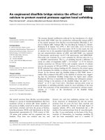
(Fig. 1) [43].
Roles of ABPs in the regulation of
muscle-specific gene expression
The cytoplasmic dynamics of the actin cytoskeleton
have been shown to regulate the subcellular localiza-
tion of some transcription factors, such as the myocar-
din-related transcription factors MRTF-A (also
referred to as MAL, MKL1 and BSAC) and MRTF-B
(also referred to as MKL2 or MAL16) [46,47], the
developmentally regulated PREP2 homeoprotein, and
the transcriptional repressor Yin-Yang 1 [48,49].
Because actin dynamics are regulated by a number of
ABPs, ABPs may play a critical role in the regulation
of transcription and gene expression [50]. Studies have
established that some ABPs induce the formation of
actin filaments by their ability to nucleate actin fila-
ment polymerization; other ABPs promote filament
breakdown by a mechanism referred to as severing.
Still other ABPs cross-link or bundle actin filaments or
prevent filament formation by their so-called sequester-
ing activity. Among the notable transcription factors
controlled by ABPs are MRTFs, which associate with
serum response factor (SRF) and stimulate SRF-
dependent transcription [46,51,52]. In addition, actin
dynamics are regulated by several signal transduction
cascades that converge on ABPs [53].
Actin
Actin
Pol II
TF
TF
TF
TBP
CTD
hnRNP U
Actin
Actin
mRNA processing
CTD
P
P
P
Pre-mRNA
Ac
Ac
Actin
polymerization
?
Activator
hnRNP U
PCAF or
P2D10
Pol II
HRP65-2
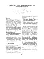
Fig. 1. Model for actin–hnRNP U-mediated control of pol II transcription elongation. Actin may modulate several steps in Pol II transcription
initiation and elongation, either as a monomer or as a polymer. Actin may modulate transcription as a monomeric component of transcription
preinitiation, chromatin-remodeling and hnRNP complexes. During transcription elongation, actin may be recruited to the elongating transcrip-
tion machinery via the hyperphosphorylated C-terminal domain and then to the nascent RNP, where actin in complex with the hnRNP U can
facilitate recruitment of PCAF or P2D10 to the active gene. Formation of actin filaments in the proximity of the Pol II C-terminal domain may
help establish a network of interactions between the various factors necessary for transcription elongation and pre-mRNA processing.
Actin and ABPs in transcription regulation B. Zheng et al.
2672 FEBS Journal 276 (2009) 2669–2685 ª 2009 The Authors Journal compilation ª 2009 FEBS
MRTF-A associates with G-actin, is predominantly
localized in the cytoplasm of NIH 3T3 cells in the
absence of serum and accumulates in the nucleus in
response to serum stimulation. MRTF-B also under-
goes nuclear translocation in response to serum stimu-
lation, although it is less responsive than MRTF-A
[54]. Upon activation of RhoA, actin becomes poly-
merized and releases MRTF-A, which in turn translo-
cates to the nucleus to associate with SRF [46].
Striated muscle activator of Rho signaling (STARS) is
a muscle-specific ABP capable of stimulating SRF-
dependent transcription via a mechanism involving
RhoA activation and actin polymerization [55].
Recently, MRTF-A and -B were shown to serve as a
link between STARS and SRF. In NIH 3T3 cells
cotransfected with expression plasmids encoding
MRTFs and STARS, the MRTFs are translocated to
the nucleus in the absence of serum. The nuclear local-
ization of myocardin is unchanged in the absence or
presence of STARS [54]. Thus, STARS may substitute
for serum stimulation and promote the nuclear translo-
cation of MRTFs with the consequent activation of
SRF-dependent transcription. Kuwahara et al. [54]
found that coexpression of STARS with a dominant-
negative myocardin mutant, which can inhibit the
transcriptional activities of myocardin and MRTF-A
and -B, can completely block the ability of STARS to
induce SRF-dependent transcription in NIH 3T3,
COS1 and 293T cells. However, STARS does not alter
the level of expression of MRTFs. These observations
suggest that STARS stimulates SRF-dependent tran-
scription solely by promoting the nuclear translocation
of MRTF-A and -B.
The STARS protein contains 375 amino acids, with
the conserved ABD contained within the C-terminal
142 residues [55]. The STARS C-terminal deletion
mutant, N233, which cannot bind actin or activate
SRF, fails to induce the nuclear accumulation of
MRTF-A and -B. By contrast, the C-terminal 142
amino acids of STARS, which bind actin and stimulate
SRF, induce the nuclear accumulation of MRTFs as
efficiently as full-length STARS. STARS N233 fails to
enhance MRTF-dependent activation of SRF-depen-
dent reporters, whereas STARS C142 synergistically
enhances MRTF-mediated transcription to the same
level as full-length STARS [55]. These results demon-
strate that the ABD of STARS is both necessary and
sufficient for the nuclear accumulation and transcrip-
tional activation of MRTFs by STARS.
The activity of STARS involves actin dynamics.
Treatment of NIH 3T3 cells with latrunculin B, which
sequesters actin monomers and prevents Rho-depen-
dent nuclear accumulation of MRTF-A and SRF
activation [46], blocks the nuclear accumulation
of MRTF-A and -B in the presence of STARS.
Conversely, cytochalasin D, which dimerizes actin, but
prevents actin polymerization and activates SRF,
strongly induces the nuclear translocation of MRTFs,
even in the absence of STARS [54]. Consistent with
these effects on MRTF nuclear import, latrunculin B
significantly blocks the stimulatory effect of STARS
on MRTF-dependent transcription, and cytochala-
sin D enhances the activity of MRTFs alone. These
results indicate that actin dynamics are involved in the
STARS-induced nuclear accumulation of MRTFs and
transcriptional activation of SRF via MRTFs.
MRTF-A was recently reported to interact directly
with G-actin [56]. Unpolymerized G-actin controls
MRTF activity [46], and STARS induces actin poly-
merization [55]. Kuwahara et al. [54] demonstrated
that expression of wild-type actin, which increases the
amount of G-actin, but does not alter the F-actin ⁄
G-actin ratio, reduced the ability of STARS to activate
MRTF-dependent transcription. Wild-type actin did
not significantly alter the activity of MRTF in the
absence of STARS. The actin mutant that favors
F-actin formation and increases the F-actin ⁄ G-actin
ratio [56] stimulates MRTF activity, even in the
absence of STARS, and abolishes further activation of
MRTFs by STARS. By contrast, the actin mutant that
is unable to polymerize and decreases the F-actin ⁄
G-actin ratio inhibits MRTF activity and also reduces
the ability of STARS to enhance MRTF activity.
These results suggest that STARS stimulates MRTF
activity by inducing the dissociation of MRTFs from
actin via depletion of the G-actin pool.
The N-terminal regions of MRTFs contain three
RPEL motifs which have been shown to sequester
MRTFs in the cytoplasm by association with actin
[46,56]. Consistent with STARS promoting the nuclear
import of MRTFs by displacing them from monomeric
G-actin, the RPEL motifs are required for the effects
of STARS on MRTFs. MRTFs are cytoplasmic, accu-
mulating in the nucleus upon activation of Rho
GTPase signaling, which alters interactions between
G-actin and the RPEL domain. Guettler et al. [57]
showed that the RPEL domain of MRTF-A binds
actin more strongly than the RPEL domain of myocar-
din, and that the RPEL motif itself is an actin-binding
element. RPEL1 and RPEL2 of myocardin bind actin
weakly compared with MRTF-A, whereas RPEL3 is
of comparable and low affinity in the two proteins.
Actin binding by all three motifs is required for
MRTF-A regulation. The differing behaviors of
MRTF-A and myocardin are specified by the RPEL1–
RPEL2 unit, whereas RPEL3 can be exchanged
B. Zheng et al. Actin and ABPs in transcription regulation
FEBS Journal 276 (2009) 2669–2685 ª 2009 The Authors Journal compilation ª 2009 FEBS 2673
between them. It has been proposed that differential
actin occupancy of multiple RPEL motifs regulates
nucleocytoplasmic transport and MRTF-A activity.
Because myocardin is insensitive to the effects of
STARS, its target genes are expected to be highly
active, irrespective of the polymerization state of actin.
However, STARS would be expected to further aug-
ment the expression of these genes via its actions on
MRTF-A and -B, which are also expressed in cardiac
muscle and which form heterodimers with myocardin.
In a yeast two-hybrid screen of a skeletal muscle
cDNA library using STARS as bait, Barrientos et al.
[58] identified two novel members of the actin-binding
LIM protein (ABLIM) family, ABLIM-2 and -3, as
STARS-interacting proteins. These novel proteins con-
tain four LIM domains and a C-terminal villin head-
piece domain, which mediates actin-binding in several
proteins, such as villin and dematin [59]. Both
ABLIM-2 and -3 show high homology with ABLIM-1.
ABLIM-1 was originally found in the human retina, as
well as in the sarcomeres of murine cardiac tissue, and
was postulated to regulate actin-dependent signaling
[60]. Similarly, ABLIM-2 and -3 are expressed in a tis-
sue-specific pattern. ABLIM-2 is highly expressed in
skeletal muscle and at lower levels in brain, spleen and
kidney. No significant expression has been detected in
the heart. In contrast to ABLIM-2, ABLIM-3 is pre-
dominantly expressed in human heart and brain,
whereas the murine ABLIM-3 homolog displays a
somewhat broader tissue distribution that also includes
lung and liver [58].
Both ABLIM-2 and -3 strongly bind F-actin and
colocalize with actin stress fibers. The interaction of
STARS with ABLIM-2 and -3 was confirmed by coim-
munoprecipitation and further supported by the colo-
calization of STARS and ABLIM-2, as detected by
immunofluorescence [58]. The complementary expres-
sion patterns of ABLIM-2 and -3 in striated muscle
imply that, in vivo, STARS interacts with ABLIM-2 in
skeletal muscle and ABLIM-3 in cardiac muscle.
Consistent with the notion that STARS activates SRF-
dependent transcription via stabilization of the actin
cytoskeleton [54], both ABLIM-2 and -3 modulate
STARS-dependent activation of a luciferase reporter
construct controlled by the SM22 promoter, which
contains two essential SRF-binding sites and is highly
sensitive to STARS activity [58]. The data suggest that
ABLIM-2 and -3 stimulate STARS activity. ABLIM-2
and -3 enhance STARS-dependent SRF-transcription
in COS cells in a dose-dependent manner [58], suggest-
ing that STARS and ABLIMs both physically interact
and functionally synergize to deliver activating signals
to SRF. The data imply that, in striated muscle,
STARS plays a critical role in the MRTF-A nuclear
translocation process; STARS promotes the nuclear
translocation of MRTFs, and thereby SRF-dependent
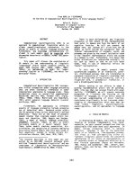
transcription (Fig. 2).
STARS activation of SRF-dependent transcription
is mediated, in part, by a Rho-dependent mechanism,
because the Rho inhibitor C3 transferase reduces SRF
activation by STARS. The ability of the Rho kinase
inhibitor, Y-27632, to diminish SRF activation by
STARS also suggests that Rho kinase is a downstream
effector of STARS [55]. The Rho family of GTPases,
including the best characterized members, Rho, Rac
and Cdc42, serve as molecular switches in the regula-
tion of a wide variety of signal transduction pathways
[61,62], in particular, actin polymerization and stress
fiber formation [63]. RhoA signaling has been shown
to induce the nuclear import of MRTF-A in smooth
muscle cells, thereby triggering smooth muscle gene
activation [64]. It is well-known that actin dynamics
and Rho signaling are involved in STARS-induced
nuclear translocation and transcriptional activation
of MRTFs, and Rho activity is crucial for actin
dynamics. Kuwahara et al. [54] showed that the
dominant-negative RhoA mutant inhibits the nuclear
accumulation of MRTFs and the stimulatory effect of
STARS on the transcriptional activity of MRTFs.
Although STARS requires Rho activity to induce actin
treadmilling and MRTF nuclear translocation, and the
inhibition of Rho activity blocks STARS activity,
assays of RhoA activity in STARS-transfected cells
did not differ from those in untransfected cells. Thus,
Fig. 2. Model of the involvement of STARS and ABLIM in actin
dynamics and SRF-dependent transcription.
Actin and ABPs in transcription regulation B. Zheng et al.
2674 FEBS Journal 276 (2009) 2669–2685 ª 2009 The Authors Journal compilation ª 2009 FEBS
STARS does not appear to function as an upstream
activator of Rho, but requires Rho–actin signaling and
changes in actin dynamics to evoke its stimulatory
effects on MRTFs and SRF activity. Taken together,
the small GTPase acts downstream of STARS, and it
seems possible that ABLIM integrates signals from the
small GTPases, Rac and RhoA (via STARS) toward
the actin cytoskeleton.
Roles of ABPs in the regulation of
nuclear receptor
Nuclear receptors regulated by ABPs include the glu-
cocorticoid receptor, estrogen receptor, androgen
receptor (AR), thyroid receptor and peroxisome prolif-
erator-activated receptor-c. Among these, the AR is
the most widely studied and well-characterized. The
AR is a ligand-activated transcription factor that con-
trols the expression of genes involved in functions such
as cell proliferation, cell growth, differentiation and
cell death [65,66]. The AR contains an N-terminal
domain harboring activation function 1, a central
DNA-binding domain (DBD) and a C-terminal ligand-
binding domain (LBD) containing activation func-
tion 2 [67–70]. Upon binding androgens, the AR LBD
undergoes conformational changes leading to dissocia-
tion from chaperones and translocation to the nucleus
[71–74]. AR binding to DNA facilitates the recruit-
ment of general transcriptional machinery and ancil-
lary factors that result in the activation or repression
of specific genes in targeted cells and tissues [75]. In
the last decade, an increasing number of proteins have
been proposed to possess AR coactivating or core-
pressing characteristics [76,77]. Cofactors facilitate AR
transcription function by histone modifications, chro-
matin remodeling and regulation of the AR N-terminal
domain, and the LBD interaction (N ⁄ C interaction)
[78–82]. All available data suggest that no single
AR-binding protein completely defines the multiple
functions of the AR in controlling cellular growth and
differentiation in normal and malignant cells [75].
Alternatively, AR pleiotropic activities are probably
mediated through its binding to specific functional pro-
tein complexes to carry out its broad biological func-
tions in mammalian cells. More than 200 nuclear
receptor coregulators have been identified since the
first nuclear receptor coactivator, SRC-1, was isolated
in 1995 [83]. Among the nuclear receptor coregulators,
ABPs and actin monomers bind to the AR, indicating
that they also play an important role in AR-mediated
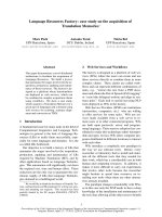
transcription (Fig. 3) [5,84]. For example, supervillin, a
nuclear ⁄ cytoplasmic F-actin-bundling protein, is able
to interact with the AR N-terminal domain and DBD–
LBD. This association is enhanced in the presence of
androgens [85]. In recent years, ABPs have been shown
to elicit increased activity in regulating AR than was
previously thought (Table 1).
Filamin, originally identified as a protein that facili-
tates nuclear transport of the AR, interacts with the
AR DBD–LBD in a ligand-independent manner
[77,86,87]. The absence of filamin hampers androgen-
induced AR transactivation. In the absence of filamin,
the receptor–Hsp90 (Hsp90 is a chaperone protein that
plays a key role in the conformational change and
transcriptional activity of the AR) complex may
remain inactive, anchored to the actin filaments, even
in the presence of steroid and an available nuclear
localization sequence on the receptor [87]. Filamin
may act as a mediator between the receptor and the
Hsp90, and control the release of activated receptor
after ligand binding in AR cytoplasmic trafficking
[87,88]. Filamin-A (FLNa) interferes with AR inter-
domain interactions and competes with the coactivator
transcriptional intermediary factor 2 (TIF2) to specifi-
cally downregulate AR function [86]. When cleaved at
the protease-cleavage site between repeats 15 and 16,
A
A
r
RE
R AR
AR
HSP
AR AR
ABPs
ABPs
Coactivators
HAT
Actin
AR nuclea
translocation
AR N/C
interaction
Coactivator
competition
HDAC chromatin
condensation
Actin
Pol II
ABPs
Fig. 3. Regulation of androgen receptor
gene transcription by actin-binding proteins.
B. Zheng et al. Actin and ABPs in transcription regulation
FEBS Journal 276 (2009) 2669–2685 ª 2009 The Authors Journal compilation ª 2009 FEBS 2675
full-length FLNa releases FLNa(16–24) [86–90]. This
naturally occurring C-terminal 100 kDa fragment of
filamin, interacting with the motor protein dynein,
may exert its inhibitory effect by interfering with inter-
actions between the N- and C-terminal domains, and
the coactivator functions of the AR [86,91]. Full-length
FLNa is bound to the actin cytoskeleton on the cell
surface and perinuclear areas of the cell via its N-ter-
minal ABD. In the absence of ligand, AR is localized
predominantly in the cytoplasm, and its hinge domain
and the LBD are tethered to the C-terminal end of
FLNa [86]. FLNa(16–24) colocalizes with liganded AR
to the nucleus. In the nucleus, FLNa(16–24) disrupts
interactions between the N- and C-termini of the AR,
and interferes with the binding of the coactivator TIF2
[86,91]. There is evidence that interaction between the
FXXLF (X = any amino acid) motif of the TAD and
the LBD reduces coactivator recruitment and binding
of the LXXLL motif of TIF2 [92]. Alternatively,
FLNa(16–24) may also directly recruit transcriptional
repressors to the target promoter or possess intrinsic
histone deacetylase activity to inhibit transcription
initiation [86]. In addition, the recent report of Rho-
regulated PAK6 as an AR hinge-interacting kinase [93]
suggests that the FLNa(16–24)–AR hinge complex
may serve as an integrator for the many cytoskeletal
signaling cascades that converge on the AR.
Supervillin (SV) was initially identified from blood
cells as an ABP and was found to be expressed in
skeletal muscles and several cancer cell lines [94].
Table 1. Role of nuclear actin-binding proteins interacting with the androgen receptor. AR, androgen receptor; LBD, ligand-binding domain.
Actin-binding
protein
Targeting
sequence Classes Role in the cytoplasm AR effect Mechanism
Direct or indirect
association
with the AR Region
Gelsolin ()) Actin filament
severing
and capping
protein
Involved in gel-to-sol
transformations;
severs and caps
polymeric actin
filaments; acts in
the actin-scavenging
system; inhibits actin
polymerization
Coactivator Promotes AR
activity in a
ligand-enhanced
manner
Direct LBD
Flightless I NLS Actin-
remodeling
proteins
Possess F-actin-serving
activity
Coactivator Does not enhance
the activity of
ARs alone, but
requires the
presence of a
p160 coactivator
Direct
a-actinin-2 ()) Bundling
proteins
Functions as scaffolds
for signaling intermediates
that stimulate actin
elongation; binding
partners for ICAM-1
Coactivator Indirect
Supervillin NLS F-actin- and
membrane-
associated
scaffolding
protein
Regulates cell-substrate
adhesion; organization
of muscle co-stameres;
stimulus-mediated
contractility of smooth
muscle and myogenic
differentiation
Coactivator Increases interaction
frequency with
the AR
Direct N- and
C-Terminal
Filamin NLS? Cross-linking
proteins
Cytoplasmic transport;
membrane integrity;
cellular adhesion
Coactivator AR cytoplasmic
trafficking
Direct Hinge
Filamin A NLS? Cross-linking
proteins
Cross-links actin filaments;
recruits F-actin into
extended networks
Corepressor Inhibits N ⁄ C,
suppresses
TIF2 activation
Direct Hinge
Transgelin ()) Cross-linking
proteins
Organizes actin
filaments into dense
meshworks
Corepressor Through ARA 54 Indirect LBD
Actin and ABPs in transcription regulation B. Zheng et al.
2676 FEBS Journal 276 (2009) 2669–2685 ª 2009 The Authors Journal compilation ª 2009 FEBS
SV is localized to the plasma membrane at sites of
intracellular contact. The nuclear localization signal is
located in the middle of this protein [95]. At low den-
sity, SV shows a punctate distribution localized to the
cytoplasm and nucleus, whereas at high density, SV is
localized almost exclusively to the plasma membrane.
SV has been identified as an AR-interacting protein,
which can interact with both N-terminal activation
function-1 and C-terminal activation function-2 of the
AR and plays a role in AR dimerization [85]. The
functional coregulator domain of SV is located at
amino acids 831–1281 of bovine origin, which has
putative actin-binding sites and nuclear localization
signals (NLS) [96]. Ting et al. [96] showed that SV
(amino acids 831–1281) has a better enhancing effect
on AR transactivation than full-length SV and SV
(amino acids 1010–1792). It is possible that by remain-
ing within the nucleus, SV may increase the interaction
frequency with the AR, resulting in a change in AR
conformation to an activated form to facilitate binding
of the androgen response element located in the target
genes. SV is relatively weak in promoting non-andro-
genic steroid-mediated AR transactivation, but is capa-
ble of coordinating with other coregulators, including
ARA55 and ARA70, to enhance AR transactivation
[96,97]. These results suggest that the final AR activity
may involve balancing and coordinating multiple
coregulators in any given cell. In addition, previous
experiments reported that actin and SV potentiate each
other in promoting AR activity [96]. Because several
putative actin-binding sites and functional NLS of SV
are important for the AR transactivation function, and
the minimal functional fragment of SV, which only
contains one actin-binding site, is located in the
nucleus, recruiting actin into the chromatin-remodeling
complex is a potential mechanism of co-regulator
activity [96]. The actin chelator, latrunculin B, which
attenuates the coregulator function of both full-length
SV and the minimal functional fragment, also identifies
this potential mechanism. Furthermore, Rac signaling
stimulates membrane ruffling that further attenuates
the coregulator activity of SV. There are two possible
explanations for this: (a) the accumulation of SV in
the membrane prevents it from associating with AR;
and (b) a decrease in the amount of actin monomer
affects SV coregulator activity, which requires actin
monomers [96]. However, SV has no effect on the
cytoplasmic–nuclear translocation of the AR, and does
not affect the half-life of the AR [85].
Gelsolin is a multifunctional ABP, implicated in cell
signaling, cell motility, apoptosis and carcinogenesis
[98,99]. Gelsolin regulates actin polymerization and
depolymerization by sequestering actin monomers, and
can sever and cap actin filaments [1]. Nishimura et al.
[100] identified gelsolin as an AR-interacting protein
that can enhance its transactivation in prostate cancer
cells. Because gelsolin lacks a nuclear localization sig-
nal, it may be cotranslocated into the nucleus upon
binding to other proteins [100]. Like filamin, gelsolin is
able to interact with AR at the time of its nuclear
localization to facilitate the nuclear translocation of
AR [87]. Increased expression of gelsolin can enhance
AR activity under hydroxyflutamide (HF) with low
levels of androgen treatment to maintain AR-mediated
growth and theh survival of tumor cells. Gelsolin itself
interacts with AR LBD via FXXFF and FXXMF
motifs and enhances its activity in the presence of
androgen. The interaction between the N- and C-ter-
mini of the AR does not affect gelsolin FXXFF bind-
ing to AR LBD, indicating that the gelsolin FXXFF
motif has a higher affinity for AR LBD [71]. Two pep-
tides, D1 (amino acids 551–600) and H1–2 (amino
acids 665–695) located within AR DBD and LBD,
respectively, can block gelsolin-enhanced AR activity
[100]. Altogether, gelsolin interacts with the AR during
nuclear translocation and enhances ligand-dependent
AR activity.
Transgelin, also termed SM22a, was first isolated
from chicken gizzard as a transformation- and shape
change-sensitive ABP [101]. Recently, Yang et al. [102]
characterized transgelin as a potential suppressor of
prostate cancer via inhibition of ARA54-enhanced AR
transactivation. ARA54, a RING finger protein, inter-
acts with AR and enhances its transcriptional activity
in a ligand-inducible manner. Transgelin does not inter-
act directly with the AR, but exerts its effects through
recruitment to ARA54. ARA54 can interact with
transgelin both in vitro and in vivo in an androgen-inde-
pendent manner [102]. The data suggest that transgelin
might need the specific interaction with ARA54 to sup-
press AR transactivation. By contrast, transgelin shows
little interaction with the AR, ARA70, ARA55, SRC-1,
supervillin, gelsolin and CREB binding protein (CBP).
Silencing of endogenous ARA54 via its siRNA can
abolish the suppressive effect of transgelin on AR
function [102]. This suggests that transgelin may be
able to suppress ARA54-enhanced AR transactivation
by interrupting the interaction between the AR and
ARA54, as well as ARA54 homodimerization, resulting
in enhanced cytoplasmic retention and impaired nuclear
translocation of ARA54 and the AR.
Flightless-1 (Fli-I) is an ABP that can be either asso-
ciated with the cytoskeleton or found in the nucleus,
but its exact physiologic functions have not been eluci-
dated [103]. Fli-I can associate directly with the AR and
function in cooperation with specific combinations of
B. Zheng et al. Actin and ABPs in transcription regulation
FEBS Journal 276 (2009) 2669–2685 ª 2009 The Authors Journal compilation ª 2009 FEBS 2677
other AR coactivators to enhance the ability of the AR
to activate the transcription of AR-regulated genes [77].
Because Fli-I does not enhance AR activity by itself,
but requires the presence of a p160 coactivator, binding
of Fli-I to the AR is apparently insufficient for Fli-I
coactivator function [104]. The contacts between Fli-I
and multiple components in the transcription complex
(AR, glucocorticoid receptor-interacting protein 1,
GRIP1, p160 and coactivator-associated arginine meth-
yltransferase 1, CARM1) may result in more efficient
recruitment of Fli-I to the promoter, a more stable
coactivator complex or a more highly functional con-
formation of the coactivator complex. Fli-I is a second-
ary coactivator in AR transcription activation [104].
a-Actinin-2 is a major structural component of sar-
comeric Z-lines in skeletal muscle, where they function
to anchor actin-containing thin filaments in a constitu-
tive manner [105]. a-Actinin-2 enhances the transacti-
vation activity of SRC-2 and serves as a primary
coactivator for the AR, acting in synergy with SRC-2
to increase AR transactivation function [106]. Huang
et al. [106] indicated that wild-type a-actinin-2 (con-
taining a LXXLL motif) and mutant a-actinin-2
(mutation of the LXXLL motif to LXXAA) both bind
to the AR, but the mutant form shows much weaker
binding than wild-type a-actinin-2. That is to say, the
LXXLL motif in a-actinin-2 has a major role in the
interaction with the AR. However, the LXXLL
motif of a-actinin-2 is dispensable for its primary coac-
tivator role in NR functions, because two truncated
a-actinin-2 fragments (encoding 281–700 and 701–894),
lacking the LXXLL motif, and mutant a-actinin-2
(LXXAA) retain the primary and secondary coactiva-
tor functions of wild-type a-actinin-2. In addition,
a-actinin-2 not only serves as a primary coactivator in
the AR, but also interacts synergistically with GRIP1
and enhances GRIP1-induced AR coactivator func-
tions in the presence of cognate ligands [106]. Further-
more, a-actinin-4 also binds to the AR and exhibits
coregulating properties. a-Actinin-4 may target the AR
for degradation and ⁄ or antagonize AR synthesis upon
the addition of androgen. In addition, a-actinin-4
negatively regulates AR-mediated transcription [75].
Roles of ABPs in the regulation of
transcription complexes
More and more experiments have identified that pro-
teins traditionally thought to be strictly cytoplasmic
structural factors can influence gene regulation. ABPs
transduced the changes in cell structure that occur dur-
ing morphogenesis to the nucleus, resulting in changes
in gene expression via either the nuclear shuttling of
transcription factors or the assembly of transcriptional
regulatory complexes [107].
ABPs can recruit multiple components to transcrip-
tion complexes through different types of interactions.
Fli-I binds both actin and the actin-like BAF53 (BAF
complex 53 kDa subunit, BRG1-associated factor), as
well as p160 co-activator [104,108]. Fli-I can help to
secure the association of an SWI ⁄ SNF complex to a
p160 coactivator complex. Fli-I thus helps to coordi-
nate the complementary ATP-dependent nucleosome-
remodeling activity of the SWI ⁄ SNF complex with the
histone acetylating (e.g. from CBP and p300) and
methylating (e.g. from CARM1 and protein arginine
methyltransferase 1) activities of the p160 coactivator
complex [109]. In addition, Fli-I and Fli-I LRR-associ-
ated protein 1 (FLAP1) have an important role in reg-
ulating transcriptional activation by b-catenin and
lymphoid enhancer factor ⁄ T-cell factor (LEF1 ⁄ TCF).
FLAP1 is a key activator, cooperating synergistically
with p300 to enhance LEF1 ⁄ TCF-mediated transcrip-
tion by b-catenin. Fli-I negatively regulates the synergy
of FLAP1 and p300 [103]. Lee & Stallcup [103] found
that Fli-I does not bind well to the p300 KIX domain
and does not appear to inhibit FLAP1–p300 binding,
suggesting that Fli-I does not interfere with the bind-
ing of FLAP1 to p300. Fli-I may exert its negative
influence by inhibiting the activity of FLAP1 and other
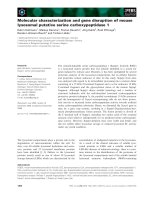
essential factors that bind to Fli-I (Fig. 4). It is also
possible that Fli-I may recruit negative regulators, such
as histone deacetylases (HDACs), CtBP, Groucho and
Chibby, to the b-catenin ⁄ LEF1 ⁄ TCF transcription
complex. Both the leucine-rich repeat (LRR) and gels-
olin-like domains of Fli-I are required for the negative
Fig. 4. Model of Fil-I participation in transcription regulation. Fli-I
protein can bind to components of the p160 coactivator complex
(p160 and CARM1), which has histone acetylating (CBP ⁄ p300) and
methylating (CARM1) activities. Fli-I can also bind to actin and the
actin-like protein BAF53, both of which are components of the
ATP-dependent nucleosome-remodeling complex SWI ⁄ SNF.
Actin and ABPs in transcription regulation B. Zheng et al.
2678 FEBS Journal 276 (2009) 2669–2685 ª 2009 The Authors Journal compilation ª 2009 FEBS
regulation of b-catenin function. Increased nuclear
levels of Fli-I presumably favor NR-mediated tran-
scription, whereas lowered nuclear levels of Fli-I or
increased levels of FLAP1 probably result in the
release of FLAP1 and activate b-catenin ⁄ LEF1 ⁄ TCF-
mediated transcription through the synergy of FLAP1
and p300. Because Fli-I acts positively on NR-medi-
ated transcription and negatively on b-catenin ⁄
LEF1 ⁄ TCF-mediated function, Fli-I may help to deter-
mine the balance between NR and b-catenin ⁄
LEF1 ⁄ TCF activity [104].
FLNa interacts with transcription factor forkhead
box C1 (FOXC1) and serves as a transcriptional bar-
rier for FOXC1 activity [107]. The proposed mecha-
nism for transcriptional regulatory activity by FLNa is
as follows. (a) In the cytoplasm, FLNa cross-links with
actin filaments to regulate actin cytoskeletal integrity.
Full-length FLNa can be localized to the nucleus.
(b) Nuclear import of transcriptional regulatory mole-
cules, such as pre-B-cell leukemia transcription factor 1
(PBX1), is regulated by FLNa. Such regulation may
be achieved by the association of FLNa with protein
kinases. That is to say, efficient nuclear localization
of PBX1 and the formation of a transcriptionally
inactive FOXC1–PBX1 complex required FLNa. (c) In
response to cell stimuli and cytoskeletal reorganization,
FLNa expression and the levels of the nuclear FLNa
pool increase. In the nucleus, FLNa acts as a scaffold
for the assembly of FOXC1 and PBX1 transcriptional
inhibitory complexes. Interaction of FOXC1 and
FLNa partitions FOXC1 to HP1a-rich condensed het-
erochromatin in the nucleus and promotes an inhibi-
tory interaction between FOXC1 and PBX1, reducing
FOXC1 transactivity. Furthermore, FOXC1–PBX1
complexes are unable to recruit coactivator complexes
and are targeted to transcriptionally inactive, HP1a-
rich heterochromatin regions of the nucleus [107,110].
That is to say, FLNa can promote the active repres-
sion of FOXC1 activity via an association with inhibi-
tory proteins, rather than simply prevent FOXC1
activation [107]. FLNa also interacts with polyoma
enhancer-binding protein (PEBP2b). FLNa retains
PEBP2b in the cytoplasm, thereby hindering its
engagement as a Runx1 partner. However, PEBP2b is
translocated into the nuclei in cells lacking FLNa,
which enhances the transcriptional activity of
PEBP2 ⁄ CBF. The interaction with FLNa is mediated
by a region within PEBP2b that includes amino acid
residues 68–93. Deletion of this region enables PEBP2b
to translocate to the nucleus [111,112].
a-Actinin-4 is capable of interacting with class II
HDACs and other transcription factors, and poten-
tiates transcription activity by myocyte enhancer
factor 2 (MEF2) [113]. First, transient transfection
data indicate that a -actinin-4 potentiates transcrip-
tional activity by MEF2. Second, overexpression of
a-actinin-4 decreases the interaction of MEF2A and
HDAC7. Third, knockdown of a-actinin-4 decreases
expression of TAF55. Fourth, MEF2C, a-actinin-4
and HDAC7 associate with the TAF55 promoter. Fur-
thermore, HDAC7 binds to amino acids 1–86 of
MEF2A, suggesting that MEF2 cannot bind HDAC7
and a-actinin-4 simultaneously. Thus, a possible com-
petition model is that MEF2 may directly recruit
a-actinin-4 to displace HDAC7 from MEF2. Alterna-
tively, HDAC7 may recruit a-actinin in response to
stimuli followed by association of a-actinin-4 with
MEF2 and activation of transcription [77,113].
Four and a half LIM domain (FHL) family mem-
bers also belong to the family of ABPs and are directly
involved in the differentiation of muscle cells. The
best-characterized member of this family is FHL2 ⁄
DRAL. FHL2 has potential transcriptional activity
and participates in a number of transcription regula-
tions [114]. Labalette et al. [115] identified that FHL2
cooperates with CBP ⁄ p300 and activates b-cate-
nin ⁄
TCF target gene cyclin D1. FHL2 also interacts
with myocardin and enhances myocardin and myocar-
din-related transcription factor (MRTF)-A-dependent
transactivation of smooth muscle a-actin, SM22a and
cardiac atrial natriuretic factor (ANF) promoters in
10T1 ⁄ 2 cells [116]. Hamidouche et al. [117] demon-
strated that FHL2 interacts with b-catenin, a key
player in bone formation induced by Wnt signaling,
which potentiates b-catenin nuclear translocation and
TCF ⁄ LEF transcription, resulting in increased Runx2
and alkaline phosphatase expression.
Human heart LIM protein (hhLIM) participates
in remodeling of the actin cytoskeleton, possibly by
promoting actin bundling [118]. hhLIM has a dual
subcellular location, depending on the context. In the
cytoplasm, hhLIM increases the stability of the actin
cytoskeleton by promoting bundling of actin filaments
[114]. In the nucleus, hhLIM interacts with Nkx2.5
(a cardiac-restricted transcription factor) via its N-ter-
minal LIM domain and enhances the ability of Nkx2.5
to bind to the NKE (Nkx2.5-binding element) boxes in
the ANF promoter. These results suggest that hhLIM
promotes specific expression of the ANF gene by
cooperating with Nkx2.5 [119]. Muscle LIM protein
(MLP) has been found in the nucleus during early
development [120], where it is a potent activator of the
myogenic regulatory factor myoD [121,122]. Lu et al.
[123] showed that MLP promotes specific expression of
the AChR gamma-subunit gene cooperatively with the
myogenin–E12 complex during myogenesis.
B. Zheng et al. Actin and ABPs in transcription regulation
FEBS Journal 276 (2009) 2669–2685 ª 2009 The Authors Journal compilation ª 2009 FEBS 2679
In addition, two ABPs, RPABC-2 and -3, are pres-
ent in all three RNA polymerases, and the solution
of the crystal structure of Pol II shows that these two
subunits are located close to each other at the surface
of the polymerase [29,124] and participate in the tran-
scription initiation. RPABC-2 and -3 form an actin-
binding patch that is common to all three RNA
polymerases and identify the same function.
Conclusions and perspectives
The findings reported clearly show that ABPs partici-
pate in muscle-specific gene expression, AR transport
and the formation of transcription complexes. This
aspect of ABPs is entirely novel and would not have
been predicted 10 years ago. As an interesting note,
modulation of nuclear ABPs on target gene expression
offers a feasible target for developing new therapeutic
agents. For example, because ABPs interact physically
with the AR to modulate its transcriptional activity,
disruption of the AR–ABP interaction may be an
important strategy by which to regulate AR-mediated
growth of prostate cancer cells. The expression of
selective ABPs may offer a growth advantage to tumor
cells in androgen ablation and ⁄ or anti-androgen ther-
apy. We also predict that future work in this field will
continue to uncover new properties of ABPs, revealing
not only unexpected roles in the nucleus, but also the
way in which they shuttle between cell compartments.
This exciting area of research will require more
detailed investigation.
Acknowledgements
This work was supported by the Program for Major
State Basic Research Development Program of China
(No. 2008CB517402), the National Natural Science
Foundation of the People’s Republic of China (No.
30770787, 30670845, 30871272), the New Century
Excellent Talents in University (No. NCET-05-0261),
the Key Project of Chinese Ministry of Education (No.
206016), and the Hebei Natural Science Foundation of
the People’s Republic of China (No. C2008001049).
This research was supported in part by the Intramural
Research Program of the NIH, National Institute on
Aging.
References
1 Chen H, Bernstein BW & Bamburg JR (2000) Regulat-
ing actin-filament dynamics in vivo. Trends Biochem Sci
25, 19–23.
2 Carlier MF, Le Clainche C, Wiesner S & Pantaloni D
(2003) Actin-based motility: from molecules to move-
ment. BioEssays 25, 336–345.
3 Sotiropoulos A, Gineitis D, Copeland J & Treisman R
(1999) Signal-regulated activation of serum response
factor is mediated by changes in actin dynamics. Cell
98, 159–169.
4 Grummt I (2006) Actin and myosin as transcription
factors. Curr Opin Genet Dev 16, 191–196.
5 Gettemans J, Van Impe K, Delanote V, Hubert T,
Vandekerckhove J & De Corte V (2005) Nuclear actin-
binding proteins as modulators of gene transcription.
Traffic 6, 847–857.
6 Percipalle P & Visa N (2006) Molecular functions of
nuclear actin in transcription. J Cell Biol 172, 967–
971.
7 Egly JM, Miyamoto NG, Moncollin V & Chambon P
(1984) Is actin a transcription initiation factor for
RNA polymerase B? EMBO J 3, 2363–2371.
8 Scheer U, Hinssen H, Franke WW & Jockusch BM
(1984) Microinjection of actin-binding proteins and
actin antibodies demonstrates involvement of nuclear
actin in transcription of lampbrush chromosomes. Cell
39, 111–122.
9 Cisterna B, Necchi D, Prosperi E & Biggiogera M
(2006) Small ribosomal subunits associate with nuclear
myosin and actin in transit to the nuclear pores.
FASEB J 20, 1901–1903.
10 Hofmann W, Reichart B, Ewald A, Muller E, Schmitt
I, Stauber RH, Lottspeich F, Jockusch BM, Scheer U,
Hauber J et al. (2001) Cofactor requirements for
nuclear export of Rev response element (RRE)- and
constitutive transport element (CTE)-containing retro-
viral RNAs. An unexpected role for actin. J Cell Biol
152, 895–910.
11 Van Impe K, De V, Eichinger L, Bruyneel E, Mareel
M, Vandekerckhove J & Gettemans J (2003) The
nucleo-cytoplasmic actin-binding protein CapG lacks a
nuclear export sequence present in structurally related
proteins. J Biol Chem 278, 17945–17952.
12 Percipalle P, Zhao J, Pope B, Weeds A, Lindberg U &
Daneholt B (2001) Actin bound to the heterogeneous
nuclear ribonucleoprotein hrp36 is associated with Bal-
biani ring mRNA from the gene to polysomes. J Cell
Biol 153, 229–236.
13 Obrdlik A, Kukalev A, Louvet E, Farrants AK, Cap-
uto L & Percipalle P (2008) The histone acetyltransfer-
ase PCAF associates with actin and hnRNP U for
RNA polymerase II transcription. Mol Cell Biol 28,
6342–6357.
14 Percipalle P, Jonsson A, Nashchekin D, Karlsson C,
Bergman T, Guialis A & Daneholt B (2002) Nuclear
actin is associated with a specific subset of hnRNP
A ⁄ B-type proteins. Nucleic Acids Res 30, 1725–1734.
Actin and ABPs in transcription regulation B. Zheng et al.
2680 FEBS Journal 276 (2009) 2669–2685 ª 2009 The Authors Journal compilation ª 2009 FEBS
15 Shen X, Ranallo R, Choi E & Wu C (2003) Involve-
ment of actin-related proteins in ATP-dependent chro-
matin remodeling. Mol Cell 12, 147–155.
16 Hofmann WA & de Lanerolle P (2006) Nuclear actin:
to polymerize or not to polymerize. J Cell Biol 172,
495–496.
17 Percipalle P, Fomproix N, Cavella
´
n E, Voigt R, Rei-
mer G, Kru
¨
ger T, Thyberg J, Scheer U, Grummt I
&O
¨
stlund Farrants AK (2006) The chromatin
remodelling complex WSTF–SNF2h interacts with
nuclear myosin 1 and has a role in RNA
polymerase I transcription. EMBO Rep 7,
525–530.
18 Yoo Y, Wu X & Guan JL (2007) A novel role of the
actin-nucleating Arp2 ⁄ 3 complex in the regulation of
RNA polymerase II-dependent transcription. J Biol
Chem 282, 7616–7623.
19 Hofmann WA, Stojiljkovic L, Fuchsova B, Vargas
GM, Mavrommatis E, Philimonenko V, Kysela K,
Goodrich JA, Lessard JL, Hope TJ et al. (2004) Actin
is part of pre-initiation complexes and is necessary for
transcription by RNA polymerase II. Nat Cell Biol 6,
1094–1101.
20 Shang Y, Myers M & Brown M (2002) Formation of
the androgen receptor transcription complex. Mol Cell
9, 601–610.
21 McDonagh B, Tyther R & Sheehan D (2005) Carbony-
lation and glutathionylation of proteins in the blue
mussel Mytilus edulis detected by proteomic analysis
and western blotting: actin as a target for oxidative
stress. Aquat Toxicol 73, 315–326.
22 Percipalle P, Fomproix N, Kylberg K, Miralles F,
Bjorkroth B, Daneholt B & Visa N (2003) An actin–
ribonucleoprotein interaction is involved in transcrip-
tion by RNA polymerase II. Proc Natl Acad Sci USA
100, 6475–6480.
23 de Lanerolle P, Johnson T & Hofmann WA (2005)
Actin and myosin I in the nucleus: what next? Nat
Struct Mol Biol 12, 742–746.
24 Blessing CA, Ugrinova GT & Goodson HV (2004)
Actin and ARPs: action in the nucleus. Trends Cell
Biol 14, 435–442.
25 Philimonenko VV, Zhao J, Iben S, Dingova H, Kysela
K, Kahle M, Zentgraf H, Hofmann WA, de Lanerolle
P, Hozak P et al. (2004) Nuclear actin and myosin I
are required for RNA polymerase I transcription. Nat
Cell Biol 6, 1165–1172.
26 Fomproix N & Percipalle P (2004) An actin–myosin
complex on actively transcribing genes. Exp Cell Res
294, 140–148.
27 Hu P, Wu S & Hernandez N (2004) A role for beta-
actin in RNA polymerase III transcription. Genes Dev
18, 3010–3015.
28 Bartholomew B, Durkovich D, Kassavetis GA &
Geiduschek EP (1993) Orientation and topography
of RNA polymerase III in transcription complexes.
Mol Cell Biol 13, 942–952.
29 Pederson T (2008) As functional nuclear actin comes
into view, is it globular, filamentous, or both.
J Cell
Biol 180, 1061–1064.
30 Schmidt EE, Bondareva AA, Radke JR & Capecchi
MR (2003) Fundamental cellular processes do not
require vertebrate-specific sequences within the TATA-
binding protein. J Biol Chem 278, 6168–6174.
31 Johnston IM, Allison SJ, Morton JP, Schramm L,
Scott PH & White RJ (2002) CK2 forms a stable com-
plex with TFIIIB and activates RNA polymerase III
transcription in human cells. Mol Cell Biol 22,
3757–3768.
32 Reiner R, Ben-Asouli Y, Krilovetzky I & Jarrous N
(2006) A role for the catalytic ribonucleoprotein
RNase P in RNA polymerase III transcription. Genes
Dev 20, 1621–1635.
33 Ye J, Zhao J, Hoffmann-Rohrer U & Grummt I (2008)
Nuclear myosin I acts in concert with polymeric actin
to drive RNA polymerase I transcription. Genes Dev
22, 322–330.
34 Santoro R, Li J & Grummt I (2002) The nucleolar
remodeling complex NoRC mediates heterochromatin
formation and silencing of ribosomal gene transcrip-
tion. Nat Genet 32, 393–396.
35 Mizuguchi G, Shen X, Landry J, Wu WH, Sen S &
Wu C (2004) ATP-driven exchange of histone H2AZ
variant catalyzed by SWR1 chromatin remodeling com-
plex. Science 303 , 343–348.
36 Eberharter A & Becker PB (2004) ATP-dependent
nucleosome remodelling: factors and functions. J Cell
Sci 117, 3707–3711.
37 Havas K, Whitehouse I & Owen-Hughes T (2001)
ATP-dependent chromatin remodeling activities. Cell
Mol Life Sci 58, 673–682.
38 Andrin C & Hendzel MJ (2004) F-actin-dependent
insolubility of chromatin-modifying components. J Biol
Chem 279, 25017–25023.
39 Miralles F & Visa N (2006) Actin in transcription and
transcription regulation. Curr Opin Cell Biol 18, 261–
266.
40 Hurlstone AF, Olave IA, Barker N, van Noort M &
Clevers H (2002) Cloning and characterization of hEL-
D ⁄ OSA1, a novel BRG1 interacting protein. Biochem J
364, 255–264.
41 Rando OJ, Zhao K & Crabtree GR (2000) Searching
for a function for nuclear actin. Trends Cell Biol 10,
92–97.
42 Park J, Wood MA & Cole MD (2002) BAF53 forms
distinct nuclear complexes and functions as a critical
c-Myc-interacting nuclear cofactor for oncogenic trans-
formation. Mol Cell Biol 22, 1307–1316.
43 Kukalev A, Nord Y, Palmberg C, Bergman T &
Percipalle P (2005) Actin and hnRNP U cooperate
B. Zheng et al. Actin and ABPs in transcription regulation
FEBS Journal 276 (2009) 2669–2685 ª 2009 The Authors Journal compilation ª 2009 FEBS 2681
for productive transcription by RNA polymerase II.
Nat Struct Mol Biol 12, 238–244.
44 Sjolinder M, Bjork P, Soderberg E, Sabri N, Farrants
AK & Visa N (2005) The growing pre-mRNA recruits
actin and chromatin-modifying factors to transcription-
ally active genes. Genes Dev 19, 1871–1884.
45 Obrdlik A, Kukalev A & Percipalle P (2007) The func-
tion of actin in gene transcription. Histol Histopathol
22, 1051–1055.
46 Miralles F, Posern G, Zaromytidou AI & Treisman R
(2003) Actin dynamics control SRF activity by regula-
tion of its coactivator MAL. Cell 113, 329–342.
47 Posern G, Miralles F, Guettler S & Treisman R (2004)
Mutant actins that stabilise F-actin use distinct mecha-
nisms to activate the SRF coactivator MAL. EMBO J
23, 3973–3983.
48 Haller K, Rambaldi I, Daniels E & Featherstone M
(2004) Subcellular localization of multiple PREP2 iso-
forms is regulated by actin, tubulin, and nuclear
export. J Biol Chem 279, 49384–49394.
49 Favot L, Hall SM, Haworth SG & Kemp PR (2005)
Cytoplasmic YY1 is associated with increased smooth
muscle-specific gene expression: implications for neona-
tal pulmonary hypertension. Am J Pathol 167, 1497–
1509.
50 dos Remedios CG, Chhabra D, Kekic M, Dedova IV,
Tsubakihara M, Berry DA & Nosworthy NJ (2003)
Actin binding proteins: regulation of cytoskeletal mi-
crofilaments. Physiol Rev 83, 433–473.
51 Cen B, Selvaraj A & Prywes R (2004) Myocar-
din ⁄ MKL family of SRF coactivators: key regulators
of immediate early and muscle specific gene expression.
J Cell Biochem 93, 74–82.
52 Han Z, Li X, Wu J & Olson EN (2004) A myocardin-
related transcription factor regulates activity of serum
response factor in Drosophila. Proc Natl Acad Sci USA
101, 12567–12572.
53 Tolias KF, Hartwig JH, Ishihara H, Shibasaki Y,
Cantley LC & Carpenter CL (2000) Type Ialpha
phosphatidylinositol-4-phosphate 5-kinase mediates
Rac-dependent actin assembly. Curr Biol 10, 153–156.
54 Kuwahara K, Barrientos T, Pipes GC, Li S & Olson
EN (2005) Muscle-specific signaling mechanism that
links actin dynamics to serum response factor. Mol Cell
Biol 25, 3173–3181.
55 Arai A, Spencer JA & Olson EN (2002) STARS, a stri-
ated muscle activator of Rho signaling and serum
response factor-dependent transcription. J Biol Chem
277, 24453–24459.
56 Posern G, Sotiropoulos A & Treisman R (2002)
Mutant actins demonstrate a role for unpolymerized
actin in control of transcription by serum response fac-
tor. Mol Biol Cell 13, 4167–4178.
57 Guettler S, Vartiainen MK, Miralles F, Larijani B &
Treisman R (2008) RPEL motifs link the serum
response factor cofactor MAL but not myocardin to
Rho signaling via actin binding. Mol Cell Biol 28, 732–
742.
58 Barrientos T, Frank D, Kuwahara K, Bezprozvannaya
S, Pipes GC, Bassel-Duby R, Richardson JA, Katus
HA, Olson EN & Frey N (2007) Two novel members
of the ABLIM protein family, ABLIM-2 and -3, asso-
ciate with STARS and directly bind F-actin. J Biol
Chem 282, 8393–8403.
59 Vermeulen W, Vanhaesebrouck P, Van Troys M, Ver-
schueren M, Fant F, Goethals M, Ampe C, Martins
JC & Borremans FA (2004) Solution structures of the
C-terminal headpiece subdomains of human villin and
advillin, evaluation of headpiece F-actin-binding
requirements. Protein Sci 13, 1276–1287.
60 Roof DJ, Hayes A, Adamian M, Chishti AH & Li T
(1997) Molecular characterization of abLIM, a novel
actin-binding and double zinc finger protein. J Cell Biol
138, 575–588.
61 Sorokina EM & Chernoff J (2005) Rho-GTPases: new
members, new pathways. J Cell Biochem 94, 225–231.
62 Etienne-Manneville S & Hall A (2002) Rho GTPases in
cell biology. Nature 420, 629–635.
63 Ridley AJ & Hall A (1992) The small GTP-binding
protein rho regulates the assembly of focal adhesions
and actin stress fibers in response to growth factors.
Cell 70, 389–399.
64 Du KL, Chen M, Li J, Lepore JJ, Mericko P & Par-
macek MS (2004) Megakaryoblastic leukemia factor-1
transduces cytoskeletal signals and induces smooth
muscle cell differentiation from undifferentiated embry-
onic stem cells. J Biol Chem 279, 17578–17586.
65 Baek SH, Ohgi KA, Nelson CA, Welsbie D, Chen C,
Sawyers CL, Rose DW & Rosenfeld MG (2006)
Ligand-specific allosteric regulation of coactivator func-
tions of androgen receptor in prostate cancer cells.
Proc Natl Acad Sci USA 103, 3100–3105.
66 Wang F, Liu XQ, Li H, Liang KN, Miner JN, Hong
M, Kallel EA, van Oeveren A, Zhi L & Jiang T (2006)
Structure of the ligand-binding domain (LBD) of
human androgen receptor in complex with a selective
modulator LGD2226. Acta Crystallogr Sect F Struct
Biol Cryst Commun 62, 1067–1071.
67 Goto K, Zhao Y, Saito M, Tomura A, Morinaga H,
Nomura M, Okabe T, Yanase T, Takayanagi R &
Nawata H (2003) Activation function-1 domain of
androgen receptor contributes to the interaction
between two distinct subnuclear compartments.
J Steroid Biochem Mol Biol 85, 201–208.
68 Toumazou C, Li J & Wong J (2009) Cofactor restric-
tion by androgen receptor N-terminal and C-terminal
interaction. Mol Endocrinol doi:10.1210/me.2006-0228.
69 Dehm SM & Tindall DJ (2006) Ligand-independent
androgen receptor activity is activation function-2-inde-
pendent and resistant to antiandrogens in androgen
Actin and ABPs in transcription regulation B. Zheng et al.
2682 FEBS Journal 276 (2009) 2669–2685 ª 2009 The Authors Journal compilation ª 2009 FEBS
refractory prostate cancer cells. J Biol Chem 281,
27882–27893.
70 Jenster G, van der Korput JA, Trapman J & Brink-
mann AO (1992) Functional domains of the human
androgen receptor. J Steroid Biochem Mol Biol 41,
671–675.
71 van de Wijngaart DJ, van Royen ME, Hersmus R,
Pike AC, Houtsmuller AB, Jenster G, Trapman J &
Dubbink HJ (2006) Novel FXXFF and FXXMF
motifs in androgen receptor cofactors mediate high
affinity and specific interactions with the ligand-binding
domain. J Biol Chem 281, 19407–19416.
72 Berrevoets CA, Umar A & Brinkmann AO (2002)
Antiandrogens: selective androgen receptor modulators.
Mol Cell Endocrinol 198, 97–103.
73 Chang CY & McDonnell DP (2002) Evaluation of
ligand-dependent changes in AR structure using pep-
tide probes. Mol Endocrinol 16, 647–660.
74 Yang CS, Xin HW, Kelley JB, Spencer A, Brautigan
DL & Paschal BM (2007) Ligand binding to the
androgen receptor induces conformational changes that
regulate phosphatase interactions. Mol Cell Biol 27,
3390–3404.
75 Jasavala R, Martinez H, Thumar J, Andaya A, Ging-
ras AC, Eng JK, Aebersold R, Han DK & Wright ME
(2007) Identification of putative androgen receptor
interaction protein modules: cytoskeleton and endo-
somes modulate androgen receptor signaling in pros-
tate cancer cells. Mol Cell Proteomics 6, 252–271.
76 Heinlein CA & Chang C (2002) Androgen receptor
(AR) coregulators: an overview. Endocr Rev 23 , 175–
200.
77 Heemers HV & Tindall DJ (2007) Androgen receptor
(AR) coregulators: a diversity of functions converging
on and regulating the AR transcriptional complex.
Endocr Rev 28, 778–808.
78 Wang GG, Allis CD & Chi P (2007) Chromatin
remodeling and cancer, part I: covalent histone modifi-
cations. Trends Mol Med 13, 363–372.
79 Matsumoto T, Shiina H, Kawano H, Sato T & Kato S
(2008) Androgen receptor functions in male and female
physiology. J Steroid Biochem Mol Biol 109, 236–241.
80 Langley E, Kemppainen JA & Wilson EM (1998)
Intermolecular NH
2
- ⁄ carboxyl-terminal interactions
in androgen receptor dimerization revealed by muta-
tions that cause androgen insensitivity. J Biol Chem
273, 92–101.
81 Karvonen U, Kallio PJ, Janne OA & Palvimo JJ
(1997) Interaction of androgen receptors with androgen
response element in intact cells. Roles of amino- and
carboxyl-terminal regions and the ligand. J Biol Chem
272, 15973–15979.
82 Bai S, Grossman G, Yuan L, Lessey BA, French FS,
Young SL & Wilson EM (2008) Hormone control and
expression of androgen receptor coregulator MAGE-11
in human endometrium during the window of receptiv-
ity to embryo implantation. Mol Hum Reprod 14, 107–
116.
83 Onate SA, Tsai SY, Tsai MJ & O’Malley BW (1995)
Sequence and characterization of a coactivator for the
steroid hormone receptor superfamily. Science 270,
1354–1357.
84 Archer SK, Claudianos C & Campbell HD (2005) Evo-
lution of the gelsolin family of actin-binding proteins
as novel transcriptional coactivators. BioEssays 27,
388–396.
85 Ting HJ, Yeh S, Nishimura K & Chang C (2002)
Supervillin associates with androgen receptor and mod-
ulates its transcriptional activity. Proc Natl Acad Sci
USA 99, 661–666.
86 Loy CJ, Sim KS & Yong EL (2003) Filamin-A frag-
ment localizes to the nucleus to regulate androgen
receptor and coactivator functions. Proc Natl Acad Sci
USA 100, 4562–4567.
87 Ozanne DM, Brady ME, Cook S, Gaughan L, Neal
DE & Robson CN (2000) Androgen receptor nuclear
translocation is facilitated by the F-actin cross-linking
protein filamin. Mol Endocrinol 14, 1618–1626.
88 Krief S, Faivre JF, Robert P, Le Douarin B, Brument-
Larignon N, Lefrere I, Bouzyk MM, Anderson KM,
Greller LD, Tobin FL et al. (1999) Identification and
characterization of cvHsp. A novel human small stress
protein selectively expressed in cardiovascular and insu-
lin-sensitive tissues. J Biol Chem 274, 36592–36600.
89 Ghosh A & Heston WD (2004) Tumor target prostate
specific membrane antigen (PSMA) and its regulation
in prostate cancer. J Cell Biochem 91, 528–539.
90 Lin JF, Xu J, Tian HY, Gao X, Chen QX, Gu Q, Xu
GJ, Song JD & Zhao FK (2007) Identification of can-
didate prostate cancer biomarkers in prostate needle
biopsy specimens using proteomic analysis. Int J
Cancer 121, 2596–2605.
91 Wang Y, Kreisberg JI, Bedolla RG, Mikhailova M,
deVere White RW & Ghosh PM (2007) A 90 kDa
fragment of filamin A promotes Casodex-induced
growth inhibition in Casodex-resistant androgen
receptor positive C4-2 prostate cancer cells. Oncogene
26, 6061–6070.
92 He B, Kemppainen JA & Wilson EM (2000) FXXLF
and WXXLF sequences mediate the NH
2
-terminal
interaction with the ligand binding domain of the
androgen receptor. J Biol Chem 275, 22986–22994.
93 Lee SR, Ramos SM, Ko A, Masiello D, Swanson KD,
Lu ML & Balk SP (2002) AR and ER interaction with
a p21-activated kinase (PAK6). Mol Endocrinol 16, 85–
99.
94 Pope RK, Pestonjamasp KN, Smith KP, Wulfkuhle
JD, Strassel CP, Lawrence JB & Luna EJ (1998) Clon-
ing, characterization, and chromosomal localization of
human superillin (SVIL). Genomics 52, 342–351.
B. Zheng et al. Actin and ABPs in transcription regulation
FEBS Journal 276 (2009) 2669–2685 ª 2009 The Authors Journal compilation ª 2009 FEBS 2683
95 Wulfkuhle JD, Donina IE, Stark NH, Pope RK,
Pestonjamasp KN, Niswonger ML & Luna EJ (1999)
Domain analysis of supervillin, an F-actin bundling
plasma membrane protein with functional nuclear
localization signals. J Cell Sci 112 (Pt 13), 2125–2136.
96 Ting HJ, Hu YC & Chang C (2004) Actin monomer
enhances supervillin-modulated androgen receptor
transactivation. Biochem Biophys Res Commun 319,
393–396.
97 Ting HJ, Bao BY, Hsu CL & Lee YF (2005) Andro-
gen-receptor coregulators mediate the suppressive effect
of androgen signals on vitamin D receptor activity.
Endocr 26, 1–9.
98 Rahman M, Miyamoto H & Chang C (2004) Androgen
receptor coregulators in prostate cancer: mechanisms
and clinical implications. Clin Cancer Res 10, 2208–
2219.
99 Mielnicki LM, Ying AM, Head KL, Asch HL & Asch
BB (1999) Epigenetic regulation of gelsolin expression in
human breast cancer cells. Exp Cell Res 249, 161–176.
100 Nishimura K, Ting HJ, Harada Y, Tokizane T,
Nonomura N, Kang HY, Chang HC, Yeh S, Miyam-
oto H, Shin M et al. (2003) Modulation of androgen
receptor transactivation by gelsolin: a newly identified
androgen receptor coregulator. Cancer Res 63, 4888–
4894.
101 Lees-Miller JP, Heeley DH & Smillie LB (1987) An
abundant and novel protein of 22 kDa (SM22) is
widely distributed in smooth muscles. Purification from
bovine aorta. Biochem J 244, 705–709.
102 Yang Z, Chang YJ, Miyamoto H, Ni J, Niu Y, Chen
Z, Chen YL, Yao JL, di Sant’Agnese PA & Chang C
(2007) Transgelin functions as a suppressor via inhibi-
tion of ARA54-enhanced androgen receptor transacti-
vation and prostate cancer cell growth. Mol Endocrinol
21, 343–358.
103 Lee YH & Stallcup MR (2006) Interplay of Fli-I and
FLAP1 for regulation of beta-catenin dependent tran-
scription. Nucleic Acids Res 34, 5052–5059.
104 Lee YH, Campbell HD & Stallcup MR (2004) Devel-
opmentally essential protein flightless I is a nuclear
receptor coactivator with actin binding activity. Mol
Cell Biol 24 , 2103–2117.
105 Mills M, Yang N, Weinberger R, Vander Woude DL,
Beggs AH, Easteal S & North K (2001) Differential
expression of the actin-binding proteins, alpha-actinin-
2 and -3, in different species: implications for the evo-
lution of functional redundancy. Hum Mol Genet 10,
1335–1346.
106 Huang SM, Huang CJ, Wang WM, Kang JC & Hsu
WC (2004) The enhancement of nuclear receptor tran-
scriptional activation by a mouse actin-binding protein,
alpha actinin 2. J Mol Endocrinol 32, 481–496.
107 Berry FB, O’Neill MA, Coca-Prados M & Walter MA
(2005) FOXC1 transcriptional regulatory activity is
impaired by PBX1 in a filamin A-mediated manner.
Mol Cell Biol 25, 1415–1424.
108 Davy DA, Campbell HD, Fountain S, de Jong D &
Crouch MF (2001) The flightless I protein colocalizes
with actin- and microtubule-based structures in motile
Swiss 3T3 fibroblasts: evidence for the involvement of
PI 3-kinase and Ras-related small GTPases. J Cell Sci
114, 549–562.
109 Kopecki Z & Cowin AJ (2008) Flightless I: an
actin-remodelling protein and an important negative
regulator of wound repair. Int J Biochem Cell Biol 40,
1415–1419.
110 Hart AW, Morgan JE, Schneider J, West K, McKie L,
Bhattacharya S, Jackson IJ & Cross SH (2006) Cardiac
malformations and midline skeletal defects in mice
lacking filamin A. Hum Mol Genet 15, 2457–2467.
111 Yoshida N, Ogata T, Tanabe K, Li S, Nakazato M,
Kohu K, Takafuta T, Shapiro S, Ohta Y, Satake M
et al. (2005) Filamin A-bound PEBP2beta ⁄ CBFbeta is
retained in the cytoplasm and prevented from function-
ing as a partner of the Runx1 transcription factor. Mol
Cell Biol 25, 1003–1012.
112 Watanabe T, Yoshida N & Satake M (2005) Biological
implications of filamin A-bound PEBP2beta ⁄ CBFbeta
retention in the cytoplasm. Crit Rev Eukaryot Gene
Expr 15, 197–206.
113 Chakraborty S, Reineke EL, Lam M, Li X, Liu Y,
Gao C, Khurana S & Kao HY (2006) Alpha-actinin 4
potentiates myocyte enhancer factor-2 transcription
activity by antagonizing histone deacetylase 7. J Biol
Chem 281, 35070–35080.
114 Muller JM, Metzger E, Greschik H, Bosserhoff AK,
Mercep L, Buettner R & Schule R (2002) The transcrip-
tional coactivator FHL2 transmits Rho signals from the
cell membrane into the nucleus. EMBO J 21, 736–748.
115 Labalette C, Nouet Y, Sobczak-Thepot J, Armengol C,
Levillayer F, Gendron MC, Renard CA, Regnault B,
Chen J, Buendia MA et al. (2008) The LIM-only
protein FHL2 regulates cyclin D1 expression and cell
proliferation. J Biol Chem 283, 15201–15208.
116 Hinson JS, Medlin MD, Taylor JM & Mack CP (2008)
Regulation of myocardin factor protein stability by the
LIM-only protein FHL2. Am J Physiol Heart Circ
Physiol 295, H1067–H1075.
117 Hamidouche Z, Hay E, Vaudin P, Charbord P, Schule
R, Marie PJ & Fromigue O (2008) FHL2 mediates
dexamethasone-induced mesenchymal cell differentia-
tion into osteoblasts by activating Wnt ⁄ b-catenin
signaling-dependent Runx2 expression. FASEB J 22,
3813–3822.
118 Zheng B, Wen JK, Han M & Zhou AR (2004) hhLIM
protein is involved in cardiac hypertrophy. Biochim
Biophys Acta 1690, 1–10.
119 Zheng B, Han M, Wen JK & Zhang R (2008) Human
heart LIM protein activates atrial-natriuretic-factor
Actin and ABPs in transcription regulation B. Zheng et al.
2684 FEBS Journal 276 (2009) 2669–2685 ª 2009 The Authors Journal compilation ª 2009 FEBS
gene expression by interacting with the cardiac-
restricted transcription factor Nkx2.5. Biochem J 409,
683–690.
120 Arber S, Halder G & Caroni P (1994) Muscle LIM
protein, a novel essential regulator of myogenesis, pro-
motes myogenic differentiation. Cell 79, 221–231.
121 Kong Y, Flick MJ, Kudla AJ & Konieczny SF (1997)
Muscle LIM protein promotes myogenesis by enhanc-
ing the activity of MyoD. Mol Cell Biol 17 , 4750–4760.
122 Hentzen ER, Lahey M, Peters D, Mathew L, Barash
IA, Friden J & Lieber RL (2006) Stress-dependent and
-independent expression of the myogenic regulatory
factors and the MARP genes after eccentric contrac-
tions in rats. J Physiol 570, 157–167.
123 Lu PY, Taylor M, Jia HT & Ni JH (2004) Muscle
LIM protein promotes expression of the acetylcholine
receptor gamma-subunit gene cooperatively with the
myogenin–E12 complex. Cell Mol Life Sci 61, 2386–
2392.
124 Pederson T & Aebi U (2005) Nuclear actin extends,
with no contraction in sight. Mol Biol Cell 16, 5055–
5060.
B. Zheng et al. Actin and ABPs in transcription regulation
FEBS Journal 276 (2009) 2669–2685 ª 2009 The Authors Journal compilation ª 2009 FEBS 2685