Báo cáo khoa học: Control of transforming growth factor b signal transduction by small GTPases pot
Bạn đang xem bản rút gọn của tài liệu. Xem và tải ngay bản đầy đủ của tài liệu tại đây (394.31 KB, 19 trang )
REVIEW ARTICLE
Control of transforming growth factor b signal
transduction by small GTPases
Dimitris Kardassis
1,2
, Carol Murphy
3
, Theodore Fotsis
3,4
, Aristidis Moustakas
5
and
Christos Stournaras
1
1 Department of Biochemistry, University of Crete Medical School, Heraklion, Greece
2 Institute of Molecular Biology and Biotechnology, Foundation for Research & Technology-Hellas, Heraklion, Greece
3 Biomedical Research Institute, Foundation for Research & Technology-Hellas, Ioannina, Greece
4 Laboratory of Biological Chemistry, University of Ioannina Medical School, Greece
5 Ludwig Institute for Cancer Research, Uppsala University, Sweden
Transforming growth factor b (TGFb) is the prototype
member of a large, evolutionarily conserved, superfam-
ily of pleiotropic cytokines that also includes activins,
bone morphogenetic proteins (BMPs) and growth and
differentiation factors, among others [1]. TGFb con-
trols various physiological processes during embryo-
genesis and is an important homeostatic regulator in
various cell types, for example, epithelial and endo-
thelial cells in adult organisms [1–3]. TGFb is a growth
suppressor because of its cytostatic program [4].
However, during the late stages of cancer and metasta-
sis, TGFb acts as a tumor promoter because of its
ability to enhance processes such as epithelial to
mesenchymal transition (EMT), cell motility and inva-
sion, immunosuppresion, angiogenesis and extracellu-
lar matrix production [4–7].
All members of the TGFb superfamily signal via a
‘canonical’ pathway that involves a heterotetrameric
complex of two type I and two type II Ser ⁄ Thr kinase
receptors on the plasma membrane and downstream
Keywords
actin cytoskeleton; activin; non-Smad
signaling; Rab ⁄ Ran ⁄ Ral; receptor
endocytosis; Rho; Smad signaling; small
GTPases; TGFb; trafficking
Correspondence
C. Stournaras, Department of Biochemistry,
School of Medicine, University of Crete,
GR-71110 Heraklion, Greece
Fax: +30 2810 394530
Tel: +30 2810 394563
E-mail:
(Received 6 February 2009, revised 11
March 2009, accepted 31 March 2009)
doi:10.1111/j.1742-4658.2009.07031.x
The integrated roles of small GTPases in executing the transforming
growth factor b (TGFb) signaling pathway have attracted increasing atten-
tion in recent years. In this review, we summarize recent findings on TGFb
signaling during receptor endocytosis, Smad trafficking and actin cytoskele-
ton remodeling, and emphasize the role of small GTPases in these pro-
cesses. First, we give an overview of the different endocytic routes taken by
TGFb receptors, their impact on active TGFb signaling versus degradation
and their regulation by the small GTPases Rab, RalA ⁄ Ral-binding protein
1 and Rap2. Second, we focus on the mechanisms and regulation of Smad
trafficking in the cytoplasm, through the nuclear pores and into the
nucleus, and the contribution of Ran GTPase to these events. Third, we
summarize the role of Rho small GTPases in early and late cytoskeleton
remodeling in various cell models and diseases, and the positive and nega-
tive cross-talk between Rho GTPases and the TGFb ⁄ Smad pathway. The
biological significance of this exciting research field, the perspectives and
critical open questions are discussed.
Abbreviations
AP1, activating protein 1; ARIP2, activin receptor interacting protein 2; BMP, bone morphogenetic proteins; CCVMR, clathrin-coated vesicle-
mediated route; CRM1, chromosome region maintenance 1; EH, Eps15 homology; EMT, epithelial to mesenchymal transition; Endofin,
endosome-associated FYVE-domain protein; GAP, GTPase activating protein; GEF, guanine exchange factor; NES, nuclear export signal;
NLS, nuclear localization signal; RalBP1, Ral-binding protein 1; ROCK, Rho coiled-coiled kinase; SARA, Smad anchor for receptor activation;
TGFb, transforming growth factor b;TbRI, TGFb type I receptor; TbRII, TGFb type II receptor.
FEBS Journal 276 (2009) 2947–2965 ª 2009 The Authors Journal compilation ª 2009 FEBS 2947
cytoplasmic effector proteins termed Smads [8,9].
TGFb promotes receptor oligomerization which leads
to the phosphorylation of its type I receptor (TbRI) by
the constitutively active type II receptor (TbRII). Acti-
vated TbRI (also called ALK5), phosphorylates Smad2
and Smad3 at their C-terminal SSXS motifs [8–10].
The R-Smads, in turn, oligomerize with the common
partner Smad4 and rapidly translocate to the nucleus
where they bind to the promoters of a large variety of
target genes and regulate their expression in a positive
or negative manner [8–10]. TGFb target genes code for
proteins involved in cell-cycle regulation, apoptotic
regulation, extracellular matrix production, cytokine
signaling, transcriptional regulation, differentiation
control and autoinhibitory loops [4]. The best under-
stood example of a negative feedback loop involves
Smad7, an inhibitory Smad, which blocks Smad phos-
phorylation by TbRI and directs receptor ubiquitina-
tion and degradation via the ubiquitin ligases Smurf1
and Smurf2, thus ensuring that the pathway is shut off
[9,11].
Proteins have been identified which recruit Smads to
the activated type I receptor for phosphorylation.
Smad anchor for receptor activation (SARA) recruits
Smad2 into the vicinity of the receptor. Phosphoryla-
tion of Smad2 increases its affinity for Smad4 and
decreases its affinity for SARA, promoting the dissoci-
ation of Smad2 from SARA, unmasking a nuclear
localization signal in Smad2 and allowing signaling to
occur [12,13]. In the BMP pathway, endosome-associ-
ated FYVE-domain protein (Endofin) functions as a
Smad anchor for receptor activation [14,15]. Interest-
ingly, both SARA and Endofin are FYVE-domain-
containing proteins [16] and localize predominantly to
the early endocytic compartment [17–20], thereby
underscoring the importance of this compartment in
the signaling cascades of both pathways. Hrs, another
FYVE domain protein, also localizes to the early end-
ocytic compartment, binds to Smad2 via its C-terminal
domain and cooperates with SARA to stimulate acti-
vin receptor-mediated signaling via the efficient recruit-
ment of Smad2 to the receptor [21]. It is therefore
evident that receptor endocytosis is an important early
step in TGFb signal transduction.
To date, five emerging transport routes for proteins
that become internalized have been identified: the
clathrin-coated vesicle-mediated route (CCVMR),
macropinocytosis ⁄ phagocytosis, the APPL route, the
caveolar route and the nonclathrin and noncaveolar
pathways [22]. Therefore, it is clear that understanding
the endocytic route followed by TGFb receptor–ligand
complexes will allow a systems-level molecular dissec-
tion of the signaling regulators of TGFb.
Since the discovery and molecular cloning of Smad
proteins, it has been known that Smads rapidly accu-
mulate in the cell nucleus upon activation of the TGFb
receptors [23–26]. The original studies gave a static
view of the pathway, whereby Smads were thought to
reside firmly in the cytoplasm and translocate rapidly
into the nucleus upon activation via receptor-mediated
phosphorylation. Twelve years later, we appreciate that
Smad proteins show a very dynamic behavior within
the cell because they constantly shuttle in to and out
of the nucleus [27].
Furthermore, both TGFb receptor endocytosis and
Smad trafficking seem to rely on interactions and
cross-talk with the cytoskeleton, including micro-
tubules and actin-based microfilaments [10]. Such
cross-talk facilitates the timely movement and accurate
transport of signaling components to their various des-
tinations. In addition, TGFb signaling has a profound
impact on the regulation of the actin cytoskeleton,
which supports various physiological and developmen-
tal processes such as cell motility, differentiation
changes and tissue organization [10]. The regulatory
enzymes of the Ras family, namely Rab, Ran and Rho
GTPases are pivotal components in the regulation of
TGFb signaling during receptor endocytosis, Smad
trafficking and cross-talk with the actin cytoskeleton,
respectively [28]. Here, we provide a detailed review
of the specific and integrated roles of small GTPases
in the control and execution of the TGFb signaling
pathway.
Interconnection between TGF b
signaling and receptor trafficking-
regulation by small GTPases
Endocytosis has long been considered a way of termi-
nating signaling processes via receptor degradation.
This was challenged in recent years when activated epi-
dermal growth factor receptors and their effectors were
found in what was considered to be the endosomal
compartment [29]. It is now evident that endomem-
brane structures serve as signaling platforms [30], and
there are signaling endosomes or hermesomes which
may be specialized for this process [31]. The endomem-
brane system is divided into functionally and composi-
tionally specialized subdomains [32,33], which
determine the strength and duration of signaling
responses by controlling recruitment of the down-
stream effectors of signaling complexes and sorting
events such as recycling and transport to the lysosomal
compartment for degradation. The endocytic pathway
itself is controlled by signaling, demonstrating
the extent to which signaling and trafficking are
TGFb signaling and small GTPases D. Kardassis et al.
2948 FEBS Journal 276 (2009) 2947–2965 ª 2009 The Authors Journal compilation ª 2009 FEBS
interlinked [34,35]. Furthermore, transport from early
to late endocytic compartments is controlled by the
cargo, and activated receptors may alter the kinetics to
modulate their signaling duration [36].
Is internalization required for TGFb family
signaling?
The presence of SARA, Hrs and Endofin in early end-
ocytic compartments questions whether signaling can
occur from the plasma membrane or whether internali-
zation is required to bring activated receptors to the
endosome which is enriched in SARA and Endofin.
This issue remains controversial, reflecting differences
in experimental approaches and their limitations.
TbRII undergoes constitutive internalization in the
absence of ligand via clathrin-coated pits. This process
is dependent on a short sequence (I
218
-I
219
-L
220
) that
conforms to the di-leucine family of internalization sig-
nals [37,38] and the direct binding of type I and II
receptors to b2-adaptin [39]. No di-leucine motifs have
been found in type I receptors. Interestingly, the
NANDOR box is well conserved throughout type I
receptors [40] and appears to play a role in type I
receptor endocytosis.
Indeed, TbRI (ALK5) is internalized rapidly via
CCVMR [41,42]. Ligand stimulation has no effect on
the initial internalization rate or receptor recycling
[42]. Using a range of techniques including potassium
(K
+
) depletion, which inhibits clathrin-mediated endo-
cytosis [43], and a dominant-negative form of the dyn-
amin GTPase, K44A dynamin II, which inhibits both
clathrin- and caveolar-mediated endocytosis [44], vari-
ous groups have addressed the requirement for inter-
nalization in TGFb signaling. Lu et al. [41] found no
involvement, however, several other groups have
demonstrated the need for internalization [45,46].
Further studies showed that TGFb receptors localize
to both raft and nonraft membrane domains and the
internalization route dictates whether signaling or deg-
radation will ensue [11,47]. Internalization of TGF b
receptors, via the CCVMR, into an EEA1- and
SARA-positive endosome promoted signaling. How-
ever, internalization via the raft–caveolar pathway,
where Smad7 and Smurf2 are localized, promoted
ubiquitin-dependent receptor degradation and inhibi-
tion of this pathway led to receptor stabilization,
suggesting that trafficking of receptors to the SARA-
positive early endosome functions to sequester recep-
tors from the rafts and caveolae, thereby stabilizing
the receptors [11].
In support of the above model, hyaluronan, an
extracellular matrix polysaccharide, attenuated TGFb
signaling by increasing the segregation of TGFb recep-
tors into a lipid raft–caveolar compartment [48],
whereas ADAM12 (a disintegrin and metalloprotein-
ase) facilitated signaling by inducing the accumulation
of TbRII in early endosomal vesicles and counteract-
ing the internalization of TbRII into a caveolin1-posi-
tive compartment [49]. Likewise, interleukin-6
augmented TGFb signaling by increasing partitioning
of TGFb receptors to the nonlipid raft fraction (early
endosomal) [50]. No significant caveolar internalization
was observed in the study by Mitchell et al. [42], in
which nystatin (used at lower, more specific doses) had
no effect on receptor internalization and degradation.
Moreover, TGFb receptors did not exhibit consi-
derable co-localization in compartments positive for
caveolin-1 [42].
What about the role of endocytosis in the signaling
of other members of the TGFb receptor family? With
regard to activin A signaling, an ALK4 mutant,
Alk4W477A, that was unable to undergo activin-
dependent internalization, retained the ability to signal,
demonstrating that ALK4 can signal without receptor
internalization [51]. However, in another detailed study
addressing the memory of Xenopus embryonic cells to
activin A exposure, the critical step in determining the
duration of activin A signaling was the time spent by
the ligand ⁄ receptor complexes in the endo-lysosomal
pathway. Activin A internalization was required for
correct signaling, suggesting that the localization of
ligand to the endosomes was also required for a
signaling step upstream of Smad2 activation. Dynam-
in-dependent endocytosis was necessary to generate
signaling complexes, whereas delayed targeting to the
lysosome ensured the persistence of signaling by such
internalized complexes [52].
In agreement with the results with endosomal signal-
ing of activin A ⁄ receptor complexes in Xenopus, work
in Drosophila has shown that mutations in spinster
(spin) [53], hrs ⁄ vps27p [54] and vps25 [55], which
impair endosome-to-lysosome trafficking, cause an
increase in BMP signaling, accompanied in some
cases by increased levels of Thick Veins (an ortholog
of ALK3 ⁄ 6). By contrast, Spichthyin (Spict), the
Drosophila ortholog of the SPG6 and ichthyin protein
family, which causes segregation (without degradation)
of Wit (an ortholog of BMPRII) in early endosomes
(Rab5-positive compartment), inhibits BMP signaling
[56]. Further work in Drosophila revealed that Nervous
Wreck interacts with Thick Veins and the endocytic
machinery to attenuate BMP signaling. Because Ner-
vous Wreck co-localizes with Rab11, the authors
suggested that Nervous Wreck might regulate the rate
at which vacant Thick Veins receptors are recycled
D. Kardassis et al. TGFb signaling and small GTPases
FEBS Journal 276 (2009) 2947–2965 ª 2009 The Authors Journal compilation ª 2009 FEBS 2949
back to the plasma membrane following activation
and internalization [57]. Indeed, as mentioned below,
TGFb receptors are recycled via a Rab11-dependent
mechanism independent of ligand binding, possibly as
a means of rapidly and dynamically regulating surface
receptor number and thus sensitivity to TGFb [42].
Recent biochemical data has shed more light on the
link between BMP signaling and endocytic trafficking.
BMPRI and BMPRII appear to be continuously inter-
nalized via CCVMR endocytosis, and BMPRII is also
endocytosed via a caveolae- and cholesterol-dependent
route [58]. Smad1 ⁄ 5 phosphorylation seems to occur at
the plasma membrane; however, continuation of
Smad1 ⁄ 5-dependent signaling requires internalization
via the CCVMR. The BMP receptor population that
resides in cholesterol-enriched, detergent-resistant
membrane fractions is required for Smad-independent
BMP signaling [58]. However, downregulation of cave-
olin-1 via siRNA resulted in a loss of BMP-dependent
Smad phosphorylation and gene regulation, and was
not linked only to Smad-independent signaling [59].
Rab GTPases
Rab GTPases are master regulators of vesicular trans-
port and are distributed in distinct intracellular com-
partments. Rab5 is a key regulator of endocytosis that,
by interacting with multiple effectors [60], regulates
organelle-tethering, fusion and motility. Rab7 localizes
to the late endocytic compartment and controls the
trafficking of late endosomes [61]. Therefore, conver-
sion of Rab5 to Rab7 controls the progression of
cargo from the early to the late endocytic compart-
ment, but the cargo itself can also modulate the kinet-
ics of this transport step [36]. Thus, inputs from the
Rab5 ⁄ 7 machinery or cargo (activated growth-factor
receptors) may modulate the extent of downstream sig-
naling by altering early ⁄ late endosome transport kinet-
ics, thereby allowing activated receptors to access
and ⁄ or reside for longer in an environment that allows
productive signaling, especially in the case of
TGFb ⁄ activin A pathways in which SARA is enriched
in the early endosome.
Indeed, RIN1, a Rab5 guanine exchange factor
(GEF), via the activation of Rab5, directs TbRs into
an endocytic pathway that promotes TGFb signaling
through Smads [62] (Fig. 1A). Silencing of RIN1, in
turn, reduces TbR signaling efficiency. A negative feed-
back loop exists, whereby TbR signaling induces
SNAI1, which in turn represses RIN1 expression.
Interestingly, RIN1 promotes clathrin-dependent endo-
cytosis of RTKs, such as MET and epidermal growth
factor receptor, through direct binding to activated
receptors and the stimulation of Rab5 proteins [62].
This serves principally to direct RTK receptors to deg-
radation, thereby leading to reduced signaling [63–65].
The differential signaling outcome of RTK versus TbR
signaling by RIN1, however, suggests that Rab5-medi-
ated endocytosis is not inextricably linked to a particu-
lar signaling outcome. Multiple endocytic complexes,
each containing RIN1 and Rab5, and also other
distinct components, may help explain different
signaling outcomes during and following receptor
internalization.
An additional consideration is the length of time
TGFb family receptors reside in early endosomes [55]
once trafficked there. This is important because the
signaling outcome is proportional to the residence time
in this compartment. Trafficking of TGFb family
receptors via early endosomes with extremely fast
kinetics will most likely have a minimal enhancing
effect on signaling compared with early endosomal
trafficking that is accompanied by blocking of further
trafficking. Indeed, several studies on activin A and
BMPs have revealed that the enhancing effect on sig-
naling of various proteins was dependent on how long
the relevant receptors resided on early endosomes [52–
54]. Whether conversion of Rab5 to Rab7 or other
mechanisms are responsible remains open. Our previ-
ous results suggest that Rab5 cycling between the GTP
and GDP forms may influence the length and intensity
of TGFb ⁄ activin signaling cascades by regulating
TGFb–activin type I ⁄ II receptor trafficking via the
early endocytic compartment [17]. Indeed, in endothe-
lial cells, Rab5S34N, a Rab5 mutant locked in the
GDP form, caused augmented Smad3-dependent tran-
scription in the absence of ligand. Because RN-tre, a
specific Rab5 GTPase-activating protein (GAP) that
blocks plasma membrane endocytosis, did not influ-
ence Smad3-dependent transcription, we concluded
that the effect of Rab5S34N should have been the con-
sequence of decreased degradative or recycling traffick-
ing, leading to an accumulation of constitutively
formed TGFb–activin type I ⁄ II receptor complexes on
early endosomal membranes.
Certainly, the station after early endosomes in the
trafficking route of TGFb family receptors is critical.
Recycling back to the plasma membrane will influence
signaling differently compared with trafficking towards
late endosomes ⁄ lysosomes. This issue has been investi-
gated by overexpressing dominant-negative forms of
Rab4 (Rab4S22N) and Rab11 (Rab11S25N) and
assessing TGFb receptor trafficking [42]. Rab4 regu-
lates recycling from sorting ⁄ early endosomes to the
plasma membrane, whereas Rab11 controls recycling
through the perinuclear recycling endosomes [66] and
TGFb signaling and small GTPases D. Kardassis et al.
2950 FEBS Journal 276 (2009) 2947–2965 ª 2009 The Authors Journal compilation ª 2009 FEBS
trans-Golgi network to plasma membrane transport
[67]. Only Rab11S25N caused significant intracellular
retention of TGFb receptors, in both the presence and
absence of ligand. Because co-localization of TGFb
receptors with Rab11 has been reported [11], it seems
that, after clathrin-dependent internalization, TGFb
receptors recycle (irrespective of their activation state)
in a Rab4-independent and Rab11-dependent manner
(Fig. 1A). To date, the effect of Rab4 and Rab11
mutants or siRNA silencing on TGFb signaling has
not been investigated. However, it is expected to influ-
ence TGFb signaling, especially its developmental
aspects.
RalA
⁄
Ral-binding protein 1
RalA is a multifunctional GTPase that is activated by
receptor-activated Ras via recruitment of Ral GEFs
[68–70]. Activated Ral associates with the Ral effector
Ral-binding protein 1 (RalBP1), a cytosolic protein
that is recruited to membranes following Ral activa-
tion [71] and activates hydrolysis of GTP bound to
Rac1 and Cdc42. RalA has been implicated in many
intracellular trafficking events [72] from the regulation
of the endocytosis of EGF and insulin receptors [73]
to secretion [74]. Indeed, RalA, via its effector protein
RalBP1, interacts with the l2 subunit of the AP-2
complex [75] as well as with REPS1 [76] and POB1
[77] which are EGF receptor substrates containing
Eps15 homology (EH) domains. POB1 interacts
directly with the EH-containing proteins epsin and
eps15, which have been reported to be involved in the
regulation of EGF and transferin receptor endocytosis
[67,78,79]. Thus, activation of RalA by EGF and insu-
lin suggests that RalA ⁄ RalBP1 and its interactions
with the l2 chain of AP-2, REPS1, POB1, epsin and
eps15 act as a scaffold that conveys signals from recep-
tors to the endocytic machinery, thereby regulating
P
Tβ I
RTβ
IIR
C
5
b
a
R5
b
a
R
laRlaR
PTGPTG
P
)4klA(
IR
t
c
ABIIRtc
A
noitadar
g
eDnoitadar
g
eD
d
n
as
i
s
otyc
o
d
nEd
n
as
i
s
otyc
o
d
nE
1NI
R
1NI
R
P
T
GP
T
G
1
1
baR11
baR
2
PIR
A
2
PIR
A
1
BOP
1
BOP
1
P
BlaR1
P
BlaR
51sp
E
51sp
E
sis
o
ty
codnE
d
n
a
s
is
o
ty
codnE
d
n
a
gnila
n
gisgnila
n
gis
n
o
ita
z
i
l
anret
n
In
o
ita
z
i
l
anret
n
I
n
is
pEn
is
pE
1IAN
S
1IAN
S
1NIR1NIR
d
n
a
gil
+des
a
e
r
ceD
P
I
R
tc
A
B
II
R
t
c
A
P
I
R
tcA
B
I
I
R
t
c
A
r
otpec
e
r
n
o
ita
darge
d
11b
aR
11b
aR
2pa
R
7
d
a
m
S
g
nila
ng
i
S
2
p
aR
11b
aR
11b
aR
fr
u
mS
7
d
a
m
S
CCVME
Caveolae
Activin/Nodal
Activin ATGFβ
Recycling
Recycling
AB
C
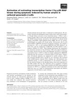
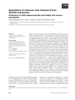
Fig. 1. Control of TGFb and activin ⁄ nodal receptor trafficking by small GTPases. (A) The role of Rab5 and Rab11 in TGFb receptor endocyto-
sis and recycling. The cycling of Rab5 between the GTP and GDP forms may influence the length and intensity of TGFb ⁄ activin signaling
cascades by regulating TGFb–activin type I ⁄ II receptor trafficking via the early endocytic compartment. RIN1, a Rab5 GEF, via activation of
Rab5, directs TbRs into an endocytic pathway that promotes TGFb signaling through Smads. SNAI1, which represses RIN1 expression, is
induced by TGFb thus creating a negative feedback loop. Following clathrin-dependent internalization, TGFb receptors recycle (irrespective of
their activation state) in a Rab4-independent and Rab11-dependent manner. (B) The role of ARIP2, RalA and RalBP1 in activin A receptor
internalization. ARIP2 interacts with ActRII and triggers their endocytosis via RalA ⁄ RalBP1 and POB1. POB1 interacts directly with the EH-
containing proteins Epsin and Eps15. This protein complex acts as a scaffold to convey signals from the activin receptor to the endocytic
machinery. (C) The role of Rap2 in activin ⁄ nodal receptor trafficking in Xenopus embryos. In the absence of ligand, Rap2 directs activin ⁄ nodal
receptors into a Rab11-dependent recycling compartment, thereby avoiding degradation and maintaining cell-surface levels of receptors.
Upon ligand addition, Rap2 competes with the Smad7 ⁄ Smurf1 complex and delays receptor degradation, thus enhancing signaling.
D. Kardassis et al. TGFb signaling and small GTPases
FEBS Journal 276 (2009) 2947–2965 ª 2009 The Authors Journal compilation ª 2009 FEBS 2951
ligand-dependent receptor-mediated endocytosis.
Moreover, REPS1 interacts with Rab11-FIP2 [80]
a Rab11 effector that may couple REPS1-containing
vesicles originating from clathrin-coated vesicles (and
the early endocytic compartment) to the recycling
endosomes.
RalA and RalBP1 appear to be involved in activin
A receptor trafficking and signaling (Fig. 1B). It has
been shown that activin receptor interacting protein 2
(ARIP2) interacts with ActRIIs and regulates their
endocytosis via a PDZ domain-mediated interaction,
concentrating them in a perinuclear compartment.
Thus, ARIP2 reduces the response to ligands by
decreasing the levels of ActRII at the plasma
membrane [81]. ARIP2 triggers the endocytosis of
ActRIIs via Ral ⁄ RalBP1. Indeed, ARIP2 associates
with ActRIIA and RALBP1 via its PDZ domain and
C-terminal region, respectively. Because ARIP2C, the
C-terminal deletion mutant of ARIP2 that does not
bind RalBP1, failed to induce ActRII endocytosis, it
appears that endocytosis of ActRIIs by ARIP2 is
RalA ⁄ RalBP1 dependent. Moreover, activin A acti-
vates GDP–GTP exchange in RalA [81]. Activation of
RalA ⁄ RalBP1 by activin A is calcium dependent, in
contrast to activation by EGF and insulin, which
occurs via a Ras-dependent cascade [73]. Interestingly,
because only ActRIIs among all the serine ⁄ threonine
kinase receptors for BMP ⁄ TGFb ⁄ activin have the
PDZ-binding sequence (ESSL for ActRIIA and ESSI
for ActRIIB) [82], PDZ protein-regulated endocytosis
and sorting is expected to influence only ActRIIs.
Because ActRIIs bind both activins and also nodal
and BMP7, ARIP2 is likely to play a role in shaping
the activin ⁄ nodal ⁄ BMP gradient by regulating the
endocytosis of ActRIIs.
Rap2
Rap2 is a member of the Ras family of small GTPases
whose effector domain is almost identical to that of
Ras, and can therefore bind most Ras effectors. Rap2
inhibits many Ras pathways including Ras-induced
Raf activation at the plasma membrane [83]. Rap2 also
binds to the Ral GEFs, Ral GDS, RGL and RLF [84].
These proteins are also Ras effectors and induce nucle-
otide exchange leading to the formation of active
RalA. As discussed above, Ral has been implicated in
activin A receptor trafficking and may be linked to the
molecular mode of action of Rap2 in Xenopus,as
explained below.
In a very elegant study in Xenopus embryos, Rap2
was shown to regulate activin ⁄ nodal signaling by mod-
ulating receptor trafficking [85] (Fig. 1C). In the
absence of ligand, Rap2 directs activin ⁄ nodal receptors
into a Rab11-dependent recycling compartment,
thereby avoiding degradation and maintaining cell-sur-
face levels of receptors. Upon ligand addition, Rap2
no longer directs the receptors for recycling, but rather
competes with Smad7 and delays receptor degradation,
thus enhancing signaling. Moreover, Rap2 is initially
enriched in the dorsal region of the blastulae, then as
gastrulation proceeds, it decreases dorsally and
increases ventrally. However, Smad7 is expressed uni-
formly across the dorso–ventral axis in early gastrula-
tion and as gastrulation proceeds, Smad7 is restricted
to the ventral region. Thus, Smad7 and Rap2 levels
appear to regulate Smad2 activation along the dorso–
ventral axis of the developing embryo.
Growing evidence links the progression of TGFb
receptor signaling to key regulatory steps in endocytic
trafficking. These steps involve the active regulation of
GDP-to-GTP exchange by various small GTPases of
the Rab ⁄ Ral and Rap families. These mechanisms
ensure optimal signal transduction from active receptor
complexes to activated Smads.
Intracellular Smad trafficking – the role
of the Ran GTPase
Most current evidence on the mechanisms that govern
the dynamic shuttling of Smad proteins in the cell is
based on the behavior of engineered GFP–Smad2 and
GFP–Smad4 fusion proteins which are stably
expressed in human cells cultured in vitro. The evi-
dence supports a model whereby Smads shuttle con-
stantly, although each specific Smad seems to obey
distinct kinetic properties during its movements [86].
Mathematical modeling of Smad protein shuttling has
recently suggested that the strength of Smad signaling
depends directly upon the length of time a certain
Smad molecule spends in the nucleus [87]. Such kinetic
analysis also emphasized that the nuclear export of
Smads is highly regulated, whereas the nuclear import
of Smads may act as a default pathway.
The evidence from the in vitro cell system is comple-
mented by pioneering in vivo studies first developed in
Xenopus embryos [88,89]. Continuous shuttling of
Smad2 could be observed in developing Xenopus and
zebrafish embryos [88]. Furthermore, Smad2 and
Smad4 proteins fused to fluorescent protein fragments
fluoresce only when a Smad2–Smad2 homo-oligomer,
Smad4–Smad4 homo-oligomer or Smad2–Smad4
hetero-oligomer forms inside the living cells of Xenopus
embryos caused by trans-complementation of the fused
fragments [89]. Cells in the developing Xenopus embryo
are responding to the TGFb members nodal or activin
TGFb signaling and small GTPases D. Kardassis et al.
2952 FEBS Journal 276 (2009) 2947–2965 ª 2009 The Authors Journal compilation ª 2009 FEBS
and show accumulation of Smad4 homo-oligomers
only in the cytoplasm, whereas Smad2 homo-oligomers
and Smad2–Smad4 hetero-oligomers accumulate in the
nucleus. These experiments demonstrated that Smad2–
Smad4 oligomers can be observed in the nuclei of
developing embryonic cells only when these cells
reached the proper developmental stage. This observa-
tion suggested that factors independent of nodal ⁄ acti-
vin signaling regulate the ‘competence’ of the
embryonic cell to accumulate nuclear Smad2–Smad4
oligomers. Smad trafficking may be classified accord-
ing to the cellular compartment where this specific
movement occurs. Thus, we can consider Smad traf-
ficking in the cytoplasm, Smad trafficking through the
nuclear pores and Smad trafficking inside the nucleus.
Smad trafficking in the cytoplasm
When Smad2 moves inside the cytoplasm it associates
with the motor protein kinesin-1 and the integrity of
the microtubular network is essential to support this
type of motility [88]. This new evidence is compatible
with an older study that first identified an inherent
ability of all Smad proteins to associate and localize
on microtubules [90]. Another motor-like protein that
associates with Smad2 is the dynein light chain km23-1,
which assists in the nuclear accumulation of Smad2,
and also regulates trafficking of the TbRI [91]. Accord-
ing to this new evidence, cytoplasmic Smads traffic
towards the signaling receptors with the help of kinesin
motors that slide on microtubules. The signaling recep-
tors most likely reside on endosomes, as discussed
above. However, cytoplasmic Smad trafficking towards
the nucleus involves the dynein motor–microtubule
machinery. Although it makes sense to consider micro-
tubules as trafficking highways that facilitate the
movement of Smad proteins, microtubules have also
been shown to act as cytoplasmic traps for Smads [92].
According to this model, connexin 43 is a regulatory
protein that competes with Smads for binding to
microtubules. However, the latter mechanism needs to
be further clarified as it is important to understand
which factor regulates the residence of Smads on
microtubules versus their mobility along microtubules
and towards neighboring cellular locations.
The association of Smads with microtubules pro-
vides additional insight into the functional regulation
of these proteins. In dividing cells, such as those of the
Xenopus embryo, Smads can associate with the spindle
and decorate the metaphase chromosomes [89]. This
evidence is compatible with a role for microtubules in
trapping Smads and protecting their integrity, thus
delivering them safely to the daughter cells after mito-
sis. It remains unclear as to whether Smad signaling
may also regulate mitosis or cytokinesis. However, in
addition to protecting Smad integrity, microtubules
may also guide a pool of Smads towards their ultimate
turnover. The site of assembly of the microtubular net-
work is known to be the centrosome, a subcellular
structure in which Smads that are phosphorylated in
their linker domain can also localize and undergo
ubiquitin-dependent proteasomal degradation [93]. It
appears that Smads may slide along microtubules to
reach the centrosomes and become degraded [94].
Interestingly, when cells divide, the pool of linker-
phosphorylated Smads that traffic towards the centro-
some segregates together with other ubiquitinated
proteins on the mitotic spindle towards only one of the
two daughter cells [94]. This mechanism ensures that
proteins targeted for disposal go to only one of the
two daughter cells, leaving the other relatively clear of
such signaling byproducts. A deeper understanding of
the role of microtubules in the regulation of Smad
trafficking and signaling is clearly warranted.
Smad trafficking through nuclear pores
The entry of Smad proteins to the nucleus is regulated
by specific interactions with transporters and nucelo-
porins. A lysine-rich nuclear localization signal (NLS)
located in the N-terminal Mad homology 1 domain of
all Smads binds to importin-b in the case of Smad3
and importin-a in the case of Smad4, while mutation
of the NLS blocks the ability of these proteins to enter
the nucleus [95–98]. Although the functional role of
the Smad2 NLS has not yet been determined, the long
Smad2 isoform that incorporates exon 3 fails to bind
to importin-b, whereas the shorter Smad2 isoform that
lacks exon 3 binds to importin-b similar to Smad3
[95]. In addition, the importin moleskin mediates the
nuclear entry of the Drosophila R-Smad Mad, and its
human orthologues, importin-7 and importin-8,
mediate the nuclear translocation of Smad1, Smad2,
Smad3 and Smad4 in human cancer cells in response
to BMP or TGFb signaling [99]. Future work may
explain why Smads utilize multiple importins for their
entry to the nucleus (Fig. 2).
Importins are known to move through the pore by
consecutive contacts with the phenylalanine ⁄ glycine
(F ⁄ G)-rich repeats of specific nucleoporins. Such step-
wise translocation is energetically demanding and
requires GTP expenditure. Similar to the role of Rab
GTPases that control the trafficking of endocytic vesi-
cles during TGFb signaling in the cytoplasm (Fig. 1),
the small GTPase Ran controls Smad3 trafficking via
the nuclear pore (Fig. 2) [95]. Ran is a small GTPase
D. Kardassis et al. TGFb signaling and small GTPases
FEBS Journal 276 (2009) 2947–2965 ª 2009 The Authors Journal compilation ª 2009 FEBS 2953
dedicated to the control of nucleocytoplasmic traffick-
ing and chromosomal segregation during mitosis [100].
A Ran activity gradient is established through the
nuclear pore with high Ran–GDP concentrations in
the cytoplasmic phase of the pore which gradually
decrease along the pore [101]. In the nuclear phase of
the pore, the Ran-specific GEF RCC1 loads Ran with
GTP, thus establishing a high Ran–GTP concentration
in the nucleus. GDP-bound Ran drives the transport
of Smad3 through the pore, whereas Ran–GTP
induces the allosteric change needed to dissociate
Smad3 from importin-b (Fig. 2) [95]. Ran also medi-
ates importin-b trafficking back into the cytoplasmic
phase of the pore [102].
In addition to binding to importins, Smad2 can also
bind directly to the F⁄ G-rich repeats of nucleoporins
Nup214 and Nup153 of the nuclear pore (Fig. 2)
[103,104]. However, whether Smad3 and Smad4 bind to
the nucleoporins directly or via the importins remains
unclear [103,104]. In addition, it would be interesting to
examine whether Smad2–nucleoporin interactions are
regulated by the Ran GTPase gradient along the
nuclear pore. Analysis of importin-7 and importin-8 as
Smad carriers suggested that continuous Smad shut-
tling in the absence of ligand activation is independent
of the action of transportins, and is presumably facili-
tated by direct contacts with nucleoporins [99]. By con-
trast, when TGFb receptor activation leads to R-Smad
phosphorylation, nuclear import seems to depend on
the activity of specific transportins. Thus, different
mechanisms of nuclear import might operate at differ-
ent stages of the TGFb signaling pathway.
The cytoplasmic distribution of Smads in the resting
cell seems to be regulated by the dominant role of Smad
nuclear export [86,87]. Upon ligand-dependent signal-
ing, nuclear Smad complexes prevail but eventually
shuttle back to the cytoplasm, thus providing a way of
dampening the strength of the signal or alternatively
replenishing the cytoplasmic pool of Smads with mole-
cules that are ready to become activated again, as long
as the receptors remain active. The importance of
nuclear export is underscored by the presence of nuclear
export signals (NES) in all Smads examined to date.
Smad4 carries a leucine-rich NES in its linker domain,
which mediates export via exportin-1 ⁄ chromosome
region maintenance 1 (CRM1) (Fig. 2) [105,106]. Muta-
tion of hydrophobic amino acids within the Smad4 NES
or exposure of cells to the pharmacological inhibitor of
CRM1 leptomycin-B, lead to an exclusive nuclear distri-
bution of Smad4, independent of the presence or
absence of ligand. Smad3 is exported from the nucleus
in a CRM1-independent manner and an extended
peptide surface of the MH2 domain has been identified
as critical for this export by exportin-4 [107]. In the case
4
p x E
1
p
x E
i
P+PDG
–
n
a
Ri
P+PDG
–
n
a
Ri
P+PDG
–
n
a
R
? p x
E
3 d a m S
3 d a
m
S
P A G n
a R
4
d
a m
S
4 d
a
m S
P A G n a R
2 d a m S
2
d
a m
S
P A G
n a R
s u
e
l
c
u n
3 5
1 p
u N
4
1
2 p u N
4 p x E
3 d
a m
S
- p
m I
β
1
8
/ 7 - p m
I
1 p x
E
4 d a m S
-
p
m I
α
8 /
7 -
p m I
PT
G
–n a R
?
p x E
2
d
a m
S
8
/
7 -
p m I
P
T
G -
n a R
P T G + 1
C
C R
P
T G
-
n a R
Cytoplasm
Nucleus
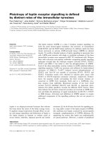
Fig. 2. Smad trafficking through nuclear pores. Smad2, Smad3 and Smad4 are shown to interact with importins (Imp) in the cytoplasm and
start their nuclear import via additional contacts with nucleoporins (Nup). Smads are released in the nucleoplasm and importins recycle back
to the cytoplasm (not shown). Nuclear Smads associate with exprotins (Exp) and Ran–GTP and translocate to the cytoplasm by making con-
tacts with nucleoporins. The cytoplasmic Smad–exportin–Ran–GTP complex is disrupted by the action of RanGAP, which releases Smad,
exportin and Ran–GDP, and free orthophosphate after the hydrolysis of GTP. Completion of the Ran cycle is shown in the middle for Smad3
because the role of Ran has only been analyzed in detail in the case of Smad3. Cytoplasmic Ran–GDP (grey symbol) diffuses through the
nuclear pore where it meets the nuclear GEF RCC1, which exchanges GDP for GTP and restores nuclear Ran–GTP (black symbol) levels.
TGFb signaling and small GTPases D. Kardassis et al.
2954 FEBS Journal 276 (2009) 2947–2965 ª 2009 The Authors Journal compilation ª 2009 FEBS
of Smad3, the role of Ran has been studied and it was
clearly demonstrated that, similar to many other
exported proteins, Ran supports the movement of
Smad3 via the nuclear pore towards the cytoplasm
(Fig. 2). The Smad3 NES has no obvious resemblance
to a bipartite leucine-rich motif identified in the MH2
domain of Smad1, the R-Smad of the BMP pathways,
which is thought to be recognized by CRM1 based on
leptomycin-B inhibitor experiments [108]. The role of
Ran in mediating the export of proteins from the
nucleus follows the inverse biochemical steps used for
import of proteins to the nucleus [100,102]. Ran–GTP
promotes the association of Smad3 with exportin-4 in
the nuclear phase of the pore [107]. Upon trafficking
via the nuclear pore, Ran–GTP in complex with cargo
is attacked by Ran GAP, which is associated on the
cytoplasmic phase of the nuclear pore, and activates
the GTPase activity of Ran so that GTP is hydrolyzed
to GDP and orthophosphate (Fig. 2) [100,102]. This
leads to conformational changes in Ran that facilitate
disruption of the complex between exportin and
its cargo, and the ultimate release of cargo to the
cytoplasm.
Smad trafficking in the nucleus
Although a growing understanding of the mechanisms
that guide bidirectional Smad trafficking in the cyto-
plasm and through the nuclear pores is now estab-
lished, nothing is known about Smads trafficking
within the nucleoplasm. Classically, the native, yet
weak, ability of Smads to bind to DNA has suggested
that upon entry to the nucleus, Smads might tether
chromatin. However, the current dynamic shuttling
model of Smads necessitates a more dynamic view of
the nuclear residence of these proteins. The dynamic
shuttling model disfavors long-lasting and very stable
tethering mechanisms, however, it allows for the highly
regulated formation of protein complexes between
Smads and nuclear residents. In fact, nuclear Smads
are known to bind to a high number of nuclear tran-
scription factors and the role of such interactions in
the timing and shuttling behavior of Smads remains
unexplored [27]. One nuclear factor that seems to fulfill
the criteria for a tethering factor and which might
coordinate the nuclear residence time of Smads and
the process of transcription is the newly reported pro-
tein transcriptional coactivator with PDZ-binding
motif (TAZ) [109]. TAZ is a transcriptional regulator
containing a WW domain and promotes the nuclear
accumulation of Smads. Loss of TAZ perturbs the
ability of Smads to accumulate in the nucleus. TAZ
binds the transcriptionally active Smad complex and
anchors it to ARC105, a central component of the
transcriptional mediator complex. TAZ has a close
homolog, the WW domain protein YAP, which might
also be involved in a similar mechanism. Thus, we
await significant developments in Smad nuclear traf-
ficking that might provide a more comprehensive view
of how the entry and exit of Smads from the nucleus
coordinates with transcription. It will also be interest-
ing to examine the role of additional nuclear small
GTPases as regulators of nuclear Smad function,
because this class of proteins offers a versatile regula-
tory system that empowers biological processes with
the ability to switch on and off.
The role of small GTPases of the Rho
subfamily in TGFb-induced actin
cytoskeleton remodeling
Actin cytoskeleton remodeling is one of the earliest
cellular responses to extracellular stimuli [110–115].
Binding of ligands to the appropriate receptors triggers
specific signaling cascades, which may generate rapid
and long-term modifications of actin polymerization
dynamics and microfilament organization [116–120].
Among the specific signaling effectors regulating actin
architecture, the family of small Rho GTPases has a
prominent role. Classically, plasma membrane recep-
tors activate specific guanine-exchange factors often
via phosphorylation, which leads to the subsequent
activation of Rho GTPases [121]. Rho GTPases have
been implicated in many cellular processes, including
actin and microtubule cytoskeleton organization, cell
division, motility, cell adhesion, cell-cycle progression,
vesicular trafficking, phagocytosis and transcriptional
regulation [122,123]. Rho proteins cycle constantly
between GTP-bound active forms and GDP-bound
inactive forms, and this process is regulated by various
factors including GEFs, guanine nucleotide dissocia-
tion inhibitors and GAPs [124]. As well as contributing
to physiological processes, Rho GTPases have been
found to contribute to pathological processes includ-
ing cancer cell migration, invasion, metastasis,
inflammation and wound repair [122,123]. Although
Rho proteins do not seem to be mutated in cancer
cells, their expression is often elevated, indicating that
Rho dysregulation promotes malignant phenotypes
[125].
Rho proteins can be subdivided into three major
groups: Rho (RhoA, RhoB, RhoC), Rac (Rac1, Rac2)
and cdc42 proteins [123]. Active Rho GTPases trans-
mit signals via downstream effectors such as Rho
coiled-coiled kinase 1 (ROCK1), p21-activated kinase
1 and neural Wiskott–Aldrich syndrome protein
D. Kardassis et al. TGFb signaling and small GTPases
FEBS Journal 276 (2009) 2947–2965 ª 2009 The Authors Journal compilation ª 2009 FEBS 2955
[126,127]. Activated p21-activated kinase 1 and
ROCK1 phosphorylate and activate LIM-kinases 1
and 2, respectively [128–132]. Eventually, LIM-kinases
1 and 2 phosphorylate actin-depolymerizing proteins
such as cofilin, destrin and actin-depolymerizing factor,
which are inactivated and thus permit actin polymeri-
zation to occur [128–130,133,134].
Mechanisms of TGFb-induced actin cytoskeleton
remodeling – short- and long-term events
The ability of TGFb to regulate actin cytoskeleton
remodeling has been demonstrated in a variety of cell
systems, and specific members of the Rho subfamily of
small GTPases including RhoA, RhoB, Rac and cdc42
have been found to play essential roles (Fig. 3). The
contribution of individual Rho GTPases and their
downstream effectors in TGFb-induced actin remodel-
ing has been studied using a variety of experimental
tools. These tools include constitutively active and
dominant-negative mutants of Rho proteins or their
target proteins, siRNA-mediated gene silencing or gen-
eral inhibition of Rho function using molecules such
as the C3 exoenzyme, which selectively ADP-ribosy-
lates and inactivates low molecular mass G proteins of
the Rho subfamily at an asparagine residue within the
effector domain. Rho GTPase activation is generally
measured by affinity precipitation using appropriate
GST–fusion peptides that bind only to GTP-bound
Rho proteins such as GTP–Rhotekin binding domain
for RhoA and RhoB or GST–p21-activated kinase and
GST–Wiskott–Aldrich syndrome protein for Rac1 and
cdc42 [135]. Changes in the actin cytoskeleton are
monitored by immunofluorescence microscopy of
rhodamin ⁄ phalloidin-labelled actin or by calculating
the ratio of total versus polymerized actin by immuno-
blotting Triton-soluble (globular actin) and Triton-
insoluble (filamentous actin) cell extracts [136].
TGFb-induced cytoskeleton rearrangements
involving Rho activation in EMT
The most extensively investigated TGFb-induced cyto-
skeleton rearrangements are the differentiation of epi-
thelial to mesenchymal cells, a process that is called
epithelial to mesenchymal transition or transdifferenr-
T β I R T β I I R
F
G
T β
l l l t E l l l t E
P
T β I R T β I I R
Cytoplasm
E x t e c a r l l u l e c a p s r a E x t e c a r l l u l e c a p s r a
1 K C O R 4 d a m S
d a m S - R
d a m S - R
2 K M I L
K P A M 8 3 p
4 d a m S
ohR
o
hR
P
D G –P D G
P
D
G
P T
G
o h R o h R PT
G
–PTG
n i l i f o C
K P A M 8 3 p
2
K R
P / N K P
d a
m S - R
)
F E
G
( 1 T E N
s
u
e l c u N
B o
h R
g n
i l e
d
o m e
r
n i t c A
2
K R
P / N K P
4 d a m S
d
a m
S - R
α A M S
-
M
S
-
C
H
M
l l e c e l c s
u
m h t o o m S l l e c e l c s
u
m h t o o m S
n o i t a i t n e r e f f i d n o i t a i t n e r e f f i d
T
M E
T
M E
:
s
r o t c a f g n i t a r e p o
o
C
F
R S
7 d a m S
M
S
-
C
H
M
a 2 2 - M S
F
R S
1 P A
A
T A G
2
F
E M
t s a l b o r b i F - t s a
l
b o r
b
i F t s a
l
b o r b
i f
o y m - t s a l b o r b i
f
o
y
m
n o
i
t a i t n e
r
e f
f
i d n o
i
t a i t n e
r
e f
f
i d
Fig. 3. The role of small Rho GTPases in short- and long-term actin cytoskeleton reorganization in response to the TGFb signaling pathway.
TGFb induces short-term actin cytoskeleton remodeling via the activation of various Rho GTPases including RhoA, RhoB, Rac and Cdc42
(generally termed Rho). Activation of these GTPases causes actin polymerization via the ROCK1 ⁄ LIMK2 ⁄ cofilin, as well as by
MAPK ⁄ PKN ⁄ PRK2 pathways. In long-term cytoskeletal reorganization, which involves nuclear events, TGFb receptor activation causes the
phosphorylation of Smads and their subsequent translocation to the nucleus. In the nucleus, R-Smad ⁄ Smad4 complexes bind to the promot-
ers of various target genes such as the smooth muscle-specific genes a-SMA, SM-22a or SM-MHC, the Rho GEF NET1, the inhibitory
Smad7 protein and the RhoB gene. Activation of cofactors such as serum response factor, AP1, GATA and myosin enhancer factor 2 via
p38 MAPK or other pathways facilitates these transcriptional responses. Actin remodeling in turn facilitates processes such as smooth
muscle cell differentiation, EMT and others.
TGFb signaling and small GTPases D. Kardassis et al.
2956 FEBS Journal 276 (2009) 2947–2965 ª 2009 The Authors Journal compilation ª 2009 FEBS
tation (EMT) [137,138]. EMT is characterized by the
dissolution of epithelial cell–cell junctions and reorga-
nization of the actin cytoskeleton with the formation
of focal adhesions and stress fibers, acquisition of a
spindle-shaped morphology, delocalization of E-cadh-
erin from cell junctions and elevated N-cadherin
expression. This process is associated with embryonic
tissue movements and also with cancer cell invasive-
ness and metastasis. The earliest event in TGFb-
induced EMT is the activation of RhoA which occurs
within 5 min of TGFb stimulation. This activation
lasts for a short time (15 min to 3 h, depending on the
cell type) and is followed by the activation of down-
stream target kinases such as ROCK. Rapid RhoA
activation was found to operate in a variety of cell
models of TGFb-induced EMT under physiological or
pathological conditions. (a) In the atrioventricular
canal of the embryonic chicken heart, TGFb was
found to promote the conversion of endothelial cells to
mesenchymal cells via a pathway that requires the acti-
vation of RhoA [139]. (b) During tubulointerstitial
fibrosis, TGFb promotes the differentiation of tubular
epithelium to mesenchymal cells via a biphasic activa-
tion of RhoA and its downstream target ROCK; a
rapid and transient elevation of RhoA-GTP levels
which was detectable as early as 1 min after TGFb
stimulation and lasted for 5 min, and a chronic eleva-
tion at 24 h of stimulation. Chronic activation was
correlated with the upregulation of a-SMA gene
expression via activating protein 1 (AP1) factors [140].
(c) In proliferative vitroretinopathy, TGFb leads to the
transformation of retinal pigment epithelial cells to
contractile fibroblasts via rapid activation of RhoA
and Rac1 GTPases and their downstram effectors
ROCK kinase, LIMK and cofilin, and the concomitant
upregulation of a-SMA gene expression [141].
TGFb-induced Rho GTPase activation and actin
remodeling in various cell systems
In addition to their role in EMT, Rho GTPases and
their downstream effectors are activated by TGFb and
contribute to cytoskeletal rearrengements in other cell
systems. (a) TGFb promotes the differentiation of neu-
ral crest cells into vascular smooth muscle cells via a
rapid (5 min) activation of RhoA and ROCK1 [142].
In this study, it was shown that inhibition of RhoA
activity blocked Smad phosphorylation by TGFb, sug-
gesting that RhoA and Smads may cooperate in TGFb
signaling responses, a concept that is dicscussed thor-
oughly below. (b) TGFb promotes the differentiation
of rat pulmonary arterial smooth muscle cells via a
rapid (2 min) activation of RhoA, which was followed
by activation of its downstream kinases ROCK,
PKN ⁄ PRK2 and p38 MAPK and the transcriptional
upregulation of smooth muscle-specific genes such as
a-SMA, SM-MHC and SM-22a via the cooperation of
serum response factor, GATA and myosin enhancer
factor 2 transcription factors [143] (Fig. 3). (c) In the
human prostate carcinoma cell line PC-3U, TGFb
induces the rapid (5 min) formation of membrane ruf-
fles via activation of RhoA and Cdc42 in a Smad-inde-
pendent manner [144]. In the same study, it was also
shown that TGFb induced long-term actin remodeling
and stress fiber formation which required an active
Smad pathway. Thus, this study revealed that the
rapid and sustained changes in actin cytoskeleton reor-
ganization that are observed in response to TGF b are
mechanistically distinct processes and could be medi-
ated by separate nongenomic and transcriptional sig-
naling pathways that are induced by the same
stimulus. Short-term and sustained actin cytoskeleton
remodeling has been investigated thoroughly in fibro-
blasts such as H-ras transformed NIH3T3 fibroblasts,
mouse embryo fibroblasts and Swiss3T3 fibroblasts
[145–148]. In fibroblasts transformed by inducible
expression of the H-Ras oncogene, TGFb induced the
formation of new stress fibers from focal adhesions as
early as 15 min post TGFb addition and this
reorganization was associated with an increase in the
polymerization state of actin and in protein levels of
RhoA and RhoB [146].
In Swiss3T3 fibroblasts, TGFb induced rapid activa-
tion of both RhoA and RhoB small GTPases as early
as 5 min post TGFb1 addition which remained high
for 3 h before decreasing [147]. Activation of RhoA
and RhoB was accompanied by phosphorylation of
the downstream effectors LIMK2 and cofilin, whereas
inhibition of ROCK1 completely blocked TGFb1-
induced LIMK2 ⁄ cofilin phosphorylation and down-
stream stress fiber formation (Fig. 3). In these cells,
TGFb induced fibroblast to myofibroblast differentia-
tion, which was evidenced by enhanced expression of
a-SMA and the subsequent incorporation of a-SMA
into microfilamentous structures [148]. Fibroblast to
myofibroblast conversion is a pathophysiological
feature of various fibrotic diseases such as idiopathic
pulmonary fibrosis, asthma and chronic obstructive
pulmonary diseases [149–151]. Given that enhanced
TGFb concentrations have been detected in various
fibrotic diseases, including idiopathic pulmonary fibro-
sis [152,153], sarcoidosis [154] and cystic fibrosis [155],
understanding the mechanism that underlies this
TGFb-induced conversion may lead to the develop-
ment of novel therapeutic approaches for these
diseases.
D. Kardassis et al. TGFb signaling and small GTPases
FEBS Journal 276 (2009) 2947–2965 ª 2009 The Authors Journal compilation ª 2009 FEBS 2957
Non-Smad and Smad pathways in Rho GTPase
activation by TGFb
Certain studies have examined the participation of
MAP kinases or the phosphatidylinositol 3-kinase as
downstream signaling effectors that cooperate with
Rho GTPases or are activated by them to achieve
TGFb-induced cytoskeleton remodeling. Edlund et al.
[144] showed that treatment of human prostate cancer
cells with an inhibitor of the p38 MAP kinase
(SB203580) at a concentration that was unable to
block the activity of TbRI, as well as ectopic expres-
sion of a kinase inactive p38 mutant, abrogated the
TGFb-induced actin reorganization. The same group
also showed that TGFb-induced membrane ruffling
and stress fiber formation in prostate cancer cells
requires an active phosphatidylinositol 3-kinase path-
way [156]. Chen et al. [142] used neural crest stem cells
to show that p38, p44 ⁄ 42 MAPK and phospatidylinos-
itol-kinase inhibitors did not counterbalance the TGFb
induction of a-SMA expression in smooth muscle cell
differentiation from stem cells. By contrast, in a differ-
ent system of smooth muscle cell differentiation,
Deaton et al. [143] showed that inhibition of p38
MAPK by SB203580 blocked the TGFb1-mediated
activation of a-SMA and other SMC marker genes
(Fig. 3).
Although the extremely rapid activation of Rho
GTPases in response to TGFb stimulation implies the
involvement of non-Smad pathways, in certain cases it
was found that the Smad pathway may also play a
role in the early activation of Rho proteins by TGFb.
By studying the signaling properties of a TbRI bearing
a mutation in its L45 loop, which contains the Smad
docking site [8], Vardouli et al. [147] demonstrated that
interaction of TbRI with R-Smads is required for
signaling towards Rho GTPases and the actin cyto-
skeleton. The role of Smads in TGFb-induced actin
remodeling is further supported by experiments in a
cellular model lacking endogenous Smad3 expression
(JEG3 choriocarcinoma cells) [148]. In addition,
TGFb-induced Rho activation and cytoskeleton orga-
nization was abolished by overexpression of the inhibi-
tory Smad7 protein which blocks the TGFb ⁄ Smad
signaling pathway [147,157]. This observation is in
contrast to a study showing that Smad7 is required for
TGFb-induced activation of Cdc42 and the concomi-
tant reorganization of the actin filament system, as
discussed below.
The ability of TGFb to affect both rapid and sus-
tained actin cytoskeleton remodeling in various cell
types [144,147,148] implies that genomic actions of
TGFb may be involved in long-term cytoskeleton reor-
ganization (Fig. 3). In support of this, it was shown that
treatment of Swiss3T3 fibroblasts with actinomycin D, a
well-established inhibitor of active gene transcription,
abolished TGFb-induced actin reorganization in
Swiss3T3 fibroblasts and HaCaT keratinocytes (E.
Vasilaki, E. Papadimitriou, C. Stournaras and D.
Kardassis, unpublished results). TGFb also activates the
expression of Rho GEF NET1 via the Smad pathway
(Fig. 3) [158]. In a recent study, it was shown that TGFb
induces the transcription of RhoB in mouse fibroblasts
and human hepatocytes (Fig. 3) [148]. In this study,
Vardouli et al. [148] demonstrated that transcriptional
upregulation by TGFb was specific for RhoB, because
the expression of RhoA was not affected, and that the
TGFb ⁄ Smad pathway activated the human RhoB but
not the RhoA promoter. Expression of the endogenous
RhoB gene in fibroblasts was also upregulated by over-
expression of TGFb-regulated Smads via adenovirus-
mediated gene transfer [148]. Similar observations have
been made in HaCaT keratinocytes (Vasilaki et al.,
unpublished results). However, this genomic effect of
TGFb on RhoB gene expression is cell type-specific
because it could not be observed in mink lung epithelial
cells. In these cells, TGFb promoted the accumulation
of RhoB protein without a concomitant increase in
RhoB mRNA levels [159].
Cross-talk between Rho GTPases and the
TGFb
⁄
Smad pathway
In addition to the established positive role of Rho
GTPases in TGFb-induced actin cytoskeleton remodel-
ing, certain Rho proteins seem to play a negative role
in TGF b ⁄ Smad signaling when their expression is
upregulated. In epithelial cells, RhoB overexpression
antagonized TGFb for the transcriptional activation of
a Smad-responsive promoter, whereas dominant-
negative RhoB mutant enhanced TGFb signaling
towards this promoter [159]. In a different study and
system, it was shown that ectopic expression of RhoB,
but not RhoA, caused a decrease in the expression
TbRII and in the activity of the TbRII promoter in
HaCaT keratinocytes and pancreatic carcinoma cells,
and antagonized the TGFb-mediated anti-proliferative
responses [160]. Downregulation of the TbRII gene by
RhoB was mediated by inhibition of AP1 transcription
factors that bind to an AP1 site in the proximal TbRII
promoter [160]. Rho proteins might also play a posi-
tive regulatory role in TGFb ⁄ Smad signaling, as dem-
onstrated by Chen et al. [142] who showed that ectopic
expression of a dominant-negative RhoA mutant in
Monc-1 neural crest stem cells blocked the phospho-
rylation of Smad2 and Smad3 by TbRI, their
TGFb signaling and small GTPases D. Kardassis et al.
2958 FEBS Journal 276 (2009) 2947–2965 ª 2009 The Authors Journal compilation ª 2009 FEBS
translocation to the nucleus and the activation of a
Smad-specific reporter gene. Chen et al. [142] also
showed that general inhibition of Rho activity by C3
exotoxin attenuated Smad-mediated transactivation.
The positive role of R-Smads and the negative role
of the inhibitory Smad7 in Rho GTPase activation by
TGFb is discussed above. A novel, positive role for
Smad7 in actin remodeling was reported by Edlund
et al. [156]. This study showed that increased expres-
sion of Smad7 in prostate cancer cells was associated
with increased mobilization of the actin filament sys-
tem and activation of the Rho GTPase Cdc42 (Fig. 3).
It also showed that the Smad7-induced rearrangement
of actin cytoskeleton required the p38 MAPK pathway
previously shown to act downstream of Cdc42 [144].
Recently, a Par6–Smurf1–RhoA pathway was shown
to operate in TGFb-induced EMT. According to this
model, Par6, a regulator of epithelial cell polarity and
tight junction assembly, interacts with TGFb receptors
at sites of tight junctions and is phosphorylated by
TbRI, an event that leads to its interaction with the
E3 ubiquitin ligase Smurf1. By an unknown mecha-
nism, this ubiquitin ligase recruits RhoA and ubiquiti-
nates it, thus causing its proteosomal degradation and
the dissolution of the tight junctions [161]. This mecha-
nism was later confirmed in TGFb-induced atrioven-
tricular cushion endocardial cell EMT [162].
Finally, recent screening of miRNA microarrays for
miRNAs that are up- or downregulated by TGFb in
epithelial NMuMG cells, identified miR-155 as the
most significantly activated miRNA. Knockdown of
miR-155 suppressed TGFb-induced EMT and tight
junction dissolution, migration and invasion of these
cells [163]. Importantly, ectopic expression of miR-155
inhibited the synthesis of RhoA. Given that miR-155
levels are frequently elevated in invasive breast cancer,
the new data indicated that miRNA-based strategies
could be used for the treatment of breast cancer [163].
Conclusions and perspectives
The balance of evidence suggests that the endocytosis
of TGFb family receptors plays an enhancing role in
TGFb family signaling. However, the magnitude and
duration of the effect on the signaling output depend
on the cell type. Embryonic stem cells and differenti-
ated cells of various types are not expected to conform
to the same mechanisms. Indeed, it has been reported
that there are fundamental differences in the endocytic
sorting of TGFb receptors between fibroblasts and epi-
thelial cells [164]. Moreover, in some cell types, endo-
cytosis of TGFb receptors might not be interconnected
with signaling, as observed in some studies. However,
many questions remain. Which endocytic routes are
taken by TGFb receptor complexes and what is the
contribution of each pathway to the final signal? Of
the five emerging transport routes, internalization of
TGFb receptors has been reported to occur via the
CCVMR and caveolar routes. There are no studies
regarding the contribution and significance of the other
routes. What dictates which route the receptor will fol-
low, and more interestingly which are the effec-
tors ⁄ regulators with which TGFb family receptors will
interact along the various endocytic routes? These
questions are more or less unanswered. It is anti-
cipated that understanding the endocytic route fol-
lowed by a receptor–ligand complex will allow for a
more detailed dissection of the molecular mechanisms
of TGFb family signaling and the functional conse-
quences thereof on cell responses.
As far as Smad trafficking is concerned, although
the dynamic nature of such nucleocytoplasmic shut-
tling has been established, critical questions remain.
These concentrate on the dynamics of the movement
of single Smads versus oligomeric Smad complexes.
The trafficking of pools of Smads that undergo specific
post-translational modifications is a major area for
future research. As explained above, nuclear trafficking
of Smads is only now beginning to be elucidated
because the dynamics of the nuclear architecture and
of chromatin interactions are now amenable to precise
experimental analysis. Finally, as the complexity and
depth of understanding of TGFb signaling increase, a
most critical aspect of the whole signaling pathway
remains the sequence of specific steps and the
establishment of the complete time-lapse history of this
cascade.
Small GTPases of the Rho ⁄ Rac ⁄ Cdc42 family con-
trol the early TGFb signaling towards actin cytoskele-
ton reorganization via non-Smad pathways, whereas
the late cytoskeletal events seem to be directed by
specific cross-talk between Smad-mediated transcrip-
tional events involving the upregulation of Rho pro-
teins (RhoB), GEFs (NET1) or cytoskeletal proteins
(i.e. a-SMA). This may be of extreme biological
significance during the premalignant to malignant
transition of cancer cells, which is characterized by
Rho-mediated increases in cell motility and invasive-
ness, or during the pathogenesis of various fibrotic
diseases. Cross-talk between TGFb ⁄ Smad signaling
and the nongenomic or the transcriptional regulation
of Rho GTPases is beginning to be elucidated.
However, the complexity of TGFb signaling towards
Rho-governed actin cytoskeleton reorganization and
cellular responses leave several exciting open ques-
tions to be addressed.
D. Kardassis et al. TGFb signaling and small GTPases
FEBS Journal 276 (2009) 2947–2965 ª 2009 The Authors Journal compilation ª 2009 FEBS 2959
Acknowledgements
DK and CS acknowledge funding by the Greek Secre-
tariat for Research and Technology (PENED03ED688)
and the Research Council of the Greek Ministry of
Health (KESY03KA2396). TF and CM acknowledge
funding from EndoTrack FP6 Integrated Project and
PENED03ED688 and thank Savvas Christoforidis for
comments on the manuscript. AM acknowledges fund-
ing by the Ludwig Institute for Cancer Research, the
Atlantic Philanthropies ⁄ Ludwig Institute for Cancer
Research Clinical Discovery Program, the Swedish
Cancer Society, the Swedish Research Council and
the Marie Curie Research Training Network (RTN)
‘EpiPlastCarcinoma’ under the European Union FP6
program.
References
1 Massague J (1998) TGF-beta signal transduction. Annu
Rev Biochem 67, 753–791.
2 Sporn MB & Roberts AB (1992) Transforming growth
factor-beta: recent progress and new challenges. J Cell
Biol 119, 1017–1021.
3 Roberts AB (1998) Molecular and cell biology of TGF-
beta. Miner Electrolyte Metab 24, 111–119.
4 Massague J & Gomis RR (2006) The logic of TGFbeta
signaling. FEBS Lett 580, 2811–2820.
5 Dumont N & Arteaga CL (2003) Targeting the TGF
beta signaling network in human neoplasia. Cancer Cell
3, 531–536.
6 Grunert S, Jechlinger M & Beug H (2003) Diverse cel-
lular and molecular mechanisms contribute to epithelial
plasticity and metastasis. Nat Rev Mol Cell Biol 4,
657–665.
7 Roberts AB & Wakefield LM (2003) The two faces of
transforming growth factor beta in carcinogenesis. Proc
Natl Acad Sci USA 100, 8621–8623.
8 Shi Y & Massague J (2003) Mechanisms of TGF-beta
signaling from cell membrane to the nucleus. Cell 113,
685–700.
9 Moustakas A, Souchelnytskyi S & Heldin CH (2001)
Smad regulation in TGF-beta signal transduction.
J Cell Sci 114, 4359–4369.
10 Moustakas A & Heldin CH (2008) Dynamic control of
TGF-beta signaling and its links to the cytoskeleton.
FEBS Lett 582, 2051–2065.
11 Di Guglielmo GM, Le Roy C, Goodfellow AF &
Wrana JL (2003) Distinct endocytic pathways regulate
TGF-beta receptor signalling and turnover. Nat Cell
Biol 5, 410–421.
12 Tsukazaki T, Chiang TA, Davison AF, Attisano L &
Wrana JL (1998) SARA, a FYVE domain protein that
recruits Smad2 to the TGFbeta receptor. Cell 95, 779–
791.
13 Xu L, Chen YG & Massague J (2000) The nuclear
import function of Smad2 is masked by SARA and
unmasked by TGFbeta-dependent phosphorylation.
Nat Cell Biol 2 , 559–562.
14 Shi W, Chang C, Nie S, Xie S, Wan M & Cao X
(2007) Endofin acts as a Smad anchor for receptor acti-
vation in BMP signaling. J Cell Sci 120, 1216–1224.
15 Murphy C (2007) Endo-fin-ally a SARA for BMP
receptors. J Cell Sci 120, 1153–1155.
16 Stenmark H & Aasland R (1999) FYVE-finger proteins
– effectors of an inositol lipid. J Cell Sci 112, 4175–
4183.
17 Panopoulou E, Gillooly DJ, Wrana JL, Zerial M, Sten-
mark H, Murphy C & Fotsis T (2002) Early endosomal
regulation of Smad-dependent signaling in endothelial
cells. J Biol Chem 277, 18046–18052.
18 Itoh F, Divecha N, Brocks L, Oomen L, Janssen H,
Calafat J, Itoh S & Dijke Pt P (2002) The FYVE
domain in Smad anchor for receptor activation
(SARA) is sufficient for localization of SARA in early
endosomes and regulates TGF-beta ⁄ Smad signalling.
Genes Cells 7, 321–331.
19 Hu Y, Chuang JZ, Xu K, McGraw TG & Sung CH
(2002) SARA, a FYVE domain protein, affects Rab5-
mediated endocytosis. J Cell Sci 115, 4755–4763.
20 Seet LF & Hong W (2001) Endofin, an endosomal
FYVE domain protein. J Biol Chem 276, 42445–42454.
21 Miura S, Takeshita T, Asao H, Kimura Y, Murata K,
Sasaki Y, Hanai JI, Beppu H, Tsukazaki T, Wrana JL
et al. (2000) Hgs (Hrs), a FYVE domain protein, is
involved in Smad signaling through cooperation with
SARA. Mol Cell Biol 20, 9346–9355.
22 Conner SD & Schmid SL (2003) Regulated portals of
entry into the cell. Nature 422, 37–44.
23 Lagna G, Hata A, Hemmati-Brivanlou A & Massague
´
J (1996) Partnership between DPC4 and SMAD
proteins in TGF-b signalling pathways. Nature 383,
832–836.
24 Macias-Silva M, Abdollah S, Hoodless PA, Pirone R,
Attisano L & Wrana JL (1996) MADR2 is a substrate
of the TGFb receptor and its phosphorylation is
required for nuclear accumulation and signaling. Cell
87, 1215–1224.
25 Nakao A, Imamura T, Souchelnytskyi S, Kawabata M,
Ishisaki A, Oeda E, Tamaki K, Hanai J, Heldin C-H,
Miyazono K et al. (1997) TGF-b receptor-mediated
signalling through Smad2, Smad3 and Smad4. EMBO
J 16, 5353–5362.
26 Zhang Y, Musci T & Derynck R (1997) The tumor
suppressor Smad4 ⁄ DPC 4 as a central mediator of
Smad function. Curr Biol 7, 270–276.
TGFb signaling and small GTPases D. Kardassis et al.
2960 FEBS Journal 276 (2009) 2947–2965 ª 2009 The Authors Journal compilation ª 2009 FEBS
27 Schmierer B & Hill CS (2007) TGFb–SMAD signal
transduction: molecular specificity and functional
flexibility. Nat Rev Mol Cell Biol 8, 970–982.
28 Wennerberg K, Rossman KL & Der CJ (2005) The
Ras superfamily at a glance. J Cell Sci 118, 843–846.
29 Baass PC, Di Guglielmo GM, Authier F, Posner BI &
Bergeron JJ (1995) Compartmentalized signal trans-
duction by receptor tyrosine kinases. Trends Cell Biol
5, 465–470.
30 von Zastrow M & Sorkin A (2007) Signaling on the
endocytic pathway. Curr Opin Cell Biol 19, 436–445.
31 Miaczynska M, Christoforidis S, Giner A, Shevchenko
A, Uttenweiler-Joseph S, Habermann B, Wilm M,
Parton RG & Zerial M (2004) APPL proteins link
Rab5 to nuclear signal transduction via an endosomal
compartment. Cell 116, 445–456.
32 Gruenberg J (2001) The endocytic pathway: a mosaic
of domains. Nat Rev Mol Cell Biol 2, 721–730.
33 Zerial M & McBride H (2001) Rab proteins as mem-
brane organizers. Nat Rev Mol Cell Biol 2, 107–117.
34 Pelkmans L, Fava E, Grabner H, Hannus M,
Habermann B, Krausz E & Zerial M (2005) Genome-
wide analysis of human kinases in clathrin- and
caveolae ⁄ raft-mediated endocytosis. Nature 436, 78–86.
35 Johannessen LE, Pedersen NM, Pedersen KW,
Madshus IH & Stang E (2006) Activation of the
epidermal growth factor (EGF) receptor induces
formation of EGF receptor- and Grb2-containing
clathrin-coated pits. Mol Cell Biol 26, 389–401.
36 Rink J, Ghigo E, Kalaidzidis Y & Zerial M (2005)
Rab conversion as a mechanism of progression from
early to late endosomes. Cell 122, 735–749.
37 Ehrlich M, Shmuely A & Henis YI (2001) A single
internalization signal from the di-leucine family is
critical for constitutive endocytosis of the type II
TGF-beta receptor. J Cell Sci 114 , 1777–1786.
38 Bonifacino JS & Traub LM (2003) Signals for sorting
of transmembrane proteins to endosomes and lyso-
somes. Annu Rev Biochem 72, 395–447.
39 Yao D, Ehrlich M, Henis YI & Leof EB (2002) Trans-
forming growth factor-beta receptors interact with AP2
by direct binding to beta2 subunit. Mol Biol Cell 13,
4001–4012.
40 Garamszegi N, Dore JJ Jr, Penheiter SG, Edens M,
Yao D & Leof EB (2001) Transforming growth factor
beta receptor signaling and endocytosis are linked
through a COOH terminal activation motif in the type
I receptor. Mol Biol Cell 12, 2881–2893.
41 Lu Z, Murray JT, Luo W, Li H, Wu X, Xu H, Backer
JM & Chen YG (2002) Transforming growth factor
beta activates Smad2 in the absence of receptor endo-
cytosis. J Biol Chem 277, 29363–29368.
42 Mitchell H, Choudhury A, Pagano RE & Leof EB
(2004) Ligand-dependent and -independent transform-
ing growth factor-beta receptor recycling regulated by
clathrin-mediated endocytosis and Rab11. Mol Biol
Cell 15, 4166–4178.
43 Larkin JM, Brown MS, Goldstein JL & Anderson RG
(1983) Depletion of intracellular potassium arrests
coated pit formation and receptor-mediated endocyto-
sis in fibroblasts. Cell 33, 273–285.
44 Vallee RB, Herskovits JS, Aghajanian JG, Burgess CC
& Shpetner HS (1993) Dynamin, a GTPase involved in
the initial stages of endocytosis. Ciba Found Symp 176,
185–193.
45 Hayes S, Chawla A & Corvera S (2002) TGF beta
receptor internalization into EEA1-enriched early endo-
somes: role in signaling to Smad2. J Cell Biol 158,
1239–1249.
46 Penheiter SG, Mitchell H, Garamszegi N, Edens M,
Dore JJ Jr & Leof EB (2002) Internalization-dependent
and -independent requirements for transforming
growth factor beta receptor signaling via the Smad
pathway. Mol Cell Biol 22, 4750–4759.
47 Felberbaum-Corti M, Van Der Goot FG & Gruenberg
J (2003) Sliding doors: clathrin-coated pits or caveolae?
Nat Cell Biol 5, 382–384.
48 Ito T, Williams JD, Fraser DJ & Phillips AO (2004)
Hyaluronan regulates transforming growth factor-beta1
receptor compartmentalization. J Biol Chem 279,
25326–25332.
49 Atfi A, Dumont E, Colland F, Bonnier D, L’Helgo-
ualc’h A, Prunier C, Ferrand N, Clement B, Wewer
UM & Theret N (2007) The disintegrin and metallo-
proteinase ADAM12 contributes to TGF-beta signaling
through interaction with the type II receptor. J Cell
Biol 178, 201–208.
50 Zhang XL, Topley N, Ito T & Phillips A (2005) Inter-
leukin-6 regulation of transforming growth factor
(TGF)-beta receptor compartmentalization and turn-
over enhances TGF-beta1 signaling. J Biol Chem 280,
12239–12245.
51 Zhou Y, Scolavino S, Funderburk SF, Ficociello LF,
Zhang X & Klibanski A (2004) Receptor internaliza-
tion-independent activation of Smad2 in activin
signaling. Mol Endocrinol 18, 1818–1826.
52 Jullien J & Gurdon J (2005) Morphogen gradient inter-
pretation by a regulated trafficking step during ligand–
receptor transduction. Genes Dev 19, 2682–2694.
53 Sweeney ST & Davis GW (2002) Unrestricted synaptic
growth in spinster – a late endosomal protein impli-
cated in TGF-beta-mediated synaptic growth
regulation. Neuron 36, 403–416.
54 Jekely G & Rorth P (2003) Hrs mediates downregula-
tion of multiple signalling receptors in Drosophila.
EMBO Rep 4, 1163–1168.
55 Thompson BJ, Mathieu J, Sung HH, Loeser E,
Rorth P & Cohen SM (2005) Tumor suppressor
properties of the ESCRT-II complex component
Vps25 in Drosophila. Dev Cell 9, 711–720.
D. Kardassis et al. TGFb signaling and small GTPases
FEBS Journal 276 (2009) 2947–2965 ª 2009 The Authors Journal compilation ª 2009 FEBS 2961
56 Wang X, Shaw WR, Tsang HT, Reid E & O’Kane CJ
(2007) Drosophila spichthyin inhibits BMP signaling
and regulates synaptic growth and axonal micro-
tubules. Nat Neurosci 10, 177–185.
57 O’Connor-Giles KM, Ho LL & Ganetzky B (2008)
Nervous wreck interacts with thickveins and the
endocytic machinery to attenuate retrograde BMP
signaling during synaptic growth. Neuron 58, 507–518.
58 Hartung A, Bitton-Worms K, Rechtman MM, Wenzel
V, Boergermann JH, Hassel S, Henis YI & Knaus P
(2006) Different routes of bone morphogenic protein
(BMP) receptor endocytosis influence BMP signaling.
Mol Cell Biol 26, 7791–7805.
59 Wertz JW & Bauer PM (2008) Caveolin-1 regulates
BMPRII localization and signaling in vascular smooth
muscle cells. Biochem Biophys Res Commun 375,
557–561.
60 Christoforidis S, McBride HM, Burgoyne RD & Zerial
M (1999) The Rab5 effector EEA1 is a core component
of endosome docking. Nature 397, 621–625.
61 Cantalupo G, Alifano P, Roberti V, Bruni CB & Bucci
C (2001) Rab-interacting lysosomal protein (RILP): the
Rab7 effector required for transport to lysosomes.
EMBO J 20, 683–693.
62 Hu H, Milstein M, Bliss JM, Thai M, Malhotra G,
Huynh LC & Colicelli J (2008) Integration of
transforming growth factor beta and RAS signaling
silences a RAB5 guanine nucleotide exchange factor
and enhances growth factor-directed cell migration.
Mol Cell Biol 28, 1573–1583.
63 Barbieri MA, Kong C, Chen PI, Horazdovsky BF &
Stahl PD (2003) The SRC homology 2 domain of Rin1
mediates its binding to the epidermal growth factor
receptor and regulates receptor endocytosis. J Biol
Chem 278, 32027–32036.
64 Kong C, Su X, Chen PI & Stahl PD (2007) Rin1 inter-
acts with signal-transducing adaptor molecule (STAM)
and mediates epidermal growth factor receptor traffick-
ing and degradation. J Biol Chem 282, 15294–15301.
65 Tall GG, Barbieri MA, Stahl PD & Horazdovsky BF
(2001) Ras-activated endocytosis is mediated by the
Rab5 guanine nucleotide exchange activity of RIN1.
Dev Cell 1, 73–82.
66 Ullrich O, Reinsch S, Urbe S, Zerial M & Parton RG
(1996) Rab11 regulates recycling through the pericentri-
olar recycling endosome. J Cell Biol 135, 913–924.
67 Chen W, Feng Y, Chen D & Wandinger-Ness A (1998)
Rab11 is required for trans-Golgi network-to-plasma
membrane transport and a preferential target for GDP
dissociation inhibitor. Mol Biol Cell 9, 3241–3257.
68 Urano T, Emkey R & Feig LA (1996) Ral-GTPases
mediate a distinct downstream signaling pathway from
Ras that facilitates cellular transformation. EMBO J
15, 810–816.
69 White MA, Vale T, Camonis JH, Schaefer E & Wigler
MH (1996) A role for the Ral guanine nucleotide
dissociation stimulator in mediating Ras-induced
transformation. J Biol Chem 271, 16439–16442.
70 Wolthuis RM, Bauer B, van ‘t Veer LJ, de Vries-Smits
AM, Cool RH, Spaargaren M, Wittinghofer A, Burger-
ing BM & Bos JL (1996) RalGDS-like factor (Rlf) is a
novel Ras and Rap 1A-associating protein. Oncogene
13, 353–362.
71 Matsubara K, Kishida S, Matsuura Y, Kitayama H,
Noda M & Kikuchi A (1999) Plasma membrane
recruitment of RalGDS is critical for Ras-dependent
Ral activation. Oncogene 18, 1303–1312.
72 van Dam EM & Robinson PJ (2006) Ral: mediator of
membrane trafficking. Int J Biochem Cell Biol 38,
1841–1847.
73 Nakashima S, Morinaka K, Koyama S, Ikeda M,
Kishida M, Okawa K, Iwamatsu A, Kishida S & Kiku-
chi A (1999) Small G protein Ral and its downstream
molecules regulate endocytosis of EGF and insulin
receptors. EMBO J 18, 3629–3642.
74 Moskalenko S, Henry DO, Rosse C, Mirey G,
Camonis JH & White MA (2002) The exocyst is a Ral
effector complex. Nat Cell Biol 4, 66–72.
75 Jullien-Flores V, Mahe Y, Mirey G, Leprince C,
Meunier-Bisceuil B, Sorkin A & Camonis JH (2000)
RLIP76, an effector of the GTPase Ral, interacts with
the AP2 complex: involvement of the Ral pathway in
receptor endocytosis. J Cell Sci 113, 2837–2844.
76 Yamaguchi A, Urano T, Goi T & Feig LA (1997) An
Eps homology (EH) domain protein that binds to the
Ral-GTPase target, RalBP1. J Biol Chem 272, 31230–
31234.
77 Ikeda M, Ishida O, Hinoi T, Kishida S & Kikuchi A
(1998) Identification and characterization of a novel
protein interacting with Ral-binding protein 1, a
putative effector protein of Ral. J Biol Chem 273,
814–821.
78 Carbone R, Fre S, Iannolo G, Belleudi F, Mancini P,
Pelicci PG, Torrisi MR & Di Fiore PP (1997) eps15
and eps15R are essential components of the endocytic
pathway. Cancer Res 57, 5498–5504.
79 Chen H, Fre S, Slepnev VI, Capua MR, Takei K,
Butler MH, Di Fiore PP & De Camilli P (1998) Epsin
is an EH-domain-binding protein implicated in
clathrin-mediated endocytosis. Nature 394, 793–797.
80 Cullis DN, Philip B, Baleja JD & Feig LA (2002)
Rab11-FIP2, an adaptor protein connecting cellular
components involved in internalization and recycling of
epidermal growth factor receptors. J Biol Chem 277,
49158–49166.
81 Matsuzaki T, Hanai S, Kishi H, Liu Z, Bao Y, Kikuchi
A, Tsuchida K & Sugino H (2002) Regulation of
endocytosis of activin type II receptors by a novel PDZ
TGFb signaling and small GTPases D. Kardassis et al.
2962 FEBS Journal 276 (2009) 2947–2965 ª 2009 The Authors Journal compilation ª 2009 FEBS
protein through Ral ⁄ Ral-binding protein 1-dependent
pathway. J Biol Chem 277, 19008–19018.
82 Tsuchida K, Nakatani M, Matsuzaki T, Yamakawa N,
Liu Z, Bao Y, Arai KY, Murakami T, Takehara Y,
Kurisaki A et al. (2004) Novel factors in regulation of
activin signaling. Mol Cell Endocrinol 225 , 1–8.
83 Ohba Y, Mochizuki N, Matsuo K, Yamashita S,
Nakaya M, Hashimoto Y, Hamaguchi M, Kurata T,
Nagashima K & Matsuda M (2000) Rap2 as a slowly
responding molecular switch in the Rap1 signaling
cascade. Mol Cell Biol 20, 6074–6083.
84 Nancy V, Wolthuis RM, de Tand MF, Janoueix-
Lerosey I, Bos JL & de Gunzburg J (1999) Identi-
fication and characterization of potential effector
molecules of the Ras-related GTPase Rap2. J Biol
Chem 274, 8737–8745.
85 Choi SC, Kim GH, Lee SJ, Park E, Yeo CY & Han
JK (2008) Regulation of activin ⁄ nodal signaling by
Rap2-directed receptor trafficking. Dev Cell 15, 49–61.
86 Schmierer B & Hill CS (2005) Kinetic analysis of Smad
nucleocytoplasmic shuttling reveals a mechanism for
transforming growth factor b-dependent nuclear accu-
mulation of Smads. Mol Cell Biol 25, 9845–9858.
87 Schmierer B, Tournier AL, Bates PA & Hill CS (2008)
Mathematical modeling identifies Smad nucleocytoplas-
mic shuttling as a dynamic signal-interpreting system.
Proc Natl Acad Sci USA 105, 6608–6613.
88 Batut J, Howell M & Hill CS (2007) Kinesin-mediated
transport of Smad2 is required for signaling in response
to TGF-b ligands. Dev Cell 12 , 261–274.
89 Saka Y, Hagemann AI, Piepenburg O & Smith JC
(2007) Nuclear accumulation of Smad complexes
occurs only after the midblastula transition in Xenopus.
Development 134, 4209–4218.
90 Dong C, Li Z, Alvarez R Jr, Feng X-H & Goldsch-
midt-Clermont PJ (2000) Microtubule binding to
Smads may regulate TGF b activity. Mol Cell 5, 27–34.
91 Jin Q, Ding W & Mulder KM (2007) Requirement for
the dynein light chain km23-1 in a Smad2-dependent
transforming growth factor-b signaling pathway. J Biol
Chem 282, 19122–19132.
92 Dai P, Nakagami T, Tanaka H, Hitomi T & Taka-
matsu T (2007) Cx43 mediates TGF- b signaling
through competitive Smads binding to microtubules.
Mol Biol Cell 18, 2264–2273.
93 Fuentealba LC, Eivers E, Ikeda A, Hurtado C, Kuroda
H, Pera EM & De Robertis EM (2007) Integrating
patterning signals: Wnt ⁄ GSK3 regulates the duration
of the BMP ⁄ Smad1 signal. Cell 131, 980–993.
94 Fuentealba LC, Eivers E, Geissert D, Taelman V & De
Robertis EM (2008) Asymmetric mitosis: unequal
segregation of proteins destined for degradation. Proc
Natl Acad Sci USA 105, 7732–7737.
95 Kurisaki A, Kose S, Yoneda Y, Heldin C-H &
Moustakas A (2001) Transforming growth factor-b
induces nuclear import of Smad3 in an importin-b1 and
Ran-dependent manner. Mol Biol Cell 12, 1079–1091.
96 Xiao Z, Latek R & Lodish HF (2003) An extended
bipartite nuclear localization signal in Smad4 is
required for its nuclear import and transcriptional
activity. Oncogene 22, 1057–1069.
97 Xiao Z, Liu X, Henis YI & Lodish HF (2000) A
distinct nuclear localization signal in the N-terminus
of Smad3 determines its ligand-induced nuclear trans-
location. Proc Natl Acad Sci USA 97, 7853–7858.
98 Xiao Z, Liu X & Lodish HF (2000) Importin b medi-
ates nuclear translocation of Smad 3. J Biol Chem 275 ,
23425–23428.
99 Xu L, Yao X, Chen X, Lu P, Zhang B & Ip YT (2007)
Msk is required for nuclear import of TGF-b ⁄ BMP-
activated Smads. J Cell Biol 178, 981–994.
100 Rensen WM, Mangiacasale R, Ciciarello M & Lavia P
(2008) The GTPase Ran: regulation of cell life and
potential roles in cell transformation. Front Biosci 13,
4097–4121.
101 Gorlich D, Seewald MJ & Ribbeck K (2003) Charac-
terization of Ran-driven cargo transport and the
RanGTPase system by kinetic measurements and
computer simulation. EMBO J 22, 1088–1100.
102 Cook A, Bono F, Jinek M & Conti E (2007) Structural
biology of nucleocytoplasmic transport. Annu Rev
Biochem 76, 647–671.
103 Xu L, Kang Y, Co
¨
l S & Massague
´
J (2002) Smad2
nucleocytoplasmic shuttling by nucleoporins
CAN ⁄ Nup214 and Nup153 feeds TGFb signaling
complexes in the cytoplasm and nucleus. Mol Cell 10,
271–282.
104 Xu L, Alarco
´
nC,Co
¨
l S & Massague
´
J (2003) Distinct
domain utilization by Smad3 and Smad4 for nucleopo-
rin interaction and nuclear import. J Biol Chem 278,
42569–42577.
105 Pierreux CE, Nicolas FJ & Hill CS (2000) Transform-
ing growth factor b-independent shuttling of Smad4
between the cytoplasm and nucleus. Mol Cell Biol 20,
9041–9054.
106 Watanabe M, Masuyama N, Fukuda M & Nishida E
(2000) Regulation of intracellular dynamics of Smad4
by its leucine-rich nuclear export signal. EMBO Rep 1,
176–182.
107 Kurisaki A, Kurisaki K, Kowanetz M, Sugino H,
Yoneda Y, Heldin C-H & Moustakas A (2006) The
mechanism of nuclear export of Smad3 involves
exportin 4 and Ran. Mol Cell Biol
26, 1318–1332.
108 Xiao Z, Watson N, Rodriguez C & Lodish HF (2001)
Nucleocytoplasmic shuttling of Smad1 conferred by its
nuclear localization and nuclear export signals. J Biol
Chem 276, 39404–39410.
109 Varelas X, Sakuma R, Samavarchi-Tehrani P, Peerani
R, Rao BM, Dembowy J, Yaffe MB, Zandstra PW &
Wrana JL (2008) TAZ controls Smad nucleocytoplasmic
D. Kardassis et al. TGFb signaling and small GTPases
FEBS Journal 276 (2009) 2947–2965 ª 2009 The Authors Journal compilation ª 2009 FEBS 2963
shuttling and regulates human embryonic stem-cell self-
renewal. Nat Cell Biol 10, 837–848.
110 Pollard TD & Borisy GG (2003) Cellular motility
driven by assembly and disassembly of actin filaments.
Cell 112, 453–465.
111 Raftopoulou M & Hall A (2004) Cell migration: Rho
GTPases lead the way. Dev Biol 265, 23–32.
112 Gourlay CW & Ayscough KR (2005) Identification of
an upstream regulatory pathway controlling actin-med-
iated apoptosis in yeast. J Cell Sci 118, 2119–2132.
113 Yamazaki D, Kurisu S & Takenawa T (2005) Regula-
tion of cancer cell motility through actin reorganiza-
tion. Cancer Sci 96, 379–386.
114 Thomas SG, Huang S, Li S, Staiger CJ & Franklin-
Tong VE (2006) Actin depolymerization is sufficient to
induce programmed cell death in self-incompatible
pollen. J Cell Biol 174, 221–229.
115 Papakonstanti EA & Stournaras C (2008) Cell
responses regulated by early reorganization of actin
cytoskeleton. FEBS Lett 582, 2120–2127.
116 Theriot JA (1994) Regulation of the actin cytoskeleton
in living cells. Semin Cell Biol 5, 193–199.
117 Papakonstanti EA & Stournaras C (2002) Association
of PI-3 kinase with PAK1 leads to actin phosphoryla-
tion and cytoskeletal reorganization. Mol Biol Cell 13,
2946–2962.
118 Papakonstanti EA & Stournaras C (2004) Tumor
necrosis factor-alpha promotes survival of opossum
kidney cells via Cdc42-induced phospholipase
C-gamma1 activation and actin filament redistribution.
Mol Biol Cell 15, 1273–1286.
119 Rivera GM, Antoku S, Gelkop S, Shin NY, Hanks
SK, Pawson T & Mayer BJ (2006) Requirement of
Nck adaptors for actin dynamics and cell migration
stimulated by platelet-derived growth factor B. Proc
Natl Acad Sci USA 103, 9536–9541.
120 Papakonstanti EA, Kampa M, Castanas E &
Stournaras C (2003) A rapid, nongenomic, signaling
pathway regulates the actin reorganization induced by
activation of membrane testosterone receptors. Mol
Endocrinol 17, 870–881.
121 Schmidt A & Hall A (2002) Guanine nucleotide
exchange factors for Rho GTPases: turning on the
switch. Genes Dev 16, 1587–1609.
122 Jaffe AB & Hall A (2005) Rho GTPases: biochemistry
and biology. Annu Rev Cell Dev Biol 21, 247–269.
123 Vega FM & Ridley AJ (2008) Rho GTPases in cancer
cell biology. FEBS Lett 582, 2093–2101.
124 Etienne-Manneville S & Hall A (2002) Rho GTPases in
cell biology. Nature 420, 629–635.
125 Prendergast GC (2001) Actin’ up: RhoB in cancer and
apoptosis. Nat Rev Cancer 1, 162–168.
126 Amano M, Fukata Y & Kaibuchi K (2000) Regulation
and functions of Rho-associated kinase. Exp Cell Res
261, 44–51.
127 Jaffer ZM & Chernoff J (2002) p21-activated kinases:
three more join the Pak. Int J Biochem Cell Biol 34,
713–717.
128 Amano T, Tanabe K, Eto T, Narumiya S & Mizuno
K (2001) LIM-kinase 2 induces formation of stress
fibres, focal adhesions and membrane blebs, depen-
dent on its activation by Rho-associated kinase-
catalysed phosphorylation at threonine-505. Biochem
J 354, 149–159.
129 Edwards DC, Sanders LC, Bokoch GM & Gill GN
(1999) Activation of LIM-kinase by Pak1 couples
Rac ⁄ Cdc42 GTPase signalling to actin cytoskeletal
dynamics. Nat Cell Biol 1, 253–259.
130 Maekawa M, Ishizaki T, Boku S, Watanabe N, Fujita
A, Iwamatsu A, Obinata T, Ohashi K, Mizuno K &
Narumiya S (1999) Signaling from Rho to the actin
cytoskeleton through protein kinases ROCK and
LIM-kinase. Science 285, 895–898.
131 Ohashi K, Nagata K, Maekawa M, Ishizaki T,
Narumiya S & Mizuno K (2000) Rho-associated kinase
ROCK activates LIM-kinase 1 by phosphorylation at
threonine 508 within the activation loop. J Biol Chem
275, 3577–3582.
132 Sumi T, Matsumoto K & Nakamura T (2001) Specific
activation of LIM kinase 2 via phosphorylation of
threonine 505 by ROCK, a Rho-dependent protein
kinase. J Biol Chem 276, 670–676.
133 Arber S, Barbayannis FA, Hanser H, Schneider C,
Stanyon CA, Bernard O & Caroni P (1998) Regulation
of actin dynamics through phosphorylation of cofilin
by LIM-kinase. Nature 393, 805–809.
134 Sumi T, Matsumoto K, Takai Y & Nakamura T
(1999) Cofilin phosphorylation and actin cytoskeletal
dynamics regulated by rho- and Cdc42-activated
LIM-kinase 2. J Cell Biol 147, 1519–1532.
135 Ren XD & Schwartz MA (2000) Determination of
GTP loading on Rho. Methods Enzymol 325, 264–272.
136 Papakonstanti EA & Stournaras C (2007) Actin cyto-
skeleton architecture and signaling in osmosensing.
Methods Enzymol 428, 227–240.
137 Moustakas A & Heldin CH (2007) Signaling networks
guiding epithelial–mesenchymal transitions during
embryogenesis and cancer progression. Cancer Sci 98,
1512–1520.
138 Yang J & Weinberg RA (2008) Epithelial–mesenchymal
transition: at the crossroads of development and tumor
metastasis. Dev Cell 14, 818–829.
139 Tavares AL, Mercado-Pimentel ME, Runyan RB &
Kitten GT (2006) TGF beta-mediated RhoA expres-
sion is necessary for epithelial–mesenchymal transition
in the embryonic chick heart. Dev Dyn 235, 1589–
1598.
140 Masszi A, Di Ciano C, Sirokmany G, Arthur WT,
Rotstein OD, Wang J, McCulloch CA, Rosivall L,
Mucsi I & Kapus A (2003) Central role for Rho in
TGFb signaling and small GTPases D. Kardassis et al.
2964 FEBS Journal 276 (2009) 2947–2965 ª 2009 The Authors Journal compilation ª 2009 FEBS
TGF-beta1-induced alpha-smooth muscle actin
expression during epithelial–mesenchymal transition.
Am J Physiol Renal Physiol 284, F911–F924.
141 Lee J, Ko M & Joo CK (2008) Rho plays a key role in
TGF-beta1-induced cytoskeletal rearrangement in
human retinal pigment epithelium. J Cell Physiol 216,
520–526.
142 Chen S, Crawford M, Day RM, Briones VR, Leader
JE, Jose PA & Lechleider RJ (2006) RhoA modulates
Smad signaling during transforming growth factor-
beta-induced smooth muscle differentiation. J Biol
Chem 281, 1765–1770.
143 Deaton RA, Su C, Valencia TG & Grant SR (2005)
Transforming growth factor-beta1-induced expression
of smooth muscle marker genes involves activation of
PKN and p38 MAPK. J Biol Chem 280, 31172–31181.
144 Edlund S, Landstrom M, Heldin CH & Aspenstrom P
(2002) Transforming growth factor-beta-induced mobi-
lization of actin cytoskeleton requires signaling by
small GTPases Cdc42 and RhoA. Mol Biol Cell 13,
902–914.
145 Akhmetshina A, Dees C, Pileckyte M, Szucs G, Sprie-
wald BM, Zwerina J, Distler O, Schett G & Distler JH
(2008) Rho-associated kinases are crucial for myofibro-
blast differentiation and production of extracellular
matrix in scleroderma fibroblasts. Arthritis Rheum 58,
2553–2564.
146 Moustakas A & Stournaras C (1999) Regulation of
actin organisation by TGF-beta in H-ras-transformed
fibroblasts. J Cell Sci 112, 1169–1179.
147 Vardouli L, Moustakas A & Stournaras C (2005)
LIM-kinase 2 and cofilin phosphorylation mediate
actin cytoskeleton reorganization induced by trans-
forming growth factor-beta. J Biol Chem 280, 11448–
11457.
148 Vardouli L, Vasilaki E, Papadimitriou E, Kardassis D
& Stournaras C (2008) A novel mechanism of
TGFbeta-induced actin reorganization mediated by
Smad proteins and Rho GTPases. FEBS J 275, 4074–
4087.
149 Pardo A & Selman M (2002) Idiopathic pulmonary
fibrosis: new insights in its pathogenesis. Int J Biochem
Cell Biol 34, 1534–1538.
150 Scotton CJ & Chambers RC (2007) Molecular targets
in pulmonary fibrosis: the myofibroblast in focus. Chest
132, 1311–1321.
151 Darby IA & Hewitson TD (2007) Fibroblast differenti-
ation in wound healing and fibrosis. Int Rev Cytol 257,
143–179.
152 Khalil N, O’Connor RN, Unruh HW, Warren PW,
Flanders KC, Kemp A, Bereznay OH & Greenberg
AH (1991) Increased production and immunohisto-
chemical localization of transforming growth factor-
beta in idiopathic pulmonary fibrosis. Am J Respir Cell
Mol Biol 5, 155–162.
153 Broekelmann TJ, Limper AH, Colby TV & McDonald
JA (1991) Transforming growth factor beta 1 is present
at sites of extracellular matrix gene expression in
human pulmonary fibrosis. Proc Natl Acad Sci USA
88, 6642–6646.
154 Salez F, Gosset P, Copin MC, Lamblin Degros C,
Tonnel AB & Wallaert B (1998) Transforming growth
factor-beta1 in sarcoidosis. Eur Respir J 12, 913–919.
155 Wojnarowski C, Frischer T, Hofbauer E, Grabner C,
Mosgoeller W, Eichler I & Ziesche R (1999) Cytokine
expression in bronchial biopsies of cystic fibrosis
patients with and without acute exacerbation. Eur
Respir J 14, 1136–1144.
156 Edlund S, Landstrom M, Heldin CH & Aspenstrom P
(2004) Smad7 is required for TGF-beta-induced activa-
tion of the small GTPase Cdc42. J Cell Sci 117, 1835–
1847.
157 Bhowmick NA, Ghiassi M, Bakin A, Aakre M, Lund-
quist CA, Engel ME, Arteaga CL & Moses HL (2001)
Transforming growth factor-beta1 mediates epithelial
to mesenchymal transdifferentiation through a RhoA-
dependent mechanism. Mol Biol Cell
12, 27–36.
158 Shen X, Li J, Hu PP, Waddell D, Zhang J & Wang
XF (2001) The activity of guanine exchange factor
NET1 is essential for transforming growth factor-beta-
mediated stress fiber formation. J Biol Chem 276,
15362–15368.
159 Engel ME, Datta PK & Moses HL (1998) RhoB is
stabilized by transforming growth factor beta and
antagonizes transcriptional activation. J Biol Chem
273, 9921–9926.
160 Adnane J, Seijo E, Chen Z, Bizouarn F, Leal M, Sebti
SM & Munoz-Antonia T (2002) RhoB, not RhoA,
represses the transcription of the transforming growth
factor beta type II receptor by a mechanism involving
activator protein 1. J Biol Chem 277, 8500–8507.
161 Ozdamar B, Bose R, Barrios-Rodiles M, Wang HR,
Zhang Y & Wrana JL (2005) Regulation of the polar-
ity protein Par6 by TGFbeta receptors controls epithe-
lial cell plasticity. Science 307, 1603–1609.
162 Townsend TA, Wrana JL, Davis GE & Barnett JV
(2008) Transforming growth factor-beta-stimulated
endocardial cell transformation is dependent on
Par6c regulation of RhoA. J Biol Chem 283,
13834–13841.
163 Kong W, Yang H, He L, Zhao JJ, Coppola D, Dalton
WS & Cheng JQ (2008) MicroRNA-155 is regulated by
the transforming growth factor beta ⁄ Smad pathway
and contributes to epithelial cell plasticity by targeting
RhoA. Mol Cell Biol 28, 6773–6784.
164 Dore JJ Jr, Yao D, Edens M, Garamszegi N, Sholl EL
& Leof EB (2001) Mechanisms of transforming growth
factor-beta receptor endocytosis and intracellular sort-
ing differ between fibroblasts and epithelial cells. Mol
Biol Cell 12, 675–684.
D. Kardassis et al. TGFb signaling and small GTPases
FEBS Journal 276 (2009) 2947–2965 ª 2009 The Authors Journal compilation ª 2009 FEBS 2965