Báo cáo khoa học: Phosphorylation-dependent binding of human transcription factor MOK2 to lamin A⁄C pot
Bạn đang xem bản rút gọn của tài liệu. Xem và tải ngay bản đầy đủ của tài liệu tại đây (953.96 KB, 11 trang )
Phosphorylation-dependent binding of human
transcription factor MOK2 to lamin A
⁄
C
Maryannick Harper, Jeanne Tillit, Michel Kress and Miche
`
le Ernoult-Lange
CNRS-FRE2937, Institut Andre
´
Lwoff, Villejuif, France
The zinc-finger transcription factor MOK2 recognizes
both DNA and RNA through its zinc-finger motifs [1].
This dual affinity suggests that MOK2 may play a role
in transcription, as well as in the post-transcriptional
regulation of specific genes. We have shown that
MOK2 represses expression of the interphotoreceptor
retinoid-binding protein (IRBP) gene [2]. IRBP contains
two MOK2-binding elements, a complete 18-bp
MOK2-binding site, located in intron 2, and the essen-
tial 8-bp core MOK2-binding site (corresponding to
the conserved 3¢-half site) which is in the IRBP pro-
moter. MOK2 can bind to the 8-bp sequence in the
IRBP promoter and repress transcription from this
promoter. In the IRBP promoter, the TAAAGGCT
MOK2-binding site overlaps with the photoreceptor-
specific Crx-binding element, suggesting that MOK2
represses transcription by competing with the cone–rod
homeobox protein for DNA binding and decreasing
transcriptional activation by the cone–rod homeobox
protein. The particular arrangement of the two
MOK2-binding sites, observed in the human IRBP
gene and also in a second human potential MOK2 tar-
get gene, Pax3, suggests that MOK2 may repress tran-
scription via a dual mechanism. Previously, we
identified lamin A ⁄ C proteins as binding partners for
hsMOK2 in a yeast two-hybrid screen [3]. A-type
lamins have been shown to bind hsMOK2 in vitro and
in vivo through the coil 2 domain common to lamin A
and lamin C, whereas the lamin A ⁄ C-binding site in
hsMOK2 has been mapped to its N-terminal acidic
domain. Divergent evolution has been observed
between human and mouse MOK2 genes which results
in the loss of this NH
2
-domain in the mouse gene [4].
An in silico search of MOK2 genes in different species
has shown that the lamin-binding site is present only
in primate MOK2 proteins. Furthermore, we have
found that a fraction of human hsMOK2 protein is
associated with the nuclear matrix. We therefore
suggested that hsMOK2 interactions with lamin A ⁄ C
and the nuclear matrix might be important for its
Keywords
Aurora A; JLP; JNK3; JSAP1; MOK2
Correspondence
M. Ernoult-Lange, CNRS-FRE2937, Institut
Andre
´
Lwoff, 7 rue Guy Mo
ˆ
quet, 94801
Villejuif, France
Fax: +33 1 49 58 33 43
Tel: +33 1 49 58 33 46
E-mail:
(Received 15 December 2008, revised 4
March 2009, accepted 31 March 2009)
doi:10.1111/j.1742-4658.2009.07032.x
Human MOK2 is a DNA-binding transcriptional repressor. Previously, we
identified nuclear lamin A ⁄ C proteins as protein partners of hsMOK2. Fur-
thermore, we found that a fraction of hsMOK2 protein was associated with
the nuclear matrix. We therefore suggested that hsMOK2 interactions with
lamin A ⁄ C and the nuclear matrix may be important for its ability to
repress transcription. In this study, we identify JNK-associated leucine zip-
per and JSAP1 scaffold proteins, two members of c-Jun N-terminal kinase
(JNK)-interacting proteins family as partners of hsMOK2. Because these
results suggested that hsMOK2 could be phosphorylated, we investigated
the phosphorylation status of hsMOK2. We identified Ser38 and Ser129 of
hsMOK2 as phosphorylation sites of JNK3 kinase, and Ser46 as a phos-
phorylation site of Aurora A and protein kinase A. These three serine resi-
dues are located in the lamin A ⁄ C-binding domain. Interestingly, we were
able to demonstrate that the phosphorylation of hsMOK2 interfered with
its ability to bind lamin A⁄ C.
Abbreviations
GST, glutathione S-transferase; IRBP, interphotoreceptor retinoid-binding protein; JIP, JNK-interacting proteins; JLP, JNK-associated leucine
zipper; JNK, c-Jun N-terminal kinase; PKA, protein kinase A.
FEBS Journal 276 (2009) 3137–3147 ª 2009 The Authors Journal compilation ª 2009 FEBS 3137
ability to repress transcription. Lamins A and C are
the major products of the LMNA gene which is
expressed in most differentiated cells [5,6]. Mutations
in the LMNA gene have been shown to cause a variety
of inherited human diseases (i.e. laminopathies). We
investigated whether missense mutations located in the
coil 2 domain of lamin A ⁄ C could affect the interac-
tion with hsMOK2 [7]. Our results showed that none
of the tested mutations was able to disrupt binding to
hsMOK2 in vitro or in vivo. However, we observed an
aberrant cellular localization of hsMOK2 into nuclear
aggregates induced by pathogenic lamin A and C
mutant proteins. These results indicated that patho-
genic mutations in lamin A ⁄ C lead to sequestration of
hsMOK2 into nuclear aggregates, which may deregu-
late MOK2 target genes.
In this study, we identify two new partners of
hsMOK2, which belong to the c-Jun N-terminal kinase
(JNK)-interacting proteins (JIP) family. The JIP family
regulates both the JNK and P38 kinase cascade [8–10].
We therefore investigated the phosphorylation status of
hsMOK2 and identified two JNK3 phosphorylation
sites. Furthermore, we also identified an Aurora A ⁄ pro-
tein kinase A (PKA) phosphorylation site on hsMOK2.
Interestingly, using phosphomimetic substitution, we
determined that phosphorylation at this site interferes
with the ability of hsMOK2 to bind lamin A ⁄ C.
Results and Discussion
hsMOK2 interacts with JNK-associated leucine
zipper and JSAP1
To identify partners of hsMOK2 that might be
involved in regulating hsMOK2 functions, we
performed a two-hybrid yeast screen, as described
previously [3]. One of the clones corresponded to the
N-terminal region of JNK-associated leucine zipper
(JLP) protein (amino acids 1–141), which is the most
recently identified member of the JIP group of scaffold
proteins [11]. To determine which region of hsMOK2
interacts with JLP, we co-transformed the yeast strain
L40 with the library pGAD–JLP 1–141 vector and
pLex containing either the nonfinger acidic domain
(pLex–NH
2
) or the finger domain (pLex–finger) of
hsMOK2, and performed b-galactosidase assays. The
JLP 1–141 domain interacted only with the finger
domain of hsMOK2 (Fig. 1A). No interaction was
found with the NH
2
-acidic domain of hsMOK2.
To corroborate the two-hybrid results and test for a
direct interaction between JLP and hsMOK2, the
JLP 1–141 domain was expressed as a glutathione
S-transferase (GST)–fusion protein in bacteria. The
GST–JLP 1–141 protein was purified, immobilized on
glutathione–agarose beads and incubated with nuclear
extracts from HeLa cells transfected with full-length
hsMOK2. Consistent with the results obtained in the
yeast two-hybrid analysis, it was found that this N-ter-
minal region of JLP protein (amino acids 1–141)
bound to hsMOK2 (Fig. 2A). To further define the
region required for interaction with hsMOK2, we con-
structed a deletion series by removing N- and C-termi-
nal amino acids residues. Similar amounts of different
GST proteins were used in the binding assay. As
shown in Fig. 2A and summarized in Fig. 2B,
hsMOK2 was bound by GST–JLP 1–101 and GST–
JLP 21–101 deletion mutants at levels similar to those
LexA
NH
2
Finger
Bait
Prey
β
β
–Gal
–
pGAD–GH
pLex–hsMOK2
NH
2
LexA
Finger
–
pLexA
LexA
pGAD–JLP (1–141)
+/–
pGAD–GH
pLex–NH
2
LexA
NH
2
–
pGAD–GH
pLex–Finger
LexA
Finger
+/–
pLex–NH
2
LexA
NH
2
pGAD–JLP (1–141)
+++
pLex–hsMOK2
pGAD–JLP (1–141)
+++
LexA
Finger
pLex–Finger
pGAD
–JLP (1–141)
A
B
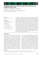
Fig. 1. Identification of JLP as a partner of hsMOK2 using the
yeast two-hybrid screen and identification of the hsMOK2 interac-
tion domain. (A) Constructs expressing full-length or the indicated
domains of hsMOK2 and the human polypeptide JLP 1–141 were
co-transformed into yeast. The specificity of the interaction
between bait and prey was determined by estimating the degree
of color development after 90 min of incubation in the filter lift
b-galactosidase assay, as described in Materials and methods.
(+++) High color blue development, (+ ⁄ )) very low color blue
development, ()) no color development. (B) Amino acid sequence
alignment of N-terminal of JLP 1–141 and JSAP1 1–146.
Phosphorylation-dependent binding of MOK2 M. Harper et al.
3138 FEBS Journal 276 (2009) 3137–3147 ª 2009 The Authors Journal compilation ª 2009 FEBS
with the GST–JLP 1–141 protein. Furthermore,
deletion of amino acids 71–101 strongly reduced inter-
action with hsMOK2, and GST–JLP 66–141 and
GST–JLP 66–101 proteins did not bind to hsMOK2.
These results demonstrated that the minimal domain
of JLP to mediate hsMOK2 binding was located from
amino acids 21 to 101 and that the region surrounding
JLP residues 71–101 was required but was not
sufficient for this interaction.
JLP exhibits sequence homology to JSAP1 (also
called JIP3) [12,13]. In particular, they share 77.3%
homology in their N-terminal region (Fig. 1B),
suggesting that hsMOK2 may also interact with
JSAP1. We therefore tested its interaction with the
N-terminal region of JSAP1 protein (amino acids
26–106), which corresponds to amino acids 21–101 of
JLP. hsMOK2 bound even more efficiently to GST–
JSAP1 26–106 protein than to GST–JLP 21–101
(Fig. 1A,D). We analyzed and compared the secondary
structure of the JLP 1-141 and JSAP1 6–106 domains
using the paircoil program [14]. This program uses
pairwise residue probabilities to detect coiled-coil
motifs in protein sequence data, and the database of
pairwise residue correlations suggests structural
A
D
B
C
E
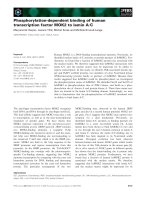
Fig. 2. The N-terminal domains of JLP and JSAP1 bind to hsMOK2 in vitro. (A) Mapping the interaction region of JLP using GST pull-down
analysis. Various JLP N-terminal regions and the homologous JSAP1 region were tested for their interaction with hsMOK2. Nuclear extracts
(20 lg) from HeLa cells transfected with the expression vector hsMOK2 were incubated with an equal amount (10 l g) of recombinant GST
fusion proteins bound to glutathione beads. After washing the beads thoroughly, the bound proteins were eluted in SDS sample buffer,
resolved by SDS ⁄ PAGE and immunoblotted with an affinity purified anti-hsMOK2 serum. The proteins were visualized by exposing the blots
to CL-Xposure film (Pierce). (B) Structure of the JLP deletion mutants. The amino acid number of the encoded proteins is indicated for each
construct. Interactions observed in (A) are summarized on the right. (C) Comparison of the predicted coiled-coil structure of JSAP1 6-146
domain (black) with wild-type (red) and mutant D68N (green) and F65L ⁄ D68N (blue) JLP 1–141 domains. The graphs of coiled-coil scores
were determined using the
PAIRCOIL program [14]. (x-axis) Residue number. (y-axis) Probability of a coiled-coil formation. (D) GST pull-down
by JSAP1 21–101 domain and wild-type or mutant of JLP 26–106 domains was performed as described in (A). (E) Bound proteins were visu-
alized with Fluor-S Max MultiImager and quantified with
QUANTITY ONE software (Bio-Rad). Results were expressed as a percentage of binding
to JSAP1 (21–101) domain (mean ± SD of three different experiments).
M. Harper et al. Phosphorylation-dependent binding of MOK2
FEBS Journal 276 (2009) 3137–3147 ª 2009 The Authors Journal compilation ª 2009 FEBS 3139
features that stabilize or destabilize coiled-coils. Analy-
sis showed that the predicted coiled-coil region in the
JSAP1 6–146 domain is more extended than in the
JLP 1–141 domain (Fig. 2C). The coiled-coil region
begins at residue 69 in the JLP 1–141 domain, whereas
it begins 15 amino acids upstream in the JSAP1 6–146
domain. Interestingly, a difference of three amino acids
was found between the two sequences of this region
(Fig. 2C). The substitution in JLP sequence of F65
and D68 amino acids by the corresponding JSAP1
residues (Leu70 and Asn73) increases the probability
of residues 54–68 forming a coil-coiled region. We
determined experimentally the effects of a single point
mutation D68N and double point mutation
F65L ⁄ D68N in JLP (amino acids 21–101) on binding
to hsMOK2. Only the double substitution within the
JLP domain significantly enhanced the association of
JLP with hsMOK2 (Fig. 2D,E) suggesting that the
single mutation D68N does not provide enough stabi-
lization of the coil-coiled region. The association with
the double point mutation F65L ⁄ D68N in JLP became
comparable with that observed with the JSAP1
domain. These results confirmed that the region
between residues 59 and 73 in JSAP1 strongly
promotes binding to hsMOK2.
To determine whether hsMOK2 interacts with
JSAP1 in mammalian cells, GST pull-down and
co-immunoprecipitation analyses were performed. The
endogenous MOK2 protein is difficult to assess
because of its very low expression level, and so we
examined the in vivo interaction in transfected cells.
Indeed, the endogenous MOK2 protein has been
detected only by electron microscopy [1]. HEK293 cells
were transfected with constructs that expressed
hsMOK2 tagged with GST in the N-terminus (GST–
hsMOK2) and JSAP1 fused to a Flag epitope in the
N-terminus (Flag–JSAP1), either together or sepa-
rately. As shown in Fig. 3A, Flag–JSAP1 protein was
strongly detected in glutathione-bound proteins from
cells co-transfected with Flag–JSAP1 and GST–
hsMOK2 (lane 3) compared with those from cells
transfected with Flag–JSAP1 alone (lane 1). In reverse
experiments, GST–hsMOK2 protein was strongly
detected in anti-Flag immunoprecipitates from cells
co-transfected with Flag–JSAP1 and GST–hsMOK2
(Fig. 3B, lane 3) compared with those from cells trans-
fected with GST–hsMOK2 alone (lane 2). To verify
equivalent recovery of GST–hsMOK2 (Fig. 3A, lower)
or Flag–JSAP1 (Fig. 3B, lower), the blots were
stripped and reprobed with anti-hsMOK2 or anti-Flag
serum, respectively. These results demonstrated that
interaction between the full-length hsMOK2 and
JSAP1 proteins occurs in mammalian cells.
hsMOK2 is phosphorylated in cells
The interaction of hsMOK2 with JLP and JSAP1 scaf-
fold proteins suggests that hsMOK2 activity could be
modulated by phosphorylation by the JNK family of
MAP kinases. We examined the in vivo phosphoryla-
tion status of transfected hsMOK2. HeLa cells trans-
fected with GST–hsMOK2 were lyzed and incubated
with glutathione–agarose to purify the GST–hsMOK2
fusion protein. As negative and positive controls, we
used GST alone and GST-tagged kinesin-12 (also
called Kif 15), respectively [15]. We used two commer-
cially available antibodies to detect phosphorylation at
serine or threonine residues in the hsMOK2 protein.
Purified GST fusion proteins were resolved in dupli-
cate SDS ⁄ PAGE gels and immunoblotted with either
A
B
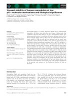
Fig. 3. Interaction of hsMOK2 and JSAP1 in human cells. Cultured
HEK293 cells were transfected with expression vector for Flag–
JSAP1 (lane 1), GST–hsMOK2 (lane 2) or co-transfected with both
vectors (lane 3). (A) Whole-cell extracts (100 lg) were incubated
with 30 lL of 50% slurry glutathione beads. After washing the
beads thoroughly, the bound proteins were eluted in SDS sample
buffer, resolved by SDS ⁄ PAGE and immunoblotted with mouse
anti-(Flag M2) mAb. The blot was stripped and reprobed with anti-
hsMOK2 serum to verify equivalent recovery of the GST fusion pro-
tein (lower). (B) Whole-cell extracts from transfected HEK293 cells
(100 lg) were immunoprecipitated with 20 lL of anti-(Flag M2) aga-
rose affinity gel. After washing the beads thoroughly, the bound
proteins were eluted in SDS sample buffer, resolved by SDS ⁄ PAGE
and immunoblotted with an affinity purified anti-hsMOK2 serum.
The blot was stripped and reprobed with Flag M2 mAb to verify
equivalent recovery of the Flag fusion protein (lower).
Phosphorylation-dependent binding of MOK2 M. Harper et al.
3140 FEBS Journal 276 (2009) 3137–3147 ª 2009 The Authors Journal compilation ª 2009 FEBS
anti-(phosphoserine Q5) or anti-(phosphothreonine
Q7) serum. None of the antibodies reacted with GST
alone (Fig. 4, lane 1). Phosphorylation of GST–
hsMOK2 was detected only by the anti-phosphoserine
serum (Fig. 4, lane 3), whereas phosphorylation of
GST–Kin-12–stalk2 was detected only by the anti-
phosphothreonine serum (Fig. 4, lane 2). An anti-GST
serum confirmed that equivalent amounts of purified
GST fusion proteins were loaded and that the bands
revealed by the anti-phosphoserine or the anti-phos-
phothreonine serum corresponded to the migration of
GST–hsMOK2 and GST–Kin-12–stalk2 (Fig. 4, left).
These results established that hsMOK2 is phosphory-
lated on serine residues in vivo.
hsMOK2 is phosphorylated by JNK3, Aurora A
and PKA kinases in vitro
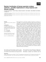
It is known that JNK kinases phosphorylate Ser ⁄ Thr-
Pro motifs in target proteins [16]. hsMOK2 contains
four of these motifs: S28P, S38P, S129P and S191P
(Fig. 5A). The sequence of human MOK2 is highly
conserved between primates, but only S129P motif is
conserved (Fig. 5A). Because JNK3 is the JNK kinase
expressed primarily in the brain like MOK2 [16,17],
we examined hsMOK2 phosphorylation by JNK3
kinase in an in vitro kinase assay. hsMOK2 was
expressed in Escherichia coli as GST fusion protein,
purified on glutathione–agarose and incubated with
activated recombinant JNK3 in the presence of
[
32
P]ATP[cP]. The result showed that recombinant
hsMOK2 was a substrate for JNK3 in vitro (Fig. 5B,
lane 1). To determine the possible contribution of the
four serine residues, we replaced individual serine resi-
dues with alanine and expressed the mutant proteins
as GST fusion in E. coli. Similar quantities of the pro-
tein (as shown in the Coomassie Brilliant Blue-stained
gel in Fig. 5B, lower), were subjected to in vitro phos-
phorylation with JNK3 kinase. The results showed
that replacement of Ser28 or Ser191 with Ala did not
decrease the phosphorylation of hsMOK2 by JNK3,
whereas the phosphorylation was markedly decreased
when Ser38 or Ser129 were replaced with Ala. The
simultaneous replacement of Ser38 and Ser129 caused
Fig. 4. hsMOK2 is a phosphoserine protein. Whole-protein extracts (500 lg) from HeLa cells transfected with GST (lane 1), GST–Kin-12–stalk2
(lane 2) or GST–hsMOK2 (lane 3) were incubated with 50 lL of 50% slurry glutathione beads. After thoroughly washing the beads, the bound
proteins were eluted in SDS sample buffer, resolved in duplicate gels by SDS ⁄ PAGE and immunoblotted with either anti-(phosphoSerine Q5)
serum (middle) or anti-(phosphoThreonine Q7) serum (right). The same blots were stripped and re-probed with anti-GST serum to confirm
equivalent loading of GST fusion protein (left). The proteins were visualized by exposing the blots to CL-Xposure film (Pierce).
A
B
Fig. 5. Phosphorylation of hsMOK2 in vitro by JNK3 kinase. (A)
Alignment of primate MOK2 proteins highlighting the potential SP
motifs for JNK kinases in bold. The percentage identity with human
is indicated in parentheses. (B) GST-tagged wild-type or mutant
hsMOK2 bound to glutathione beads were incubated with recombi-
nant JNK3 kinase in the presence of [
32
P]ATP[cP]. The proteins
were then separated on SDS ⁄ PAGE. The gel was subjected to
Coomassie Brilliant Blue staining (lower) followed by autoradio-
graphy (upper).
M. Harper et al. Phosphorylation-dependent binding of MOK2
FEBS Journal 276 (2009) 3137–3147 ª 2009 The Authors Journal compilation ª 2009 FEBS 3141
a larger decrease in phosphorylation. The data demon-
strate that JNK3 phosphorylates hsMOK2 on Ser38
and Ser129 in vitro.
Because JSAP1 is expressed along with JNK3 and
MOK2 in the brain, JSAP1 could bring together
MAPKs and hsMOK2 in this tissue. Such a function
of JIP proteins has been previously proposed for two
other transcription factors, c-Myc and Max [11]. Inter-
estingly, the region of JLP that binds Myc is similar to
the region of JSAP1 binding hsMOK2. It is tempting
to speculate that JSAP1 may enhance hsMOK2 phos-
phorylation by JNK3. Unfortunately, our attempts to
demonstrate such an effect were unsuccessful. The
JSAP1 protein had no effect when added to our in vitro
phosphorylation assay (data not shown), which may
be because the recombinant JNK3 is an activated form
of the kinase. To address this issue in vivo, HEK293
cells, which do not express JSAP1, were transiently
transfected with tagged hsMOK2, with and without
JSAP1. JSAP1 did not stimulate hsMOK2 phosphory-
lation, even after activation of endogeneous JNK
kinases following incubation of the cells with sorbitol.
This does not preclude that JSAP1 may play a role in
hsMOK2 phosphorylation in brain tissue.
A computer search for other potential phosphoryla-
tion sites indicated the existence of several putative
PKA, protein kinase C and caseine kinase II phos-
phorylation sites spread along the hsMOK2 sequence,
as well as two Aurora phosphorylation sites in the
lamin A ⁄ C-binding N-terminal acidic domain of
hsMOK2. Because these two Aurora sites, centered on
amino acids Ser46 and Ser146, are strictly conserved
between primates [18] (Fig. 6A), we tested whether
they could be phosphorylated by Aurora A kinase.
In vitro phosphorylation experiments showed that
hsMOK2 was a substrate for recombinant Aurora A
kinase (Fig. 6B). To determine which serine is
phosphorylated, we replaced Ser46 or Ser146, or both
Ser46 and Ser146, with alanine in GST–hsMOK2
constructs. Incubation of these fusion proteins with
recombinant Aurora A revealed a minor reduction in
phosphorylation of the mutant containing only the
S146A mutation (Fig. 6B, lane 3), compared with the
wild-type, and a remarkably reduced phosphorylation
of the two mutants containing the S46A mutation
(Fig. 6B, upper, lanes 2, and 4). We concluded that
only Ser46 is a major Aurora A phosphorylation site
on hsMOK2. Recently, it has been reported that
human Aurora A and Aurora B kinases prefer
substrate sequences with an arginine residue at the
position )2. [19,20]. Accordingly, only the sequence
surrounding Ser46 in hsMOK2 conforms to this
preference (RDSV). Lastly, because the consensus for
Aurora A is reminiscent of that of PKA, we examined
the ability of PKA to phosphorylate hsMOK2 protein
in vitro. We obtained the same phosphorylation pattern
of wild-type and mutant hsMOK2 as observed with
Aurora A kinase (Fig. 6C). We conclude that hsMOK2
is efficiently phosphorylated in vitro by Aurora A
kinase and PKA at residue Ser46.
Analysis of phosphomimetic mutations
on hsMOK2 capacity to bind DNA
Binding of hsMOK2 to DNA may be affected, either
positively or negatively, by phosphorylation. There-
fore, to determine whether serine phosphorylation
would affect the ability of hsMOK2 to bind DNA, we
introduced phosphomimetic mutations in hsMOK2 by
replacing individual serine residues by aspartic acid.
A
B
C
Fig. 6. Phosphorylation of hsMOK2 in vitro by Aurora A and PKA. (A)
Alignment of primate MOK2 proteins highlighting the two conserved
Aurora phosphorylation motifs in bold. (B) GST-tagged wild-type or
mutant hsMOK2 bound to glutathione beads were incubated with
recombinant Aurora A in the presence of [
32
P]ATP[cP]. The proteins
were then separated on SDS ⁄ PAGE. The gel was subjected to
Coomassie Brilliant Blue staining (lower) followed by autoradiography
(upper). (C) GST-tagged wild-type or mutant hsMOK2 bound to
glutathione beads were incubated with recombinant PKA, in the
presence of [
32
P]ATP[cP] and visualized as in (B).
Phosphorylation-dependent binding of MOK2 M. Harper et al.
3142 FEBS Journal 276 (2009) 3137–3147 ª 2009 The Authors Journal compilation ª 2009 FEBS
Gel-shift experiments were performed with nuclear
extracts of HeLa cells expressing wild-type or mutant
hsMOK2 proteins and a [
32
P]-labeled double-stranded
oligonucleotide, corresponding to the 18-bp MOK2-
binding site present in hsIRBP intron 2. Immunoblot
analysis attested that similar amounts of the wild-type
and mutant proteins were used in the gel-shift assay
(data not shown). As shown in Fig. 7, phosphomimetic
substitutions at positions Ser46, Ser38 and Ser129 had
no effect on protein–DNA complex abundance at any
o the NaCl concentrations tested. Hence, the two
phosphomimetic mutants retained the ability to bind
the specific DNA sequence with high affinity, indicat-
ing that this binding is not regulated by phosphoryla-
tion at Aurora A ⁄ PKA or JNK sites.
Effect of hsMOK2 phosphorylation on its capacity
to bind lamin A
⁄
C
The two JNK phosphorylation sites of hsMOK2 are
located in the lamin A ⁄ C-binding N-terminal acidic
domain. We therefore examined whether phosphoryla-
tion of hsMOK2 at JNK3 or Aurora A ⁄ PKA sites had
an impact on the interaction with lamin A ⁄ C. Nuclear
extracts from HeLa cells expressing wild-type or
mutant hsMOK2 proteins were prepared and incu-
bated with an equal amount of GST–DlaminC bound
to glutathione beads. Phosphomimetic substitutions at
Ser38, Ser129 or the double Ser38Ser129 mutation did
not markedly decrease binding to DlaminC (Fig. 8A,
upper). By contrast, phosphomimetic substitution at
Ser46 markedly decreased the binding, although ala-
nine substitution at Ser46 had no effect (Fig. 8A,
lower). The data indicated that the phosphorylation of
hsMOK2 at the Aurora A ⁄ PKA site interfered with its
ability to bind lamin A ⁄ C in vitro. We then sought evi-
dence that a similar effect occurs in vivo. We used the
characteristic that mutations in lamin A ⁄ C lead to
sequestration of hsMOK2 in nuclear aggregates
(Fig. 8B) [7]. HeLa cells were co-transfected with the
expression vector for the Q294P lamin C mutant and
wild-type hsMOK2 or hsMOK2 mutated at position
S46. The nonphosphorylatable hsMOK2–S46A protein
was found in the nuclear aggregates induced by the
Q294P lamin C mutant like hsMOK2–WT protein
(Fig. 8B), whereas the phosphomimetic hsMOK2–
S46D protein exhibited a homogeneous nuclear pattern
(Fig. 8B). The hsMOK2–S46D protein was therefore
not displaced in nuclear aggregates, demonstrating that
phosphomimetic substitution at Ser46 also prevents the
interaction with lamin A ⁄ C in vivo.
To confirm that phosphorylation in vivo can disrupt
the interaction between hsMOK2 and lamin A ⁄ C, we
examined the localization of hsMOK2–WT in cells
treated with the phosphatase inhibitor orthovanadate
to enhance cellular phosphorylation. HeLa cells
co-transfected with expression vector for lamin
C–Q294P and hsMOK2–WT were incubated with
1mm sodium orthovanadate for 8 h. In this condition,
no sequestration of hsMOK2 by mutant lamin A ⁄ C
was observed, confirming the importance of phosphor-
ylation for hsMOK2 and lamin A ⁄ C interaction
(Fig. 8C, upper). However, the same observation was
made using hsMOK2–S46A (Fig. 8C, lower), indicat-
ing that the effect of orthovanadate treatment can be
mediated by phosphorylation at another position.
Human MOK2 is a DNA-binding transcriptional
repressor and its interaction with lamin A ⁄ C and the
nuclear matrix may be important for its ability to
repress transcription. Such involvement of lamins A ⁄ C
has been proposed previously for the transcriptional
activator pRb [21–23]. pRb controls cell-cycle progres-
sion by negatively regulating the E2F transcription fac-
tor in a phosphorylation-dependent manner [24]. The
active (hypophosphorylated) form of pRb co-localizes
with lamins A ⁄ C at the nuclear periphery in vivo and
binds to lamins in vitro [21]. Thus, transcriptional
repression by pRb correlates with its lamin-binding
activity. Similarly, transcriptional repression by
hsMOK2 might be correlated with its lamin-binding
Fig. 7. Effects of hsMOK2 phosphomimetic mutations on DNA
binding activity. EMSA was performed on whole-cell extracts
derived from HeLa cells transfected with various hsMOK2 expres-
sion plasmids as indicated. The amount of extracts was adjusted to
obtain equal level of the various hsMOK2 proteins. The
32
P-labeled
double-stranded oligonucleotide corresponds to the 18-bp MOK2
binding site of human IRBP gene. The binding reaction was
performed in buffer containing various concentrations of NaCl.
M. Harper et al. Phosphorylation-dependent binding of MOK2
FEBS Journal 276 (2009) 3137–3147 ª 2009 The Authors Journal compilation ª 2009 FEBS 3143
activity. The simplest scenario is one in which the
lamin A ⁄ C–hsMOK2 complex stabilizes a repressive
complex on DNA, preventing gene activation. To
allow gene activation, hsMOK2 would be phosphory-
lated and then released from lamin A ⁄ C. This regula-
tion may take place following activation of kinases
such as PKA in response to various signaling
pathways. In addition, Aurora A kinase is specifically
activated before mitosis [25,26]. Mitotic nuclear
envelope breakdown requires disassembly of the
nuclear lamina. Lamins A and C are rapidly released
throughout the nucleoplasm in early prophase [27,28].
hsMOK2 dissociation from lamin A ⁄ C at hsMOK2-
regulated loci in early mitosis may contribute to the
dispersion of lamins A ⁄ C into the cytoplasm.
Materials and methods
Plasmid constructs
The recombinant pLex–hsMOK2, pLex–NH
2,
pLex–finger,
pCMV–hsMOK2, pCMV–laminC(Q294P), pGEX–hsMOK2,
pGEX–DlaminC and pGEX–Kin-12-stalk2 vectors have
been described previously [2,3,7,15]. Point mutations were
introduced into hsMOK2 constructs using the Quick-
Change XL site-directed mutagenesis kit (Stratagene,
Amsterdam, The Netherlands). The prokaryotic expression
vector pET29, containing Aurora A cDNA, was a kind gift
from C. Prigent (Faculte
´
de Me
´
decine, Rennes, France).
The pGEX–JLP(1–141) and truncation mutants were
generated from pGAD–JLP(1–141) by PCR using 5¢
A
B
C
Fig. 8. Effect of hsMOK2 phosphorylation on its capacity to bind lamin A ⁄ C. (A) For in vitro interaction, nuclear extracts from HeLa cells
overexpressing wild-type or mutant hsMOK2 were incubated with 10 lg of GST–DlaminC or GST alone bound to glutathione beads. The
amount of nuclear extracts was adjusted to obtain equal levels of the various hsMOK2 proteins. Input lanes correspond to 5% (upper) and
10% (lower) of the extracts used for binding reaction. After thoroughly washing the beads, the bound proteins were eluted in SDS sample
buffer, resolved by SDS ⁄ PAGE and immunoblotted with affinity purified anti-hsMOK2 serum. The proteins were visualized by exposing the
blots to CL-Xposure film (Pierce). (B) For in vivo interaction, HeLa cells were co-transfected with expression vector for lamin C–WT or
lamin C–Q294P mutant and hsMOK2–WT, hsMOK2–S46A or hsMOK2–S46D. After 36 h, cells were fixed and double stained sequentially
with lamin A ⁄ C mAb and anti-hsMOK2 serum. Cells were observed with a Leica DMR microscope and an Apochromat 63 · 1.32 oil
immersion objective. (C) HeLa cells were co-transfected with expression vector for lamin C–Q294P and hsMOK2–WT or hsMOK2–S46A.
Sixteen hours after transfection, the cells were treated with 1 m
M sodium orthovanadate for 8 h, fixed and double-stained sequentially with
lamin A ⁄ C mAb and anti-hsMOK2 serum.
Phosphorylation-dependent binding of MOK2 M. Harper et al.
3144 FEBS Journal 276 (2009) 3137–3147 ª 2009 The Authors Journal compilation ª 2009 FEBS
primers and 3¢ primers containing an EcoRI site and a
SalI site, respectively. After digestion with EcoRI and
SalI, the products were cloned into the corresponding
sites of the pGEX–6P1 vector (Amersham Pharmacia
Biotech, Orsay, France). The mammalian expression
pcDNA3–Flag–JSAP1 plasmid containing the entire coding
sequence of mouse JSAP1 was kindly provided by
K. Yoshioka [29].
Cell culture, transfections and protein extracts
Human HeLa or HEK293 cells were routinely maintained
in Dulbecco’s modified Eagle’s medium supplemented
with 10% fetal calf serum. For transient transfections,
10
5
cells were plated on a 35-mm Petri dish containing a
glass coverslip, 24 h prior to transfection with 2 lgof
plasmid, using calcium phosphate precipitation, as
described previously [30]. For whole-protein extracts,
transfected HeLa cells from three 100-mm Petri dishes
were scraped into NaC ⁄ P
i
, resuspended in 600 lL Hepes
buffer (25 mm Hepes, pH 7.5, 150 m m NaCl, 10% glyc-
erol, 10 lm ZnSO
4
and 0.1% Nonidet P-40) supplemented
with a protease inhibitor cocktail without EDTA (Roche
Diagnostics, Meylan, France) and disrupted by sonication.
After centrifugation at 15 000 g for 10 min, the total
extracts were frozen at )80 °C. Whole-protein extracts
from one 100-mm Petri dish plated with transfected
HEK293 cells were prepared in 500 lLof20mm
Tris ⁄ HCl (pH 7.5), 150 mm NaCl, 1% Triton X-100,
0.5% sodium deoxycholate and 1 mm EDTA,
supplemented with protease inhibitor cocktail, as
described for transfected HeLa cells. Nuclear extracts
were prepared according to the Dignam method [31] and
dialyzed against Hepes buffer at 4 °C for 4 h. After
dialysis and centrifugation, the nuclear extract was frozen
at )80 °C. The protein concentration was determined by
the Coomassie Brilliant Blue protein assay (Pierce,
Brebieres, France).
Antibodies
Affinity-purified rabbit polyclonal anti-hsMOK2 serum
was obtained as described previously [2]. Mouse anti-
(lamin A ⁄ C(636)) mAb was purchased from Santa Cruz
Biotechnology, Heidelberg, Germany). Mouse monoclonal
anti-(Flag M2) and anti-(Flag M2) agarose affinity gel
were purchased from Sigma (St-Quentin Fallavier,
France). Mouse anti-(phosphoThreonine Q7) and anti-
(phosphoSerine Q5) sera were purchased from Qiagen
(Courtaboeuf, France). Rhodamine (TRITC)-conjugated
goat anti-(mouse IgG), CY2
TM
-conjugated goat anti-(rab-
bit IgG) and peroxidase-conjugated rabbit anti-(mouse
IgG) sera were purchased from Jackson Immunoresearch
Laboratories (Bar Harbor, ME, USA).
Yeast two-hybrid screen
Yeast two-hybrid screening using human hsMOK2 as bait
was described previously [3]. A human HeLa S3 Match-
maker cDNA library (BD Clontech, St-Germain-en-Laye,
France), constructed in the pGAD–GH vector expressing
the GAL4 activation domain fusion protein, was trans-
formed into L40 containing the pLex–hsMOK2 construct.
The cDNA inserts of positive clones were isolated by direct
PCR of yeast colonies. The cDNA inserts were further
characterized by sequencing and searching for gene
sequence similarity in the GenBankÔ database with the
program blast.
Immunofluorescence microscopy
Cells were fixed in )20 °C methanol for 3 min and incu-
bated with primary anti-hsMOK2 or anti-(lamin A ⁄ C) sera,
followed by incubation with the secondary antibody. The
incubations were for 1 h each and were carried out sequen-
tially (with washes in NaCl ⁄ P
i
after each step). The cellular
DNA was labeled with 0.12 lgÆmL
)1
4¢-6-diamidino-2-
phenylindole for 1 min. The slides were mounted in antifa-
dent AF1 ⁄ glycerol ⁄ NaCl ⁄ P
i
mounting medium (Citifluor
Ltd, London, UK). Immunofluorescence microscopy was
performed using a Leica DMR microscope (Leica,
Heidelberg, Germany) and an Apochromat 63 · 1.32 oil
immersion objective. Photographs were taken using a
Micromax (Princeton Instruments, Evry, France) CCD
camera and metaview (Universal Imaging Corp.) software.
Purification of GST fusion proteins and kinase
assay
For bacteria expressing GST fusion proteins, crude
protein extracts were prepared and purified as described
previously [3]. The purity and amount of the recombinant
proteins were determined by examining SDS ⁄ PAGE gel
staining with Coomassie Brilliant Blue. The GST fusion
proteins bound to glutathione beads were used as 50%
slurry in appropriate buffer. The N-terminal His
6
-tagged
full-length human Aurora A protein was expressed in
E. coli strain BL21(DE3) and purified using Ni
2+
⁄ NAT
agarose as described by Cremet et al. [32]. The Aurora A
protein solution was concentrated using a centricon 10
(Millipore, St-Quentin-en Yvelines, France) at 1 mg Æ mL
)1
and stored at )80 °C. The N-terminal His
6
-tagged
full-length human JNK3 ⁄ SPAK1b active protein was
purchased from Upstate (St-Quentin-en Yvelines, France)
and the N-terminal His tagged human catalytic subunit
of PKA was purchased from Calbiochem (Nottingham,
UK).
The kinase assays were performed in 25 lLof50mm
Tris ⁄ HCl pH 7.5, 0.1% 2-mercaptoethanol, 1 mm EGTA,
M. Harper et al. Phosphorylation-dependent binding of MOK2
FEBS Journal 276 (2009) 3137–3147 ª 2009 The Authors Journal compilation ª 2009 FEBS 3145
0.2 mm dithiothreitol, 0.2 mm sodium orthovanadate,
15 mm MgCl
2,
100 lm ATP containing 10 lCi [
32
P]ATP
[cP] 3000 CiÆmmol
)1
(Amersham Pharmacia Biotech),
200 ng Aurora A or 50 ng JNK3 ⁄ SPAK1b or 10 ng PKA
and 10–15 lL of a 50% slurry of GST fusion proteins
bound to the beads. The reactions were incubated at
30 °C for 10 min. Ten microliters of 5· Laemmli sample
buffer was added and the reaction was heated at 100 °C
for 5 min. The proteins were then separated on a Nupage
4–12% Bis-Tris gels (Invitrogen, Illkirch, France). The gel
was subjected to Coomassie Brilliant Blue staining, dried
and analysed using a Phosphoimager apparatus (Mole-
cular Dynamics, Orsay, France).
GST pull-down assay and
co-immunoprecipitation
For in vitro GST pull-down assay, 10 lg of GST fusion
protein, immobilized on glutathione–agarose beads was
added to 20 l g of nuclear proteins from transfected HeLa
cells, in a total volume of 400 lL Hepes buffer. For in vivo
GST pull-down assay or co-immunoprecipitation, whole-
cell extracts (100 lg) from transfected HEK293 were incu-
bated either with 30 lL of 50% slurry glutathione beads or
with 20 lL of anti-(Flag M2) agarose affinity gel. After 2 h
at 4 °C, the beads were extensively washed and eluted by
boiling in Laemmli buffer. Bound proteins were separated
by SDS ⁄ PAGE and analyzed by immunoblotting with the
indicated antibodies using the Supersignal West Pico
Chemiluminescent Signal kit (Pierce). The proteins were
visualized by exposing the blots to CL-XPosure film
(Pierce) or using a Fluor-S Max MultiImager with
quantity one software (Bio-Rad, Marne-la-Coquette,
France).
Electrophoretic mobility shift assay
A 25-bp oligonucleotide corresponding to the sequence of
human IRBP intron 2 containing the 18-bp MOK2-binding
site (5¢-CTGCAGGACTTGTCAGGGCCTTTAA-3¢) was
used as a probe. The double-strand oligonucleotide was
labeled with T4 polynucleotide kinase (Biolabs, Ipswich,
MA, USA) in the presence of [
32
P]ATP[cP] and purified on
a 15% polyacrylamide gel. End-labeled oligonucleotides
(0.2 ng) were incubated for 20 min at room temperature in
20 lL Hepes buffer containing 2 lg poly(dI–dC), various
concentrations of NaCl and whole-protein extracts from
HeLa cells transfected with wild-type or mutant hsMOK2.
The amount of extract was adjusted to obtain an equivalent
level of the transfected hsMOK2 proteins. Complexes were
analyzed by electrophoresis on a nondenaturing premigrat-
ed 6% polyacrylamide gel (acrylamide ⁄ bis ratio 19 : 1) in
0.5· TB buffer (45 mm Tris borate, pH 8.3) at 4 °Cat
200 V. EDTA was omitted in all binding and electrophore-
sis buffers to avoid denaturing hsMOK2.
Acknowledgements
We thank Katsuji Yoshioka for providing the
pcDNA3–Flag–JSAP1 expression vector and Claude
Prigent for pET29–Aurora A vector. We also thank
Vanessa Philipot for technical assistance and Domi-
nique Weil for critical reading of the manuscript. This
research was supported by grants from the Centre
National de la Recherche Scientifique, the Fondation
Raymonde et Guy Strittmatter and the association
Retina France.
References
1 Arranz V, Harper F, Florentin Y, Puvion E, Kress M
& Ernoult-Lange M (1997) Human & mouse MOK2
proteins are associated with nuclear ribonucleoprotein
components & bind specifically to RNA & DNA
through their zinc finger domains. Mol Cell Biol 17,
2116–2126.
2 Arranz V, Dreuillet C, Crisanti P, Tillit J, Kress M &
Ernoult-Lange M (2001) The zinc finger transcription
factor, MOK2, negatively modulates expression of the
interphotoreceptor retinoid-binding protein (IRBP)
gene. J Biol Chem 276, 11963–11969.
3 Dreuillet C, Tillit J, Kress M & Ernoult-Lange M
(2002) In vivo & in vitro interaction between human
transcription factor MOK2 & nuclear lamin A ⁄ C.
Nucleic Acids Res 30, 4634–4642.
4 Ernoult-Lange M, Arranz V, Leconiat M, Berger R &
Kress M (1995) Human & mouse Kruppel-like (MOK2)
orthologue genes encode two different zinc finger pro-
teins. J Mol Evol 41, 784–794.
5 Fisher DZ, Chaudhary N & Blobel G (1986) cDNA
sequencing of nuclear lamins A & C reveals primary &
secondary structural homology to intermediate filament
proteins. Proc Natl Acad Sci USA 83, 6450–6454.
6 McKeon FD, Kirschner MW & Caput D (1986)
Homologies in both primary & secondary structure
between nuclear envelope & intermediate filament
proteins. Nature 319, 463–468.
7 Dreuillet C, Harper M, Tillit J, Kress M & Ernoult-
Lange M (2008) Mislocalization of human transcription
factor MOK2 in the presence of pathogenic mutations
of lamin A ⁄ C. Biol Cell 100, 51–61.
8 Dhanasekaran DN, Kashef K, Lee CM, Xu H & Reddy
EP (2007) Scaffold proteins of MAP-kinase modules.
Oncogene 26, 3185–3202.
9 Morrison DK & Davis RJ (2003) Regulation of MAP
kinase signaling modules by scaffold proteins in mam-
mals. Annu Rev Cell Dev Biol 19, 91–118.
10 Yoshioka K (2004) Scaffold proteins in mammalian
MAP kinase cascades. J Biochem 135, 657–661.
11 Lee CM, Onesime D, Reddy CD, Dhanasekaran N &
Reddy EP (2002) JLP: a scaffolding protein that tethers
Phosphorylation-dependent binding of MOK2 M. Harper et al.
3146 FEBS Journal 276 (2009) 3137–3147 ª 2009 The Authors Journal compilation ª 2009 FEBS
JNK ⁄ p38MAPK signaling modules & transcription
factors. Proc Natl Acad Sci USA 99, 14189–14194.
12 Ito M, Yoshioka K, Akechi M, Yamashita S, Takama-
tsu N, Sugiyama K, Hibi M, Nakabeppu Y, Shiba T &
Yamamoto KI (1999) JSAP1, a novel jun N-terminal
protein kinase (JNK)-binding protein that functions as
a Scaffold factor in the JNK signaling pathway. Mol
Cell Biol 19, 7539–7548.
13 Kelkar N, Gupta S, Dickens M & Davis RJ (2000)
Interaction of a mitogen-activated protein kinase signal-
ing module with the neuronal protein JIP3. Mol Cell
Biol 20, 1030–1043.
14 Berger B, Wilson DB, Wolf E, Tonchev T, Milla M &
Kim PS (1995) Predicting coiled coils by use of pairwise
residue correlations. Proc Natl Acad Sci USA 92, 8259–
8263.
15 Buster DW, Baird DH, Yu W, Solowska JM, Chauviere
M, Mazurek A, Kress M & Baas PW (2003) Expression
of the mitotic kinesin Kif15 in postmitotic neurons:
implications for neuronal migration & development.
J Neurocytol 32, 79–96.
16 Davis RJ (2000) Signal transduction by the JNK group
of MAP kinases. Cell 103, 239–252.
17 Ernoult-Lange M, Kress M & Hamer D (1990) A gene
that encodes a protein consisting solely of zinc finger
domains is preferentially expressed in transformed
mouse cells. Mol Cell Biol 10, 418–421.
18 Cheeseman IM, Anderson S, Jwa M, Green EM, Kang J,
Yates JR III, Chan CS, Drubin DG & Barnes G (2002)
Phospho-regulation of kinetochore–microtubule
attachments by the Aurora kinase Ipl1p. Cell 111,
163–172.
19 Ferrari S, Marin O, Pagano MA, Meggio F, Hess D, El
Shemerly M, Krystyniak A & Pinna LA (2005)
Aurora-A site specificity: a study with synthetic peptide
substrates. Biochem J 390, 293–302.
20 Ohashi S, Sakashita G, Ban R, Nagasawa M, Matsuzaki
H, Murata Y, Taniguchi H, Shima H, Furukawa K &
Urano T (2006) Phospho-regulation of human
protein kinase Aurora-A: analysis using anti-
phospho-Thr288 monoclonal antibodies. Oncogene 25,
7691–7702.
21 Mancini MA, Shan B, Nickerson JA, Penman S & Lee
WH (1994) The retinoblastoma gene product is a cell
cycle-dependent, nuclear matrix-associated protein. Proc
Natl Acad Sci USA 91, 418–422.
22 Ozaki T, Saijo M, Murakami K, Enomoto H, Taya Y
& Sakiyama S (1994) Complex formation between
lamin A & the retinoblastoma gene product:
identification of the domain on lamin A required for
its interaction. Oncogene 9, 2649–2653.
23 Shan B, Zhu X, Chen PL, Durfee T, Yang Y, Sharp D
& Lee WH (1992) Molecular cloning of cellular genes
encoding retinoblastoma-associated proteins: identifica-
tion of a gene with properties of the transcription factor
E2F. Mol Cell Biol 12, 5620–5631.
24 Chellappan SP, Hiebert S, Mudryj M, Horowitz JM &
Nevins JR (1991) The E2F transcription factor is a
cellular target for the RB protein. Cell 65, 1053–1061.
25 Bischoff JR, Anderson L, Zhu Y, Mossie K, Ng L,
Souza B, Schryver B, Flanagan P, Clairvoyant F,
Ginther C et al. (1998) A homologue of Drosophila
aurora kinase is oncogenic & amplified in human
colorectal cancers. EMBO J 17, 3052–3065.
26 Stenoien DL, Sen S, Mancini MA & Brinkley BR
(2003) Dynamic association of a tumor amplified
kinase, Aurora-A, with the centrosome & mitotic spin-
dle. Cell Motil Cytoskeleton 55, 134–146.
27 Georgatos SD, Meier J & Simos G (1994) Lamins &
lamin-associated proteins. Curr Opin Cell Biol 6
, 347–
353.
28 Beaudouin J, Gerlich D, Daigle N, Eils R & Ellenberg
J (2002) Nuclear envelope breakdown proceeds by
microtubule-induced tearing of the lamina. Cell 108,
83–96.
29 Bayarsaikhan M, Shiratsuchi A, Gantulga D, Nakanishi
Y & Yoshioka K (2006) Selective expression of the scaf-
fold protein JSAP1 in spermatogonia & spermatocytes.
Reproduction 131, 711–719.
30 Arranz V, Kress M & Ernoult-Lange M (1994) The
gene encoding the MOK-2 zinc-finger protein: charac-
terization of its promoter & negative regulation by
mouse Alu type-2 repetitive elements. Gene 149,
293–298.
31 Dignam JD, Lebovitz RM & Roeder RG (1983) Accu-
rate transcription initiation by RNA polymerase II in a
soluble extract from isolated mammalian nuclei. Nucleic
Acids Res 11, 1475–1489.
32 Cremet JY, Descamps S, Verite F, Martin A & Prigent
C (2003) Preparation & characterization of a human
aurora-A kinase monoclonal antibody. Mol Cell
Biochem 243, 123–131.
M. Harper et al. Phosphorylation-dependent binding of MOK2
FEBS Journal 276 (2009) 3137–3147 ª 2009 The Authors Journal compilation ª 2009 FEBS 3147