Báo cáo khoa học: The transcription factor ZBP-89 suppresses p16 expression through a histone modification mechanism to affect cell senescence doc
Bạn đang xem bản rút gọn của tài liệu. Xem và tải ngay bản đầy đủ của tài liệu tại đây (586.15 KB, 10 trang )
The transcription factor ZBP-89 suppresses p16 expression
through a histone modification mechanism to affect
cell senescence
Yunpeng Feng*, Xiuli Wang*, Liang Xu, Hong Pan, Shan Zhu, Qian Liang, Baiqu Huang
and Jun Lu
Institute of Genetics and Cytology, Northeast Normal University, and the Key Laboratory of Molecular Epigenetics of Ministry of Education
(MOE), Northeast Normal University, Changchun, China
Introduction
ZBP-89 is a ubiquitously expressed four-zinc finger
transcription factor that binds to the GC-rich DNA
elements, functioning either as a repressor or as an
activator of the known target genes. For instance,
when acting as an activator, ZBP-89 recruits the
coactivator p300 to the p21 promoter, resulting in
Keywords
histone deacetylase 3 (HDAC3); histone
deacetylase 4 (HDAC4); p16; senescence;
ZBP-89
Correspondence
J. Lu, Institute of Genetics and Cytology,
and the Key Laboratory of Molecular
Epigenetics of MOE, Northeast Normal
University, 5268 Renmin Street, Changchun
130024, China
Fax: +86 431 85099768
Tel: +86 431 85099798
E-mail:
*These authors contributed equally to this
work
(Received 1 April 2009, revised 29 May
2009, accepted 3 June 2009)
doi:10.1111/j.1742-4658.2009.07128.x
The transcription factor ZBP-89 has been implicated in the induction of
growth arrest and apoptosis. In this article, we demonstrate that ZBP-89
was able to restrain senescence in NCI-H460 human lung cancer cells,
through epigenetically regulating p16
INK4a
expression. Specifically, our
results indicate that knockdown of ZBP-89 by RNA interference stimulated
cellular senescence in NCI-H460 cells, as judged by the senescence-
associated b-galactosidase activity assay and senescence-associated hetero-
chromatin foci assay, and this process could be reversed by RNA
interference-mediated p16
INK4a
silencing. We also show that histone deacet-
ylase (HDAC) 3 and HDAC4 inhibited p16
INK4a
promoter activity in a
dose-dependent manner. Furthermore, chromatin immunoprecipitation
assays verified that HDAC3 was recruited to the p16
INK4a
promoter by
ZBP-89 through an epigenetic mechanism involving histone acetylation
modification. Moreover, immunofluorescence and coimmunoprecipitation
assays revealed that ZBP-89 and HDAC3 formed a complex. These data
suggest that ZBP-89 and HDAC3, but not HDAC4, can work coordinately
to restrain cell senescence by downregulating p16
INK4a
expression through
an epigenetic modification of histones.
Structured digital abstract
l
MINT-7144512: HDAC4 (uniprotkb:P56524) physically interacts (MI:0914) with ZBP-89
(uniprotkb:
Q9UQR1)byanti tag coimmunoprecipitation (MI:0007)
l
MINT-7144482, MINT-7144499: ZBP-89 (uniprotkb:Q9UQR1) physically interacts (MI:0914)
with HDAC3 (uniprotkb:
O15379)byanti tag coimmunoprecipitation (MI:0007)
l
MINT-7144469: ZBP-89 (uniprotkb:Q9UQR1) and HDAC3 (uniprotkb:O15379) colocalize
(
MI:0403)byfluorescence microscopy (MI:0416)
Abbreviations
CDK, cyclin-dependent kinase; ChIP, chromatin immunoprecipitation; Co-IP, coimmunoprecipitation; DAPI, 4¢,6-diamidino-2-phenylindole;
GFP, green fluorescent protein; HAT, histone acetyltransferase; HDAC, histone deacetylase; RNAi, RNA interference; SAHF, senescence-
associated heterochromatin foci; SA-b-gal, senescence-associated b-galactosidase; siRNA, small interfering RNA; TRITC,
tetramethylrhodamine isothiocyanate.
FEBS Journal 276 (2009) 4197–4206 ª 2009 The Authors Journal compilation ª 2009 FEBS 4197
upregulation of the gene [1]. Bai and Merchant also
reported that elevated expression of ZBP-89 induced
growth arrest and apoptosis through promoting p21
expression upon treatment with the histone deacetylase
(HDAC) inhibitor butyrate, or through stabilizing p53
protein, indicating that ZBP-89 plays a role in cell
cycle progression [2]. Recently, Wu et al. [3] reported
that ZBP-89 functioned as a repressor by recruiting
HDAC1 to the vimentin promoter. ZBP-89 shares with
Sp1 and other Sp-like factors the ability to recognize
GC-rich sequences in target genes. To depict this over-
lapping DNA recognition, a competitive model of inhi-
bition has been proposed, in which ZBP-89 represses
gene transcription by displacing proteins such as Sp1
and Sp3 [4,5]. An analysis of the proximal promoter of
the ornithine decarboxylase gene revealed that Sp1 and
ZBP-89 bound to the GC elements in a mutually
exclusive manner [6]. In other cases, ZBP-89 appears
to inhibit gene activity by binding to DNA indepen-
dently of Sp1 [7].
Reversible acetylation of internal lysine residues of
the N-terminal domains of nucleosomal histones and
the resultant changes in the chromatin structure are
important epigenetic mechanisms in the regulation of
gene transcription. The interplay between histone acet-
yltransferases (HATs) and HDACs is critical to the
dynamics of chromatin structure and function, thus
regulating gene expression in eukaryotes [8]. Several
HATs have been identified that act as transcriptional
coactivators. In contrast, HDACs form part of tran-
scriptional corepressor complexes [9].
The INK4A locus encodes a cyclin-dependent kinase
(CDK) inhibitor, p16
INK4a
(hereafter p16), which func-
tions as a negative regulator of cyclin–CDK com-
plexes. It binds preferentially to CDK4 and CDK6,
and prevents their association with D-type cyclins,
thus inhibiting retinoblastoma protein phosphorylation
and blocking cell cycle progression [10,11]. Expression
of p16 is regulated primarily at the transcriptional
level. The p16 promoter lacks a distinct TATA box,
and is GC-rich. The GC-rich regions represent the
putative binding sites for the ubiquitously expressed
Sp1 transcription factor [12]. As ZBP-89 also binds to
the GC-rich DNA elements, it raises the question of
whether ZBP-89 participates in p16 transcriptional reg-
ulation. In this article, we present experimental data
showing that knockdown of ZBP-89 in human lung
cancer cells by a specific small interfering RNA (siR-
NA) vector (ZBP-89i) increased expression of p16 and
induced cell senescence. Moreover, overexpression of
HDAC3 and HDAC4 resulted in repression of p16
expression, and HDAC3 was recruited to the p16 pro-
moter through ZBP-89. On the basis of these data, we
discuss the possible mechanisms of the functional
interactions among ZBP-89, HDAC3 and HDAC4 in
p16 transcriptional inhibition and their effects on cell
senescence.
Results
Knockdown of endogenous ZBP-89 promoted
NCI-H460 cell senescence
Previously, a study showed that ZBP-89 was able to
induce cell growth arrest and apoptosis [2]. However,
whether ZBP-89 affects cancer cell senescence has not
been investigated. To test this effect, we constructed an
siRNA vector specific to ZBP-89 (ZBP-89i) to knock
down ZBP-89 expression in the human lung cancer cell
line NCI-H460. Western blots verified the exogenous
expression of the ZBP-89 vector (Fig. 1A), and the
efficiency of inhibition of ZBP-89 expression by ZBP-
89i (Fig. 1B). The transfected NCI-H460 cells were
then lysed and assayed for the activity of senescence-
associated b-galactosidase (SA-b-gal; pH 6.0), a bio-
marker that is tightly associated with senescence in
human cells [13]. As shown in Fig. 1C, a 1.5-fold
increase in SA-b-gal activity was seen after 7 days of
ZBP-89i transfection, whereas overexpression of ZBP-
89 led to a reverse effect. In addition, cells transfected
with ZBP-89i exhibited phenotypic changes that are
typical of cells undergoing replicative senescence. These
changes include increased SA-b-gal staining, flattened
cell morphology, and enlarged cell size (Fig. 1D).
Meanwhile, the senescence-associated heterochromatin
foci (SAHF) assays were performed using antibodies
against 3MeK9H3 and HP1 proteins, and the reactions
of these antibodies were visualized by confocal micros-
copy. As shown in Fig. 1E, both marker proteins were
localized to the specific heterochromatic foci in cells
transfected with ZBP-89i. Also, 3MeK9H3 and HP1
proteins were found to be colocalized in discrete foci
in the senescent cells, as observed by confocal micros-
copy. Together, these data implied that ZBP-89 played
a role in restraint of human lung cancer NCI-H460 cell
senescence.
ZBP-89 interacted with the p16 promoter to
repress its transcription
It has been well documented that p16 plays a critical
role in inducing cell senescence; we were therefore curi-
ous to know whether ZBP-89 induced senescence
through p16 regulation in NCI-H460 cells. We used a
p16 siRNA vector (p16i) to knock down p16 expres-
sion [14]. The results showed that, as compared trans-
ZBP-89 affects senescence through p16 Y. Feng et al.
4198 FEBS Journal 276 (2009) 4197–4206 ª 2009 The Authors Journal compilation ª 2009 FEBS
fection with ZBP-89i alone, cotransfection of cells with
ZBP-89i and p16i vectors failed to induce NCI-H460
cell senescence (Fig. 2A). Western blotting demon-
strated that p16 protein expression was decreased on
ZBP-89 ectopic expression, whereas it was enhanced
by knockdown of the endogenous ZBP-89 in NCI-
H460 cells (Fig. 2B). Furthermore, overexpression of
ZBP-89 greatly inhibited p16 promoter activity
(Fig. 2C). Also, it can be seen from Fig. 2D that the
p16 mRNA level was decreased on ectopic expression
of ZBP-89, but increased by knockdown of endoge-
nous ZBP-89. To determine whether ZBP-89 was truly
present at the p16 promoter to regulate the gene as a
transcription factor, we designed a series of primers
coordinate to the three regions in the p16 promoter
for chromatin immunoprecipitation (ChIP) assays
(Fig. 2E). P1 locates far upstream of the p16 promoter
()1800 bp) as a negative control, whereas P2 and P3
locate downstream of the p16 promoter at )700 and
)400 bp, which represent the important regulatory
regions of the p16 gene. The ChIP data shown in
Fig. 2F reveal that ZBP-89 was enriched at the P2 and
P3 regions of the p16 promoter upon ZBP-89 overex-
pression. These results suggest that ZBP-89 was able
to inhibit p16 expression at the promoter activity,
mRNA and protein levels.
HDAC3 and HDAC4 downregulated p16 by
inducing histone hypoacetylation
We previously reported that the HAT p300 stimulated
p16 expression [14], and this prompted us to speculate
whether HDAC(s) also plays a role in p16 regulation
as the opposing enzyme(s) to the HATs. To test this
assumption, we transfected 293T cells with the p16
promoter reporter together with the expression vectors
of HDAC1–HDAC6; of the six HDACs tested,
HDAC3 and HDAC4 had much more prominent
effects on p16 repression (Fig. 3A). Also, p16
promoter activity was inhibited by HDAC3 and
HDAC4 overexpression in a dose-dependent manner
(Fig. 3B,C). The endogenous p16 mRNA level was
also decreased upon HDAC3 and HDAC4 overexpres-
sion, as revealed by real-time PCR (Fig. 3D). Addi-
tionally, ChIP assays with antiacetylated histone H3
and histone H4 antibodies showed that the acetylation
level of histone H3 was significantly changed by exoge-
nous expression of HDAC4, whereas the acetylation of
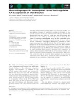
Fig. 1. Knockdown of endogenous ZBP-89
promoted human lung cancer NCI-H460 cell
senescence. Western blot analysis of the
ZBP-89 protein in NCI-H460 cells transfect-
ed with ZBP-89–Flag, or pcDNA3.1 as a
control (A), or ZBP-89i, or ZBP-89–Flag plus
ZBP-89i vectors, and an irrelevant siRNA
vector as a control (B). (C) ZBP-89i
increased the SA-b-gal activity. NCI-H460
cells transfected with ZBP-89 or ZBP-89i
vectors were lysed and tested for SA-b-gal
activity, using o-nitrophenyl-
D-galactopyrano-
side as substrate at pH 6.0. The controls
were the pcDNA3.1 empty vector and
an irrelevant siRNA vector.
**
P < 0.01,
*
P < 0.05 (n = 3). (D) Representative photo-
micrographs of the SA-b-gal staining at
day 7 post-ZBP-89i transfection. The irrele-
vant siRNA vector was used as the control.
(E) NCI-H460 cells were transfected with
ZBP-89i for 7 days. Cells were stained with
DAPI, and heterochromatic foci were visual-
ized by fluorescence microscopy. 3MeK9H3
was immunostained in red, and HP1 in
green. The nuclei were counterstained with
DAPI (blue). It can be seen that HP1 and
3MeK9H3 were colocalized in senescent
cells in discrete SAHF (white and yellow
spots), as shown by confocal microscopy.
Y. Feng et al. ZBP-89 affects senescence through p16
FEBS Journal 276 (2009) 4197–4206 ª 2009 The Authors Journal compilation ª 2009 FEBS 4199
histone H4 was markedly affected by overexpression
of HDAC3 (Fig. 3E,F). These experiments demon-
strate that repression of p16 expression by HDAC3
and HDAC4 coincided with histone hypoacetylation.
HDAC3 interacted with ZBP-89
We next sought to investigate whether physical interac-
tions among HDAC3 ⁄ HDAC4 and ZBP-89 occur. In
293T cells cotransfected with HDAC3 ⁄ 4–green fluores-
cent protein (GFP) and ZBP-89, HDAC3 and ZBP-89
were colocalized in the nuclei, but HDAC4 and ZBP-89
were not colocalized remarkably, as revealed by confocal
laser scanning microscopy (Fig. 4A). Moreover, coim-
munoprecipitation (Co-IP) assays revealed that com-
plexes containing HDAC3–GFP ⁄ HDAC4–GFP and
ZBP-89–Flag were precipitated by antibodies against
GFP and Flag, and they were detected in immunoblots
by antibodies against Flag and GFP (Fig. 4B), suggest-
ing that HDAC3 and ZBP-89 were present in the same
complexes, but not HDAC4. These data provide evi-
dence that the transcription factor ZBP-89 and the core-
pressor HDAC3 interacted and worked coordinately to
contribute to the repression of p16 expression.
HDAC3 was recruited to the p16 promoter by
ZBP-89
To determine whether HDAC3 and HDAC4 were
recruited to the p16 promoter by ZBP-89, we examined
the binding of HDAC3 and HDAC4 in different
regions of the p16 promoter upon knockdown of the
endogenous ZBP-89, and the results showed that the
binding of HDAC3 was indeed significantly reduced by
knockdown of the endogenous ZBP-89, whereas that of
HDAC4 was not affected (Fig. 5A). We then analyzed
the relationship between histone H3 ⁄ H4 acetylation
and ZBP-89 expression. The ChIP data indicated that
the histone H4 acetylation level was decreased by over-
expression of ZBP-89, whereas that of histone H3 was
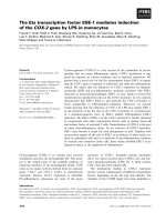
Fig. 2. ZBP-89 restrained cancer cell senescence by repressing p16 expression. (A) ZBP-89 restrained senescence of NCI-H460 cells
through p16 repression. Representative photomicrographs of the SA-b-gal staining at day 7 after ZBP-89i transfection, or ZBP-89i plus p16i
transfection. An irrelevant siRNA vector was used as the control. (B) ZBP-89 repressed p16 protein expression. Western blot analysis of the
p16 protein in NCI-H460 cells transfected with ZBP-89 or ZBP-89i vector. (C) ZBP-89 inhibited p16 promoter activity. 293T cells were trans-
fected with ZBP-89 vector, and the p16 promoter activity was examined by luciferase reporter assay. The control was the pcDNA3.1 empty
vector.
**
P < 0.01 (n = 3). (D) ZBP-89 repressed endogenous p16 mRNA. 293T cells were transfected with ZBP-89 or ZBP-89i vector. Total
RNA was isolated and reverse transcribed, and p16 mRNA was measured by PCR. b-Actin was used as an internal control. (E) Diagram of
the 5¢-flanking region of p16 gene. Lines denote the three regions of the p16 promoter (P1, P2, and P3) amplified by specific primers in ChIP
analysis. (F) Binding of ZBP-89 on the p16 promoter. ChIP assays with antibody against Flag in 293T cells transfected with the ZBP-89–Flag
expression vector. No Ab: samples with no antibody. Input: DNA prior to immunoprecipitation.
ZBP-89 affects senescence through p16 Y. Feng et al.
4200 FEBS Journal 276 (2009) 4197–4206 ª 2009 The Authors Journal compilation ª 2009 FEBS
not affected (Fig. 5B). Moreover, the acetylation levels
of both histone H3 and histone H4 were increased by
ZBP-89i transfection (Fig. 5C). Furthermore, we mea-
sured the binding of endogenous HDAC3 and
HDAC4, as well as ZBP-89, at the p16 promoter in
NCI-H460 cells. The results showed that the binding of
ZBP-89 and HDAC3 was reduced by knockdown of
the endogenous ZBP-89 (Fig. 5D). Thus, these data
clearly indicate that HDAC3, but not HDAC4, was
recruited to the p16 promoter via ZBP-89.
Discussion
ZBP-89 is a four zinc finger transcription factor that
either represses or activates several target genes [15],
although it is more commonly known as a transcrip-
tional repressor [16]. This transcription factor contains
a transcription activation domain at its C-terminus
and a repression domain at its N-terminus [17]. It has
been shown that ZBP-89 binds to the GC-rich pro-
moter elements of genes that are involved in cell
growth regulation, e.g. genes coding for gastrin, orni-
thine decarboxylase, and the CDK inhibitor p21
[1,4,6,18]. It was reported that elevated expression of
ZBP-89 induced growth arrest and apoptosis through
promoting p21 expression upon treatment with the
HDAC inhibitor butyrate, or through p53 protein sta-
bilization [2]. We report here that ZBP-89 was capable
of restraining human lung cancer NCI-H460 cell senes-
cence, and this process could be reversed by inhibition
of p16 expression through RNA interference (RNAi)
(Fig. 2A). Our data also show that ZBP-89 was able
to decrease both p16 promoter activity (Fig. 2C) and
the endogenous p16 mRNA level (Fig. 2D), as well as
to decrease the p16 protein level (Fig. 2B). These
experimental results supported our assumption that the
cell senescence induced by ZBP-89 might be p16-
dependent. A number of previous reports suggested
that p16 was required for cellular senescence in normal
human fibroblasts [19]. A more recent study by Herbig
et al. [20] indicated that p16 and p21 acted through
independent pathways to influence cellular senescence.
Taking together all of these data, we speculate that
ZBP-89 is a multiple-function factor that participates
in a variety of cell processes by regulating different
genes. These functions include the induction of apop-
tosis through p21 and p53 [2], and the restraint of cell
senescence through p16, as shown in this study.
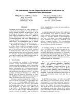
Fig. 3. HDAC3 and HDAC4 downregulated p16 by inducing histone hypoacetylation. (A) HDAC3 and HDAC4 downregulated p16 promoter
activity. 293T cells were transfected with p16 luciferase plasmid together with HDAC constructs expressing HDAC1–HDAC6. Results are
shown as fold repression relative to that of the cells transfected with empty plasmid. (B, C) One microgram of the p16 reporter vector, plus
different amounts of HDAC3 (B) or HDAC4 (C), were cotransfected into 293T cells. Luciferase activity was determined 24 h after transfec-
tion and normalized to the Renilla activity. The pcDNA3.1 vector was used as the control. **P < 0.01, *P < 0.05 (n = 3). (D) Quantitative
estimation of p16 mRNA level. Cells were transfected with HDAC3 or HDAC4 vector. Total RNA was isolated and reverse transcribed, and
p16 mRNA was measured by real-time PCR. b-Actin was used as an internal control. **P < 0.01, *P < 0.05 (n = 3). (E, F) HDAC3 and
HDAC4 participated in p16 regulation by inducing histone hypoacetylation. Cells were transfected with HDAC3 or HDAC4. The presence of
acety-H3 (E) or acety-H4 (F) in each region was measured by real-time PCR. The input was used as an internal control. Input: DNA prior to
immunoprecipitation. **P < 0.01, *P < 0.05 (n = 3).
Y. Feng et al. ZBP-89 affects senescence through p16
FEBS Journal 276 (2009) 4197–4206 ª 2009 The Authors Journal compilation ª 2009 FEBS 4201
It has been suggested that, as a transcription factor,
ZBP-89 can function through multiple mechanisms.
These mechanisms include the competition of ZBP-89
with transcription activators such as Sp1 for over-
lapping binding sites, thereby decreasing promoter
activity and transcription intensity [4]. Others include
the ability of ZBP-89 to recruit the coactivator p300 to
the promoter of the target gene, resulting in upregula-
tion of gene expression [1]. A third model suggests that
ZBP-89 recruits a corepressor to a promoter, and that
this corepressor either negatively regulates other fac-
tors that are present, or alters the local chromatin
structure, through factors such as HDAC1 [3]. How-
ever, the precise link between ZBP-89 and the chroma-
tin-modifying factors, e.g. the HDACs, has not been
extensively investigated prior to this study. Here, we
discovered that the siRNA-mediated knockdown of
endogenous ZBP-89 expression markedly reduced the
enrichment of HDAC3 on the p16 promoter
(Fig. 5A,D). Our experimental evidence also supports
important roles of the HDAC activity of HDAC3 in
repression of p16 expression (Fig. 3). It is likely that
the inhibition of on p16 gene expression by ZBP-89 fits
the model described by Wu et al. [3], which involves
the recruitment of corepressor and chromatin modifiers
to the gene promoter. Our Co-IP evidence for the
coexistence of HDAC3 and ZBP-89 in the same com-
plex (Fig. 4B) further supports this notion, and the
data in Fig. 5A,D show that knockdown of ZBP-89
failed to decrease the binding of HDAC4 to the p16
promoter. We suspect that the interaction between
Fig. 4. Interactions among ZBP-89 and HDAC3 ⁄ 4. (A) Colocalization
of ZBP-89 and HDAC3 ⁄ 4. 293T cells were plated onto glass slides
and transfected with HDAC3–GFP or HDAC4–GFP plus ZBP-89–
Flag. Cells were fixed in formaldehyde and stained with antibody
against FLAG and then a TRITC secondary antibody, and visualized
under a fluorescence microscope. ZBP-89 was immunostained in
red and HDAC3 ⁄ 4 in green. The nuclei were counterstained with
Hoechst 33342 (blue). Cells were examined under a Confocal Laser
Scanning Microscope (Olympus, FV-1000, Japan). (B) Co-IP assays
for the association of HDAC3 ⁄ 4 with ZBP-89. The cell nuclear
extracts were prepared and precipitated with antibodies against
Flag and GFP, and detected by using immunoblotting with respec-
tive antibodies. Lanes 1, 3, 5 and 7: cells were transfected with
HDAC3–GFP and ZBP-89–Flag vectors. Lanes 2, 4, 6 and 8: cells
were transfected with HDAC4–GFP and ZBP-89–Flag vectors.
Lanes 1 and 2: input and with anti-GFP serum. Lanes 3 and 4:
immunoprecipitation (IP) with anti-Flag serum, immuno-blotting (IB)
with anti-GFP serum. Lanes 5 and 6: input and with anti-Flag
serum. Lanes 7 and 8: IP with anti-Flag serum, IB with anti-GFP
serum. Input: protein prior to immunoprecipitation.
Fig. 5. HDAC3 was recruited to the p16 promoter by ZBP-89. (A)
HDAC3 was recruited to the p16 promoter by ZBP-89. 293T cells
were transfected with ZBP-89i vector together with HDAC3–Flag or
HDAC4–Flag vector. Samples were immunoprecipitated with anti-
body against Flag. DNA was then amplified using PCR. (B, C) ChIP
assays for detection of the presence of acetylated histone H3 and
histone H4 on the p16 promoter. 293T cells were transfected with
ZBP-89 (B) or ZBP-89i vector (C). Cells were then harvested, and
DNA was sheared and immunoprecipitated with antibodies against
acetylated histone H3 and acetylated histone H4. Input: DNA prior
to immunoprecipitation. (D) NCI-H460 cells were transfected with
ZBP-89i vector. Samples were immunoprecipitated with antibodies
against HDAC3, HDAC4, or ZBP-89. DNA was then amplified using
PCR. No Ab: samples with no antibody.
ZBP-89 affects senescence through p16 Y. Feng et al.
4202 FEBS Journal 276 (2009) 4197–4206 ª 2009 The Authors Journal compilation ª 2009 FEBS
ZBP-89 and HDAC4 might be indirect. There have
been indications that HDAC3 can interact with
HDAC4 [26]. These data have led us to speculate that
ZBP-89 may interact with HDAC4 through HDAC3,
thus forming a complex that works coordinately to
contribute to the repression of p16 expression.
HDACs are expressed in a variety of tissue types. In
mammalian cells, HDACs normally function as repres-
sors of gene expression by forming large protein com-
plexes [21]. Also, HDACs could directly interact with
transcription factors to repress gene expression. For
instance, HDAC1 can directly interact with the tran-
scription factor MyoD to silence p21 gene expression
[22]. HDAC2 and HDAC4 interact with the tran-
scription factor YY1 to repress gene expression [23,24],
and HDAC3 interacts with c-Jun to mediate AP-1-
dependent gene repression [25]. HDAC1 is recruited to
vimentin’s proximal promoter by ZBP-89 [3]. In this
study, HDAC3, but not HDCA4, was found to be
the specific deacetylase recruited to the p16 promoter,
further verifying the gene specificity of HDACs.
To summarize, we demonstrate in this article, for
the first time, that the multifunctional transcription
factor ZBP-89 was able to restrain human lung cancer
NCI-H460 cell senescence through inhibition of p16
expression, and this process involved the recruitment
of HDAC3 to the p16 promoter by ZBP-89. Moreover,
we provide experimental evidence that ZBP-89 and
HDAC3 coexisted in the same complex and worked
coordinately to contribute to the repression of p16
expression, which, in turn, induced cell senescence.
Noticeably, current data indicate that, as a bifunc-
tional transcription factor, ZBP-89 can interact with
p300 ⁄ CBP on the p21 promoter to enhance gene activ-
ity [1], or with HDAC3 on the p16 promoter to sup-
press p16 expression, as shown in this study. Further
experiments will be required to fully elucidate the
details of the regulatory mechanisms of ZBP-89.
Experimental procedures
Cell culture, transfection, and luciferase reporter
assay
The human lung cancer cell line NCI-H460 and the human
embryonic kidney cell line 293T were maintained in IMDM
supplemented with 10% fetal bovine serum, 100 mg ÆmL
)1
penicillin and 100 mgÆmL
)1
streptomycin in a humidified
atmosphere containing 5% CO
2
at 37 °C. The 293T cells
were transfected using a standard calcium phosphate
method. Cells were then incubated for 5 h before the culture
medium was changed. After another 24 or 48 h, cells were
harvested for luciferase activity, RT-PCR, western blot or
ChIP assays. The luciferase activities were measured on a
Turner Designs TD-20 ⁄ 20 Luminometer in the Dual-Lucif-
erase Assay System (Promega, Madison, WI, USA) mode,
which uses a second luciferase gene from Renilla reniformis,
providing constitutive activity as an internal control. The
NCI-H460 cells were transfected using Fu GENE HD trans-
fection reagent (Roche, Basel, Switzerland).
Plasmid constructs
The p16 promoter reporter ()869 to +1 bp from the ATG
translation initiation site) ligated to the luciferase reporter
gene (pGL2 basic; Promega) was provided by E. Hara
(Imperial Cancer Research Fund Laboratories, London,
UK). Plasmids expressing human HDAC3 and HDAC4
(fused to the FLAG-epitope) were gifts from W. C. Greene
(Gladstone Institute of Virology and Immunology, San
Francisco, CA, USA). Flag-ZBP-89-myc was provided by
J. L. Merchant (Department of Internal Medicine and
Physiology, University of Michigan, USA). The plasmids
expressing human HDAC4 (fused to the GFP-epitope) were
generously provided by R. Bassel-Duby (Department of
Molecular Biology, University of Texas Southwestern
Medical Center, Dallas, TX, USA).
RNA extraction and real-time quantitative PCR
Total cellular RNA was extracted from the 293T cells
according to the Promega Total RNA Isolation System man-
ual. RNA was resuspended in RNase-free water and quanti-
tated by spectrophotometry before being reverse transcribed.
PCR products were resolved in 2% agarose gel. Real-time
quantitative PCR analyses for mRNA levels were performed
using an ABI Prism 7000 Sequence Detection System
(Applied Biosystems, Foster City, CA, USA) with an SYBR
Green kit (Toyobo, Osaka, Japan). The primer pairs for p16
were as follows: sense, 5¢-TTCCTGGACACGCTGGT-3¢;
and antisense, 5¢-CAATCGGGGATGTCTGAG-3¢. The
b-actin primer pairs were as follows: sense, 5¢-TCGTGCGT
GACATTAAGGAG-3¢; and antisense, 5¢-ATGCCAGGGT
ACATGGTGGT-3¢. The 25 lL reaction mixture contained
1nm each primer. Data were analyzed by using the 2
)DDCt
method [26].
ChIP
The protocol for ChIP has been described previously [27].
Briefly, the chromatin solution was precleared with 50 lLof
protein A–agarose beads (Upstate Biotechnology, Santa
Cruz, CA, USA). The soluble fraction was collected, and
5 lg of antibodies against acetyl-histone H3 (Upstate Bio-
technology), acetyl-histone H4 (Upstate Biotechnology),
HDAC3 (Santa Cruz; sc-11417), HDAC4 (Santa Cruz
Biotechnology, Santa Cruz, CA, USA; sc-11418) or Flag
Y. Feng et al. ZBP-89 affects senescence through p16
FEBS Journal 276 (2009) 4197–4206 ª 2009 The Authors Journal compilation ª 2009 FEBS 4203
(Sigma, St Louis, MO, USA; F3165) were added. The immu-
noprecipitated chromatin DNAs were analyzed by PCR or
real-time quantitative PCR. The sequences of the primers
used were as follows: P1 sense, 5¢-AGTTTCGCTCTTGTCT
CCCAG-3¢; P1 antisense, 5¢-ATGGCGAAACCCTGTCTC
TAC-3¢; P2 sense, 5¢-AGACAGCCGTTTTACACGCAG-3¢;
P2 antisense, 5¢-CACCGAGAAATCGAAATCACC-3¢;P3
sense, 5 ¢-TAGGAAGGTTGTATCGCGGAGG-3¢; and P3
antisense, 5¢-CAAGGAAGGAGGACTGGGCTC-3¢ [28].
The locations of P1, P2 and P3 at the p16 promoter are
illustrated in Fig. 2E. All of the PCR experiments were
repeated at least three times, and one of the representative
results is shown.
Western blot and Co-IP assays
NCI-H460 and 293T cells were harvested after treatments,
and 1 · 10
6
cells were digested and lysed in the lysis buffer
for 30 min at 4 °C. Total cell extracts were separated by
12% SDS ⁄ PAGE, and then transferred to a poly(vinylidene
difluoride) membrane. The membrane was incubated with
antibodies against p16 (Santa Cruz; sc-468), Flag, ZBP-89
(Santa Cruz; sc-19408), or b-actin (Sigma; A1978), and
visualized by using the chemiluminescent substrate method
with the SuperSignal West Pico kit provided by Pierce Co.,
(Rockford, IL, USA). b-Actin was used as an internal
control for normalizing the loading materials.
Coprecipitation was performed in 293T cells, using a pro-
tocol described elsewhere [24]. Total cell extracts were precle-
ared with 40 lL of protein A–agarose at 4 °C for 1 h. The
supernatant was incubated with the antibodies against Flag
and GFP (Upstate; 06-896) with gentle shacking for 1 h at
4 °C, and this was followed by the addition of 40 lL of pro-
tein A–agarose for another 3 h. The beads were resuspended
in 100 lLof2· loading buffer and boiled for 10 min. The
proteins were separated on a 12% SDS ⁄ PAGE gel and then
transferred to a poly(vinylidene difluoride) membrane for
immunoblot detection with antibodies against Flag or GFP.
RNAi
The ZBP-89-targeting and p16-targeting siRNAs were
synthesized according to published data. The target RNAi
sequence for ZBP-89 was 5¢-GAGCAGAAGCAGGTG
CAGA-3¢ [29]. The p16-targeting siRNA sequence was
5¢-GAGGAGGTGCGGGCGCTGC-3¢ [18]. An oligo-
nucleotide that represents the small hairpin RNA targeting
the ZBP-89 sequence was designed and cloned into the pSli-
encer2.0-U6 vector (Ambion, Austin, TX, USA) between
the BamHI and HindIII restriction sites, according to the
manufacturer’s instructions. Cells were seeded in six-well
plates, cultured for 18 h, and then transfected with 5 lgof
ZBP-89 siRNA, p16 siRNA, or control vectors. Cells were
incubated for another 48 h, and collected for immunoblot-
ting analysis.
Immunofluorescence staining and SAHF assay
The treated 293T cells were washed twice in NaCl ⁄ P
i
, fixed
in 4% paraformaldehyde for 15 min, permeabilized with
0.2% Triton X-100 at room temperature, and then
quenched in ice-cold NaCl ⁄ P
i
. After blocking with 5% BSA,
collected cells were incubated with rabbit anti-Flag serum
for 1 h and stained with tetramethylrhodamine isothio-
cyanate (TRITC)-conjugated goat anti-(rabbit serum) as
secondary antibody (Zhongshan, Beijing, China) for 45 min
at 4 °C. Cells were examined under an Olympus FV1000
(Olympus, Tokyo, Japan) confocal microscope. For SAHF
assay, cells were incubated with rabbit anti-HP1 serum for
1 h and stained with fluorescein isothiocyanate-conjugated
goat anti-(rabbit serum) as secondary antibody, incubated
with rat anti-3MeK9H3 serum and stained with TRITC-
conjugated goat anti-(rat serum) as secondary antibody, and
finally stained with 4¢,6-diamidino-2-phenylindole (DAPI).
Cells were visualized under an Olympus FV1000 (Olympus,
Japan) confocal microscope.
Senescence-associated galactosidase activity
assay
Cells were lysed in reporter lysis buffer. Cell lysates
containing equal amounts of protein were diluted in equal
volumes of 2· assay buffer containing 1.33 mgÆmL
)1
o-nitrophenyl-d-galactopyranoside, 2 mm MgCl
2
and 100 lL
of 2-mercaptoethanol in 200 mm phosphate buffer (pH
6.0), and incubated at 37 °C for 4 h. The absorbance at
420 nm was measured after the addition of an equal
volume of 1 m Na
2
CO
3
.
NCI-H460 cells were transfected with the ZBP-89i vec-
tors and p16i vectors, or an irrelevant siRNA vector as the
control. At day 7 after transfection, cells were processed
using a Senescence b-Galactosidase Staining Kit (Cell Sig-
naling Technology, Danvers, MA, USA). These experi-
ments were repeated three times, and one of the
representative results is shown.
Acknowledgements
This work was supported by grants from The National
Basic Research Program of China (2005CB522404 and
2006CB910506), the Program for Changjiang Scholars
and Innovative Research Team (PCSIRT) in Universi-
ties (IRT0519), and the National Natural Science
Foundation of China (30800557 and 30671184).
References
1 Bai L & Merchant JL (2000) Transcription factor ZBP-
89 cooperates with histone acetyltransferase p300 during
butyrate activation of p21waf1 transcription in human
cells. J Biol Chem 275, 30725–30733.
ZBP-89 affects senescence through p16 Y. Feng et al.
4204 FEBS Journal 276 (2009) 4197–4206 ª 2009 The Authors Journal compilation ª 2009 FEBS
2 Bai L & Merchant JL (2001) ZBP-89 promotes growth
arrest through stabilization of p53. Mol Cell Biol 21,
4670–4683.
3 Wu Y, Zhang X, Salmon M & Zehner ZE (2007) The
zinc finger repressor, ZBP-89, recruits histone deacety-
lase 1 to repress vimentin gene expression. Genes Cells
12, 905–918.
4 Merchant JL, Iyer GR, Taylor BR, Kitchen JR,
Mortensen ER, Wang Z, Flintoft RJ, Michel JB &
Bassel-Duby R (1996) ZBP-89, a Kruppel-like zinc
finger protein, inhibits epidermal growth factor induc-
tion of the gastrin promoter. Mol Cell Biol 16, 6644–
6653.
5 Cheng PY, Kagawa N, Takahashi Y & Waterman MR
(2000) Three zinc finger nuclear proteins, Sp1, Sp3, and
a ZBP-89 homologue, bind to the cyclic adenosine
monophosphate-responsive sequence of the bovine adre-
nodoxin gene and regulate transcription. Biochemistry
39, 4347–4357.
6 Law GL, Itoh H, Law DJ, Mize GJ, Merchant JL &
Morris DR (1998) Transcription factor ZBP-89
regulates the activity of the ornithine decarboxylase
promoter. J Biol Chem 273, 19955–19964.
7 Zhang X, Diab IH & Zehner ZE (2003) ZBP-89
represses vimentin gene transcription by interacting with
the transcriptional activator, Sp1. Nucleic Acids Res 31,
2900–2914.
8 Ajamian F, Salminen A & Reeben M (2004) Selective
regulation of class I and class II histone deacetylase
expression by inhibitors of histone deacetylases in cul-
tured mouse neural cells. Neurosci Lett 365, 64–68.
9 Barnes PJ, Adcock IM & Ito K (2005) Histone acetyla-
tion and deacetylation: importance in inflammatory
lung diseases. Eur Respir J 25, 552–563.
10 Sherr CJ & Roberts JM (1999) CDK inhibitors: positive
and negative regulators of G1-phase progression. Genes
Dev 13, 1501–1512.
11 Ohtani N, Zebedee Z, Huot TJ, Stinson JA,
Sugimoto M, Ohashi Y, Sharrocks AD, Peters G &
Hara E (2001) Opposing effects of Ets and Id proteins
on p16INK4a expression during cellular senescence.
Nature 409, 1067–1070.
12 Gizard F, Amant C, Barbier O, Bellosta S, Robillard
R, Percevault F, Sevestre H, Krimpenfort P, Corsini A,
Rochette J et al. (2005) PPAR alpha inhibits vascular
smooth muscle cell proliferation underlying intimal
hyperplasia by inducing the tumor suppressor
p16INK4a. J Clin Invest 115, 3228–3238.
13 Xu HJ, Zhou Y, Ji W, Perng GS, Kruzelock R, Kong
CT, Bast RC, Mills GB, Li J & Hu SX (1997) Reex-
pression of the retinoblastoma protein in tumor cells
induces senescence and telomerase inhibition. Oncogene
15, 2589–2596.
14 Wang X, Pan L, Feng Y, Wang Y, Han Q, Han L,
Han S, Guo J, Huang B & Lu J (2008) p300 plays a
role in p16(INK4a) expression and cell cycle arrest.
Oncogene 27, 1894–1904.
15 Bai L & Merchant JL (2003) Transcription factor ZBP-
89 is required for STAT1 constitutive expression.
Nucleic Acids Res 31, 7264–7270.
16 Feo S, Antona V, Barbieri G, Passantino R, Cali L &
Giallongo A (1995) Transcription of the human beta
enolase gene (ENO-3) is regulated by an intronic mus-
cle-specific enhancer that binds myocyte-specific enhan-
cer factor 2 proteins and ubiquitous G-rich-box binding
factors. Mol Cell Biol 15, 5991–6002.
17 Passantino R, Antona V, Barbieri G, Rubino P, Mel-
chionna R, Cossu G, Feo S & Giallongo A (1998) Neg-
ative regulation of beta enolase gene transcription in
embryonic muscle is dependent upon a zinc finger factor
that binds to the G-rich box within the muscle-specific
enhancer. J Biol Chem 273, 484–494.
18 Hasegawa T, Takeuchi A, Miyaishi O, Isobe K & de
Crombrugghe B (1997) Cloning and characterization of
a transcription factor that binds to the proximal pro-
moters of the two mouse type I collagen genes. J Biol
Chem 272
, 4915–4923.
19 Brown JP, Wei W & Sedivy JM (1997) Bypass of senes-
cence after disruption of p21CIP1 ⁄ WAF1 gene in nor-
mal diploid human fibroblasts. Science 277, 831–834.
20 Herbig U, Jobling WA, Chen BP, Chen DJ & Sedivy
JM (2004) Telomere shortening triggers senescence of
human cells through a pathway involving ATM, p53,
and p21(CIP1), but not p16(INK4a). Mol Cell 14,
501–513.
21 Laherty CD, Yang WM, Sun JM, Davie JR, Seto E &
Eisenman RN (1997) Histone deacetylases associated
with the mSin3 corepressor mediate mad transcriptional
repression. Cell 89, 349–356.
22 Mal A, Sturniolo M, Schiltz RL, Ghosh MK & Harter
ML (2001) A role for histone deacetylase HDAC1 in
modulating the transcriptional activity of MyoD: inhibi-
tion of the myogenic program. EMBO J 20 , 1739–1753.
23 Yang WM, Inouye C, Zeng Y, Bearss D & Seto E
(1996) Transcriptional repression by YY1 is mediated
by interaction with a mammalian homolog of the yeast
global regulator RPD3. Proc Natl Acad Sci USA 93,
12845–12850.
24 Han S, Lu J, Zhang Y, Cheng C, Han L, Wang X,
Li L, Liu C & Huang B (2006) Recruitment of histone
deacetylase 4 by transcription factors represses inter-
leukin-5 transcription. Biochem J 400, 439–448.
25 Weiss C, Schneider S, Wagner EF, Zhang X, Seto E &
Bohmann D (2003) JNK phosphorylation relieves
HDAC3-dependent suppression of the transcriptional
activity of c-Jun. EMBO J 22, 3686–3695.
26 Livak KJ & Schmittgen TD (2001) Analysis of relative
gene expression data using real-time quantitative PCR
and the 2(-Delta Delta C(T)) Method. Methods 25,
402–408.
Y. Feng et al. ZBP-89 affects senescence through p16
FEBS Journal 276 (2009) 4197–4206 ª 2009 The Authors Journal compilation ª 2009 FEBS 4205
27 Lu J, Sun H, Wang X, Liu C, Xu X, Li F & Huang B
(2005) Interleukin-12 p40 promoter activity is regulated
by the reversible acetylation mediated by HDAC1 and
p300. Cytokine 31, 46–51.
28 Kondo Y, Shen L & Issa JP (2003) Critical role of
histone methylation in tumor suppressor gene silencing
in colorectal cancer. Mol Cell Biol 23, 206–215.
29 Sui G, elAffar B, Shi Y, Brignone C, Wall NR, Yin P,
Donohoe M, Luke MP, Calvo D, Grossman SR et al.
(2004) Yin Yang 1 is a negative regulator of p53. Cell
117, 859–872.
ZBP-89 affects senescence through p16 Y. Feng et al.
4206 FEBS Journal 276 (2009) 4197–4206 ª 2009 The Authors Journal compilation ª 2009 FEBS