Báo cáo khoa học: NMR study of complexes between low molecular mass inhibitors and the West Nile virus NS2B–NS3 protease ppt
Bạn đang xem bản rút gọn của tài liệu. Xem và tải ngay bản đầy đủ của tài liệu tại đây (546.81 KB, 12 trang )
NMR study of complexes between low molecular mass
inhibitors and the West Nile virus NS2B–NS3 protease
Xun-Cheng Su
1
, Kiyoshi Ozawa
1
, Hiromasa Yagi
1
, Siew P. Lim
2
, Daying Wen
2
,
Dariusz Ekonomiuk
3
, Danzhi Huang
3
, Thomas H. Keller
2
, Sebastian Sonntag
2
,
Amedeo Caflisch
3
, Subhash G. Vasudevan
2,
* and Gottfried Otting
1
1 Research School of Chemistry, Australian National University, Canberra, Australia
2 Novartis Institute for Tropical Diseases, Singapore
3 Department of Biochemistry, University of Zu
¨
rich, Switzerland
Introduction
West Nile virus (WNV) encephalitis is a mosquito-
borne disease that infects mainly birds, but also
animals and humans. It occurs in Africa, Europe and
Asia and, since 1999, has also been spreading in North
America, causing several thousand cases per year, with
a fatality rate of 5%, as reported by the US Depart-
ment of Health [1].
WNV is a member of the flavivirus genus along with
yellow fever virus, dengue virus and Japanese encepha-
litis virus, all of which cause human diseases. There is
no vaccine or specific antiviral therapy currently in
existence for WNV encephalitis in humans. During
infection, the flavivirus RNA genome is translated
into a polyprotein, which is cleaved into several
Keywords
drug development; inhibitors; NMR
spectroscopy; NS2B–NS3 protease; West
Nile virus
Correspondence
G. Otting, Research School of Chemistry,
Australian National University, Canberra,
ACT 0200, Australia
Fax: +61 2 612 50750
Tel: +61 2 612 56507
E-mail:
*Present address
Program in Emerging Infectious Diseases,
Duke-NUS Graduate Medical School,
Singapore
Note
Xun-Cheng Su and Kiyoshi Ozawa
contributed equally to this work
(Received 28 February 2009, revised 9 April
2009, accepted 4 June 2009)
doi:10.1111/j.1742-4658.2009.07132.x
The two-component NS2B–NS3 protease of West Nile virus is essential for
its replication and presents an attractive target for drug development. Here,
we describe protocols for the high-yield expression of stable isotope-
labelled samples in vivo and in vitro. We also describe the use of NMR
spectroscopy to determine the binding mode of new low molecular
mass inhibitors of the West Nile virus NS2B–NS3 protease which were
discovered using high-throughput in vitro screening. Binding to the sub-
strate-binding sites S1 and S3 is confirmed by intermolecular NOEs and
comparison with the binding mode of a previously identified low molecular
mass inhibitor. Our results show that all these inhibitors act by occupying
the substrate-binding site of the protease rather than by an allosteric mech-
anism. In addition, the NS2B polypeptide chain was found to be positioned
near the substrate-binding site, as observed previously in crystal structures
of the protease in complex with peptide inhibitors or bovine pancreatic
trypsin inhibitor. This indicates that the new low molecular mass com-
pounds, although inhibiting the protease, also promote the proteolytically
active conformation of NS2B, which is very different from the crystal
structure of the protein without inhibitor.
Abbreviations
BPTI, bovine pancreatic trypsin inhibitor; Bz-nKRR-H, benzoyl-norleucine-lysine-arginine-arginine-aldehyde; HTS, high-throughput screen;
WNV, West Nile virus.
4244 FEBS Journal 276 (2009) 4244–4255 ª 2009 The Authors Journal compilation ª 2009 FEBS
components. Nonstructural protein 3 (NS3) is responsi-
ble for proteolysis of the polyprotein through its serine
protease N-terminal domain (NS3pro), in conjunction
with a segment of 40 residues from the NS2B protein
acting as a co-factor. NS3 is essential for viral replica-
tion and therefore presents an attractive drug target.
The C-terminal two-thirds of NS3, which contain a
nucleotide triphosphatase, an RNA triphosphatase and
a helicase, have been shown to have little influence on
protease activity [2], although the 3D structure of
the full-length dengue virus DENV-4 NS3 protease–
helicase suggests that the protease domain assists the
binding of nucleotides to the helicase and may also
participate in RNA unwinding [3].
Crystal structures of WNV NS2B–NS3pro have
been reported in the absence of inhibitor [4] and in the
presence of peptide inhibitors [5,6] or bovine pancre-
atic trypsin inhibitor (BPTI) [4]. In the absence of
inhibitor, the structure shows the b-hairpin of NS2B
positioned far (almost 40 A
˚
) from the active site.
Because the C-terminal residues of NS2B are not only
essential for full catalytic activity of WNV NS2B–
NS3pro [7,8], but are also found near the active site
in the structures with peptide inhibitors and BPTI,
the proteolytically most active conformations are
thought to be represented by the structures observed
with inhibitors rather than the one without inhibitor.
The function of the protease is preserved in a 28 kDa
construct in which NS2B and NS3pro are fused via a
Gly
4
–Ser–Gly
4
linker (Fig. 2) [2,9].
A number of low molecular mass nonpeptidic inhibi-
tors have been generated in hit-to-lead activities fol-
lowing a high-throughput screen (HTS) directed
against dengue virus NS2B–NS3 protease (C. Bodenre-
ider et al., manuscript in preparation). Because of the
high sequence homology between dengue virus and
WNV, many of the compounds found to inhibit the
dengue virus protease also inhibited WNV protease,
albeit with different affinities (C. Bodenreider et al.,
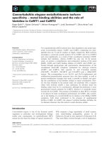
manuscript in preparation). Figure 1 shows three of
the inhibitors found. Compounds 1 and 2 originated
from the HTS, whereas compound 3 was discovered
using the crystal structure of WNV NS2B–NS3pro
with bound tetrapeptide [5] in an in silico screening
approach [10]. Compounds 1 and 2 showed inhibition
constants in the low micromolar range, but no related
compounds could be found with inhibition constants
below 1 lm (C. Bodenreider et al., manuscript in prep-
aration).
The results of two other published HTS efforts
confirmed that discovery of high-affinity inhibitors for
WNV NS2B–NS3pro is nontrivial. In one study,
competitive inhibitors with an inhibition constant of
3 lm were found and their binding to WNV NS2B–
NS3pro modelled [11]. In another, noncompetitive
inhibitors with IC
50
values of 0.1 lm were found, but
these were prone to hydrolysis with deactivation half-
lives of 1–2 h. The latter are thought to bind to
NS3pro, displacing the C-terminal b-hairpin of NS2B
from NS3pro [12]. HTS campaigns against the WNV
replicon, in which the target protein is unknown, also
failed to discover nonpeptidic inhibitors with inhibi-
tory activities much below 1 lm [13,14], with an EC
50
value of 0.85 lm being reported for the most active
compound [15].
In order to improve our understanding of the action
of compounds 1–3 against WNV NS2B–NS3pro, struc-
tural information about their binding modes must be
obtained. Despite many efforts, however, no crystal
structure of the protease could be determined in com-
plex with compounds 1–3 or any other low molecular
mass inhibitor. In view of the ability of NS2B to
undergo a large structural change between proteolyti-
cally deactivated and fully active states, as observed in
crystal structures [4,5], competitive inhibition may con-
ceivably be achieved by binding to an allosteric site
rather than to the active site. We therefore turned to
solution NMR spectroscopy to identify the binding
sites of 1–3 to WNV NS2B–NS3pro.
We have previously described a model of 3 bound
to WNV NS2B–NS3pro, obtained by automatic
computational docking, which is in agreement with the
Fig. 1. Synthetic inhibitors 1–3 of WNV NS2B–NS3pro studied.
Individual atoms are numbered as reference for NMR resonance
assignments.
X C. Su et al. NMR analysis of the West Nile virus protease
FEBS Journal 276 (2009) 4244–4255 ª 2009 The Authors Journal compilation ª 2009 FEBS 4245
intermolecular NOEs reported here [10]. Ekonomiuk
et al. [10] also presented the dissociation constant of 3
measured by NMR and, as additional proof for bind-
ing of 3 to the substrate-binding site, demonstrated
changes in cross-peak positions for residues lining the
substrate-binding site, without discussing the complete
resonance assignment.
In the following, we report protocols for the expres-
sion of isotope-labelled WNV NS2B–NS3pro in high
yields in Escherichia coli in vivo and by cell-free syn-
thesis, the first virtually complete assignments of the
15
N-HSQC spectrum, structure analysis of WNV
NS2B–NS3pro with bound inhibitor, and identification
of intermolecular NOEs between the inhibitors and the
protease.
Results
Sample preparation
The original construct of NS2B–NS3pro (construct 1,
Fig. 2) was toxic to E. coli, leading to cell lysis on
plates prepared with rich media as well as in large-
scale preparations. Improved protein yields were
obtained by a modified protocol, where E. coli colonies
grown on M9 media plates were selected prior to
large-scale expression. In this way, 9.3 mg of purified
uniformly
15
N ⁄
13
C-labelled protein were obtained per
litre of a
15
N ⁄
13
C-labelled rich medium (induction by
isopropyl b-d-thiogalactoside), whereas an autoinduc-
tion protocol [16] yielded as much as 59 mg of purified
15
N-labelled protein per litre of cell culture (Materials
and methods).
Construct 1 equally produced hardly any protein in
our cell-free protein synthesis system [17,18]. This
problem was overcome by construct 2 which starts
with the first six codons from T7 gene 10 and which
expresses well in cell-free systems. A clone in a high-
copy number T7 plasmid [19] facilitated the prepara-
tion of large quantities of DNA required for the
cell-free synthesis. Typical yields were close to 1 mg of
purified protein per mL of cell-free reaction mixture.
Although acceptable
15
N-HSQC spectra could be
recorded without purification of the protein [20,21],
complex formation with the inhibitors required puri-
fied protein because compounds 1 and 2 also bound to
components of the cell-free mixture.
The NS2B–NS3pro construct 1 in Fig. 2 was suscepti-
ble to gradual self-cleavage by the protease at two sites,
following the first glycine in the linker after Lys96
NS2B
and Lys15
NS3
(Fig. 2) [5,22], resulting in release of the
intermittent peptide from the protein. Because variable
extents of cleavage led to sample heterogeneity, later
work employed the mutant Lys96
NS2B
fi Ala (con-
struct 3) which prevented cleavage at either site [23]. The
K96A mutant turned out to be much less toxic to
E. coli, producing high yields even when overexpression
was induced by isopropyl b-d-thiogalactoside. The
K96A mutant retained full proteolytic activity in the
assay used (C. Bodenreider et al., manuscript in prepa-
ration) to measure the inhibition constant of different
ligands (data not shown).
Inhibitor binding monitored by NMR
spectroscopy
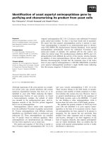
In the absence of inhibitors, assignment of the NMR
resonances for WNV NS2B–NS3pro was difficult
because many signals were broadened beyond detec-
tion and the spectral resolution was poor (Fig. 3A).
Over 100 different compounds that had been suggested
by high-throughput docking calculations with a large
library of molecules [10] or had appeared as hits in the
in vitro high-throughput screens were tested for bind-
ing to WNV NS2B–NS3pro by NMR spectroscopy
using
15
N-labelled protein. 1D
1
N NMR spectra were
used to assess any line broadening experienced by the
low molecular mass compounds and
15
N-HSQC spec-
tra were recorded to detect responses in the protein.
Most of the compounds showed broad lines in the
presence of protein without noticeably changing the
15
N-HSQC spectrum. This situation was interpreted as
nonspecific binding. Other compounds were barely sol-
uble in water. Compounds 1 and 2, however, improved
the
15
N-HSQC spectra of the protein dramatically
in a manner similar to compound 3. In addition to
Fig. 2. Amino acid sequence of the WNV NS2B–NS3pro constructs used. In addition to the sequence shown, constructs contained the
N-terminal sequences MGSSHHHHHHSSGLVPRGSHM (construct 1) or MASMTGHHHHHH (construct 2; Materials and methods). A third
construct (construct 3) contained the mutation Lys96
NS2B
fi Ala with N-terminal MASMTGHHHHHH peptide [WNV NS2B–NS3pro(K96A)].
All constructs ended at residue 187 of NS3. Vertical lines identify two autocatalyic cleavage sites [23]. The K96A mutation prevents
self-cleavage at either site. Residues without backbone resonance assignments (disregarding proline) are highlighted in orange.
NMR analysis of the West Nile virus protease X C. Su et al.
4246 FEBS Journal 276 (2009) 4244–4255 ª 2009 The Authors Journal compilation ª 2009 FEBS
improved spectral dispersion, the
15
N-HSQC spectra
of the complexes with 2 and 3 (Fig. S1) showed
marked similarities, indicating that both compounds
stabilize the same structure of the enzyme.
Compound 1 originated from the in vitro screen (C.
Bodenreider et al., manuscript in preparation). It was
the first found to improve the NMR spectrum of
WNV NS2B–NS3pro in a manner very similar to the
inhibitor benzoyl-norleucine-lysine-arginine-arginine-
aldehyde (Bz-nKRR-H) [24], which has been used for
crystallization [5]. Hence, the first resonance assign-
ments of the protease by 3D NMR spectroscopy were
performed using the complex with 1. Compound 2 was
designed to improve the solubility of 1 and lift its two-
fold symmetry in order to facilitate the assignment
of intermolecular NOEs. 2 bound to WNV NS2B–
NS3pro with similar affinity to 1 (IC
50
of 11 versus
25 lm) (C. Bodenreider et al., manuscript in prepara-
tion). Compound 3 inhibited WNV NS2B–NS3 by
35% when tested at 25 lm and had a K
d
value of
40 lm as measured by NMR [10].
Similar to 3 [10], as 1 or 2 were added to the enzyme
some of the
15
N-HSQC peaks shifted, indicative of
chemical shift averaging by chemical exchange on a
time scale of tens of milliseconds, whereas others
appeared at new positions, as expected for slow
exchange in the limit of large chemical shift differences
between the free and complexed protein (Fig. S2). The
15
N-HSQC spectra did not change significantly when
the inhibitors were used in excess.
Resonance assignments
The quality of the
15
N-HSQC spectra obtained in the
presence of 1, 2 or 3 was sufficient for sequential reso-
nance assignments using conventional triple-resonance
3D NMR experiments. NMR spectra of NS2B–
NS3pro and NS2B–NS3pro(K96A) were closely
similar, as expected for a point mutation in a mobile
segment of the polypeptide chain. Increased mobility
of the segment surrounding residue 96 in NS2B had
been suggested by the absence of electron density for
the linker peptide between NS2B and NS3 following
Asp90 in the crystal structure with BPTI [4] and was
confirmed by narrow NMR line shapes.
The resonances of the complex with 1 were assigned
using NS2B–NS3pro, whereas the 3D NMR experi-
ments of the complexes with 2 and 3 employed the
WNV NS2B–NS3pro(K96A) mutant. The resonance
assignments of the complexes with 1 and 3 were sup-
ported by combinatorial
15
N-labelling (Fig. S3). The
assignments of the backbone amide cross-peaks are
shown in Fig. S1. Resonance assignments were
obtained for the backbone amides of the segments
comprising residues 50–96 of NS2B and 17–187 of
NS3pro, with the exception of prolines and a few resi-
dues with very broad amide peaks. The resonances of
the peptide connecting NS2B and NS3pro appeared at
chemical shifts characteristic of random coil confor-
mation and were not assigned.
Conformation of WNV NS2B–NS3pro induced by
inhibitors
NOEs between NS2B and NS3pro observed for the
complex with 2 showed that NS2B docks to NS3pro
as in the crystal structures with peptidic inhibitors
(Table 1) [4–6]. Furthermore, the similarity of the
backbone amide chemical shifts seen in complexes with
1, 2 and 3 (Fig. S1) indicated that NS2B assumes the
same conformation in the presence of any of the three
compounds. The crystal structures of NS2B–NS3pro
A
B
Fig. 3.
15
N-HSQC spectra of WNV NS2B–NS3pro(K96A) in the
absence and presence of inhibitor 2 at 25 °C. The samples con-
tained 0.9 m
M protein in 90% H
2
O ⁄ 10% D
2
O containing 20 mM
Hepes buffer (pH 7.0) and 2 mM dithiothreitol. The complex with 2
was prepared by adding 15 lL of 100 m
M solutions of inhibitor in
d
6
-dimethylsulfoxide to the protein solution. The spectra were
recorded at a
1
N NMR frequency of 800 MHz. (A)
15
N-HSQC spec-
trum in the absence of inhibitor. (B)
15
N-HSQC spectrum in the
presence of compound 2 (3 m
M).
X C. Su et al. NMR analysis of the West Nile virus protease
FEBS Journal 276 (2009) 4244–4255 ª 2009 The Authors Journal compilation ª 2009 FEBS 4247
in complex with peptide inhibitors or BPTI [4–6]
are thus suitable starting points for modelling the
complexes with the low molecular mass inhibitors of
this study.
Inhibitor binding sites
Because the NMR spectra of the protease complexes
with 1 and 2 were very similar, both compounds must
bind in the same way. Therefore, we only studied the
binding of the nonsymmetric and more soluble com-
pound 2 using intermolecular NOEs. In the 1 : 1 com-
plex with the protease, the proton resonances of the
phthalazine ring of 2 were too broad to be observable.
(1 behaved in the same way.) Therefore, we used 2 in
an approximately three-fold excess over the protease in
order to measure intermolecular NOEs. The maximal
solubility of 2 in water was 3mm, but aggregation
occurred at much lower concentrations. Thus, even at
0.3 mm, the NMR line widths of 2 were broader than
expected for a monomeric compound (Fig. S4).
Furthermore, negative intramolecular NOEs were
observed for a sample at 0.7 mm, indicating an effec-
tive molecular mass of > 500 Da. The possibility of
self-association made it harder to interpret the inter-
molecular NOEs observed between the protease and 2.
Consequently, we used the NOE data with 3 to sup-
port the assignment of intermolecular NOEs with 2.
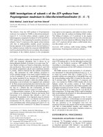
Figure 4 shows intermolecular NOEs observed
between WNV NS2B–NS3pro(K96A) and 3. Although
most NOEs could readily be assigned, the difficulty of
obtaining complete side-chain resonance assignments
for the protein prompted us to seek additional verifica-
tion that 3 binds to the substrate-binding site of the
protease.
In the first experiment, we compared the
15
N-HSQC
spectra of WNV NS2B–NS3pro(K96A) in the presence
of 3 and in the presence of the Bz-nKRR-H inhibitor
used in one of the crystal structure determinations [5].
As expected for closely related binding sites, the spec-
Table 1. NOEs observed between NS2B and NS3pro in the pres-
ence of 2 or 3.
NS2B NS3 Distance ⁄ A
˚
a
Trp53 H
N
Thr27 H
a
3.7
Ala58 H
N
Val22 H
N
3.1
Asp59 H
a
Val22 H
N
3.6
Ser72 H
a
Gly114 H
N
2.8
Arg74 H
a
Val115 H
N
2.6
Val77 H
N
Lys117 H
N
3.3
Gly83 H
N
Lys73 H
a
2.8
a
Distance in the crystal structure with tetrapeptide inhibitor
(2FP7) [5].
Fig. 4. 2D NOESY spectrum with
13
C(x
2
) ⁄
15
N(x
2
) half-filter of WNV NS2B–
NS3pro(K96A) in complex with 3. Parame-
ters: 0.9 m
M protein and 2 mM 3 in 90%
H
2
O ⁄ 10% D
2
O containing 20 mM Tris ⁄ HCl
buffer (pH 7.2) and 2 m
M dithiothreitol,
25 °C, mixing time 120 ms, t
1max
= 34 ms,
t
2max
= 86 ms, 800 MHz
1
N NMR
frequency. Intermolecular NOEs with the
aromatic ring protons of 3 are marked with
their assignments. Several of the NOEs are
also observed with the methyl groups of 3
at 2.3 p.p.m.
NMR analysis of the West Nile virus protease X C. Su et al.
4248 FEBS Journal 276 (2009) 4244–4255 ª 2009 The Authors Journal compilation ª 2009 FEBS
tra were very similar except for chemical shift changes
for some of the residues lining the substrate-binding
site (Fig. S5).
In another experiment, selectively
15
N-Gly-labelled
samples of WNV NS2B–NS3pro were prepared of the
wild-type protein and the Gly151Ala mutant. Gly151
is located in close proximity to the active-site histidine
residue and mutation to alanine should interfere with
both enzyme activity and with inhibitors that target
the substrate-binding site. Indeed, the G151A mutant
was inactive in the enzymatic assay [25] and unable to
bind 3 (Fig. S6).
Having established that compound 3 occupies the
substrate-binding site, we used the INPHARMA strat-
egy [26] to verify that compound 2 is also residing in
the substrate-binding site. A NOESY spectrum of 2
and 3 in the presence of a small quantity of protease
revealed an intermolecular cross-peak between the
methyl group of 3 and the phthalazine ring of 2,as
expected for an overlapping binding site (Fig. 5).
Table 2 compiles the intermolecular NOEs observed
with 2 and 3. The NOEs with Ile155 were most readily
assigned because of their characteristic chemical shifts,
whereas other NOEs were assigned using the assump-
tion that the protease fold was that observed in the
crystal structures with peptide inhibitors. The fact that
all intermolecular NOEs observed with the aromatic
ring proton of 3 were also observed with the methyl
group was, in most cases, probably a consequence of
spin-diffusion. Relaxation during the half-filter delays
and the twofold symmetry of 3 further impeded accu-
rate distance measurements.
The data show that both inhibitors are in proximity
of Thr132 and Ile155. There are, however, also signifi-
cant differences between the binding modes of the two
compounds. For example, 3 contacts the side chain of
His51 in the active site, whereas no equivalent interac-
tion could be found for 2. No intermolecular NOE
with NS2B could be observed because of the difficulty
of observing proton resonances of amino and guanidi-
nium groups.
Model building
Docking of compound 2 was performed automatically
by daim ⁄ seed ⁄ ffld [27–31] using the PDB coordinate
set 2FP7 [5], as described previously for 3 [10]. For
each compound, a total of 50 poses was kept upon
clustering. The pose which best satisfied the inter-
molecular NOEs (Table 2) was selected as the final
model. Not all cross-peaks observed for 2 (Table 2)
could be explained as direct NOEs with the protease.
This may be because of spin-diffusion during the mix-
ing time of the NOESY experiment, movements of the
ligand in the binding pocket or differences in side-
chain orientations between the crystal and solution
Fig. 5. 2D NOESY spectrum of 0.6 mM 2 and 0.5 mM 3 in the
presence of 0.03 m
M WNV NS2B–NS3pro(K96A) in D
2
Oat25°C.
Under these conditions, the signals of 2 were sufficiently narrow to
be observable (Fig. S4C). Other parameters: mixing time 150 ms,
t
1max
= 35 ms, t
2max
= 71 ms. The cross-peak between 3 H3 and 2
H6 or H6¢ is assigned as well as the intramolecular NOE between
3 H3 and H1.
Table 2. Intermolecular NOEs between West Nile virus (WNV)
NS2B–NS3pro(K96A) and inhibitors 2 and 3.
Protons of WNV NS3pro Compound 2
a
Compound 3
His51 H
d2
H1 and CH
3
Tyr130 H
d
H6 ⁄ H6¢
Thr132 C
!2
H
3
H6 ⁄ H6¢ H1 and CH
3
Thr132 H
a
H1 and CH
3
Thr134 C
!2
H
3
H6 ⁄ H6¢
Tyr150 H
d
H6 ⁄ H6¢
Asn152 C
b
H
2
H6 ⁄ H6¢
Gly153 H
N
H1
Val154 C
!
H
3
H1, H2, H5 ⁄ H5¢ H1 and CH
3
Ile155 C
d1
H
3
H1, H2, H3, H4 H1 and CH
3
Tyr161 H
d
H1 and CH
3
a
NOEs identified in Fig. 6 are underlined.
X C. Su et al. NMR analysis of the West Nile virus protease
FEBS Journal 276 (2009) 4244–4255 ª 2009 The Authors Journal compilation ª 2009 FEBS 4249
structure. [For example, the side chain of Ile155
is differently oriented in the structure with BPTI
(v
1
= )66°) [4] than in the structure used for Fig. 6
(v
1
= )180°) [5], and the intermolecular NOEs
observed with Ile155 are in much better agreement
with v
1
= )180° than v
1
= )66°.] In the case of
aggregation-prone compound 2, binding of more than
a single molecule may have confounded the interpreta-
tion of intermolecular NOEs. Nonetheless, the model
in Fig. 6A satisfies most NOEs. It places the positively
charged cyclic amidine group near the negatively
charged side chain of Asp129 which interacts with the
positively charged side chain of the P1 residues of
Bz-nKRR-H [5] and BPTI [4]. The primary amino
group of 2 points towards the C-terminal b-hairpin of
NS2B which carries three aspartate residues in a row
in positions 80–82. Although 2 belongs to a different
class of compounds than 3, the binding modes of both
compounds are not dissimilar (Fig. 6).
Discussion
Competitive inhibition is usually accepted as strong
indication that the binding sites of two inhibitors are
at least partially overlapping. In the case of the WNV
NS2B–NS3 protease, the C-terminal b-hairpin of
NS2B is essential for catalytic activity, but has been
found far away from the substrate-binding site in the
absence of inhibitor [4]. In addition, the substrate-
binding site changes significantly between the
A
B
Fig. 6. Stereoviews of models of 2 and 3
bound to WNV NS2B–NS3pro. The protein
structure is that by Erbel et al. [5], with
NS2B drawn as a grey ribbon. Heavy atom
representations of 2 and 3 are drawn in
black. The side chains of residues for
which intermolecular NOEs are reported in
Table 2 are shown in a stick representation.
(A) Complex with 2. Selected intermolecular
NOEs (Table 2) are highlighted with
magenta lines. (B) Complex with 3 reported
in Ekonomiuk et al. [10].
NMR analysis of the West Nile virus protease X C. Su et al.
4250 FEBS Journal 276 (2009) 4244–4255 ª 2009 The Authors Journal compilation ª 2009 FEBS
structures with and without inhibitor, so that competi-
tive inhibition may conceivably be achieved by binding
to a site that prevents NS2B from correct association
with the substrate-binding site. In this situation, NMR
spectroscopy provides an important tool for the identi-
fication of the inhibitor binding site.
No sequence-specific NMR resonance assignments
have been reported for the WNV NS2B–NS3 protease.
The poor quality of the NMR spectrum of WNV
NS2B–NS3pro in the absence of inhibitors is reminis-
cent of the situation in the homologous NS2B–NS3pro
construct from dengue virus type 2, in which selec-
tively
15
N ⁄
13
C-labelled samples show a great variation
in NMR line-width, prohibiting conventional assign-
ment strategies by multidimensional NMR spectro-
scopy [32]. The dramatic improvement in spectral
quality observed upon formation of complexes with
our inhibitors is readily explained by a shift in confor-
mational exchange equilibria towards a single con-
former. NOEs between NS2B and NS3 indicate that
this conformer is related to the conformation observed
in the crystal structures of the complex with peptidic
inhibitors [4–6], in which the C-terminal b-hairpin of
NS2B is positioned near the substrate-binding site
rather than far away as in the crystal structure in the
absence of inhibitor [4]. We were able to obtain this
result without optimized engineering of the NS2B part
that had been required to obtain an acceptable NMR
spectrum of the closely related dengue virus NS2B–
NS3 protease [33].
The NMR data clearly show that the small synthetic
inhibitors 1–3 bind to the substrate-binding site of
WNV NS2B–NS3pro. Competitive inhibition with
established peptide inhibitors is thus effected by direct
competition rather than by indirect competition via an
allosteric inactivation mechanism. Considering the
apparent ease with which the C-terminal b-hairpin of
NS2B is brought into the vicinity of the active site,
our results indicate that the crystal structures of the
protease–peptide complexes are valid starting points
for the search for low molecular mass inhibitors.
Indeed, compound 3 is the first inhibitor of WNV
NS2B–NS3pro that has been discovered by a computer
search using the crystal structure with a tetrapeptide
inhibitor as a template [5,10]. An important implica-
tion is that the only available crystal structure of the
corresponding dengue virus protease [5] is not a suit-
able starting point, because it positions the C-terminal
b-hairpin of NS2B far from the substrate-binding site.
Although compounds 1–3 induce a more uniform
structure of WNV NS2B–NS3pro, they are not able to
suppress all conformational exchange. For example,
we could not assign the backbone amides of Thr132,
Gly133 and Gly151 even in the presence of 1, 2 or 3,
and the backbone resonances of neighbouring residues
were broad. All three residues line the substrate-bind-
ing pocket. In order to find improved inhibitors, it is
thus relevant to explore the conformational space of
the protease in a molecular dynamics simulation rather
than relying exclusively on the structures observed
in the solid state. Intriguingly, the Thr132–Gly133
peptide bond was found to flip spontaneously in the
course of two 80-ns and one 40-ns molecular dynamics
simulations performed recently [34]. A flip of this
peptide bond also presents the main difference in
backbone conformation of the substrate-binding site
between the crystal structures 2IJO and 2FP7 [4].
The Gly
4
–Ser–Gly
4
linker connecting NS2B and
NS3pro is highly flexible in solution because the corre-
sponding signals appeared in an intense cluster of peaks
at a chemical shift characteristic of a random coil pep-
tide chain. Structural variability of these residues has
initially been suggested by the absence of electron den-
sity for the linker residues and the C-terminal residues
of NS2B following Asn89 in the WNV NS2B–
NS3pro(K96A) mutant in complex with BPTI [4]. Also,
the recent structure of the protease in complex with a
tripeptide inhibitor misses electron density for, respec-
tively, three or all of the residues of the Gly
4
–Ser–Gly
4
linker in the two conformers reported [6]. The high
mobility observed by NMR for the peptide linker in
solution provides a firm explanation for the finding that
the covalent linkage between NS2B and NS3 does not
restrain the function of the protease [2,9].
In conclusion, compounds 1 and 2 target the sub-
strate-binding site of the WNV NS2B–NS3 protease.
Their binding site overlaps with that of compound 3
(Fig. 6). Remarkably, even these small, nonpeptide
inhibitors can stabilize the conformation of NS2B
observed in crystal structures with peptides. This result
provides crucial validation for the use of computa-
tional approaches that start from the crystal structures
obtained with peptide inhibitors [10]. It also underpins
the success of further computations that, by taking
into account the conformations sampled by molecu-
lar dynamics simulations, led to nonpeptidic lead
compounds with low-micromolar affinity [35].
Materials and methods
Materials
Compounds 1 and 2 were synthesized in-house. Compound
3 was obtained from Maybridge (Tintagel, UK) (Cat#
S01870SC). Spectra 9 (
13
C,
15
N) media was obtained from
Spectra Stable Isotopes (Columbia, MD, USA).
15
NH
4
Cl,
X C. Su et al. NMR analysis of the West Nile virus protease
FEBS Journal 276 (2009) 4244–4255 ª 2009 The Authors Journal compilation ª 2009 FEBS 4251
13
C ⁄
15
N-Silantes (OD2) media,
15
N-glycine,
13
C ⁄
15
N-tyro-
sine and
13
C ⁄
15
N-phenylalanine were purchased from Cam-
bridge Isotope Laboratories (Andover, MA, USA). E. coli
strains Rosetta::kDE3 ⁄ pRARE and BL21 Star::kDE3
were obtained from Novagen (Gibbstown, NJ, USA) and
Invitrogen (Carlsbad, CA, USA), respectively. Synthetic
oligonucleotides were purchased from GeneWorks (Hind-
marsh, Australia). Sequences of oligonucleotides used are
listed in the Supporting Information. Vent DNA polymer-
ase and Phusion DNA polymerase were obtained from
New England BioLabs (Ipswich, MA, USA). Qiaquick
PCR purification and Qiaquick gel extraction kits were
purchased from Qiagen (Hilden, Germany).
Preparation of uniformly
15
N-labelled WNV
NS2B–NS3pro
The E. coli strain Rosetta::k DE3 ⁄ pRARE was transformed
with the plasmid pET15b–WNV CF40GlyNS3pro187 (con-
struct 1 of Fig. 2) [5] on Luria–Bertani plates containing
100 lgÆmL
–1
ampicillin and 50 lgÆmL
–1
chloramphenicol. A
single transformant colony (10
8
cells) was diluted with
Luria–Bertani media to 10
7
cells in 1 mL of Luria–
Bertani and 100 lL batches of the diluted cells were plated
on 15 M9 minimal media plates, containing 5 mm glucose,
0.2% (w ⁄ v) glycerol, 100 lgÆmL
–1
ampicillin and
50 lgÆmL
–1
chloramphenicol. Following growth for 2 days
at 37 °C, the colonies were collected and resuspended in
small volumes of M9 media. Approximately 100 D
595
units
of cells were used to inoculate 500 mL of
15
N-autoinduc-
tion media containing 0.5 gÆL
–1 15
NH
4
Cl, 100 lgÆmL
)1
ampicillin and 50 lgÆmL
)1
chloramphenicol [16]. Four con-
ical 2-L flasks, each containing 500 mL of
15
N-autoinduc-
tion cultures, were shaken at room temperature at 200 rpm
for 2 days up to an D
595
value of 5, yielding 16.6 g of
cells. The cells were suspended in 80 mL of buffer A
(50 mm Hepes, pH 7.5, 300 mm NaCl, 5% glycerol, 20 mm
imidazole) and lysed by a French press (12 000 psi, two
passes). After centrifuging the lysate at 15 000 g for 1 h,
the supernatant was filtered through a 0.45 lm Millipore
filter. The filtrate was directly loaded on a 5 mL Ni-NTA
column (Amersham Biosciences, Uppsala, Sweden). The
bound
15
N-WNV NS2B–NS3pro was eluted with an imid-
azole gradient of 20–500 mm in buffer A. The overall yield
of purified protein was 118 mg per 2 L of culture. The pro-
tein concentration was determined spectrophotometrically
at 280 nm, using a calculated e
280
value of 55 760 [36] and
the purity checked by SDS ⁄ PAGE.
For subsequent testing of different compounds by
15
N-HSQC spectra in 3 mm NMR tubes, the protein was
subdivided into over 100 batches of 200 lL each, contain-
ing 7 mgÆmL
)1
protein in NMR buffer [20 mm Hepes ⁄
KOH, pH 6.98, 90% H
2
O ⁄ 10% D
2
O, 1 mm tris(2-carboxy-
ethyl)phosphine or 2 mm dithiothreitol]. A sample was pre-
pared for each individual compound by injecting 3 lLof
100 mm solutions of compound in d
6
-dimethylsulfoxide into
200 lL of aqueous protein solution in a 3 mm NMR tube.
Preparation of uniformly
13
C/
15
N-labelled WNV
NS2B–NS3pro
13
C ⁄
15
N-labelled WNV NS2B–NS3pro was prepared using
the same protocol as for
15
N-labelled WNV NS2B–NS3pro,
except that 2 · 500 mL of
13
C ⁄
15
N-Silantes media (OD2)
were used which were supplemented with 100 lgÆmL
–1
ampicillin and 33 lgÆmL
)1
chloramphenicol. The cells were
grown at 37 °C and 200 r.p.m. for 6 h before induction
with 0.6 mm isopropyl b-d-thiogalactoside at D
595
= 0.95.
The induced cells were grown at room temperature over-
night to D
595
= 1.1, yielding 1.8 g of cells which were
suspended in 20 mL buffer A for purification as described
above. The final yield of
13
C ⁄
15
N-labelled protease was
9.3 mg in NMR buffer. The sample used for 3D NMR
experiments was 0.4 mm in protein in a 5 mm NMR tube.
Preparation of uniformly
13
C/
15
N-labelled WNV
NS2B–NS3pro(K96A)
A
13
C ⁄
15
N-labelled sample of the K96A mutant of WNV
NS2B–NS3pro (construct 3, Fig. 2) was prepared using the
same protocol as for
13
C ⁄
15
N-labelled WNV NS2B–NS3pro,
except that 2 · 500 mL of
13
C ⁄
15
N-Spectra 9 media was
used, which was supplemented with 100 lgÆmL
)1
ampicillin
and 50 lgÆmL
)1
chloramphenicol. Cells were grown at 37 °C
and 200 rpm for 3 h before induction with 0.6 mm isopropyl
b-d-thiogalactoside at D
595
= 1. The induced cells were
grown at room temperature overnight to D
595
= 1.9, yield-
ing 4.4 g of cells which were suspended in 50 mL buffer A
for purification on a 5 mL Ni-NTA column as described
above. Following elution from the column, the protein was
dialysed against 1 L of 50 mm Tris ⁄ HCl (pH 7.6). The
dialysate was loaded on a 7.4 mL DEAE-Toyopearl 650M
column (2.5 · 1.5 cm; Tosoh Bioscience, Montgomeryville,
PA, USA) and the bound protease eluted by a NaCl gradient
of 0 mm to 1 m in a buffer of 50 mm Tris ⁄ HCl (pH 7.6) and
1mm dithiothreitol. The final yield of
13
C ⁄
15
N-labelled
protease was 48.4 mg in NMR buffer. NMR samples were
0.9 mm in protein.
Cell-free synthesis of WNV NS2B–NS3pro
Construct 2 (Fig. 2) was designed for optimum expression
yields in a cell-free system. Primers 1307 and 1308
(Table S1) were used to amplify the protease gene by PCR
from the template plasmid pET15b-WNV CF40glyN-
S3pro187 using Phusion DNA polymerase. Following
digestion by NdeI and EcoRI, the PCR fragment was trans-
ferred into the corresponding site of the pRSET-5b vector
[19]. The resulting vector (pRSET-WNV MASMTGH
6
-
NMR analysis of the West Nile virus protease X C. Su et al.
4252 FEBS Journal 276 (2009) 4244–4255 ª 2009 The Authors Journal compilation ª 2009 FEBS
CF40glyNS3pro187) was used for cell-free protein synthesis
using a cell extract from E. coli.
S30 cell extracts were prepared from the E. coli strains
Rosetta::kDE3 ⁄ pRARE and BL21 Star::kDE3 as described
previously [17,18,37], including concentration with poly-
ethylene glycol 8000 [38] and heat treatment of the concen-
trated extracts at 42 °C [39].
Cell-free protein synthesis was performed for 6–7 h either
using an autoinduction system with plasmid pKO1166 for
in situ production of T7 RNA polymerase [40] or using
a standard protocol with purified T7 RNA polymerase
at 37 or 30 °C [18,21]. The reactions were performed
with lgÆmL
)1
target plasmid. Site-directed mutants were
produced from 5 to 10 lgÆmL
)1
PCR-amplified DNA tem-
plates. Following cell-free synthesis, the reaction mixtures
were clarified by centrifugation (30 000 g, 1 h) at 4 °C.
Cell-free synthesis of combinatorially
15
N-labelled
WNV NS2B–NS3pro
Five sets of
15
N-combinatorially labelled samples [41,42]
of construct 2 (Fig. 2) were produced by cell-free protein
synthesis. Synthesis was performed using 1 mL reaction
mixtures for sets 1–4 and 2 mL for set 5. Set 5 was the only
reaction containing
15
N-glutamate. This set was prepared
using 100 mm potassium succinate in the reaction mixture
instead of the usual 208 mm potassium glutamate buffer.
Cell-free protein synthesis was performed at 37 °C for 6 h.
Following centrifugation, the supernatants were diluted with
5–10 mL of buffer A and the proteins purified by a 1 mL
Ni-NTA column (Pharmacia) using a 20–500 mm imidazole
gradient in buffer A. The buffer of the samples was
exchanged to 20 mm Hepes ⁄ KOH (pH 7.0) and 1 mm tris(2-
carboxyethyl)phosphine using Millipore Ultra-4 centrifugal
filters (molecular mass cutoff 10 000), followed by concen-
tration to a final volume of 0.2 mL. D
2
O was added to a
final concentration of 10% (v ⁄ v) prior to NMR measure-
ments, resulting in a protein concentration of 50 lm.
Cell-free synthesis of
15
N-Gly labelled wild-type
and mutant WNV NS2B–NS3pro
Wild-type and mutant (Gly151Ala) samples of selectively
15
N-Gly labelled WNV NS2B–NS3pro (construct 2) were
produced by cell-free synthesis from cyclized PCR tem-
plates [32] using primers 1314, 1315 and 1131–1134
(Table S1). The synthesis was performed in 1 mL reaction
mixtures, using the same conditions and purification proto-
col as for the combinatorially labelled samples.
NMR measurements
All NMR spectra were recorded at 25 °C using Bruker 800
and 600 MHz Avance NMR spectrometers equipped
with TCI cryoprobes. Samples of complexes contained
an approximately three-fold excess of inhibitor in order to
facilitate the observation of intermolecular NOEs. 3D spec-
tra recorded included HNCA, HN(CO)CA, CC(CO)NH,
(H)CCH-TOCSY and NOESY-
15
N-HSQC (mixing time
60 ms). NOESY spectra with
13
C(x
2
) ⁄
15
N(x
2
) half-filters
(mixing time 120 ms) were used to suppress intramolecular
NOEs of the protease and observe intermolecular NOEs.
For unambiguous identification of intraligand NOEs, the
experiment was also recorded with a
13
C-BIRD sequence in
the middle of the mixing time which suppressed any NOE
from
13
C-bound protons of the protein. A 3D
13
C-HMQC-
NOESY spectrum with
13
C ⁄
15
N(x
2
) half-filter (mixing time
150 ms) facilitated the assignment of the intermolecular
NOEs by comparison with the (H)CCH-TOCSY spectrum.
The chemical shifts have been deposited in the BioMagRes-
Bank (accession number 11053).
Acknowledgements
This work was supported by the Australian Research
Council. Docking calculations were performed on the
Matterhorn computer cluster at the University of
Zu
¨
rich.
References
1 Hayes EB & Gubler DJ (2006) West Nile virus: epide-
miology and clinical features of an emerging epidemic
in the United States. Annu Rev Med 57, 181–194.
2 Chappell KJ, Stoermer MJ, Fairlie DP & Young PR
(2007) Generation and characterization of proteolyti-
cally active and highly stable truncated and full-length
recombinant West Nile virus NS3. Protein Expr Purif
53, 87–96.
3 Luo D, Xu T, Hunke C, Gruber G, Vasudevan SG &
Lescar J (2008) Crystal structure of the NS3 protease–
helicase from Dengue virus. J Virol 82, 173–183.
4 Aleshin AE, Shiryaev SA, Strongin AY & Liddington
RC (2007) Structural evidence for regulation and
specificity of flaviviral proteases and evolution of the
Flaviviridae fold. Protein Sci 16, 795–806.
5 Erbel P, Schiering N, D’Arcy A, Renatus M, Kroemer
M, Lim SP, Yin Z, Keller TH, Vasudevan SG &
Hommel U (2006) Structural basis for the activation of
flaviviral NS3 proteases from dengue and West Nile
virus. Nat Struct Mol Biol 13, 372–373.
6 Robin G, Chappell K, Stoermer MJ, Hu S, Young PR,
Fairlie DP & Martin JL (2009) Structure of West Nile
virus NS3 protease: ligand stabilization of the catalytic
conformation. J Mol Biol 385, 1568–1577.
7 Radichev I, Shiryaev SA, Aleshin AE, Ratnikov BI,
Smith JW, Liddington RC & Strongin AY (2008) Struc-
ture-based mutagenesis identifies important novel deter-
X C. Su et al. NMR analysis of the West Nile virus protease
FEBS Journal 276 (2009) 4244–4255 ª 2009 The Authors Journal compilation ª 2009 FEBS 4253
minants of the NS2B cofactor of the West Nile virus
two-component NS2B–NS3 proteinase. J Gen Virol 89,
636–641.
8 Chappell KJ, Stoermer MJ, Fairlie DP & Young PR
(2008) Mutagenesis of the West Nile virus NS2B cofac-
tor domain reveals two regions essential for protease
activity. J Gen Virol 89, 1010–1014.
9 Leung D, Schroder K, White H, Fang NX, Stoermer MJ,
Abbenante G, Martin JL, Young PR & Fairlie DP (2001)
Activity of recombinant dengue 2 virus NS3 protease in
the presence of a truncated NS2B co-factor, small
peptide substrates, and inhibitors. J Biol Chem 276,
45762–45771.
10 Ekonomiuk D, Su XC, Ozawa K, Bodenreider C,
Lim SP, Yin Z, Keller TH, Beer D, Patel V, Otting G
et al. (2009) Discovery of a non-peptidic inhibitor of
West Nile virus NS3 protease by high-throughput
docking. PLoS Negl Trop Dis 3, e356.
11 Mueller NH, Pattabiraman N, Ansarah-Sobrinho C,
Viswanathan P, Pierson TC & Padmanabhan R (2008)
Identification and biochemical characterization of small
molecule inhibitors of West Nile Virus serine protease
by a high throughput screen. Antimicrob Agents Chemo-
ther 52, 3385–3393.
12 Johnston PA, Phillips J, Shun TY, Shinde S, Lazo JS,
Huryn DM, Myers MC, Ratnikov BI, Smith JW, Su Y
et al. (2007) HTS identifies novel and specific uncom-
petitive inhibitors of the two-component NS2B–NS3
proteinase of West Nile virus. Assay Drug Dev Technol
5, 737–750.
13 Goodell JR, Puig-Basagoiti F, Forshey BM, Shi PY &
Ferguson DM (2006) Identification of compounds with
anti-West Nile virus activity. J Med Chem 49, 2127–2137.
14 Gu B, Ouzunov S, Wang L, Mason P, Bourne N,
Cuconati A & Block TM (2006) Discovery of small
molecule inhibitors of West Nile virus using a high-
throughput sub-genomic replicon screen. Antiviral Res
70, 39–50.
15 Noueiry AO, Olivo PD, Slomczynska U, Zhou Y,
Buscher B, Geiss B, Engle M, Roth RM, Chung KM,
Samuel M et al. (2007) Identification of novel small-
molecule inhibitors of West Nile virus infection. J Virol
81, 11992–12004.
16 Studier FW (2005) Protein production by auto-induc-
tion in high-density shaking cultures. Protein Expr Purif
41, 207–234.
17 Ozawa K, Dixon NE & Otting G (2005) Cell-free syn-
thesis of
15
N-labelled proteins for NMR studies.
IUBMB Life 57, 615–622.
18 Apponyi M, Ozawa K, Dixon NE & Otting G (2008)
Cell-free protein synthesis for analysis by NMR spec-
troscopy. In Methods in Molecular Biology, Vol. 426,
Structural Proteomics: High-throughput Methods (Kobe
B, Guss M & Huber T, eds), pp. 257–268. Humana
Press, Totowa, NJ.
19 Schoepfer R (1993) The pRSET family of T7 promoter
expression vectors for Escherichia coli. Gene 124, 83–85.
20 Guignard L, Ozawa K, Pursglove SE, Otting G &
Dixon NE (2002) NMR analysis of in vitro-synthesized
proteins without purification: a high-throughput
approach. FEBS Lett 524, 159–162.
21 Ozawa K, Headlam MJ, Schaeffer PM, Henderson BR,
Dixon NE & Otting G (2004) Optimization of an Esc-
herichia coli system for cell-free synthesis of selectively
15
N-labelled proteins for rapid analysis by NMR spec-
troscopy. Eur J Biochem 271, 4084–4093.
22 Shiryaev SA, Ratnikov BI, Chekanov AV, Sikora S,
Rozanov DV, Godzik A, Wang J, Smith JW, Huang Z,
Lindberg I et al. (2006) Cleavage targets and the
d-arginine-based inhibitors of the West Nile virus NS3
processing proteinase. Biochem J 393, 503–511.
23 Shiryaev SA, Aleshin AE, Ratnikov BI, Smith JW,
Liddington RC & Strongin AY (2007) Expression and
purification of a two-component flaviviral proteinase
resistant to autocleavage at the NS2B–NS3 junction
region. Protein Expr Purif 52, 334–339.
24 Yin Z, Patel SJ, Wang WL, Chan WL, Rao KRR,
Wang G, Ngew X, Patel V, Beer D, Knox JE et al.
(2006) Peptide inhibitors of dengue virus NS3 protease.
Part 2: SAR study of tetrapeptide aldehyde inhibitors.
Bioorg Med Chem Lett 16, 40–43.
25 Li J, Lim SP, Beer D, Patel V, Wen D, Tumanut C,
Tully DC, Williams JA, Jiricek J, Priestle JP et al.
(2005) Functional profiling of recombinant NS3 prote-
ases from all four serotypes of dengue virus using tetra-
and octa-peptide substrate libraries. J Biol Chem 280,
28766–28774.
26 Sa
´
nchez-Pedregal VM, Reese M, Meiler J, Blommers
MJJ, Griesinger C & Carlomagno T (2005) The IN-
PHARMA method: protein-mediated interligand NOEs
for pharmacophore mapping. Angew Chem Int Ed 44,
4172–4175.
27 Kolb P & Caflisch A (2006) Automatic and efficient
decomposition of two-dimensional structures of small
molecules for fragment-based high-throughput docking.
J Med Chem 49, 7384–7392.
28 Majeux N, Scarsi M, Apostolakis J, Ehrhardt C &
Caflisch A (1999) Exhaustive docking of molecular
fragments on protein binding sites with electrostatic
solvation. Protein Struct Funct Genet 37, 88–105.
29 Majeux N, Scarsi M & Caflisch A (2001) Efficient
electrostatic solvation model for protein-fragment
docking. Protein Struct Funct Genet 42, 256–268.
30 Budin N, Majeux N & Caflisch A (2001) Fragment-
based flexible ligand docking by evolutionary optimiza-
tion. Biol Chem 382, 1365–1372.
31 Cecchini M, Kolb P, Majeux N & Caflisch A (2004)
Automated docking of highly flexible ligands by genetic
algorithms: a critical assessment. J Comput Chem 25,
412–422.
NMR analysis of the West Nile virus protease X C. Su et al.
4254 FEBS Journal 276 (2009) 4244–4255 ª 2009 The Authors Journal compilation ª 2009 FEBS
32 Wu PSC, Ozawa K, Lim SP, Vasudevan SG, Dixon NE
& Otting G (2007) Cell-free transcription ⁄ translation
from PCR amplified DNA for high-throughput NMR
studies. Angew Chem Int Ed 46, 3356–3358.
33 Melino S, Fucito S, Campagna A, Wrubl F, Gamarnik
A, Cicero DO & Paci M (2006) The active essential
CFNS3d protein complex – a new perspective for the
structural and kinetic characterization of the NS2B–
NS3pro complex of dengue virus. FEBS J 273, 3650–
3662.
34 Ekonomiuk D & Caflisch A (2009) Activation of the
West Nile virus NS3 protease: molecular dynamics
evidence for a conformational selection mechanism.
Protein Sci 18, 1003–1011.
35 Ekonomiuk D, Su XC, Ozawa K, Bodenreider C, Lim
SP, Otting G, Huang D & Caflisch A (2009) Flaviviral
protease inhibitors identified by fragment-based library
docking into a structure generated by molecular
dynamics. J Med Chem (in press).
36 Gill SC & von Hippel PH (1989) Calculation of protein
extinction coefficients from amino acid sequence data.
Anal Biochem 182, 319–326.
37 Pratt JM (1984) Coupled transcription–translation in
prokaryotic cell-free systems. in Transcription and
Translation (Hames BD & Higgins SJ, eds), pp.
179–209. IRL Press, Oxford.
38 Kigawa T, Yabuki T, Yoshida Y, Tsutsui M, Ito Y,
Shibata T & Yokoyama S (1999) Cell-free production
and stable-isotope labelling of milligram quantities of
proteins. FEBS Lett 442, 15–19.
39 Klammt C, Lo
¨
hr F, Scha
¨
fer B, Haase W, Do
¨
tsch V,
Ru
¨
terjans H, Glaubitz C & Bernhard F (2004) High
level cell-free expression and specific labelling of
integral membrane proteins. Eur J Biochem 271,
568–580.
40 Ozawa K, Jergic S, Crowther JA, Thompson PR,
Wijffels G, Otting G & Dixon NE (2005) Cell-free
in vitro protein synthesis in an autoinduction system for
NMR studies of protein–protein interactions. J Biomol
NMR 32, 235–241.
41 Wu PSC, Ozawa K, Jergic S, Su XC, Dixon NE &
Otting G (2006) Amino-acid type identification in
15
N-HSQC spectra by combinatorial selective
15
N-label-
ling. J Biomol NMR 34, 13–21.
42 Ozawa K, Wu PSC, Dixon NE & Otting G (2006)
15
N-labelled proteins by cell-free protein synthesis: strat-
egies for high-throughput NMR studies of proteins and
protein–ligand complexes. FEBS J 273, 4154–4159.
Supporting information
The following supplementary material is available:
Fig. S1. Assigned
15
N-HSQC spectra of 0.9 mm solu-
tions of
15
N-labelled WNV NS2B–NS3pro(K96A) at
25 °C, pH 7.0, in the presence of 3 mm 2 or 3.
Fig. S2. Selected spectral region from
15
N-HSQC spec-
tra showing the effect of increasing concentrations
of 2 on the NMR spectrum of WNV NS2B–NS3pro
(K96A).
Fig. S3.
15
N-HSQC spectra of combinatorially
15
N-
labelled samples of WNV NS2B–NS3pro in the
presence of 1.
Fig. S4. 800 MHz 1D
1
H NMR spectra of the com-
pounds 2 and 3 in the absence and presence of WNV
NS2B–NS3pro(K96A) in D
2
O solution containing
1.5% d
6
-dimethylsulfoxide.
Fig. S5. Superimposition of
15
N-HSQC spectra of
0.3 mm WNV NS2B–NS3pro(K96A) in the presence
of 0.5 mm 3 or 0.2 mm 3 + 0.4 mm Bz-nKRR-H.
Fig. S6. Superimposition of
15
N-HSQC spectra of
0.1 mm solutions of selectively
15
N-Gly labelled WNV
NS2B–NS3pro(G151A) in the absence and presence of
0.2 mm 3.
Table S1. PCR primers used in this study to produce
different variants of WNV NS2B–NS3pro.
This supplementary material can be found in the
online article.
Please note: As a service to our authors and readers,
this journal provides supporting information supplied
by the authors. Such materials are peer-reviewed and
may be re-organized for online delivery, but are not
copy-edited or typeset. Technical support issues arising
from supporting information (other than missing files)
should be addressed to the authors.
X C. Su et al. NMR analysis of the West Nile virus protease
FEBS Journal 276 (2009) 4244–4255 ª 2009 The Authors Journal compilation ª 2009 FEBS 4255