Báo cáo khoa học: Subcellular compartmentalization of FADD as a new level of regulation in death receptor signaling pdf
Bạn đang xem bản rút gọn của tài liệu. Xem và tải ngay bản đầy đủ của tài liệu tại đây (836.29 KB, 10 trang )
Subcellular compartmentalization of FADD as a new level
of regulation in death receptor signaling
Niko Fo
¨
ger
1
, Silvia Bulfone-Paus
1
, Andrew C. Chan
2
and Kyeong-Hee Lee
1
1 Department of Immunology and Cell Biology, Research Center Borstel, Leibniz Center for Medicine and Biosciences, Germany
2 Department of Immunology, Genentech, Inc., San Francisco, CA, USA
Introduction
CD95 (Fas ⁄ Apo-1 ⁄ TNFRSF6) is a prototypic death
receptor belonging to the tumor necrosis factor recep-
tor superfamily. CD95 is expressed on the surface of
cells as preassociated homotrimers and, upon CD95L
binding, undergoes a conformational change to reveal
its cytoplasmic death domain (DD) to favor homotypic
interactions with other DD-containing proteins. Fas-
associated protein with DD (FADD) is the most proxi-
mal adaptor molecule transmitting the death signal
mediated by CD95 [1]. As a DD-containing and
death effector domain-containing proapoptotic adaptor
molecule, FADD is essential to recruit the initiator
caspases-8 and -10 to instigate formation of the death-
inducing signal complex (DISC), which mediates
death receptor-induced apoptosis [2,3]. Expression of a
dominant-negative form of FADD, consisting of the
N-terminal DD only, impairs the relay of the apoptotic
signal from death receptors [4]. Moreover, FADD-
deficient mice display profound defects in apoptotic
pathways, particularly in the immune system [5]. FADD
is a multifunctional protein that, in addition to its
prominent role in cell death, has also been implicated
in the regulation of cell survival ⁄ proliferation and
cell cycle progression, as well as embryonic develop-
ment [5–7].
In our previous work, we demonstrated that CD95
internalization plays a role in CD95-induced apoptosis
[8]. Upon ligand binding, CD95 is internalized and
delivered to endosomal compartments, which then
serve as major sites for CD95-mediated DISC forma-
tion and caspase-8 activation. Given that the key role
of FADD in apoptotic signaling is efficient DISC
Keywords
apoptosis; CD95; compartmentalization;
FADD; nuclear trafficking
Correspondence
K H. Lee, Department of Immunology and
Cell Biology, Research Center Borstel,
Leibniz Center for Medicine and
Biosciences, Parkallee 22, 23845 Borstel,
Germany
Fax: +49 4537 1884904
Tel: +49 4537 188585
E-mail:
(Received 30 April 2009, accepted 4 June
2009)
doi:10.1111/j.1742-4658.2009.07134.x
Fas-associated protein with death domain (FADD) is an essential adaptor
protein in death receptor-mediated signal transduction. During apoptotic
signaling, FADD functions in the cytoplasm, where it couples activated
receptors with initiator caspase-8. However, in resting cells, FADD is pre-
dominantly stored in the nucleus. In this study, we examined the modalities
of FADD intracellular trafficking. We demonstrate that, upon CD95 acti-
vation, FADD redistributes from the nucleus to the cytoplasm. This induc-
ible nuclear–cytoplasmic translocation of FADD is independent of CD95
internalization, formation of the death-inducing signaling complex, and
caspase-8 activation. In contrast to nuclear export of FADD, its subse-
quent recruitment and accumulation at endosomes containing internalized
CD95 requires a caspase-8-dependent feedback loop. These data indicate
the existence of differential pathways directing FADD nuclear export and
cytoplasmic trafficking, and identify subcellular compartmentalization of
FADD as a novel regulatory mechanism in death receptor signaling.
Abbreviations
BFA, brefeldin A; DAPI, 4¢,6-diamidino-2-phenylindole; DD, death domain; DISC, death-inducing signaling complex; EEA-1, early endosome
antigen 1; FADD, Fas-associated protein with death domain; GFP, green fluorescent protein; INP54p, Saccharomyces cerevisiae inositol
polyphosphate 5-phosphatase; PtdIns(4,5)P
2,
phosphatidylinositol 4,5-bisphosphate.
4256 FEBS Journal 276 (2009) 4256–4265 ª 2009 The Authors Journal compilation ª 2009 FEBS
assembly at endosomal structures, FADD is expected
to function within the cytoplasm. However, FADD
carries strong nuclear localization and nuclear export
signals, and has been reported to primarily localize to
the nucleus in a variety of different cell types
[9–12]. This raises the question of how a predomi-
nantly nuclear protein such as FADD is involved in
DISC formation occurring at endosomes in the
cytoplasm.
Here, we demonstrate that CD95 stimulation
induces translocation of nuclear FADD to the cyto-
plasm. Employing a combination of biochemical, cell
biological and genetic methods, we investigated the
role of ‘classic’ apoptotic signal transduction events in
the nuclear–cytoplasmic relocalization of FADD and
its subsequent recruitment to endosomal compart-
ments, where FADD promotes efficient DISC forma-
tion. The regulation of the subcellular localization of
FADD adds a new level of complexity to the apoptotic
signaling cascade.
Results
Nuclear–cytoplasmic redistribution of FADD in
response to CD95L stimulation
To explore whether FADD shuttles between the
nucleus and the cytoplasm in response to an apoptotic
stimulus, we analyzed the subcellular distribution of
FADD in resting versus CD95L-treated BJAB cells, a
human B-cell Burkitt’s lymphoma cell line (Fig. 1A).
In agreement with previous reports on other cell lines,
FADD colocalizes with the nuclear stain 4¢,6-diamidi-
no-2-phenylindole (DAPI) in resting BJAB cells, as
well as in human peripheral blood CD4
+
T-lympho-
cytes, indicating preferential nuclear localization of
FADD (Fig. 1A,B, left panels). In response to CD95
receptor triggering, however, FADD redistributed
from a predominantly nuclear to a nuclear and cyto-
plasmic pattern. In BJAB cells, within 5 min of
CD95L treatment, a significant proportion of FADD
relocalized from the nucleus to the cytoplasm and
exhibited dispersed fine punctuate patterns in the cyto-
plasm (Fig. 1A, middle panel). These structures
became more pronounced and enlarged at 15–30 min
after CD95L stimulation (Fig. 1A, right panel). A
similar redistribution of FADD was also observed
in human peripheral blood CD4
+
T-lymphocytes
(Fig. 1B, right panel).
These observations indicate that FADD undergoes
regulated redistribution from the nucleus to the cyto-
plasm in response to CD95 triggering. Notably, we did
not observe recruitment of FADD to the plasma mem-
brane, but, instead, FADD relocalized to vesicular
structures in the cytoplasm. This specific vesicular
localization of FADD is probably due to functional
association of FADD with internalized CD95, which
predominantly occurs at endosomal compartments and
constitutes an essential step in CD95-mediated apop-
totic signaling [8].
0 minCD95L: 15 min
FADD
B
FADD/DAPI
0 minCD95L: 5 min
15 min
A
FADD/DAPI
FADD
Fluorescence intensity
Low
High
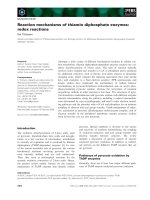
Fig. 1. Nuclear–cytoplasmic translocation of
FADD in response to CD95 stimulation. (A)
BJAB cells were stimulated with CD95L for
the indicated times. Cells were stained for
FADD (red), and nuclei were counterstained
with DAPI (blue). Overlay fluorescence is
shown in the upper panel. Quantitative
image analysis with relative pixel intensities
recorded for FADD fluorescence signals is
shown in the lower panel. (B) Activated
human peripheral blood CD4
+
T-cells were
stimulated with CD95L for 15 min. Single
FADD staining (upper panel, red) and overlay
fluorescence (lower panel) of FADD and
DAPI are shown. Fluorescence images were
generated by deconvolution microscopy.
The data shown are representative of
> 150 cells analyzed.
N. Fo
¨
ger et al. FADD trafficking and CD95 signaling
FEBS Journal 276 (2009) 4256–4265 ª 2009 The Authors Journal compilation ª 2009 FEBS 4257
Expression of a plasma membrane-localized
phosphatidylinositol 4,5-bisphosphate
[PtdIns(4,5)P
2
]-specific 5¢-phosphatase inhibits
CD95 endocytosis and apoptosis, but not the
nuclear–cytoplasmic translocation of FADD
As FADD translocation from the nucleus to the cyto-
plasm occurred within 2–5 min following CD95L stim-
ulation, prior to significant CD95 internalization, we
analyzed whether FADD translocation required CD95
internalization. To this end, we utilized Saccharomy-
ces cerevisiae inositol polyphosphate 5-phosphatase
(INP54p), an enzyme that hydrolyzes PtdIns(4,5)P
2
to
phosphatidylinositol 4-phosphate [13]. Cellular levels of
PtdIns(4,5)P
2
are tightly regulated, and it plays impor-
tant roles in a multitude of cellular functions, including
clathrin-mediated endocytosis [14–16]. Expression of a
green fluorescent protein (GFP)-tagged plasma mem-
brane-targeted INP54p (FynC–GFP–INP54p) in BJAB
cells specifically reduces PtdIns(4,5)P
2
levels in the
plasma membrane, and results in the inhibition of
CD95L-induced CD95 receptor endocytosis and apop-
tosis [8] (Fig. 2A,B). BJAB cells transfected with
FynC–GFP–INP54p did, however, still relocalize
FADD from the nucleus to the cytoplasm in response
to CD95L stimulation (Fig. 2C). Whereas the overall
degree of the CD95L-induced FADD nuclear–cytoplas-
mic translocation was similar between FynC–GFP–
INP54p
+
cells and control cells, the pattern of FADD
staining was qualitatively distinct in FynC–GFP–
INP54p
+
cells. At 15 min following CD95 activation,
FynC–GFP–INP54p
+
cells (Fig. 2C, middle panel)
showed only a diffuse staining pattern of cytoplasmic
FADD and did not exhibit the intense coalescence of
FADD with larger endocytic structures that is observed
in FynC–GFP–INP54p
)
cells (Fig. 2C, right panel).
This may reflect a lack of internalized CD95 to concen-
trate FADD within endocytic vesicles.
0
CD95L: 0 min 15 min 15 min
10
20
30
40
50
60
70
80
90
100
GFP – GFP + GFP – GFP +
FynC–GFP–INP54 FynC–GFP
A
10
0
10
1
10
2
10
3
10
4
10
0
10
1
10
2
10
3
10
4
FynC–GFP+
FynC
–GFP–INP54+
Annexin V
Cell count
B
CD95 internalization (%)
C
GFP
FADD
DAPI
FADD
Low
Hi
g
h
Fluorescence intensity
6
5
4
3
1
2
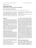
Fig. 2. FADD translocation into the cytoplasm is independent of CD95 internalization. (A, B) BJAB cells transiently expressing FynC–GFP–
INP54p, a PtdIns(4,5)P
2
-specific 5¢-phosphatase–GFP fusion construct, or the control construct FynC–GFP were analyzed for CD95 internali-
zation (A) and apoptosis (B) following CD95L stimulation for 30 min (A) and 6 h (B), respectively. (A) The remaining surface CD95 was
detected by FACS analysis, and the percentage of CD95 downregulation was calculated for the GFP
+
and GFP
)
populations. (B) Apoptosis
in GFP
+
(red) cells was assessed by annexin V staining and FACS analysis. Nonstimulated cells are shown in gray. The data shown are
representative of three experiments. (C) BJAB cells transiently transfected with FynC–GFP–INP54p were stimulated with CD95L for 0 min
(1, 4) and 15 min (2, 3, 5, 6). Panels 1, 2, 4, 5 show FynC–GFP–INP54p-expressing cells (GFP
+
), and FynC–GFP–INP54p-non-expressing
cells are shown in the right panel (3, 6). Cells were stained for DAPI (blue) and FADD (red). Overlay fluorescence is shown in the upper
panel (1–3), and quantitative image analysis of CD95 fluorescence signals is shown in the lower panel (4–6). The data shown are representa-
tive of > 50 cells analyzed.
FADD trafficking and CD95 signaling N. Fo
¨
ger et al.
4258 FEBS Journal 276 (2009) 4256–4265 ª 2009 The Authors Journal compilation ª 2009 FEBS
CD95 internalization promotes endosomal
targeting of FADD
To investigate whether internalized CD95 provides a
docking signal to recruit FADD to endosomes, we
analyzed the subcellular localization of FADD and
CD95 in CD95L-activated FynC–GFP–INP54p
+
and
FynC–GFP–INP54p
)
BJAB cells. Following stimula-
tion for 30 min with CD95L, colocalization of cyto-
plasmic FADD with internalized CD95 was readily
detected at intracellular compartments in FynC–
GFP–INP54p
)
cells (Fig. 3A, panels 5–8). In con-
trast, in CD95-activated but endocytosis-defective
FynC–GFP–INP54p
+
BJAB cells, CD95 had formed
microaggregates in the plasma membrane, and no
significant colocalization between FADD and CD95
was observed, although FADD could be readily
detected in the cytoplasm (Fig. 3A, panels 1–4).
There was minimal overlap of staining for FADD
with the early endosome marker early endosome
antigen 1 (EEA-1) in resting cells (Fig. 3B, panels
1–3). Overlap of staining for FADD and EEA-1
was, however, readily detected in CD95L-stimulated
control FynC–GFP–INP54p
)
cells (Fig. 3B, panels
9–11), whereas in FynC–GFP–INP54p
+
BJAB cells,
FADD largely failed to accumulate at EEA-1
+
en-
dosomes (Fig. 3B, panels 5–8).
An internalization-defective CD95 mutant
disrupts apoptotic signaling but still induces
FADD nuclear–cytoplasmic translocation
To further analyze the interrelationship between CD95
internalization and FADD nuclear–cytoplasmic relo-
calization, we specifically interfered with CD95
receptor endocytosis by employing the internalization-
defective CD95(Y291F) mutant [8]. The ability of this
mutant form of CD95, in which Tyr291 within the
consensus AP-2-binding motif of CD95 has been
mutated to Phe, to internalize in murine A20 B-lym-
phoma cells following stimulation with a mAb against
human CD95 (CH-11) was significantly reduced as
compared to wild-type CD95 (Fig. 4A). Concomi-
tantly, the ability of CD95(Y291F)-expressing cells to
activate caspase-8 in response to CD95 stimulation
was similarly compromised (Fig. 4C). However, despite
the relative inability of CD95(Y291F) to internalize
and to induce classic proximal apoptotic signaling
B
FADD
EEA-1
FADD / EEA-1 FADD / DAPI
1
2
3
8
4
7
6
5
9
10
11
12
CD95L
0 min
(GFP +)
30 min
(GFP +)
30 min
(GFP–)
A
GFP
CD95
FADD
CD95 / FADD
4
3
2
1
CD95L
30 min
30 min
8
7
6
5
Fig. 3. CD95 internalization promotes
endosomal targeting of FADD. (A) BJAB
cells were transfected with FynC–GFP–
INP54p and stimulated with CD95L for
30 min. Cells were stained for CD95 (red)
and FADD (blue). Panels 1–4 represent a
FynC–GFP–INP54p-expressing (GFP
+
) cell,
and panels 5–8 show a FynC–GFP–INP54p-
non-expressing (GFP
)
) cell. The data shown
are representative of > 50 cells analyzed.
(B) BJAB cells were transfected with
FynC–GFP–INP54p and stimulated with
CD95L for 0 min (1–4) and 30 min (5–12).
Cells were stained for FADD (green), EEA-1
(red), and DAPI (blue). Individual and
merged fluorescence images were obtained
by deconvolution microscopy. FynC–GFP–
INP54p-expressing cells (GFP
+
) are shown
in panels 1–8, and a FynC–GFP–INP54p-non-
expressing cell (GFP
)
) is shown in pan-
els 9–12. The data shown are representative
of > 100 cells analyzed.
N. Fo
¨
ger et al. FADD trafficking and CD95 signaling
FEBS Journal 276 (2009) 4256–4265 ª 2009 The Authors Journal compilation ª 2009 FEBS 4259
events, stimulation of CD95(Y291F) still induced
nuclear–cytoplasmic relocalization of FADD (Fig. 4A,B).
FADD was preferentially localized within the nucleus
of resting cells expressing CD95(Y291F). In response
to CD95 stimulation, FADD exhibited a nuclear and
cytoplasmic distribution in cells expressing either wild-
type CD95 or CD95(Y291F). However, whereas in
wild-type human CD95-expressing cells FADD
concentrated and colocalized with internalized CD95
at EEA-1-positive endosomal compartments, in cells
expressing the internalization mutant CD95(Y291F)
FADD remained in a diffuse cytoplasmic pattern and
showed no significant colocalization with EEA-1. The
data on the nuclear–cytoplasmic relocalization of
FADD, as observed by deconvolution microscopy,
were further confirmed by biochemical subcellular frac-
tionation experiments. Little to no FADD protein was
detected in the cytoplasmic fraction of nonstimulated
cells transfected with either wild-type human CD95 or
CD95(Y291F) (Fig. 4D, lanes 3 and 6). Triggering of
human CD95 for 15–30 min induced a significant
increase in the amount of FADD in the cytoplasmic
fraction of cells expressing wild-type CD95 (Fig. 4D,
lanes 4 and 5). A similar increase in cytoplasmic
FADD was also observed in CD95(Y291F)-expressing
cells stimulated with antibody against human CD95
(Fig. 4D, lanes 7 and 8).
Together, these data indicate that CD95L-induced
FADD translocation to the cytoplasm occurs indepen-
dently of CD95 internalization. However, internalized
CD95 then probably serves as a scaffold to amplify
and ⁄ or stabilize FADD assembly at endosomal com-
partments.
Inhibition of caspase-8 activation allows for
transient nuclear–cytoplasmic shuttling of FADD
and results in the recycling of CD95
To further investigate whether inhibition of apoptotic
signaling affects the subcellular localization of CD95
and ⁄ or FADD, BJAB cells were treated with the
caspase-8 inhibitor z-IETD, and FADD localization
B
Y291F
Y291F
WT
FADD EEA-1 FADD/EEA-1
1 2 3
6 5 4
7 8 9
C
WT Y291F
WB:
Cas-8
hCD95
CD95 : 0’ 15’ 30’ 60’ 0’ 15’ 30’ 60’
CD95:
0’ 0’ 0’
15’
30’
30’
0’
15’
1 2 3 4 5 6 7 8
WB:
FADD
Laminin
GDI-Rho
WT Y291F WT Y291F
Nuclear Cytoplasmic
D
1 2 3 4 5 6 7 8
A
Y291F
WT
FADD CD95 CD95/FADD
1
2 3
6 5 4
Fig. 4. Cytoplasmic translocation of FADD in cells expressing the
internalization mutant of human CD95 (hCD95). (A) A20 cells
expressing the internalization mutant hCD95(Y291F) (1–3) or wild-
type (WT) hCD95 (4–6) were activated for 30 min with mAb against
hCD95 (CH-11). Cells were subsequently stained for FADD (green),
CD95 (red), and DAPI (blue). (B) A20 cells expressing
hCD95(Y291F) (1–6) or wild-type hCD95 (7–9) were activated with
CH-11 for 0 min (1–3) or 30 min (4–9). Cells were stained for FADD
(green), EEA-1 (red), and DAPI (blue). Images were obtained by
deconvolution microscopy. The data shown are representative of
> 60 cells analyzed. (C) A20 cells were transfected with wild-type
hCD95 or hCD95(Y291F) and stimulated with biotinylated CH-11 for
the indicated times. Human CD95-associated signaling complexes
were isolated using streptavidin-conjugated beads. Association of
caspase-8 with hCD95 was analyzed by immunoblotting for cas-
pase-8 and hCD95. (D) A20 cells were transfected with wild-type
hCD95 or hCD95(Y291F) and stimulated with CH-11 for the indi-
cated times. Nuclear (lanes 1 and 2) and cytoplasmic (lanes 3–8)
fractions were prepared from total cellular lysates and were immu-
noblotted using antibody against FADD. Effective separation of
nuclear and cytoplasmic fractions was controlled for by immuno-
blotting for laminin (nuclear marker) and GDI-Rho (cytosolic
marker).
FADD trafficking and CD95 signaling N. Fo
¨
ger et al.
4260 FEBS Journal 276 (2009) 4256–4265 ª 2009 The Authors Journal compilation ª 2009 FEBS
was investigated. In unstimulated BJAB cells either
treated or not treated with the caspase-8 inhibitor z-
IETD, FADD was predominantly detected in the
nucleus (Fig. 5A, panels 1–3 and 13–15). Within 2 min
of stimulation with CD95L, FADD could readily be
detected in the cytoplasm of z-IETD-treated cells
(Fig. 5A, panels 16–18), as in control cells. In
untreated control cells, FADD remained in the cyto-
plasm after 30 and 60 min of CD95 stimulation, and
cells started to exhibit signs of apoptosis (Fig. 5A,
panels 7–12). In contrast, in z-IETD-treated cells,
which do not undergo apoptosis, significant amounts
of cytoplasmic FADD could only be detected within
30 min of CD95L stimulation (Fig. 5A, panels 19–20).
At 60 min, only minimal amounts of FADD had
remained in the cytoplasm of z-IETD-treated cells
(Fig. 5A, panels 22–24). Thus, inhibition of caspase-8
activation does not affect the initial nuclear–cytoplas-
mic translocation of FADD; however, FADD relocal-
ization to the cytoplasm is not persistent under these
conditions. Whether, in the absence of caspase-8 acti-
vation, FADD shuttles back to the nucleus or is
degraded in the cytoplasm remains to be investigated.
As treatment of cells with caspase inhibitors has
been reported to be required for CD95 internalization
following receptor activation [17], we next analyzed the
kinetics with which caspase inhibition may affect
receptor internalization. Treatment of BJAB cells with
the inhibitors z-IETD (caspase-8 selective), z-VAD (a
general caspase inhibitor) or z-DEVD (caspase-3 selec-
tive) did not affect ligand-mediated CD95 internaliza-
tion at 15 min and had moderate effects at 30 min as
compared to untreated cells (Fig. 5B,C). Between
30 min and 60 min, control cells further downregulated
CD95, whereas in cells treated with caspase inhibitors
an increase in CD95 surface expression was observed.
These kinetics were further supported by microscopy
studies, in which CD95 was detected within the cyto-
plasm within 30 min following CD95L stimulation,
even in the presence of z-IETD (Fig. 5A, panel 20). At
60 min following CD95L stimulation, when CD95 had
maximally internalized and cells already demonstrated
morphological changes associated with apoptosis
(Fig. 5A, panel 11), CD95 was detected almost exclu-
sively at the cell surface in cells treated with caspase
inhibitors (Fig. 5A, panel 23; Fig. 5B,C), as previously
reported [17].
To analyze the potential contributions of CD95
recycling to the plasma membrane, cells were treated
with brefeldin A (BFA), a fungal metabolite that
blocks protein transport from the endoplasmic reticu-
lum to the Golgi and protein recycling, in the presence
or absence of z-VAD. Whereas cells incubated with
z-VAD alone again demonstrated significant down-
regulation of surface CD95 expression at 30 min
followed by an increase at 60 min, cells treated with
z-VAD and BFA continued to downregulate CD95
without any subsequent increase in surface CD95
expression (Fig. 5D,E). Thus, CD95 internalization
following receptor engagement is not dependent on
caspase activation, and a significant proportion of the
surface expression of CD95 observed at 30 min and
60 min following receptor engagement in the presence
of caspase inhibitors appears to be a consequence of
CD95 receptor recycling when cells are unable to
undergo apoptosis. Microscopic analysis of CD95-
stimulated cells treated with both BFA and the cas-
pase-8 inhibitor z-IETD showed that CD95 largely
accumulated in the cytoplasm and significant amounts
of FADD localized to the cytoplasm, but CD95 and
FADD failed to interact with each other under these
conditions (Fig. 5F). These data indicate that nuclear–
cytoplasmic shuttling of FADD is independent of cas-
pase-8 activity. Further recruitment of FADD to
CD95-containing endosomal compartments, however,
seems to require an activation loop involving active
caspase-8.
Discussion
FADD is an essential adaptor protein in the CD95-
mediated apoptotic signaling cascade that couples
activated receptors with the activation of initiator
caspase-8 [1,18,19]. Here, we demonstrate that, in
response to CD95 receptor activation, a significant
amount of FADD relocalizes from the nucleus to the
cytoplasm.
Our data indicate that CD95 receptor triggering
induces membrane proximal signals to induce nuclear
export of FADD that are independent of CD95 inter-
nalization and ‘classic’ apoptotic signaling events, such
as DISC formation and caspase-8 activation. We
employed two different experimental systems to inhibit
CD95 internalization: modulation of PtdIns(4,5)P
2
levels by INP54p, and the internalization mutant
CD95(Y291F). In these systems, CD95-induced DISC
formation, caspase-8 activation and apoptosis are
severely compromised [8], whereas CD95 triggering still
induces translocation of FADD from the nucleus to the
cytoplasm. Subsequent recruitment and concentration
of FADD to endosomal compartments, where DISC is
stabilized and amplified, however, requires CD95 inter-
nalization. Consequently, in endocytosis-defective cells,
FADD did not accumulate at endosomal structures in
response to CD95 stimulation, but exhibited more dif-
fuse localization in the cytoplasm. Thus, internalized
N. Fo
¨
ger et al. FADD trafficking and CD95 signaling
FEBS Journal 276 (2009) 4256–4265 ª 2009 The Authors Journal compilation ª 2009 FEBS 4261
0
10
20
30
40
50
60
70
80
90
0 204060
No inhibitor
z-IETD (Cas-8)
z-VAD (general)
z-DEVD (Cas-3 & Cas-7)
(min)
MFI
No inhibitor
z-IETD
z-VAD
z-DEVD
Cell count
CD95
0 min
5 min
30 min
CD95
Cell count
z-VAD
z-VAD
+ BFA
0 min
5 min
30 min
BFA
No inhibitor
0
20
40
60
80
100
120
0 204060
No inhibitor
BFA
z-VAD
z-VAD + BFA
(min)
MFI
FADD CD95 CD95/FADD
CD95L (30 min)
z-IETD
BFA
CD95L (30 min)
BFA
z-IETD (caspase-8 inhibitor)No inhibitor
CD95L
A
BC
D
E
F
0 min
2 min
30 min
60 min
FADD
CD95 CD95/FADD
FADD
CD95 CD95/FADD
123
456
789
10 11 12
13 14 15
16 17 18
19 20 21
22 23 24
Fig. 5. FADD translocation is independent of caspase-8 activation. (A) BJAB cells were stimulated with CD95L for the indicated times in the
absence (left, 1–12) or presence (right, 13–24) of 50 l
M caspase-8 inhibitor z-IETD. Cells were stained for FADD (green), CD95 (red), and
DAPI (blue). Images were obtained by deconvolution microscopy. The data shown are representative of > 30 cells analyzed. (B, C) BJAB
cells were pretreated with the caspase inhibitor zIETD-fmk, zVAD-fmk or zDEVD-fmk for 1 h. Cells were then stimulated with CD95L for the
indicated times, and surface CD95 expression was assessed by FACS. Changes in mean fluorescence intensity (MFI) are quantified in (C).
(D, E) BJAB cells were pretreated with either BFA (10 lgÆmL
)1
), 50 lM z-VAD-fmk or both for 30 min. Cells were then stimulated with
CD95L for the indicated times, and surface CD95 expression was assessed by FACS (D). Changes in MFI are quantified in (E). The data
shown are representative of three independent experiments. (F) BJAB cells were stimulated with CD95L for 30 min in the presence of BFA
(10 lgÆmL
)1
) (upper panel) or with the combination of BFA (10 lgÆmL
)1
) and 50 lM caspase-8 inhibitor z-IETD (lower panel). Cells were
stained for FADD (green), CD95 (red), and DAPI (blue). Individual and merged fluorescence images were obtained by deconvolution micros-
copy. The data shown are representative of > 50 cells analyzed.
FADD trafficking and CD95 signaling N. Fo
¨
ger et al.
4262 FEBS Journal 276 (2009) 4256–4265 ª 2009 The Authors Journal compilation ª 2009 FEBS
CD95 within the endosome appears to provide a local-
izing signal for further recruitment of FADD. This
specific recruitment of FADD to internalized CD95
is, however, severely compromised in the presence of
a caspase-8 inhibitor, even when accumulation of
internalized CD95 is forced by treatment of cells with
BFA. Hence, CD95 internalization is required, but is
not sufficient, for endosomal accumulation of FADD.
Noteworthy, in BJAB cells treated with caspase-8
inhibitors, internalized CD95 appears to recycle to the
cell surface, and CD95-induced FADD shuttling to the
cytoplasm is only of a transient nature.
Our data suggest a sequential model of signaling in
which CD95 receptor activation generates early signals
at the plasma membrane that lead to the translocation
of nuclear FADD to the cytoplasm. In a process that
depends on a positive feedback loop involving caspase-
8 activation, cytoplasmic FADD is then further
recruited to internalized CD95 at endosomal struc-
tures, leading to efficient DISC assembly and amplifi-
cation and eventually to apoptotic cell death.
Nuclear localization of FADD can be regulated by
phosphorylation at Ser194, which is required for the
interaction of FADD with the nuclear–cytoplasmic
transport receptor exportin-5 [10]. The phosphoryla-
tion of FADD does not, however, appear to play a
significant role in the induction of apoptosis by CD95
[20], but is, rather, involved in the nonapoptotic
functions of FADD, such as regulation of cell cycle
progression [21,22]. Another signaling event potentially
involved in the translocation of FADD from the
nucleus to the cytoplasm is CD95-induced generation
of ceramide. A recent report has implicated ceramide
in the regulation of nucleocytoplasmic trafficking in
smooth muscle cells [23]. It is currently unclear
whether CD95-induced ceramide exhibits a similar
regulatory function during apoptosis. Also, whether or
not CD95-mediated ceramide generation, like CD95-
mediated FADD translocation, is independent of
caspase-8 activation is still controversial [24–26]. Thus,
the molecular mechanisms involved in the regulation
of FADD subcellular localization during apoptotic
signaling await further investigation.
What is the biological function of nuclear FADD
and its nuclear–cytoplasmic translocation? Functional
DISC assembly and activation of caspase-8 is generally
considered to be a ‘point of no return’ in the apoptotic
signaling cascade. Thus, trapping FADD in the nucleus
and away from the cytoplasm, where the other compo-
nents of DISC can be found, may serve as a safety
mechanism to protect cells from unwanted spontaneous
DISC formation and apoptosis. Mutation of the
nuclear export signal within FADD, such that FADD
is retained within the nucleus, reduces the death-
inducing efficacy of FADD. Only upon specific
CD95-induced signals does FADD relocalize to the
cytoplasm, promoting CD95–FADD association, which
in turn leads to DISC assembly, caspase-8 activation,
and apoptotic cell death. In addition, nuclear FADD
may be involved in other, nonapoptotic functions of
FADD, such as the control of cell cycling and prolifer-
ation of lymphoid cells or embryonic development
[5,7,21,27]. Nuclear FADD has also been implicated in
genome surveillance through its association with the
DNA repair molecule MBD4 [10]. Like FADD, the
tumor necrosis factor receptor 1-associated DD-
containing adaptor protein TRADD also rapidly shut-
tles between the nucleus and the cytoplasm. Whereas
cytoplasmic TRADD mediates apoptosis through
FADD and caspase-8 activation, nuclear TRADD acts
through a mitochondrial apoptosis pathway [28].
Our study provides, for the first time, experimental
evidence for the regulation of nuclear cytoplasmic shut-
tling of FADD by CD95-mediated signals, suggesting a
new level of regulation in death receptor signaling. As
the specific relocalization of FADD from the nucleus
to the cytoplasm is independent of CD95 receptor
internalization, DISC assembly at endosomes and cas-
pase activation, our data indicate that CD95 triggering
induces additional, plasma membrane proximal signals.
The elucidation of the molecular pathways involved in
connecting CD95 signaling to the compartmentaliza-
tion of FADD will help us to better understand the
regulatory mechanisms in death receptor signaling and
may lead to new avenues in apoptosis research.
Experimental procedures
Cells
Human Burkitt lymphoma BJAB cells and murine A20
B-lymphoma cells were cultured in RPMI-1640 supple-
mented with 10% fetal bovine serum, penicillin ⁄ streptomycin
(50 lgÆmL
)1
each) and 2 mml-glutamine (RPMI standard
medium). Cells were maintained in 5% CO2 at 37 °C. CD4
+
human peripheral blood T-lymphocytes were isolated from
heparinized blood of healthy donors with the Rosette Sep
Kit (Stem Cell Technologies, Vancouver, Canada) and
subsequent Ficoll-Hypaque density centrifugation. Freshly
isolated CD4+ human peripheral blood T-lymphocytes
were activated with mAbs against CD3 (1 lgÆmL
)1
, UCHT1;
BD Pharmingen, Franklin Lakes, NJ, USA) and CD28
(5 lgÆmL
)1
, CD28.2; BD Pharmingen), and maintained in
RPMI-1640 standard medium containing recombinant
human interleukin-2 (R&D Systems, Minneapolis, MN,
USA; 25 UÆmL
)1
).
N. Fo
¨
ger et al. FADD trafficking and CD95 signaling
FEBS Journal 276 (2009) 4256–4265 ª 2009 The Authors Journal compilation ª 2009 FEBS 4263
DNA constructs and transfection
The DNA constructs have been described previously [8].
The catalytic domain of INP54p was cloned into the modi-
fied pEGFP-C1 (Clontech, Mountain View, CA, USA)
vector following the C-terminus of GFP. The first 10 amino
acids of Fyn were engineered in frame N-terminal to GFP
(FynC–GFP–INP54p). Human CD95 was inserted into
pcDNA4 ⁄ TO (Invitrogen, Carlsbad, CA, USA), and the
specific amino acid mutation (Y291F) was generated using
the QuickChange site-directed mutagenesis kit (Stratagene,
La Jolla, CA, USA). Plasmids were transfected using the
Nucleofector (Lonza, Ko
¨
ln, Germany) transfection system
according to the manufacturer’s instructions.
Cell stimulation and apoptosis assay
For induction of apoptosis, cells were cultured with
50 ngÆmL
)1
recombinant human CD95L (AXXORA,
Lo
¨
rrach, Germany) or 200 ngÆmL
)1
antibody against human
CD95 (CH-11) for the time periods described in the figure
legends. Apoptosis was determined by annexin V ⁄ 7-AAD
staining according to the manufacturer’s instructions (BD
Pharmingen). Apoptotic cells were quantified on a FACS
Calibur flow cytometer and analyzed using cellquest
software (Becton Dickinson, Franklin Lakes, NJ, USA).
CD95 receptor downregulation
Cells were incubated with CD95L on ice for 30 min in the
presence or absence of caspase inhibitors (Biozol, Eching,
Germany) and ⁄ or BFA (Epicenter Technologies, Madison,
WI, USA). Cells were then stimulated by subjecting them to
a temperature of 37 °C for the time periods described in the
figure legends. Stimulation-induced internalization was ter-
minated by adding ice-cold 0.5% azide containing RPMI
medium and placing the cells on ice. Nonspecific interactions
were blocked by preincubation with isotype-matched IgG
1
,
and cell surface CD95 was stained with a mAb against
human CD95 (DX2; BD Pharmingen) on ice. Cells were
then fixed with 2% paraformaldehyde for analysis by flow
cytometry. Alternatively, cells were stimulated with Alexa
647-labeled CH-11 at 37 °C and analyzed by fluorescence
microscopy.
Immunofluorescence microscopy
Cells were fixed with 4% PFA and permeabilized with either
0.2% Triton X-100 for detection of FADD and CD95 or
0.2% Triton X-100 and 0.2% sodium citrate for EEA-1
detection. Immunofluorescence labeling was performed
according to standard procedures, using specific mAbs
against FADD [clone 1 (Becton Dickinson) or clone A66-2
(BD Pharmingen)], CD95 (CH-11; MBL, Woburn, MA,
USA), and EEA-1 (clone 14; Becton Dickinson). All primary
antibodies were directly labeled with Alexa 488, Alexa 546,
or Alexa 647, or biotinylated according to the manufac-
turer’s recommendations (Invitrogen). To block nonspecific
staining, cells were preincubated with isotype-matched mouse
IgG
1
or IgG
2a
prior to staining with specific antibodies.
Alexa 546-conjugated or Alexa 647-conjugated streptavidin
and DAPI were purchased from Invitrogen.
Images were obtained using a deconvolution microscope
(Applied Precision, Issaquah, WA, USA) equipped with
inverted fluorescence optics and a CCD camera. Deconvo-
luted images from 60 z-serial sections were subsequently
generated by softworx software (Applied Precision).
Quantitative analysis of images to determine relative pixel
values of fluorescence intensity was performed using iVision
software (Biovision Technologies, Exton, PA, USA).
Immunoprecipitation and western blotting
Cells were stimulated for the indicated times with
500 ngÆmL
)1
CH-11 (MBL) at 37 °C, and lysed with buffer
containing 50 mm Tris ⁄ HCl (pH 7.4), 150 mm NaCl, 1%
NP-40, 1 mm Na
3
Vo
4
,10mm NaF, and complete protease
inhibitor cocktail (Boehringer, Mannheim, Germany). To
isolate the CD95-associated signaling complex, cell lysates
were immunoprecipitated using specific antibody against the
DD of human CD95 (G254-274; BD Pharmingen) and pro-
tein A ⁄ G plus agarose (Thermo Fisher Scientific, Rockford,
IL, USA). Immunoprecipitates were subjected to western
blot analysis using antibodies against human CD95 (C20;
Santa Cruz Biotechnology, Heidelberg, Germany), FADD
[clone 1F7 (Millipore, Schwalbach, Germany) or H-181
(Santa Cruz Biotechnology)], caspase-8 (C15; Alexis Bio-
chemicals, Farmingdale, NY, USA), Laminin A ⁄ C (clone 14;
Millipore), and GDI-Rho (clone 16; BD Pharmingen).
Membrane fractionation
A20 cells expressing either full-length human CD95 or
mutant human CD95(Y291F) were incubated with
500 ngÆmL
)1
antibody against human CD95 (CH11; MBL)
for the indicated times at 37 °C. Stimulation was termi-
nated by adding ice-cold homogenization buffer (BioVision,
Mountain View, CA, USA) containing 0.5% azide. Nuclear
and cytoplasmic membrane fractions were subsequently
separated using a nuclear ⁄ cytosol protein extraction kit
(BioVision), according to the manufacturer’s instructions.
References
1 Chinnaiyan AM, O’Rourke K, Tewari M & Dixit VM
(1995) FADD, a novel death domain-containing
protein, interacts with the death domain of Fas and
initiates apoptosis. Cell 81, 505–512.
FADD trafficking and CD95 signaling N. Fo
¨
ger et al.
4264 FEBS Journal 276 (2009) 4256–4265 ª 2009 The Authors Journal compilation ª 2009 FEBS
2 Ashkenazi A & Dixit VM (1998) Death receptors:
signaling and modulation. Science 281, 1305–1308.
3 Tibbetts MD, Zheng L & Lenardo MJ (2003) The death
effector domain protein family: regulators of cellular
homeostasis. Nat Immun 4, 404–409.
4 Chinnaiyan AM, Tepper CG, Seldin MF, O’Rourke K,
Kischkel FC, Hellbardt S, Krammer PH, Peter ME &
Dixit VM (1996) FADD ⁄ MORT1 is a common media-
tor of CD95 (Fas ⁄ APO-1) and tumor necrosis factor
receptor-induced apoptosis. J Biol Chem 271, 4961–
4965.
5 Zhang J, Cado D, Chen A, Kabra NH & Winoto A
(1998) Fas-mediated apoptosis and activation-induced
T-cell proliferation are defective in mice lacking
FADD ⁄ Mort1. Nature 392, 296–300.
6 Newton K, Harris AW, Bath ML, Smith KG &
Strasser A (1998) A dominant interfering mutant of
FADD ⁄ MORT1 enhances deletion of autoreactive
thymocytes and inhibits proliferation of mature T
lymphocytes. EMBO J 17, 706–718.
7 Walsh CM, Wen BG, Chinnaiyan AM, O’Rourke K,
Dixit VM & Hedrick SM (1998) A role for FADD in T
cell activation and development. Immunity 8, 439–449.
8 Lee KH, Feig C, Tchikov V, Schickel R, Hallas C,
Schu
¨
tze S, Peter ME & Chan AC (2006) The role of
receptor internalization in CD95 signaling. EMBO J 25,
1009–1023.
9Go
´
mez-Angelats M & Cidlowski J (2003) Molecular
evidence for the nuclear localization of FADD. Cell
Death Differ 10, 791–797.
10 Screaton RA, Kiessling S, Sansom OJ, Millar CB,
Maddison K, Bird A, Clarke AR & Frisch SM (2003)
Fas-associated death domain protein interacts with
methyl-CpG binding domain protein 4: a potential link
between genome surveillance and apoptosis. Proc Natl
Acad Sci USA 100, 5211–5216.
11 Bhojani MS, Chen G, Ross BD, Beer DG & Rehemtul-
la A (2005) Nuclear localized phosphorylated FADD
induces cell proliferation and is associated with aggres-
sive lung cancer. Cell Cycle 4, 1478–1481.
12 Chen G, Bhojani MS, Heaford AC, Chang DC,
Laxman B, Thomas DG, Griffin LB, Yu J, Coppola
JM, Giordano TJ et al. (2005) Phosphorylated FADD
induces NF-kappaB, perturbs cell cycle, and is associ-
ated with poor outcome in lung adenocarcinomas. Proc
Natl Acad Sci USA 102, 12507–12512.
13 Stolz LE, Huynh CV, Thorner J & York JD (1998)
Identification and characterization of an essential family
of inositol polyphosphate 5-phosphatases (INP51,
INP52 and INP53 gene products) in the yeast Saccharo-
myces cerevisiae. Genetics 148, 1715–1729.
14 Itoh T, Koshiba S, Kigawa T, Kikuchi A, Yokoyama S
& Takenawa T (2001) Role of the ENTH domain in
phosphatidylinositol-4,5-bisphosphate binding and
endocytosis. Science 291, 1047–1051.
15 Itoh T & Takenawa T (2004) Regulation of endocytosis
by phosphatidylinositol 4,5-bisphosphate and ENTH
proteins. Curr Top Microbiol Immunol 282, 31–47.
16 Jost M, Simpson F, Kavran JM, Lemmon MA &
Schmid SL (1998) Phosphatidylinositol-4,5-bisphosphate
is required for endocytic coated vesicle formation. Curr
Biol 8, 1399–1402.
17 Algeciras-Schimnich A & Peter ME (2003) Actin depen-
dent CD95 internalization is specific for type I cells.
FEBS Lett 546, 185–188.
18 Boldin MP, Goncharov TM, Goltsev YV & Wallach D
(1996) Involvement of MACH, a novel MORT1 ⁄
FADD-interacting protease, in Fas ⁄ APO-1- and TNF
receptor-induced cell death. Cell 85, 803–815.
19 Kischkel FC, Hellbardt S, Behrmann I, Germer M,
Pawlita M, Krammer PH & Peter ME (1995) Cytotox-
icity-dependent APO-1 (Fas ⁄ CD95)-associated proteins
form a death-inducing signaling complex (DISC) with
the receptor. EMBO J 14, 5579–5588.
20 Scaffidi C, Volkland J, Blomberg I, Hoffmann I, Kr amme r
PH & Peter ME (2000) Phosphorylation of FADD ⁄
MORT1 at serine 194 and association with a 70-kDa cell
cycle-regulated protein kinase. J Immunol 164, 1236–1242.
21 Alappat EC, Feig C, Boyerinas B, Volkland J, Samuels
M, Murmann AE, Thorburn A, Kidd VJ, Slaughter
CA, Osborn SL et al. (2005) Phosphorylation of FADD
at serine 194 by CKIalpha regulates its nonapoptotic
activities. Mol Cell 19, 321–332.
22 Alappat EC, Volkland J & Peter ME (2003) Cell cycle
effects by C-FADD depend on its C-terminal phosphor-
ylation site. J Biol Chem 278, 41585–41588.
23 Faustino RS, Cheung P, Richard MN, Dibrov E,
Kneesch AL, Deniset JF, Chahine MN, Lee K, Black-
wood D & Pierce GN (2008) Ceramide regulation of
nuclear protein import. J Lipid Res 49, 654–662.
24 Grullich C, Sullards MC, Fuks Z, Merrill AH &
Kolesnick R (2000) CD95(Fas ⁄ APO-1) signals ceramide
generation independent of the effector stage of apopto-
sis. J Biol Chem 275, 8650–8656.
25 Grassme H, Jekle A, Riehle A, Schwarz H, Berger J,
Sandhoff K, Kolesnick R & Gulbins E (2001) CD95
signaling via ceramide-rich membrane rafts. J Biol
Chem 276, 20589–20596.
26 Wagenknecht B, Roth W, Gulbins E, Wolburg H &
Weller M (2001) C2-ceramide signaling in glioma cells:
synergistic enhancement of CD95-mediated, caspase-
dependent apoptosis. Cell Death Differ 8, 595–602.
27 Kabra NH, Kang C, Hsing LC, Zhang J & Winoto A
(2001) T cell-specific FADD-deficient mice: FADD is
required for early T cell development. Proc Natl Acad
Sci USA 98, 6307–6312.
28 Bender LM, Morgan MJ, Thomas LR, Liu ZG &
Thorburn A (2005) The adaptor protein TRADD acti-
vates distinct mechanisms of apoptosis from the nucleus
and the cytoplasm. Cell Death Differ 12, 473–481.
N. Fo
¨
ger et al. FADD trafficking and CD95 signaling
FEBS Journal 276 (2009) 4256–4265 ª 2009 The Authors Journal compilation ª 2009 FEBS 4265