Báo cáo khoa học: Caenorhabditis elegans metallothionein isoform specificity – metal binding abilities and the role of histidine in CeMT1 and CeMT2 potx
Bạn đang xem bản rút gọn của tài liệu. Xem và tải ngay bản đầy đủ của tài liệu tại đây (954.04 KB, 17 trang )
Caenorhabditis elegans metallothionein isoform
specificity – metal binding abilities and the role of
histidine in CeMT1 and CeMT2
Roger Bofill
1,
*, Rube
´
n Orihuela
1,
*, Mı
´
riam Romagosa
2,
*, Jordi Dome
`
nech
2,
*, Sı
´
lvia Atrian
2
and
Merce
`
Capdevila
1
1 Departament de Quı
´
mica, Facultat de Cie
`
ncies, Universitat Auto
`
noma de Barcelona, Spain
2 Departament de Gene
`
tica, Facultat de Biologia, Universitat de Barcelona and IBUB (Institut Biomedicina de la Universitat de Barcelona),
Spain
Introduction
Caenorhabditis elegans is one of the foremost model
organisms in molecular and developmental biology
studies and, consequently, its metallothionein (MT)
system has also been the subject of special attention [1].
MTs comprise a large superfamily of small cysteine-
rich, metal-binding polypeptides, present in all
Eukaryota [2] and as also reported in Eubacteria [3,4].
They most likely evolved through a web of duplication,
Keywords
Caenorhabditis elegans; differentiation;
isoform specificity; metal–histidine
coordination; metallothionein
Correspondence
S. Atrian, Departament de Gene
`
tica,
Facultat de Biologia, Universitat de
Barcelona, Avinguda Diagonal 645, 08028
Barcelona, Spain
Fax: +34 93 4034420
Tel: +34 93 4021501
E-mail:
*These authors contributed equally to this
work
(Received 15 May 2009, revised 17
September 2009, accepted 30
September 2009)
doi:10.1111/j.1742-4658.2009.07417.x
Two metallothionein (MT) isoforms have been identified in the model nem-
atode Caenorhabditis elegans: CeMT1 and CeMT2, comprising two poly-
peptides that are 75 and 63 residues in length, respectively. Both isoforms
encompass a conserved cysteine pattern (19 in CeMT1 and 18 in CeMT2)
and, most significantly, as a result of their coordinative potential, CeMT1
includes four histidines, whereas CeMT2 has only one. In the present
study, we present a comprehensive and comparative analysis of the metal
[Zn(II), Cd(II) and Cu(I)] binding abilities of CeMT1 and CeMT2, per-
formed through spectroscopic and spectrometric characterization of the
recombinant metal–MT complexes synthesized for wild-type isoforms
(CeMT1 and CeMT2), their separate N- and C-terminal moieties
(NtCeMT1, CtCeMT1, NtCeMT2 and CtCeMT2) and a DHisCeMT2
mutant. The corresponding in vitro Zn ⁄ Cd- and Zn ⁄ Cu-replacement and
acidification ⁄ renaturalization processes have also been studied, as well as
protein modification strategies that make it possible to identify and quan-
tify the contribution of the histidine residues to metal coordination. Over-
all, the data obtained in the present study are consistent with a scenario
where both isoforms exhibit a clear preference for divalent metal ion
binding, rather than for Cu coordination, although this preference is more
pronounced towards cadmium for CeMT2, whereas it is markedly clearer
towards Zn for CeMT1. The presence of histidines in these MTs is revealed
to be decisive for their coordination performance. In CeMT1, they contrib-
ute to the binding of a seventh Zn(II) ion in relation to the M(II)
6
–CeMT2
complexes, both when synthesized in the presence of supplemented
Zn(II) or Cd(II). In CeMT2, the unique C-terminal histidine abolishes the
Cu-thionein character that this isoform would otherwise exhibit.
Abbreviations
DEPC, diethyl pyrocarbonate; GST, glutathione S-transferase; ICP-AES, inductively coupled plasma atomic emission spectroscopy; MT,
metallothionein.
7040 FEBS Journal 276 (2009) 7040–7056 ª 2009 The Authors Journal compilation ª 2009 FEBS
functional differentiation and convergence events that
yielded the existing scenario, which is particularly
complicated in terms of molecular evolution and
physiological function assignment [5] and beyond the
universally accepted role in metal detoxification. Their
putative basic function, globally assumed to be related
to metal homeostasis and ⁄ or metal-redox metabolism,
may have been at the root of the appearance of MTs in
living organisms [6], and also one of the factors driving
MT differentiation and specialization events through
their evolution. In an attempt to relate MT functional
performance at the molecular level (metal-binding
abilities) and the role of MT at the physiological level
(metabolic role), we proposed the consideration of
two groups of MTs: Zn-thioneins (or divalent-metal-
thioneins) versus Cu-thioneins [7], a classification that
we recently extended to a stepwise gradation between
these two extreme types [8]. The sorting criteria are
based on the stoichiometric and spectroscopic features
of the Zn–, Cd– and Cu–MT complexes rendered by
MT recombinant synthesis, which are indicative of the
ability to coordinate one specific type of metal ion.
Most significantly, this classification is fully coincident
with the particular induction pattern (type of metal-
inducer) of each gene for MT, highlighting the idea that
MT functional specialization was most probably
achieved through both promoter responsiveness and the
MT function properties regarding a given metal. The
most interesting examples of MT specialization are
found among the invertebrates and unicellular
Eukaryota and, to date, we have defined the MT metal
binding features of the Arthropoda (crustacea [7] and
diptera [9]), Mollusc (bivalve) [10], Protozoa (ciliates)
[11] and yeast (Saccharomyces cerevisiae) [12] MTs in
accordance with this approach.
In C. elegans, two distinct MT peptides were isolated
after cadmium exposure: CeMT1 and CeMT2 [13]
(Uniprot accession numbers P17511 and P17512,
respectively) and, recently, the C. elegans genome pro-
ject confirmed that no further MTs were encoded in
this organism [14]. The CeMT1 (mtl-1) and CeMT2
(mtl-2) genes appear to share a common origin if we
consider the equivalent position of their small intron
[15]. The corresponding cDNAs were shown to code
for the CeMT1 and CeMT2 polypeptides, which are 75
and 63 residues in length, respectively [16,17]. This dis-
similarity is a result of 15 additional amino acids in the
C-terminal region of CeMT1 (Table 1). The region
common to both isoforms exhibits 67.7% sequence
identity and includes 18 cysteine residues in conserved
positions, whereas CeMT1 harbors an additional cyste-
ine in its exclusive C-terminal segment. Furthermore,
both peptides contain one tyrosine, which is a rather
Table 1. Amino acid sequences of all the CeMT peptides investigated in the present study. Wild-type isoforms are CeMT1 (Uniprot P17511) and CeMT2 (Uniprot P17512). The coordinat-
ing and putative coordinating residues are highlighted (Cys in grey shadow, His and Tyr in bold) and the total content in each peptide is indicated. The initial GS residues derive from the
expression system used for recombinant synthesis and were previously demonstrated not to influence the binding properties of MT [25].
R. Bofill et al. C. elegans CeMT1 and CeMT2 metallothioneins
FEBS Journal 276 (2009) 7040–7056 ª 2009 The Authors Journal compilation ª 2009 FEBS 7041
uncommon trait in MTs and, highly noteworthy in
view of their coordinative potential, CeMT1 includes
four histidines, whereas CeMT2 only has a terminal
one. In the absence of a comprehensive analysis of the
metal-binding abilities of CeMT1 and CeMT2, the cur-
rently available information is provided by three lines
of evidence: the expression pattern of CeMT genes,
some scattered data on metal–CeMT complexes, and
the analysis of the phenotypes exhibited by CeMT-
devoid knockouts. Hence, both CeMT genes are
strongly induced by cadmium in intestinal cells [18],
which already indicates a preference for divalent metal
binding (Zn-thionein character), although detailed
analyses of the regulation patterns of the two genes
have yielded interesting suggestions of differential
behaviour [16]. On the one hand, CeMT1 is also
transcribed constitutively, from a TATA-less
promoter, in pharyngeal cells. On the other hand, a
strictly cadmium-inducible promoter controls CeMT2
expression, which is restricted to intestinal cells. Sig-
nificantly, CeMT promoters show almost no response
to Zn or Cu [19]. Regarding the purified CeMT poly-
peptides, stable, native Cd–CeMT1 and Cd–CeMT2
complexes were recovered upon cadmium feeding,
although it was significant that the former contained
20% Zn(II) [13], suggesting some differential metal
coordination trends between the isoforms. For
CeMT2, the native homometallic species were
identified as Cd
6
–CeMT2 complexes [16] and their
recombinant synthesis yielded complexes that were
spectroscopically and stoichiometrycally equivalent to
the native species, exhibiting the common spectro-
scopic features of Cd–MT complexes [20,21]. Addi-
tionally, Zn
6
–CeMT2 species were identified as
resulting from the in vitro reconstitution of the corre-
sponding CeMT2 apo-form. Finally, the construction
of single and double MT-knockout C. elegans strains
revealed that the MT-null organisms showed an unex-
pected decrease in biological fitness, with reduced
body volume and litter size, even in the absence of
any metal surplus [22]. Furthermore, the alteration of
these phenotypical effects, even more acutely than the
increased cadmium sensitivity, was more marked in
DCeMT1 than in DCeMT2. Thus, the overall available
information suggests that: (a) C. elegans MTs are
most likely involved in basic biological processes and
(b) the role of CeMT1 in global metabolism is more
critical than that of CeMT2. MTs appear to comprise
only one of three strategies developed by C. elegans to
prevent cadmium intoxication, with the other two
consisting of phytochelatins [23] and the selective
pumping of Cd(II) ions to lysosomes that generate the
deposit granules known as cadmosomes [24].
Against this background, we considered the study of
the C. elegans MT system at the protein function level
to be of the highest interest, in order to shed light on
the possible physiological functions of MTs in this
organism and to further the understanding of the
forces driving MT isoform differentiation, both of
which are aspects that were recently claimed to be
awaiting analysis [1]. Consequently, in the present
study, we present a thorough characterization of the
metal binding abilities of the two CeMT isoforms in
accordance with our rationale, which includes the com-
parative spectroscopic and spectrometric analysis of
the Zn–, Cd– and Cu–MT complexes recombinantly
synthesized in Escherichia coli, for wild-type isoforms
(CeMT1 and CeMT2), their separate N- and C-termi-
nal moieties (NtCeMT1, CtCeMT1, NtCeMT2 and
CtCeMT2) and a DHisCeMT2 mutant. Additionally,
we also present the analysis of the in vitro Zn ⁄ Cd- and
Zn ⁄ Cu-replacement processes undergone by the corre-
sponding Zn-peptides, as well as a study of the puta-
tive contribution of their histidine residues to metal
coordination. Overall, the data obtained indicate that
both isoforms exhibit a clear preference for divalent
metal ion binding, rather than Cu(I). Nevertheless, this
preference is more pronounced towards cadmium for
CeMT2, whereas it is markedly clearer towards Zn for
CeMT1. These metal-binding features are in full con-
cordance with an involvement of CeMT1 in the global
metabolism of physiological Zn, as well as the contri-
bution of CeMT2 to ingested cadmium detoxification.
Results and Discussion
Identity and integrity of the recombinant CeMT1
and CeMT2 polypeptides
Recombinant synthesis from the pGEX expression
constructs yielded CeMT1 and CeMT2 whose identity,
purity and integrity were confirmed by ESI-MS of the
respective apo-forms obtained by acidification at pH
2.4 of the Zn–MT complexes. In all cases, a single
polypeptide of the expected molecular neutral mass
was detected: 3108.6 Da for NtCeMT1, 5287.9 Da
for CtCeMT1, 8262.4 Da for CeMT1, 3397.0 Da for
NtCeMT2, 3502.0 Da for CtCeMT2, 6737.7 Da for
CeMT2 and 6600.6 Da for DHisCeMT2. The bound-
aries between two putative metal binding domains
were defined according to an alignment with mamma-
lian MT1, considering that the two moieties main-
tained an equivalent number of cysteines (cf. sequences
shown in Table 1). None of the CD spectra of the
seven apo-peptides exhibited absorptions in the 220–
400 nm range, which is especially significant because it
C. elegans CeMT1 and CeMT2 metallothioneins R. Bofill et al.
7042 FEBS Journal 276 (2009) 7040–7056 ª 2009 The Authors Journal compilation ª 2009 FEBS
indicates that the CeMT1 and CeMT2 tyrosine residue
is CD silent. Equally, and as reported previously [20],
the presence of tyrosine caused an absorption maxi-
mum at approximately 280 nm in the corresponding
UV-visible spectra of both isoforms (data not shown).
The metal–CeMT complexes were recovered in the
concentration range of 0.5–2 · 10
)4
m for Zn– and
Cd–CeMT, and 0.5–1 · 10
)4
m for Cu–CeMT, indicat-
ing an average of 1 mg of pure metal–MT complex in
1LofE. coli culture.
Zn(II)-binding abilities of CeMT1 and CeMT2
Recombinant synthesis of CeMT1 yielded a unique
Zn
7
–CeMT1 species. Conversely, under the same con-
ditions, CeMT2 and DHisCeMT2 gave rise to mixtures
of homonuclear Zn(II) complexes with Zn
6
as the
major species, in concordance with the results of an
in vitro reconstitution of apo-CeMT2 [20], but also
with a significant contribution of Zn
5
and Zn
4
(Fig. 1
and Table 2). The three preparations showed similar,
although atypical, CD profiles because the exciton cou-
pling centered at approximately 240 nm associated
with the Zn-Cys chromophores exhibited an inverse
chirality in relation to conventional Zn–MTs [25]
(Fig. 2). To our knowledge, Zn(II)–MTO is the only
case with a similar CD fingerprint [26]. Small differ-
ences in the CD spectra of Zn(II)–CeMT2 and Zn(II)–
DHisCeMT2 (Fig. 2), together with the Raman results,
suggest that the C-terminal CeMT2 histidine can
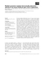
Fig. 1. ESI-TOF-MS spectra recorded at pH 7.0 of the recombinant CeMT1 (A) and CeMT2 (C) synthesized in Zn-, Cd- and Cu-supplemented
E. coli cultures. Spectra recorded after incubation with DEPC are shown for Zn– and Cd–CeMT1 (B) and Zn– and Cd–CeMT2 (D). In the final
column of (B) and (D), the spectra of the Cu–CeMT preparations recorded at pH 2.8 are shown.
R. Bofill et al. C. elegans CeMT1 and CeMT2 metallothioneins
FEBS Journal 276 (2009) 7040–7056 ª 2009 The Authors Journal compilation ª 2009 FEBS 7043
participate in Zn(II) binding. However, because both
preparations rendered identical major stoichiometries
(Table 2), it is sensible to conclude that this would
only apply to a small subset of the Zn(II)–CeMT2
complexes present in the preparation.
The higher Zn(II)-binding capacity of CeMT1 ver-
sus CeMT2 correlates well with the results obtained
for their separate putative metal-binding domains.
The highly similar N-terminal moieties (NtCeMT1
and NtCeMT2) rendered equivalent mixtures of
species, with major Zn
3
complexes. Conversely, the
C-terminal peptides (CtCeMT1 and CtCeMT2)
yielded mixtures with different major species: Zn
4
–
CtCeMT1 versus Zn
3
–CtCeMT2 (Table 2). The CD
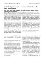
Fig. 2. Comparison between the CD and UV-visible spectra of recombinant CeMT1 (black), CeMT2 (red) and DHisCeMT2 (green) synthe-
sized in Zn- and Cd-supplemented media.
Table 2. Analytical characterization of the recombinant preparations of the Zn complexes yielded by CeMT1, CeMT2, their N-term and
C-term moieties and the DHisCeMT2 mutant. ESI-MS data comprise theoretical and experimental molecular masses of the Zn–CeMT
peptides. Zn contents were calculated from the mass difference between holo- and apo-proteins.
Peptide
Zn-peptide molar ratio
(ICP-AES)
ESI-MS
Major species
Minor species MW
theoretical
MW
experimental
CeMT1 6.5 Zn Zn
7
–CeMT1 8706.0 8708.1 ± 0.4
CeMT2 5.0 Zn Zn
6
–CeMT2 7118.1 7117.2 ± 0.8
Zn
5
–CeMT2 7054.7 7051.2 ± 1.4
Zn
4
–CeMT2 6991.3 6986.4 ± 0.2
CtCeMT1 2.1 Zn Zn
4
–CtCeMT1 5541.4 5541.4 ± 0.6
Zn
2
–CtCeMT1 5414.7 5411.2 ± 0.5
Zn
1
–CtCeMT1 5351.3 5344.4 ± 0.3
NtCeMT1 1.8 Zn Zn
3
–NtCeMT1 3298.7 3298.0 ± 0.1
Zn
1
–NtCeMT1 3172.0 3166.2 ± 0.2
CtCeMT2 2.2 Zn Zn
3
–CtCeMT2 3692.2 3691.8 ± 0.4
Zn
2
–CtCeMT2 3628.8 3626.8 ± 0.7
NtCeMT2 2.6 Zn Zn
3
–NtCeMT2 3587.2 3587.1 ± 0.1
Zn
2
–NtCeMT2 3523.8 3522.4 ± 0.2
DHisCeMT2 4.7 Zn Zn
6
–DHisCeMT2 6981.0 6980.7 ± 0.3
Zn
5
–DHisCeMT2 6917.6 6917.6 ± 0.1
Zn
4
–DHisCeMT2 6854.2 6854.0 ± 0.1
C. elegans CeMT1 and CeMT2 metallothioneins R. Bofill et al.
7044 FEBS Journal 276 (2009) 7040–7056 ª 2009 The Authors Journal compilation ª 2009 FEBS
fingerprints of the Zn(II) complexes of NtCeMT1 and
NtCeMT2 (Fig. 3) were highly atypical and difficult
to interpret, especially the absence of a CD signal at
approximately 240 nm for Zn(II)–NtCeMT2, whereas
those of CtCeMT1 and CtCeMT2 displayed a Gauss-
ian band centered at approximately 240(–) nm, resem-
bling more those of the respective entire MTs.
Finally, it is worth noting that, despite the apparent
additivity of the stoichiometries of the complexes ren-
dered by the separate moieties of CeMT1 and
CeMT2, the summation of their CD spectra did not
give rise in any case to spectra close to those of the
entire Zn(II)–CeMT preparations, which is indicative,
for both CeMTs, of a strong moiety interaction when
binding Zn(II) ions.
Overall, the differences between Zn(II)–CeMT1 and
Zn(II)–CeMT2 suggested a higher Zn binding capacity
of the former, reflected both in the stoichiometry and
the homogeneity of their preparations. These differ-
ences are a result of the different coordination capaci-
ties of the respective C-terminal moieties and are
attributable to the four additional putative coordinat-
ing residues (one cysteine and three histidine) of
CtCeMT1 compared to CtCeMT2. These results
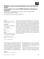
Fig. 3. Comparison between the CD spectra of recombinant CeMT1 and CeMT2 (black), NtCeMT1 and NtCeMT2 (red) and CtCeMT1 and
CtCeMT2 (green) synthesized in Zn- and Cd-supplemented media.
Table 3. Analytical characterization of the recombinant preparations of the Cd complexes yielded by CeMT1, CeMT2, their N-term and
C-term moieties and the DHisCeMT2 mutant. ESI-MS data comprise theoretical and experimental molecular masses of the Cd–CeMT
peptides. Zn and Cd contents were calculated from the mass difference between holo- and apo-proteins.
Peptide
Metal-peptide molar ratio
(ICP-AES)
ESI-MS
Major species
Minor species MW
theoretical
MW
experimental
CeMT1 0.9 Zn Cd
6
Zn
1
–CeMT1 8988.2 8989.1 ± 0.5
6.5 Cd
CeMT2 5.7 Cd Cd
6
–CeMT2 7400.2 7399.0 ± 0.5
CtCeMT1 0.6 Zn Cd
3
Zn
1
–CtCeMT1 5682.5 5683.2 ± 0.9
2.9 Cd
NtCeMT1 0.1 Zn Cd
3
–NtCeMT1 3439.8 3438.9 ± 0.6
2.9 Cd Cd
3
Zn
1
–NtCeMT1 3503.2 3502.4 ± 0.5
CtCeMT2 2.9 Cd Cd
3
–CtCeMT2 3833.2 3833.1 ± 0.1
NtCeMT2 0.1 Zn Cd
3
–NtCeMT2 3728.2 3729.0 ± 0.1
2.3 Cd Cd
3
Zn
1
–NtCeMT2 3791.6 3790.8 ± 0.1
DHisCeMT2 5.5 Cd Cd
6
–DHisCeMT2 7263.0 7262.5 ± 0.1
R. Bofill et al. C. elegans CeMT1 and CeMT2 metallothioneins
FEBS Journal 276 (2009) 7040–7056 ª 2009 The Authors Journal compilation ª 2009 FEBS 7045
strongly suggest the participation of the histidine resi-
dues of CeMT1 in Zn(II) coordination, allowing an
MT peptide with only 19 cysteines to stably coordinate
up to seven Zn(II). Unfortunately, the similarities
between the CD spectra of Zn(II)–CeMT1 and Zn(II)–
CeMT2 preclude the assignment of the putative
His-Zn(II) chromophores to defined CD absorptions,
which would have been highly informative regarding
the presence of Zn-His bonds.
In vivo and in vitro Cd(II)-binding abilities of
CeMT1 and CeMT2
Unlike the results obtained for Zn(II) coordination,
the biosynthesis in Cd-supplemented cultures of the
two wild-type CeMT1 and CeMT2 forms, as well as of
DHisCeMT2, invariably gave rise to a single species,
although of different stoichiometry, for each isoform
(Fig. 1 and Table 3). Most interestingly, CeMT1
rendered a heterometallic Cd
6
Zn
1
–CeMT1 species, in
contrast to the homometallic Cd
6
–CeMT2 and
Cd
6
–DHisCeMT2 complexes. ESI-MS results for the
separate CeMT1 moieties were highly informative
because they revealed formation of a unique Cd
3
Zn
1
–
complex for CtCeMT1, along with a major Cd
3
–
NtCeMT1 species (Table 3), suggesting that the Zn(II)
ion of Cd
6
Zn
1
–CeMT1 is located within its C-terminal
domain. By contrast, synthesis of NtCeMT2 and
CtCeMT2 gave rise to practically pure Cd
3
species,
which is also fully concordant with the entire
Cd
6
–CeMT2 complex.
The CD and UV-visible fingerprints of the Cd(II)–
CeMT1, Cd(II)–CeMT2 and Cd(II)–DHisCeMT2 prep-
arations (Fig. 2) were highly similar, showing the
typical absorptions at approximately 250 nm of con-
ventional Cd-SCys chromophores, which additionally
discarded the presence of sulfide-containing aggregates.
Our data coincided with the UV-visible absorption
spectra previously reported for the native and recombi-
nant Cd(II)–CeMT2 isoform [16,20]. The slight blue-
shift of the spectrum of Cd
6
Zn
1
–CeMT1 in relation to
that of Cd
6
–CeMT2 is attributable to the influence of
the Zn(II) ion present in the complex. The four CeMT
moiety peptides showed atypical CD envelopes
(Fig. 3), whose summation in no case reproduced that
of the corresponding full-length proteins, despite the
additivity of their metal contents (Table 3), suggesting,
as for Zn(II), clear interactions between domains when
binding Cd(II). The two N-terminal segments (of simi-
lar sequence and comparable speciation) also gave rise
to almost equivalent CD fingerprints, although of dif-
ferent intensity, which could be interpreted by assum-
ing the characteristic Cd-SCys signals at 250 nm, plus
the possible contribution of the weak absorption of
minor sulfide-containing species at approximately
280 nm. By contrast, the CD envelopes of the C-termi-
nal moieties are difficult to rationalize, especially in
the case of Cd
3
Zn
1
–CtCeMT1, where we expected the
influence of Zn(II) to be similar to that in the full-
length CeMT1. Although the CD profiles of these two
Cd(II) complexes coincide in the 240–250 nm region
(Fig. 3), Cd
3
Zn
1
–CtCeMT1 shows absorptions at
260(–) nm that are absent in the full length protein
spectrum. One possible explanation for this, and also
for the faint shoulder observed at approximately
270(+) nm for CeMT1, would be the contribution of
the multiple histidines to metal binding (see below).
Finally, the comparison of the CD spectra of the
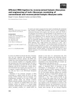
Fig. 4. CD (A), UV-visible (B) and UV-visible difference (C) spectra corresponding to the titration of a 10 lM solution of Zn–CeMT1 and
Zn–CeMT2 with Cd(II) at pH 7.0.
C. elegans CeMT1 and CeMT2 metallothioneins R. Bofill et al.
7046 FEBS Journal 276 (2009) 7040–7056 ª 2009 The Authors Journal compilation ª 2009 FEBS
recombinant Zn(II)–CeMT1 and Zn(II)–CeMT2 com-
plexes with the respective Cd(II) complexes shows their
inverse chirality, which makes it possible to propose
that they do not share the same 3D architecture,
despite their equivalent stoichiometry (M
7
–CeMT1 and
M
6
–CeMT2; M = Zn or Cd) (Fig. 2).
As well as recombinantly, Cd(II) complexes of all the
studied CeMT peptides were obtained in vitro by two
different procedures: (a) Cd(II) titration of the recombi-
nant Zn(II)–MT forms and (b) acidification plus subse-
quent reneutralization of the recombinant Cd(II)–MT
preparations. The key results of these experiments show
that, in all cases, the titration of the Zn(II)–CeMT prep-
arations with Cd(II) allowed reproduction of the spec-
trometric and spectropolarimetric features of the
biosynthesized Cd(II)–MT forms, after the addition of
the expected number of Cd(II) equivalents [i.e. six
Cd(II) equivalents for the full length proteins (Fig. 4)
and three Cd(II) equivalents for the fragments (data not
shown)]. Most interestingly, the Zn ⁄ Cd replacement
process on CeMT1 yielded Cd
6
Zn
1
–CeMT1, even after
the addition of a significant excess of Cd(II). Also, the
in vivo heteronuclear Cd
6
Zn
1
–CeMT1 complex did not
exchange the Zn(II) ion upon addition of excess Cd(II).
Acidification ⁄ reneutralization of all biosynthesized
Cd(II)–CeMT complexes revealed that the initial species
were recovered after this process. For CeMT1, these
experiments also supported the participation of histidine
residues in metal coordination because acidification of
Cd
6
Zn
1
–CeMT1, as well as of Cd
3
Zn
1
–CtCeMT1 (from
pH 7.0 to pH 1.0) did not induce important variations
in the respective CD envelopes precisely until approxi-
mately pH 4.5, with this coinciding with the particular
pK
a
value that this amino acid exhibits in MT polypep-
tides [27,28]. Furthermore, after this acidification stage,
UV-visible difference spectra revealed a loss of absor-
bance at wavelengths of approximately 240 nm (Fig. 5),
whereas the ESI-MS data indicated that, at pH 4.2,
most of the complexes lost their Zn(II) ion because the
major species present in the sample were Cd
6
–CeMT1
and Cd
3
–CtCeMT1, respectively. Consequently, it is
sensible to deduce that the coordination of the Zn(II)
ion bound at the C-terminal moiety of CeMT1 is con-
tributed to by histidines, and the number of these
involved in metal binding is analyzed below.
Thus, the overall results reveal that equivalent Cd
complexes of CeMT1 and CeMT2, as well as those of
their putative domains, are obtained in vivo (by recom-
binant synthesis) and in vitro (by Zn ⁄ Cd replacement or
acidification ⁄ reneutralization). Our data also demon-
strate that CeMT1 forms heteronuclear Cd
6
Zn
1
com-
plexes when folding in the presence of high cadmium,
and that this Zn(II) ion is bound into its C-terminal
moiety, in a coordination environment most probably
contributed to by histidine residues. By contrast,
CeMT2 folds into homonuclear, canonical Cd
6
complexes, with equivalent features regardless of their
origin, recombinant synthesis, or in vitro Zn ⁄ Cd replace-
ment, acidification ⁄ reneutralization or Cd(II) recon-
stitution of apo-forms (J. H. R. Ka
¨
gi, personal
communication). Therefore, although the CeMT2 poly-
peptide exhibits an optimal Cd(II)-binding ability that
accounts for the formation of homometallic Cd-contain-
ing complexes under excess Cd(II) conditions, the
CeMT1 isoform exhibits a metal binding behavior that
is clearly conditioned by its property to form well-folded
Zn(II) complexes, and Cd(II) complexes that retain,
under all the physiologically comparable conditions,
one Zn(II) ion [8]. This also explains the constant pres-
ence of Zn(II) in the Cd(II)–CeMT1 complexes purified
from cadmium intoxicated organisms [13].
In relation to the metal complex architecture, the
results obtained in the present study are compatible
with a two-domain folding when coordinating Zn(II)
Fig. 5. CD (A), UV-visible (B), and UV-visible difference (C) corresponding to the acidification of a 10 lM solution of Cd–CeMT1 and a 20 lM
solution of Cd–CtCeMT1.
R. Bofill et al. C. elegans CeMT1 and CeMT2 metallothioneins
FEBS Journal 276 (2009) 7040–7056 ª 2009 The Authors Journal compilation ª 2009 FEBS 7047
or Cd(II), defining N-terminal and C-terminal seg-
ments with additive metal binding capacity but not
additive structural features in relation to the full-length
polypeptides. It is worth noting that the precise differ-
ences in metal binding abilities between the isoforms
arise from their highly dissimilar C-terminal moieties,
in concordance with their amino acid sequence differ-
ences and peculiarities (i.e. a longer CtCeMT1 with
one cysteine and three extra histidine residues in rela-
tion to CtCeMT2). Hence, CeMT1 is able to bind
seven divalent metal ions, whereas CeMT2 only yields
M(II)
6
species. In the case of Zn, this implies
Zn
7
–CeMT1 versus major Zn
6
–CeMT2 complexes,
although, significantly, for cadmium, this entails
Cd
6
Zn
1
–CeMT1 versus Cd
6
–CeMT2 species. This
Zn(II) ion in Cd
6
Zn
1
–CeMT1 probably plays a struc-
tural role because even a clear excess of Cd(II) is
unable to remove it from the complex.
Quantification of the histidine residues involved
in metal coordination in the Zn– and Cd–CeMT1
and Zn– and Cd–CeMT2 complexes
Diethyl pyrocarbonate (DEPC) modification allows the
identification and quantification of the histidine resi-
dues of proteins that are not protected in some way
[29]. In the case of the reaction with histidine, DEPC
produces a 72.06 Da carboxyethyl adduct at the imid-
azole (e)-NH position [30] and, although DEPC also
reacts with other nucleophilic residues (Cys, Lys, Tyr,
Ser, Thr, Arg) and a-amino groups, this reaction pro-
ceeds with markedly lower efficiency [31,32]. Therefore,
to evaluate the number of CeMT1 and CeMT2 histi-
dines contributing to divalent metal ion coordination,
the Zn and Cd preparations of both C. elegans CeMT1
and CeMT2, and the Cd complexes of CtCeMT1 and
CtCeMT2, were incubated with DEPC and the respec-
tive results were evaluated by ESI-TOF-MS (Fig. 1),
using the Zn(II)–DHisCeMT2 and Cd(II)–NtCeMT1
peptides as negative controls because they do not
encompass any histidine.
The results obtained indicated that these two
His-devoid peptides [Zn(II)–DHisCeMT2 and Cd(II)–
NtCeMT1] were mono-carboxyethylated. Conse-
quently, under the conditions assayed, the reaction of
their free terminal a-NH
2
groups with DEPC should
be assumed as that most likely being responsible for
their single modification because these two peptides
differ greatly in terms of the number of other less
likely modifiable residues (Lys, Tyr, Ser and Thr) and
cysteines remain inaccessible due to the binding of
metal ions. Of special significance is the result with
DHisCeMT2 because CeMT2 yields a two-carboxye-
thylated derivative. Furthermore, because the only dif-
ference between these two peptides is the C-terminal
histidine, it has to be assumed that this residue is the
one responsible for the second DEPC binding, and
thus that this histidine is free (non-metal coordinating)
in the corresponding metal complex. Consequently,
regarding the CeMT1 isoform, and taking into account
the two DEPC modifications, one is attributable to its
N-terminal amino group (i.e. with the conclusion being
drawn from a comparison with the NtCeMT1 negative
control) and only one is attributable to histidine modi-
fication. Therefore, of the four histidines present in the
full-length CeMT1 peptide, one is free to react with
DEPC, and three would be protected by metal coordi-
nation, or at least inaccessible to the reactant. By anal-
ogy with the results obtained with the CeMT2 peptide,
it is logical to conclude that the terminal CeMT2 histi-
dine is that which remains free for DEPC reaction,
and therefore is not involved in metal binding. How-
ever, should this precise residue not be the metal-free
histidine, the conclusion that three of the four histi-
dines of CeMT1 are involved in divalent metal coordi-
nation, would remain equally valid.
Our subsequent results lead to the proposal that
CeMT1 and CeMT2 histidine residues not only partic-
ipate in metal coordination, but also comprise the
most responsible elements for their metal binding
behavior. With respect to CeMT1, the data suggest
the contribution of three out of four histidines (prob-
ably excluding the C-terminal histidine) in the coordi-
nation of the seventh M(II) ion, precisely the Zn(II)
of Cd
6
Zn
1
–CeMT1. Unfortunately, this Zn-NHis
coordination is not detectable by spectropolarimetric
methods. In the case of CeMT2, the single C-terminal
histidine appears to play no major role in divalent
metal coordination, although there is some hint of
partial participation in a subset of the metal com-
plexes present in our preparations. The role of histi-
dine in metal ion coordination in MTs is a subject
that has gathered increasing importance in the field,
especially because the 3D structure of the Zn and Cd
complexes of cyanobacteria (SmtA) [33] and plant
wheat-Ec-1) [28,34] MTs have been solved. The conse-
quences of the presence of histidines in MTs were
analyzed comprehensively in a recent review [35],
which clearly illustrates that they act as modulators of
the reactivity of these peptides towards Zn, conferring
the specific properties that allow them to perform
functions more related to Zn metabolism and homeo-
stasis than to cadmium detoxication. Therefore, our
assumption that the four-histidine-containing CeMT1
isoform should be related to housekeeping Zn metab-
olism fits perfectly in this scenario.
C. elegans CeMT1 and CeMT2 metallothioneins R. Bofill et al.
7048 FEBS Journal 276 (2009) 7040–7056 ª 2009 The Authors Journal compilation ª 2009 FEBS
In vivo and in vitro Cu(I)-binding abilities of
CeMT1 and CeMT2
The synthesis of CeMT1 and CeMT2 in Cu-supple-
mented cultures provided equivalent results: a mixture
of heteronuclear Zn,Cu complexes, with major M
8
and
M
9
species, which were identified as Cu
4
- and Cu
8
-con-
taining complexes by ESI-MS at pH 2.4, in full
concordance with the mean Cu(I) and Zn(II) content
per MT measured by inductively coupled plasma
atomic emission spectroscopy (ICP-AES) (Fig. 1 and
Table 4). Conversely, DHisCeMT2 synthesized under
the same conditions yielded homometallic Cu com-
plexes with a major Cu
8
–DHisCeMT2 species. Both
NtCeMT moieties also gave rise to homonuclear Cu
5
preparations. Under these conditions, low Zn contents
Table 4. Analytical characterization of the recombinant preparations of the Cu complexes yielded by CeMT1, CeMT2, their N-term and
C-term moieties and the DHisCeMT2 mutant, obtained under normal aeration conditions. ESI-MS data comprise theoretical and experimental
molecular masses of the Cu–CeMT peptides. In the case of Zn,Cu mixed-metal species, the theoretical molecular masses correspond to the
homometallic Cu
x
and Zn
x
species, respectively, and the metal-to-protein stoichiometries deduced at pH 7.0 are indicated as M
x
(M is Zn or
Cu). Cu contents at pH 2.4 were calculated from the mass difference between holo- and apo-proteins.
Peptide
Metal-peptide molar ratio
(ICP-AES)
ESI-MS
Major species
Minor species MW
theoretical
MW
experimental
CeMT1 2.2 Zn pH 7.0 M
8
–CeMT1 8762.7–8769.4 8761.6 ± 1.4
M
9
–CeMT1 8825.3–8832.8 8823.2 ± 0.7
M
6
–CeMT1 8637.7–8642.7 8635.0 ± 4.4
M
5
–CeMT1 8575.1–8579.3 8573.4 ± 9.3
4.6 Cu pH 2.4 Cu
4
–CeMT1 8512.6 8506.4 ± 1.1
Cu
8
–CeMT1 8762.7 8758.2 ± 0.3
CeMT2 2.5 Zn pH 7.0 M
8
–CeMT2 7238.1–7245.0 7237.5 ± 1.5
M
9
–CeMT2 7300.7–7308.4 7300.4 ± 5.5
M
6
–CeMT2 7113.0–7118.2 7107.6 ± 1.8
M
5
–CeMT2 7050.4–7054.8 7046.0 ± 2.0
4.3 Cu pH 2.4 Cu
4
–CeMT2 6987.9 6976.2 ± 0.1
Cu
8
–CeMT2 7238.1 7232.4 ± 4.0
CtCeMT1 0.8 Zn pH 7.0 M
4
–CtCeMT1 5538.1–5541.4 5534.4 ± 0.4
M
5
–CtCeMT1 5600.7–5604.8 5598.0 ± 0.5
3.7 Cu pH 2.4 Cu
4
–CtCeMT1 5538.1 5534.5 ± 0.5
NtCeMT1 0.0 Zn pH 7.0 Cu
5
–NtCeMT1 3421.3 3419.3 ± 0.5
4.4 Cu pH 2.4 Cu
5
–NtCeMT1 3421.3 3418.4 ± 0.5
CtCeMT2 0.5 Zn pH 7.0 M
4
–CtCeMT2 3752.2–3755.7 3753.0 ± 2.0
3.5 Cu pH 2.4 Cu
4
–CtCeMT2 3752.2 3753.2 ± 1.5
NtCeMT2 0.0 Zn pH 7.0 Cu
5
–NtCeMT2 3709.8 3706.5 ± 0.1
4.4 Cu pH 2.4 Cu
5
–NtCeMT2 3709.8 3707.5 ± 0.6
DHisCeMT2 0.0 Zn pH 7.0 Cu
8
–DHisCeMT2 7101.0 7096.8 ± 0.3
Cu
9
–DHisCeMT2 7163.5 7160.8 ± 0.4
8.7 Cu pH 2.4 Cu
8
–DHisCeMT2 7101.0 7099.6 ± 0.3
Cu
9
–DHisCeMT2 7163.5 7163.8 ± 1.2
Fig. 6. Comparison between the CD spectra of recombinant CeMT1 (black), CeMT2 under normal oxygenation conditions (red), CeMT2
under low oxygenation conditions (green), DHisCeMT2 (kaki) (A); CeMT1 (black), NtCeMT1 (red) and CtCeMT1 (green) (B); and CeMT2
(black), NtCeMT2 (red) and CtCeMT2 (green) (C) synthesized in Cu-supplemented media.
R. Bofill et al. C. elegans CeMT1 and CeMT2 metallothioneins
FEBS Journal 276 (2009) 7040–7056 ª 2009 The Authors Journal compilation ª 2009 FEBS 7049
were detected in the preparations of the Cu complexes
of the CtCeMT segments, which rendered major M
4
(Cu
4
for CtCeMT2) and additional minor M
5
(Cu
4
Zn
1
for CtCeMT1). To further extend the Cu binding pref-
erence analyses of the two isoforms, their synthesis
was repeated in Cu-supplemented media but under low
aeration conditions. Interestingly, although the results
obtained for CeMT2 were fully comparable with those
obtained for DHisCeMT2 at regular oxygenation (i.e.
homonuclear Cu complexes), the resulting Cu–CeMT1
preparations gave extremely poor spectroscopic and
spectrometric data, revealing indiscernible mixtures of
Cu species. These data were consistent with a more
pronounced character of Zn-thionein for CeMT1 and
partial Cu-thionein for CeMT2, which would behave
similar to a proper Cu-thionein if not for its C-termi-
nal histidine. The CD fingerprints of all these prepara-
tions (Fig. 6) showed the characteristic signals
associated with the Cu–MT species, although the com-
plexity of their envelopes is difficult to rationalize in
view of the mixtures of complexes obtained and the
distinct coordination environments that Cu(I) ions can
show.
For both CeMT1 and CeMT2, the different behav-
iour of the separate fragments with respect to the full
length peptides is in accordance with the non-additivity
of their respective CD fingerprints (Fig. 6), which sug-
gests the existence of cooperativity between moieties
when binding Cu(I), as described both for Zn(II) and
for Cd(II) binding. Significantly, however, this depen-
dence entails a striking consequence because the clear
Cu-binding preference of the N-terminal moieties is
turned into a definite Zn-thionein character for the
full-length proteins.
Concerning the in vitro studies, it should be noted
that the addition of Cu(I) to either Zn
7
–CeMT1, Zn
6
–
CeMT2 or Zn
6
–DHisCeMT2 gave rise to a continued
increase in absorbance at the studied wavelength range
until eight or nine Cu(I) equivalents were added, when
the spectra (Fig. 7) become invariable, indicating
saturation, in good concordance with the Cu(I) con-
tents observed in the in vivo preparations. It is difficult
Fig. 7. CD (A), UV-visible (B) and UV-visible difference (C) spectra corresponding to the titration of a 10 lM solution of Zn(II)–CeMT1 and
Zn(II)–CeMT2 with Cu(I) at pH 7.0.
C. elegans CeMT1 and CeMT2 metallothioneins R. Bofill et al.
7050 FEBS Journal 276 (2009) 7040–7056 ª 2009 The Authors Journal compilation ª 2009 FEBS
to correlate the variations in the CD envelopes
observed during the titrations with the corresponding
ESI-MS data because the latter revealed the coexis-
tence of multiple metal–MT species at all stages of the
titrations, with the presence of major M
8
– and M
9
–
CeMT from the outset, which resulted in Cu
4
– and
Cu
8
–CeMT species when acidified at pH 2.4. It is
worth noting that at the end of the titration, even after
addition of excess Cu(I), some species still retained
Zn(II) ions.
The discussion of the Cu(I) coordination behavior
becomes more complex in view of the difficulties
involved in calculating the exact Zn(II) and Cu(I) stoi-
chiometry of the corresponding species by ESI-MS.
However, and despite this drawback, the sole analysis
of the presence ⁄ absence of Zn(II) ions in the recombi-
nant complexes synthesized in Cu-supplemented cul-
tures provides enough information to confirm that
histidines are determinants of the presence of Zn(II),
and therefore of the Zn- or Cu-thionein character of
the polypeptides. Hence, the two full-length peptides,
CeMT1 and CeMT2, give rise to mixtures of com-
plexes, with major species of relative low nuclearity
(M
8
– and M
9
–CeMT). The behavior of the respective
N-terminal moieties is clear and similar, yielding
homonuclear Cu
5
complexes, and thus the N-terminal
peptides constitute typical Cu-thioneins although of a
low Cu(I) : MT ratio (5 : 1 for a nine cysteine MT).
By contrast, the histidine-containing CtCeMT moieties
both render heteronuclear Zn,Cu complexes with an
even lower Cu(I) : MT ratio than the N-terminal seg-
ments (4 : 1 for 14 or ten coordinating residues in
CtCeMT1 and CtCeMT2, respectively), therefore tend-
ing towards Zn-thionein behavior. The presence of
Zn(II) in CtCeMT1 was clearly demonstrated after the
detection of a Cu
4
Zn
1
–CtCeMT1 species. This is a new
case that is comparable to the mammalian MT1 iso-
form, where the combination of a Cu-thionein domain
(bMT1) with a Zn-thionein fragment (aMT1) renders
a full-length MT of Zn-thionein character [36]. Fur-
thermore, the Zn-thionein character of CeMT2 could
be neatly attributable to its C-terminal histidine
because the corresponding DHisCeMT2 mutant was
capable of folding into homonuclear Cu(I) complexes
when synthesized by bacteria grown under regular
aeration in Cu-supplemented cultures, whereas the
wild-type form was not.
Conclusions
The data obtained in the present study are indicative
of a differential metal binding behavior for the two
C. elegans MT isoforms. Although they exhibit a clear
preference for divalent-metal binding rather than a
Cu-thionein character, CeMT1 shows optimal behavior
when binding Zn(II), whereas CeMT2 is highly profi-
cient for Cd(II) coordination. Indeed, CeMT1 occupies
the more extreme position in our recent proposal for a
Fig. 8. Protein distance trees of CeMT1, CeMT2 and N-terminal and C-terminal separate moieties. Neighbor-joining trees constructed with
the entire CeMT1 (Ce1) and CeMT2 (Ce2) polypeptides (A) and with their N-terminal and C-terminal separate moieties (NtMT1, NtMT2,
CtMT1 and CtMT2). (B). Protein sequences were aligned by
CLUSTALW [46a], and the alignments were used as inputs to construct Neighbor-
joining trees, by the Fitch–Margoliash algorithm (
PHYLIP software) [40]. Call1, Call2 and CalliCu, Callinectes sapidus, 1, 2 and Cu isoforms (Uni-
prot accession numbers Q548Y3, Q548Y2 and Q9U620, respectively); Scy1 and Scy2, Scylla serrata isoforms 1 and 2 (Uniprot P02805 and
P02806); Carci, Carcinus maenas (Uniprot P55948); Astfl, Astacus astacus (Uniprot P55951); Pacifastacus, Pacifastacus leniusculus (Uniprot
Q9U623); Homa, Homarus americanus (Uniprot Q95P38); Potpo, Potamon potamios (Uniprot P55952); Cup1, S. cerevisiae Cup1 (Uniprot
P07215); Mtn, Drosophila melanogaster MtnA (Uniprot P04357); and Mto, Drosophila melanogaster MtnB (Uniprot P11956).
R. Bofill et al. C. elegans CeMT1 and CeMT2 metallothioneins
FEBS Journal 276 (2009) 7040–7056 ª 2009 The Authors Journal compilation ª 2009 FEBS 7051
step gradation from Zn- to Cu-thioneins [8]. These
results are in full concordance with CeMT1 being con-
stitutively expressed in pharyngeal cells, where it would
develop some background role related to physiological
metal (mainly Zn) metabolism, whereas the unique
synthesis of CeMT2 after cadmium induction confers a
basic detoxification role to this isoform. This hypothe-
sis is also concordant with the effects observed in the
fitness of C. elegans MT-knockout organisms, where
the lack of CeMT1 is more deleterious in the absence
of metals than that of CeMT2. No response has been
described for the CeMT promoters with regard to Cu
overload, nor do MTs appear to comprise a major sys-
tem for Cu tolerance in this organism [37], with this
also being in agreement with the fact that, according
to our classification, none of the CeMT isoforms
display proper Cu-thionein features, although
Cu(I)–CeMT2 complexes are certainly more stable
than Cu(I)–CeMT1 species.
In conclusion, the presence of their histidine residues
precludes these MTs behaving as Cu-thioneins, as
would otherwise be the case according to their global
protein sequence similarities. Precisely these analyses
(Fig. 8), which we have shown to yield results consis-
tent with the metal binding preferences of other MTs
[7,38], position both entire CeMT1 and CeMT2
peptides, as well as their moieties, in the subset of
Cu(I)-thioneins. According to our metal binding
preference analysis, this is true for the N-terminal
fragments and DHisCeMT2, and almost true for the
C-terminal moieties, but is obviously not the case for
the entire CeMTs. In sum, the CeMT system analysis
has revealed noteworthy metal-coordination peculiari-
ties, mainly derived from the unusual presence of
histidines in their protein sequences, which enlarges
the list of MTs where this amino acid plays a decisive
role [35,39,40]. Therefore, the ultimate details will
undoubtedly be revealed when the 3D structures of the
corresponding metal complexes become available.
Materials and methods
Construction of the expression vectors for the
C. elegans MT1 (CeMT1) and MT2 (CeMT2)
wild-type and mutant forms
All the metal–MT complexes investigated in the present
study were recombinantly synthesized in E. coli through
cloning of their cDNAs into the pGEX-4T2 plasmid (GE
Healthcare, Little Chalfont, UK) to yield primary glutathi-
one S-transferase (GST)-MT fusions from which the corre-
sponding metal–MT complexes were subsequently purified
[25].
Three kZAPII phage clones (yk120h8, yk364c6 and
yk656b5), including cDNAs for C. elegans MT1, were
kindly provided by Dr Y. Kohara (Genome Biology Unit
of the National Institute of Genetics, Mishima, Japan). For
the expression of the whole-length CeMT1 peptide, the cor-
responding ORF was amplified by a PCR reaction that
respectively added a BamHI and SalI site to the 5¢ and 3¢
ends of the coding sequence, using purified CeMT1-kZAPII
DNA as template and the primers: 5¢-GGCGGATCC
ATGGCTTGCAAGTGT-3¢ (upstream) and 5¢-GTTTTC
GTCGACTTAATGAGCCGCAGCAGT-3¢ (downstream).
cDNAs encoding for the independent CeMT1 moieties
were obtained by PCR amplification with the primers:
N-terminal fragment (residues 1–27), 5¢-CGTGGATCCAT
GGCTTGCAAGTGT-3¢ (upstream) and 5¢-GCTCGAGT
CGACTTACTCACAACACTTGTC-3¢ (downstream) and
C-terminal fragment (residues 28–75), 5¢-CCGCGT
GGA
TCCAAGTACTGCTGT-3¢ (upstream) and 5¢-CGACTCG
AGTTAATGAGCCGCAGC-3¢ (downstream). Note that
the C-terminal fragment was inserted in pGEX by a
3¢-introduced XhoI site instead of SalI as a result of
repeated cloning problems when using the latter.
The synthetically constructed CeMT2 cDNA [20] was
kindly provided by Professor J. H. R. Ka
¨
gi (Institute of
Biochemistry of the University of Zurich, Switzerland) and
also used as a template for the construction of regions
coding for the deletion mutant CeDHis and the independent
N-terminal and C-terminal CeMT2 domains. All the PCR
reactions were designed to introduce 5¢ BamHI and 3¢SalI
restriction sites for subcloning purposes. The wild-type
CeMT2 cDNA was amplified using the upstream primer
5¢-CGGGGATCCATGGTCTGCAAG-3¢ and the down-
stream primer 5¢-ACGCGTCGACCTAATGAGCAGC-3¢.
The region coding for the N-terminal CeMT2 segment,
encompassing Met1 to Glu30, was amplified using the
upstream primer 5¢-CGGGGATCCATGGTCTGCAAG-3¢
and the downstream primer 5¢-ACGCGTCGACCTACTC
ACAGCACTTG-3¢. The region coding for the C-terminal
CeMT2, which comprises the Gln31 to His63 CeMT2 resi-
dues, was amplified with the upstream primer 5¢-CGGG
GATCCCAGTACTGCTGC-3¢ and the downstream primer
5¢-ACGCGTCGACCTAATGAGCAGC-3¢. For the con-
struction of a cDNA encoding the CeMT2 peptide lacking
its C-terminal histidine residue, the wild-type cDNA was
amplified using the same upstream primer as for the entire
cDNA (5¢-CGGGGATCCATGGTCTGCAAG-3¢) but
5¢-ACGCGTCGACCTAAGCAGCCTG-3¢ as the down-
stream primer.
All the PCR reactions consisted of 30-cycle amplifica-
tions, performed with 1 U of thermo resistant Vent DNA
polymerase (New England Biolabs, Hitchin, UK), 0.2 mm
dNTPs and 100 pmol of the required primers at 2 mm
MgCl
2
(final concentration), in a final volume of 100 lL,
under the conditions: 45 s at 95 ºC (denaturation), 30 s at
55–60 °C (hybridization) and 45 s at 72 °C (elongation).
C. elegans CeMT1 and CeMT2 metallothioneins R. Bofill et al.
7052 FEBS Journal 276 (2009) 7040–7056 ª 2009 The Authors Journal compilation ª 2009 FEBS
Elongation conditions were maintained for 5 min after the
30 cycles. The final products were analyzed by agarose gel
electrophoresis ⁄ ethidium bromide staining; the band with
the expected size was excised and subcloned into the
pGEX-4T2 vector. Before recombinant protein synthesis,
all coding sequences were confirmed by automated DNA
sequencing. To this end, the pGEX-derived constructions
were transformed in E. coli DH5a cells, and sequenced
using the ABIPRISM Dye Terminator-Cycle Sequencing
Ready reaction kit (Perkin Elmer, Waltham, MA, USA) in
an ABIPRISM 310 Automatic Sequencer (Applied Biosys-
tems, Foster City, CA, USA). In all cases, the expected
sequence was corroborated, except for position 26 of the
CeMT1 peptide, which corresponded to an AGG (Arg)
instead of the reported AAG (Lys) codon. All the CeMT1
clones sequenced showed the same alteration, suggesting
that a polymorphism, in relation to the published sequence,
is the most likely explanation. Taking into account the con-
servative nature of this change and the fact that we had
recently established that an opposite charge substitution
(Lys for Glu, position 34) had no effect at all on the bind-
ing abilities of the Crs5 MT of S. cerevisiae [12], we decided
to continue our studies with the obtained clones.
Recombinant synthesis and purification of the
C. elegans MT1 (CeMT1) and MT2 (CeMT2)
wild-type and mutant forms
The obtained pGEX constructs were transformed into
E. coli BL21 cells for the expression of the cloned cDNAs
as fusions with GST. The recombinant peptides were
biosynthesized in 3 L-LB cultures, inoculated with 300 mL
of overnight pre-cultures. Induction with isopropyl thio-b-
d-galactoside was performed at D
600
= 0.8, and cultures
were grown for an additional 3 h in the presence of 500 lm
CuSO
4
, 300 lm ZnCl
2
or 300 lm CdCl
2
, aiming to recover
the corresponding Cu–, Zn– or Cd–MT complexes, respec-
tively. Cu-supplemented cultures were grown under two dif-
ferent conditions (normal and low aeration) in view of the
reported influence of oxygenation in the recovered final
complexes [12]. Cells were harvested by centrifugation
(15 min at 9600 g), resuspended in NaCl ⁄ Pi and lysed by
sonication (0.6 Hz; Branson Sonifier 250; Branson Ultra-
sonics, Danbury, CT, USA) in the presence of 0.5% b-mer-
captoethanol to avoid protein oxidation. Subsequently, all
procedures were carried out using Ar (pure grade 5.6)-satu-
rated buffers. After sonication, cellular debris was pelleted
by centrifugation (20 min at 20 000 g) and the GST-MT
fusions isolated from the supernatant by Glutathione-
Sepharose 4B (GE Healthcare) affinity chromatography.
Metal–MT complexes were excised from the fusion con-
structs by thrombin cleavage and batch affinity chromatog-
raphy. The sample concentration was attained by several
rounds of centrifugation in Centriprep Microcon 3 (Am-
icon, Millipore, MA, USA). The metal–MT complexes were
finally purified through FPLC in a Superdex75 (GE Health-
care) column equilibrated with 50 mm Tris-HCl (pH 7.0).
Selected fractions were kept at )70 °C until further use.
Analysis and characterization of the recombinant
metal peptide complexes
The S, Zn, Cd and Cu content of the Zn–, Cd– and
Cu–CeMT preparations was analyzed by means of ICP-
AES in a Polyscan 61E (Thermo Jarrell Ash, Franklin,
MA, USA) spectrometer, measuring S at 182.040 nm, Zn at
213.856 nm, Cd at 228.802 nm and Cu at 324.803 nm.
Samples were prepared in accordance with a previously
described method [41] and, in parallel, were also incubated
in 1 m HCl at 65 °C for 5 min prior to measurements to
eliminate possible traces of labile sulfide ions [42]. In all
cases, the protein concentration was calculated from the
acid ICP-AES sulfur measure, assuming that the sulfur con-
tent of the sample was contributed to by the MT peptides:
20 SÆmol
)1
for CeMT1 (one Met, 19 Cys), 19 SÆmol
)1
for
CeMT2 or DHisCeMT2 (one Met, 18 Cys), 10 SÆmol
)1
for
NtCeMT1 and NtCeMT2 (one Met, nine Cys), 10 SÆmol
)1
for CtCeMT1 (ten Cys) and 9 SÆmol
)1
for CtCeMT2 (nine
Cys).
In vitro Zn-, Cd- and Cu-binding studies of CeMT1
and CeMT2
The titration of all Zn–CeMT complexes with Cd(II) or
Cu(I) at pH 7 were carried out in accordance with previ-
ously described procedures [43,44], using CdCl
2
or
[Cu(CH
3
CN)
4
]ClO
4
solutions, respectively. The in vitro
acidification ⁄ reneutralization experiments were also per-
formed by adapting a previously described procedure [40].
Essentially, 10–20 lm preparations of the Cd peptides were
acidified from neutral pH (7.0) to acid pH (2.0) with
1–10
)3
m HCl depending on the stage of the titration. CD
and UV-visible spectra were recorded at pH 7.0, 4.5, 4.0,
3.0 and 2.0, both immediately after acid addition and
10 min later, with identical results being obtained. Finally,
the samples were kept at pH 2.0 for 20 min and then reneu-
tralized to pH 7.0 with 1–10
)3
m NaOH, also depending on
the stage of the titration. CD and UV-visible spectra were
recorded at pH 2.0, 2.5 and 7.0. All the results were cor-
rected for dilution effects. During all experiments, strict
oxygen-free conditions were maintained by saturation of
the solution with Ar.
Spectroscopic measurements
A Jasco spectropolarimeter (Model J-715; Jasco Inc., Eas-
ton, MD, USA) interfaced to a computer (J700 software)
was used for CD measurements at a constant temperature
of 25 °C maintained by a Peltier PTC-351S apparatus (TE
R. Bofill et al. C. elegans CeMT1 and CeMT2 metallothioneins
FEBS Journal 276 (2009) 7040–7056 ª 2009 The Authors Journal compilation ª 2009 FEBS 7053
Technology Inc., Traverse City, MI, USA). Electronic
absorption measurements were performed on an HP-8453
Diode array UV-visible spectrophotometer (Hewlett-Pack-
ard, Palo Alto, CA, USA). All spectra were recorded
with 1 cm capped quartz cuvettes, corrected for the dilu-
tion effects and processed using the grams 32 software
(grams ⁄ ai v.7.02, Thermo Scientific, Waltham, MA, USA).
ESI-TOF-MS analyses
Molecular mass determinations were performed by ESI-
TOF-MS in a MicroTof-Q instrument (Bruker Daltonics,
Billerica, MA, USA). Calibration was attained with NaI
(0.2 g NaI in 100 mL of a 1 : 1 H
2
O ⁄ isopropanol mixture).
Divalent metal-protein samples were analyzed: 20 lL of the
sample was injected through a polyether heteroketone col-
umn (1.5 m · 0.18 mm inner diameter) at 40 lLÆmin
)1
under the conditions: capillary counterelectrode voltage,
5000 V; dry temperature, 90–110 °C; dry gas, 6 LÆmin
)1
;
m ⁄ z range, 800–2000. The running buffer was a 5 : 95 mix-
ture of acetonitrile and 15 mm ammonium acetate ⁄ ammo-
nia (pH 7). The monovalent metal-protein samples were
analyzed: 20 lL of the sample were injected at 30 lLÆmin
)1
under the conditions: capillary counterelectrode voltage,
4000 V; dry temperature, 80 °C; dry gas, 6 LÆmin
)1
; m ⁄ z,
range 800–2000. The running buffer was a 10 : 90 mixture
of acetonitrile and 15 mm ammonium acetate ⁄ ammonia
(pH 7). Although it is possible to determine the
Zn : Cd : MT ratio in the heterometallic Zn,Cd–CeMT spe-
cies, it should be noted that the proximity between the
atomic weights of Zn and Cu and the ESI-MS experimental
error range prevents the determination of the Zn : Cu ratio
in the heterometallic Zn,Cu–CeMT species; however, ESI-
MS analysis of the samples at acid pH makes it possible to
delimit the Cu species present in the sample. For the analy-
sis of the apo-MT forms, obtained from recombinant
Zn–MT forms, and of the heterometallic Zn,Cu–MT
species, 20 lL of the sample at pH 7 were injected under
the same conditions as described for the holo-forms, with
the following exceptions for releasing Zn(II) ions but not
Cu(I) ions from the complexes: the liquid carrier was a
5 : 95 mixture of acetonitrile and ammonium for-
mate ⁄ ammonia (pH 2.4). All samples were injected at least
twice to ensure reproducibility. In all cases, molecular
masses were calculated as described previously [12,45].
DEPC protein modification assays
Covalent modification experiments with DEPC were per-
formed for 30–120 min at pH 7 under a molar excess of
DEPC aiming to avoid both the hydrolysis of DEPC (half
time of 9 min at 25 °C and pH 7) and ⁄ or additional carb-
oxyethylation of histidine at the (d)-N position [46]. There-
fore, a fresh DEPC solution in absolute ethanol
(DEPC : ethanol 1 : 200) was allowed to react with a
100 lL solution of the tested metal–CeMT complexes
(range 0.2–2.1 · 10
)4
m)in50mm Tris-HCl buffer (pH 7.0)
for 20 min at room temperature. The resulting DEPC :
protein ratios used were 8 : 1 for Zn(II)–CeMT1 and
Cd(II)–CeMT1 and 5 : 1 for Zn(II)– and Cd(II)–CeMT2,
Zn(II)– and Cd(II)–CtCeMT1 and Zn(II)– and Cd(II)–
CtCeMT2. Additionally, Zn(II)–DHisCeMT2 and Cd(II)–
NtCeMT1 were also incubated with five molar equivalents
of DEPC under the same conditions and used as negative
controls as a result of the lack of histidine in their
sequence. After incubation, all samples were immediately
analyzed by ESI-TOF MS, as described above.
Acknowledgements
This work was supported by Spanish Ministerio de
Ciencia y Tecnologı
´
a grants BIO2006-14420-C02-01
(to S.A.) and BIO2006-14420-C02-02 (to M.C.). R.O.
received a pre-doctoral fellowship from the Departa-
ment de Quı
´
mica, Universitat Auto
`
noma de Barcelona.
We are grateful to Dr Y. Kohara (Genome Biology,
National Institute of Genetics, Mishima, Japan) for
providing the CeMT1 cDNA. We are deeply indebted
to Professor J. Ka
¨
gi (Institute of Biochemistry of the
University of Zurich, Switzerland) for the CeMT2
cDNA, as well as for communication of unpublished
results and fruitful discussions. We thank the Serveis
Cientı
´
fico-Te
`
cnics de la Universitat de Barcelona (GC-
FPD, ICP-AES, DNA sequencing) and the Servei
d’Ana
`
lisi Quı
´
mica (SAQ) de la Universitat Auto
`
noma
de Barcelona (CD, UV-vis, ESI-MS) for allocating
instrument time. We are also grateful to Professor
Claudio Ferna
´
ndez (Universidad Nacional de Rosario,
Argentina) who advised us on the DEPC modification
of histidine residues. Note that any further information
about the spectroscopic and ⁄ or spectrometric data
recorded during the study of the CeMT peptides not
reported in the paper is available upon request to the
authors by e-mail.
References
1 Stu
¨
rzenbaum SR (2009) Earthworm and nematode
metallothioneins. In Metal Ions in Life Sciences:
Metallothioneins and Related Chelators (Sigel A, Sigel
H & Sigel RKO eds), pp 183–198. RSC, Cambridge.
2 Sigel A, Sigel H & Sigel RKO (2009) Metal ions in life
sciences. In Metallothioneins and Related Chelators
(Sigel A, Sigel H & Sigel RKO eds), vol. 5, pp 1–514.
RSC, Cambridge.
3 Huckle JW, Morby AP, Turner JS & Robinson NJ
(1993) Isolation of prokaryotic metallothionein locus
and analysis of transcriptional control by trace metal
ions. Mol Microbiol 7, 177–187.
C. elegans CeMT1 and CeMT2 metallothioneins R. Bofill et al.
7054 FEBS Journal 276 (2009) 7040–7056 ª 2009 The Authors Journal compilation ª 2009 FEBS
4 Gold B, Deng H, Bryk R, Vargas D, Eliezer D, Rob-
erts J, Jiang X & Nathan C (2008) Identification of a
copper-binding metallothionein in pathogenic myco-
bacteria. Nature Chem Biol 4, 609–616.
5 Palmiter R (1998) The elusive function of metallothio-
neins. Proc Natl Acad Sci USA 95, 8428–8430.
6 Robinson NJ (2008) A bacterial copper metallothion-
ein. Nature Chem Biol 4, 582–583.
7 Valls M, Bofill R, Gonzalez-Duarte R, Gonzalez-
Duarte P, Capdevila M & Atrian S (2001) A new
insight into metallothionein (MT) classification and
evolution. The in vivo and in vitro metal binding fea-
tures of Homarus americanus recombinant MT. J Biol
Chem 276, 32835–32843.
8 Bofill R, Capdevila M & Atrian S (2009) Independent
metal-binding features of recombinant metallothioneins
convergently draw a step gradation between Zn- and
Cu-thioneins. Metallomics 1, 229–234.
9 Egli D, Dome
`
nech J, Selvaraj A, Balamurugan K, Hua
H, Capdevila M, Georgiev O, Schaffner W & Atrian S
(2006) The four members of the Drosophila metal-
lothionein family exhibit distinct yet overlapping roles
in heavy metal homeostasis and detoxification. Genes
to Cells 11, 647–658.
10 Orihuela R, Dome
`
nech J, Bofill R, You C, Mackay
EA, Ka
¨
gi JHR, Capdevila M & Atrian S (2008)
Metal-binding features of a polyvalent metallothionein:
the recombinant mussel Mytilus edulis MT-10-IV. J
Biol Inorg Chem 13, 801–812.
11 Dome
`
nech J, Bofill R, Tinti A, Torreggiani A, Atrian
S & Capdevila M (2008) Comparative insight into the
Zn(II)-, Cd(II)- and Cu(I)-binding features of
the protozoan Tetrahymena pyriformis MT1
metallothionein. Biochim Biophys Acta 1784, 693–704.
12 Pagani A, Villarreal L, Capdevila M & Atrian S (2007)
The Saccharomyces cerevisiae Crs5 metallothionein
metal-binding abilities and its role in response to zinc
overload. Mol Microbiol 63, 256–269.
13 Maruyama K, Hori R, Nishihara T & Kondo M
(1986) Isolation and characterization of metallothion-
ein from nematode (Caenorhabditis elegans). Eisei Ka-
gaku 32, 22–27.
14 The C. elegans Sequencing Consortium (1998) Genome
sequence of the nematode C. elegans: a platform for
investigating biology. Science 282, 2012–2018.
15 Freedman JH, Slice LW, Dixon D, Fire A & Rubin
CS (1993) The novel metallothionein genes of Caenor-
habditis elegans – structural organization and induc-
ible, cell-specific expression. J Biol Chem 268, 2554–
2564.
16 Slice LW, Freedman JH & Rubin CS (1990) Purifi-
cation, characterization, and cDNA cloning of a
novel metallothionein-like, cadmium binding protein
from Caenorhabditis elegans. J Biol Chem 265, 256–
263.
17 Imagawa M, Onozawa T, Okumura K, Osada S,
Nishihara T & Kondo M (1990) Characterization of
metallothionein cDNAs induced by cadmium in the
nematode Caenorhabditis elegans. Biochem J, 268, 237–
240.
18 Liao VHC & Freedman JH (1998) Cadmium-regulated
genes from the nematode Caenorhabditis elegans –
identification and cloning of new cadmium-responsive
genes by differential display. J Biol Chem 273, 31962–
31970.
19 Swain SC, Keusekotten K, Baumeister R &
Stu
¨
rzenbaum SR (2004) C. elegans metallothioneins:
new insights into the phenotypic effects of cadmium
toxicosis. J Mol Biol 341, 951–959.
20 You C, Mackay EA, Gehrig PM, Hunziker PE &
Ka
¨
gi JHR (1999) Purification and characterization of
recombinant Caenorhabditis elegans metallothionein.
Arch Biochem Biophys 372, 44–52.
21 Gehrig PM, You C, Dallinger R, Gruber C, Brouwer
M, Ka
¨
gi JHR & Hunziker PE (2000) Electrospray ion-
ization mass spectrometry of Zn, Cd and Cu metallo-
thioneins: evidence for metal-binding cooperativity.
Protein Sci 9, 395–402.
22 Hugues S & Stu
¨
rzenbaum SR (2007) Single and double
metallothionein knockout in the nematode C. elegans
reveals cadmium dependant and independent toxic
effects on life history traits. Environ Pollut 145, 395–
400.
23 Vatamaniuk OK, Bucher EA, Ward JT & Rea PA
(2001) A new pathway for heavy metal detoxification
in animals. Phytochelatin synthase is required for
cadmium tolerance in Caenorhabditis elegans. J Biol
Chem 276, 20817–20820.
24 Liao VHC, Dong J & Freedman JH (2002) Molecular
characterization of a novel, cadmium-inducible gene
from the nematode Caenorhabditis elegans. A new gene
that contributes to the resistance to cadmium toxicity.
J Biol Chem 277, 42049–42059.
25 Cols N, Romero-Isart N, Capdevila M, Oliva B, Gon-
za
`
lez-Duarte P, Gonza
`
lez-Duarte R & Atrian S (1997)
Binding of excess cadmium(II) to Cd
7
-metallothionein
from recombinant mouse Zn
7
-metallothionein 1.
UV-VIS absorption and circular dichroism studies and
theoretical location approach by surface accessibility
analysis. J Inorg Biochem 68, 157–166.
26 Dome
`
nech J, Palacios O, Villarreal L, Gonza
`
lez-
Duarte P, Capdevila M & Atrian S (2003) MTO: the
second member of a Drosophila dual copper-thionein
system. FEBS Lett 533, 72–78.
27 Romero-Isart N, Cols N, Termansen M, Gelpi JL,
Gonzalez-Duarte R, Atrian S, Capdevila M &
Gonzalez-Duarte P (1999) Replacement of terminal cys-
teine with histidine in the metallothionein a and b
domain maintains its binding capacity. Eur J Biochem
259, 519–527.
R. Bofill et al. C. elegans CeMT1 and CeMT2 metallothioneins
FEBS Journal 276 (2009) 7040–7056 ª 2009 The Authors Journal compilation ª 2009 FEBS 7055
28 Leszczyszyn OI, Schmid R & Blindauer CA (2007)
Toward a property ⁄ function relationship for metallo-
thioneins: histidine coordination and unusual cluster
composition in a zinc-metallothionein from plants.
Proteins 68, 922–935.
29 Li C & Rosenberg RC (1993) Carbethoxylation of
coordinated histidine by diethylpyrocarbonate. J Inorg
Biochem 51, 727–735.
30 Miles EW (1977) Modification of histidyl residues in
proteins by diethylpyrocarbonate. Methods Enzymol
47, 431–442.
31 Qin K, Yang Y, Mastrangelo P & Westaway D (2002)
Mapping Cu(II) binding sites in prion proteins by
diethyl pyrocarbonate modification and matrix-assisted
laser desorption ionization-time of flight (MALDI-
TOF) mass spectrometric footprinting. J Biol Chem
277, 1981–1990.
32 Binolfi A, Lamberto GR, Duran R, Quintanar L,
Bertoncini CW, Souza JM, Cerven
˜
ansky C, Zweck-
stetter M, Griesinger C & Ferna
´
ndez CO (2008)
Site-specific interactions of Cu(II) with alpha and
beta-synuclein: bridging the molecular gap between
metal binding and aggregation. J Am Chem Soc 130,
11801–11812.
33 Blindauer CA, Harrison MD, Parkinson JA, Robinson
AK, Cavet JS, Robinson NJ & Sadler PJ (2001) A
metallothionein containing a zinc finger within a
four-metal cluster protects a bacterium from zinc
toxicity. Proc Natl Acad Sci USA 98, 9593–9598.
34 Peroza EA, Schmucki R, Gu
¨
ntert P, Freisinger E &
Zerbe O (2009) The ß
E
-domain of wheat E
C
-1 metallo-
thionein: a metal-binding domain with a distinctive
structure. J Mol Biol 387, 207–218.
35 Blindauer CA (2008) Metallothioneins with unusual
residues: histidines as modulators of zinc affinity and
reactivity. J Inorg Biochem 102, 507–521.
36 Bofill R, Capdevila M, Cols N, Atrian S & Gonza
`
lez-
Duarte P (2001) Zn(II) is required for the in vivo and
in vitro folding of mouse Cu-metallothionein in two
domains. J Biol Inorg Chem 6, 405–417.
37 Calafato S, Swain S, Hughes S, Kille P & Stu
¨
rzen-
baum S (2008) Knock down of Caenorhabditis elegans
cutc-1 exacerbates the sensitivity toward high levels of
copper. Toxicol Sci 106 , 384–391.
38 Tio L, Villarreal L, Atrian S & Capdevila M (2004)
Functional differentiation in the mammalian metallo-
thionein gene family. J Biol Chem 279 , 24403–24413.
39 Villarreal L, Tio L, Capdevila M & Atrian S (2006)
Comparative metal binding and genomics analysis of
the avian (chicken) metallothionein vs. mammalian
forms. FEBS J 273, 523–535.
40 Dome
`
nech J, Orihuela R, Mir G, Molinas M, Atrian S
& Capdevila M (2007) The Cd(II)-binding abilities of
recombinant Quercus suber metallothionein, QsMT:
bridging the gap between phytochelatins and metallo-
thioneins. J Biol Inorg Chem 12, 867–882.
41 Bongers J, Walton CD, Richardson DE & Bell JU
(1988) Micromolar protein concentrations and metallo-
protein stoichiometries obtained by inductively coupled
plasma. Atomic emission spectrometric determination
of sulfur. Anal Chem 60, 2683–2686.
42 Capdevila M, Dome
`
nech J, Pagani A, Tı
´
o L, Villarreal
L & Atrian S (2005) Zn- and Cd-metallothionein
recombinant species from the most diverse phyla may
contain sulfide (S
2)
) ligands. Angew Chem Int Ed 44,
4618–4622.
43 Capdevila M, Cols N, Romero-Isart N, Gonzalez-
Duarte R, Atrian S & Gonzalez-Duarte P (1997)
Recombinant synthesis of mouse Zn
3
-b and Zn
4
-a
metallothionein 1 domains and characterization of
their cadmium(II) binding capacity. Cell Mol Life Sci
53, 681–688.
44 Bofill R, Palacios O, Capdevila M, Cols N, Gonza
´
lez-
Duarte R, Atrian S & Gonza
´
lez-Duarte P (1999) A
new insight into the Ag
+
and Cu
+
binding sites in the
metallothionein b domain. J Inorg Biochem 73, 57–64.
45 Fabris D, Zaia J, Hathout Y & Fesenlau C (1996)
Retention of thiol protons in two classes of protein zinc
ion coordination centers. J Am Chem Soc 118, 12242–
12243.
46 Mendoza VL & Vachet RW (2008) Protein surface
mapping using diethylpyrocarbonate with mass
spectrometric detection. Anal Chem 80, 2895–2904.
46a Thompson JD, Higgins DG & Gibson TJ (1994)
Clustal W: improving the sensitivity of progressive
multiple sequence alignment through sequence weight-
ing, position-specific gap penalties and weight matrix
choice. Nucleic Acids Res 22, 4673–4680.
C. elegans CeMT1 and CeMT2 metallothioneins R. Bofill et al.
7056 FEBS Journal 276 (2009) 7040–7056 ª 2009 The Authors Journal compilation ª 2009 FEBS