Báo cáo khoa học: Increased susceptibility of b-glucosidase from the hyperthermophile Pyrococcus furiosus to thermal inactivation at higher pressures pptx
Bạn đang xem bản rút gọn của tài liệu. Xem và tải ngay bản đầy đủ của tài liệu tại đây (424.41 KB, 9 trang )
Increased susceptibility of b-glucosidase from the
hyperthermophile Pyrococcus furiosus to thermal
inactivation at higher pressures
Marieke E. Bruins
1
, Filip Meersman
2
, Anja E. M. Janssen
1
, Karel Heremans
2
and Remko M. Boom
1
1 Food and Bioprocess Engineering Group, Department of Agrotechnology and Food Sciences, Wageningen University and Research Centre,
The Netherlands
2 Department of Chemistry, Katholieke Universiteit Leuven, Belgium
b-Glucosidases catalyse the hydrolysis of b-O-gluco-
sidic bonds with broad substrate specificity [1]. The
b-glucosidase from the hyperthermophile Pyrococ-
cus furiosus is one of the most thermostable enzymes
known to date. It has a high kinetic stability, with a
half-life of 85 h at 100 °C and maximal activity
between 102 and 105 °C at pH 5.0 [2]. This high ther-
mal stability presumably originates from its tetrameric
structure, which has been observed for all hypertherm-
ophilic members of family 1 b-glucosidases, whereas
mesophilic and thermophilic family 1 enzymes are
mainly active as monomers or dimers [3]. The structure
of the b-glucosidase is a tetramer with four identical
58 kDa subunits [2]. Each subunit consists of a single
domain of 472 amino acids, with 18 a-helices and 16
b-strands. The centre of the monomer is formed by a
(ba)
8
-barrel or TIM-barrel, a fold that has been
observed for all family 1 glycosyl hydrolases. The
sequence and structure of the b-glucosidase from P. fu-
riosus resemble those of the b-glucosidase of Sulfolo-
bus solfataricus. They share 53% and 56% sequence
identity at the amino acid and the DNA level, respec-
tively; they also have a similar catalytic mechanism
and substrate specificity. However, the molecular basis
of the high thermostability appears to be different. A
biochemical comparison suggested that the b-glucosi-
dase from P. furiosus is mainly stabilized by hydropho-
bic interactions, whereas salt bridge interactions are
crucial for the stability of the b-glucosidase from
S. solfataricus [1].
In this study, we explored the stability of b-glucosi-
dase from P. furiosus at different temperatures and
Keywords
enzyme stability; FTIR spectroscopy; high
hydrostatic pressure; intermediate;
thermophile
Correspondence
A. E. M. Janssen, Food and Bioprocess
Engineering Group, Department of
Agrotechnology and Food Sciences,
Wageningen University and Research
Centre, PO Box 8129, 6700 EV,
Wageningen, The Netherlands
Fax: +31 317 482237
Tel: +31 317 482231
E-mail:
(Received 26 August 2008, revised 2
October 2008, accepted 24 October 2008)
doi:10.1111/j.1742-4658.2008.06759.x
The stability of b-glucosidase from the hyperthermophile Pyrococcus furio-
sus was studied as a function of pressure, temperature and pH. The confor-
mational stability was monitored using FTIR spectroscopy, and the
functional enzyme stability was monitored by inactivation studies. The
enzyme proved to be highly piezostable and thermostable, with an unfold-
ing pressure of 800 MPa at 85 °C. The tentative pressure–temperature sta-
bility diagram indicates that this enzyme is stabilized against thermal
unfolding at low pressures. The activity measurements showed a two-step
inactivation mechanism due to pressure that was most pronounced at lower
temperatures. The first part of this inactivation took place at pressures
below 300 MPa and was not visible as a conformational transition. The
second transition in activity was concomitant with the conformational tran-
sition. An increase in pH from 5.5 to 6.5 was found to have a stabilizing
effect.
Abbreviation
DAC, diamond anvil cell.
FEBS Journal 276 (2009) 109–117 ª 2008 The Authors Journal compilation ª 2008 FEBS 109
pressures. Previous studies have focused on the ther-
mal stability, revealing that the secondary structure of
the enzyme remains intact up to the upper limit of the
investigated temperature range (99 °C) [1,4] To our
knowledge the pressure stability of this protein has not
been investigated so far, although a previous pressure
study of S. solfataricus b-glucosidase up to 250 MPa
found that this enzyme is highly piezostable, with a
half-life of 91 h at 60 °C and 250 MPa. This seems to
confirm the notion that thermostable proteins are usu-
ally also very piezostable [5–8]. Knowledge of the pres-
sure stability of an enzyme is of practical importance.
In previous research, we studied the use of pressure as
a tool to increase the product concentration in equilib-
rium reactions. To study shifts in the equilibrium, rela-
tively low pressures can be applied (50–200 MPa), but
our calculations showed that for process optimization,
much higher pressures (up to 1000 MPa) have to be
used. This illustrates the need for more pressure-stable
enzymes. The b-glucosidase from P. furiosus was previ-
ously used to modify oligosaccharide yields under
pressure, where it remained sufficiently active at
500 MPa [9].
In this work, we continued our study of the stability
of the hyperthermophilic b-glycosidase from P. furio-
sus. To assess the stability, we monitored the changes
in secondary structure with FTIR spectroscopy and
enzyme inactivation. For practical applications,
enzyme activity is the most important parameter, but
the loss of structure can be measured over a wider
range of temperature and pressure. Very often, enzyme
inactivation that is due to a small change in the active
site is coupled to a conformational change in the pro-
tein. In addition to pressure and temperature, the
chemical composition of the protein solution (pH,
salts) will also influence the stability of the enzyme. In
particular, the effect of pH is also considered here, as
the pH of the solvent is both pressure and temperature
dependent [10]. On the basis of our data, we present a
tentative pressure–temperature phase diagram, which
reflects the pressure–temperature conditions in which
the enzyme is active.
Results and Discussion
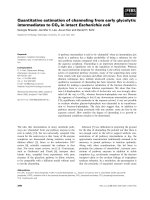
The influence of constant pressure on enzyme
inactivation
A solution of b-glucosidase of P. furiosus was pressur-
ized up to the desired pressure, temperature equili-
brated, and subsequently kept at constant pressure for
1 h. The increase in enzyme inactivation after 1 h (A
70
)
was measured and compared to the blank, which had
only been pressurized and equilibrated for temperature
changes (A
10
). The results at 25 °C and pH 6.0 are
depicted as squares in Fig. 1.
The results show increased enzyme inactivation at
pressures from 100 to 300 MPa. At a constant pressure
of 400 MPa, no enzyme inactivation occurred. When a
higher constant pressure was used, enzyme inactivation
increased again. The same trend was also visible at 40
and 60 °C, when no enzyme inactivation was measured
after 1 h at 400 MPa. It can be concluded that keeping
the enzyme solution at 100 or 400 MPa does not result
in any loss of activity as compared to the activity
immediately after pressurization. In these cases, the
inactivation equilibrium was reached within 10 min.
For some other samples, the situation was less clear,
as there was a difference in activity after 10 and
70 min. Here, the enzyme was still inactivated in time.
Figure 1 also shows the extent of the inactivation
during pressurization and temperature equilibration.
This inactivation was considerable, suggesting fast
inactivation, as half of the enzyme was already inacti-
vated after pressurization and equilibration at
400 MPa. We therefore plotted the enzyme activity as
a function of pressure when compared to the unpres-
surized sample. This is illustrated in Fig. 2A for
T =25°C. The difference between Figs 1 and 2A is
the inclusion of the pressurization time in Fig 2A.
Activity (A
70
) is compared to the untreated blank (A
0
)
instead of to the pressurized blank (A
10
). From the dif-
ference between the two figures, one can see that a
large part of the inactivation already occurred in the
first 10 min of the experiment. This inactivation was
not due to a temperature rise during pressurization.
The b-glucosidase from P. furiosus does not become
inactivated after weeks of storage at temperatures
below 60 °C at atmospheric pressure [11], and it can
0
20
40
60
80
100
120
0 200 400 600 800 1000
P (MPa)
Activity (%)
Fig. 1. Influence of pressure on the enzyme activity at constant
pressure (A
70
⁄ A
10
)() and under pressurization (A
10
⁄ A
0
)(e)of
P. furiosus b-glucosidase at 25 °C and pH 6.0.
Stability of Pyrococcus furiosus glucosidase M. E. Bruins et al.
110 FEBS Journal 276 (2009) 109–117 ª 2008 The Authors Journal compilation ª 2008 FEBS
be concluded that a higher temperature helps to stabi-
lize the enzyme against pressure denaturation. There-
fore, when fast denaturation occurs, as is the case in
our experiments, enzyme stability should be compared
to that of unpressurized samples.
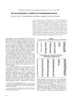
The influence of pressure treatment on enzyme
inactivation
The inactivation of b-glucosidase was measured after
pressure release as a function of the incubation
temperature, pH and pressure. Activity measurements
were compared to those of untreated sample. A signifi-
cant part of the inactivation took place at pressures
£ 300 MPa (Fig. 2). At higher pressures, a plateau
could be seen between 300 and 600 MPa, where no
further enzyme inactivation occurred, suggesting the
existence of a pressure intermediate. Pressure interme-
diates have also been reported for other proteins, e.g.
lysozyme, ribonuclease, a-lactalbumin, apomyoglobin
[12], tropomyosin [13] or synthetic proteins [14].
Hydrostatic pressure is increasingly being used in the
study of protein folding, misfolding, aggregation and
transitions. In comparison with other methods of
denaturation, such as temperature or chemical agents,
pressure induces more subtle changes in protein con-
formation, allowing the stabilization of partially folded
states that are often not significantly populated under
more drastic conditions [15]. The inactivation plateau
that indicates an intermediate state was less clear at
25 °C. At pressures above 600 MPa, inactivation
increased again. Complete inactivation occurred
between 700 and 800 MPa.
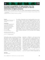
Several of the samples from this experiment were
also loaded on a native gel to detect possible dissocia-
tion or aggregation of the protein. On the gel (Fig. 3),
one band was found for all samples; this band proba-
bly corresponded to the native enzyme. The samples
that were treated at 600 and 700 MPa also showed a
second band with lower mobility. This protein with
higher molecular mass could be an aggregated form of
the enzyme. Aggregation may therefore be a cause of
inactivation at higher pressures. No dissociation into
subunits was observed. One similar example from the
literature showed that inactivation of the dimeric
almond b-glucosidase was not a result of unfolding,
dissociation or aggregation of the intact enzyme [16].
Influence of temperature on enzyme inactivation
by pressure treatment
The influence of temperature on the inactivation of
pressure-treated samples can be seen when comparing
Fig. 2A–C, which show results obtained at different
temperatures. Clearly, the enzyme is more pressure sta-
ble at higher temperatures. Comparison of the mea-
surements made at pH 6.0 shows that after pressure
treatment at 25 °C, 36% activity was left at 500 MPa,
at 40 °C the same activity was still present at
650 MPa, and at 60 °C, 36% residual enzyme activity
was found at 700 MPa. The maximum temperature for
pressure stabilization may very well not have been
reached; however, we were not able to use the high-
pressure equipment at higher temperatures.
0
20
40
60
80
100
120
A
B
C
P (MPa)
Activity (A
70
/A
0
) (%)Activity (A
70
/A
0
) (%)Activity (A
70
/A
0
) (%)
0
20
40
60
80
100
120
P (MPa)
0
20
40
60
80
100
120
0 200 400 600 800 1000
0 200 400 600 800 1000
P (MPa)
0 200 400 600 800 1000
Fig. 2. Influence of pressure on the enzyme activity (A
70
⁄ A
0
) of the
b-glucosidase after 70 min of pressure treatment including pressuri-
zation. Temperatures used were 25 °C (A), 40 °C (B) and 60 °C (C)
at pH 5.5 (e), pH 6.0 (
) and pH 6.5 ( ).
M. E. Bruins et al. Stability of Pyrococcus furiosus glucosidase
FEBS Journal 276 (2009) 109–117 ª 2008 The Authors Journal compilation ª 2008 FEBS 111
At 60 °C, the b-glucosidase from Pyrococcus is very
piezostable as compared to other b-glucosidases.
Almond b-glucosidase has a residual activity of only
20% after 1 h at 200 MPa and 60 °C [16]. The ther-
mophilic b-glucosidase from S. solfataricus is more
piezostable at 60 °C, with a 50% inactivation at
250 MPa and 60 °C [16]. Under these conditions, the
residual enzyme activity of the Pyrococcus b-glucosi-
dase is estimated to be about 70%, making it the
most piezostable of these three enzymes at higher
temperatures. At lower temperatures, however, pres-
sure-assisted cold-induced changes in the structure
cause denaturation and make the enzyme less stable,
but still comparable to, for example, the almond
b-glucosidase or the b-galactosidase from Escherichia
coli [17].
Influence of pH on enzyme inactivation by
pressure treatment
The pressure–temperature dependence of the pH of the
Mes buffer (pH 6.0) was calculated in the relevant
range for the enzyme inactivation experiments, using
the equation of Elyanov & Hamann [18]. The tempera-
ture dependence of this buffer is )0.011 DpH unitÆ°C
)1
[19], and the reaction volume (DV
0
) is 3.9 cm
3
Æmol
)1
[20]. At higher temperatures, the pH will decrease, and
at higher pressures, it will decrease. The pH varies
from 5.6 to 6.3 in the pressure–temperature plane of
measurements for the inactivation studies when start-
ing with a buffer of pH 6.0 at ambient conditions (for
a graph of the pH of Mes buffer plotted as a function
of pressure and temperature, see [10]).
From Fig. 2, we can conclude that the enzyme is
more pressure stable at higher pH values over the
whole pressure and temperature range used in the inac-
tivation experiments. This is in agreement with previ-
ous inactivation measurements at atmospheric pressure
and 95 °C [10]. Here, measurements were conducted as
a function of time. A decrease in pH of 0.5 units
caused the enzyme inactivation constant to increase by
a factor of 2–3.
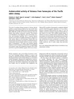
Temperature dependence of the FTIR spectra of
b-glucosidase
FTIR spectroscopy was used to follow the thermally
induced changes in the secondary structure of b-glu-
cosidase from P. furiosus. As previous reports sug-
gested that b-glucosidase unfolds at temperatures
> 100 °C (at 0.1 MPa) [1,4], the heat denaturation
was investigated using the variable-temperature cell,
where a low pressure was applied to keep water in
the liquid state. Figure 4 shows the effect of tempera-
ture on the deconvoluted amide I¢ band (1600–
1700 cm
)1
), which is the conformationally most sensi-
tive vibrational mode. At 25 °C, two peaks at 1654
and 1636 cm
)1
, indicative of a-helix and b-sheet
structures, respectively, can be observed [4]. As tem-
perature increases, the native peaks disappear, and
concomitantly one can observe the appearance of two
peaks at 1618 and 1683 cm
)1
, which are typical of
the formation of an intermolecular antiparallel b-sheet
aggregate [21].
The thermal stability of b-glucosidase was assessed
by plotting the temperature dependence of the peak
intensity at 1618 cm
)1
(Fig. 4B). The melting point of
the enzyme was estimated to be 122 °C at 50 MPa
in Tris (pH 7.5), which is in close agreement with the
value of 108 °C found in a sodium phosphate buffer
(pH 6) at 0.3 MPa by differential scanning calorimetry
[22]. Clearly, this enzyme from a hyperthermophile is
more stable than those from mesophiles [16,17]. The
downward trend above 127 °C is indicative of the
dissociation of the aggregates at higher temperatures,
as observed previously in the case of myoglobin and
lysozyme [21,23].
Thermal stability up to 80 °C was also investigated
at 200 and 400 MPa. Under these conditions, thermal
unfolding could not be observed.
Fig. 3. Native gel electrophoresis of the pressure-treated enzyme.
The pressure in MPa is given below the lanes.
Stability of Pyrococcus furiosus glucosidase M. E. Bruins et al.
112 FEBS Journal 276 (2009) 109–117 ª 2008 The Authors Journal compilation ª 2008 FEBS
Pressure dependence of the FTIR spectra of
b-glucosidase
To determine whether pressure inactivation is corre-
lated with a conformational change, the secondary
structure of b-glucosidase was also monitored by FTIR
spectroscopy during compression. Figure 5 illustrates
the conformational changes observed at different tem-
peratures. The loss of the intensity at 1654 cm
)1
is
accompanied by an increase in absorbance around
1621 cm
)1
. The latter peak can be attributed to the
pressure-induced solvation of a-helices [24]. In addi-
tion, the band at 1.0 GPa in Fig. 5C does not
resemble the broad, featureless band typical of an
unfolded protein. Taken together, these observations
suggest that the unfolding at the level of the secondary
structure is incomplete, with the pressure-unfolded
state having molten globule-like characteristics. Consis-
tent with previous work, this pressure-unfolded state is
highly aggregation prone at high temperatures, as
evidenced by the appearance of the spectral bands at
1683 and 1618 cm
)1
upon decompression (Fig. 5C)
[25].
The changes in absorbance at 1654 cm
)1
have been
plotted as a function of pressure at different tempera-
tures (Fig. 6). A cooperative transition can be seen at
most temperatures. However, at the low and high ends
of the temperature range investigated (10–105 °C), the
change in absorption was very gradual and no clear
transition was measured (Fig. 6A). A reduced cooper-
ativity at low temperature was previously also
observed for myoglobin [26]. It most likely reflects the
fact that close to the low and high unfolding tempera-
tures, the native state is already more heterogeneous.
Hence, it was not possible to determine the pressure
midpoint at these temperatures.
On the basis of the above results, a tentative pres-
sure–temperature stability diagram can be drawn
(Fig. 7). Note that this graph also includes the points
A
B
Fig. 4. The effect of increasing temperature from 25 °C up to 127 °C
on the normalized deconvoluted amide I¢ band of b-glucosidase at
atmospheric pressure (A) with DA
1618
at several temperatures (B).
The arrows indicate the direction of the temperature-induced
changes.
A
B
C
Fig. 5. Effect of pressure on the deconvoluted amide I¢ band of
b-glucosidase (A) at 10 °C, pressure range from 0.1 to 1.1 GPa, (B)
at 30 °C at 0.1 MPa (solid line, bold) and at 740 MPa (solid line)
and (C) at 85 °C at 0.1 MPa (solid line, bold), at 1.0 GPa (solid line)
and after pressure release (dotted line). The arrows indicate the
direction of the pressure-induced changes.
M. E. Bruins et al. Stability of Pyrococcus furiosus glucosidase
FEBS Journal 276 (2009) 109–117 ª 2008 The Authors Journal compilation ª 2008 FEBS 113
determined from the inactivation measurements, as
well as the melting point at 0.3 MPa taken from Bauer
& Kelly [22]. The diagram shows that at low pressures,
b-glucosidase is stabilized by pressure against thermal
denaturation, which has also been observed for other
glucosidases [27]. The enzyme becomes less pressure
sensitive as temperature increases, with an optimum at
85 °C. Finally, the pressure at which the protein
unfolds coincides with that at which the second transi-
tion in the inactivation measurements of Fig. 2 occurs.
The absence of a transition at lower pressures suggests
that the initial inactivation (100–400 MPa) is not cor-
related with a change in secondary structure. This is
consistent with the finding that at pH 10 and 75 °C,
the inactivation of b-glucosidase also does not involve
any loss of secondary structure [1].
Conclusions
The stability of the b-glucosidase from the hyperther-
mophile P. furiosus was studied as a function of tem-
perature, pressure and pH. As well as the expected
high thermostability, the enzyme proved to be highly
piezostable as well. This may be a more general feature
of hyperthermophilic enzymes [5,28]. An increase in
pH from 5.5 to 6.5 and possibly also higher values was
also shown to have a positive effect on the stability of
the enzyme.
A biochemical study by Ausili et al. on the hyperther-
mostability of the b-glucosidase from P. furiosus sug-
gested that the enzyme is mainly stabilized by
hydrophobic interactions, and that it has a very com-
pact protein core with only a few, small internal cavities
[4]. The absence of cavities is an important factor con-
tributing to the pressure stability of the enzyme [29].
Another striking feature of P. furiosus b-glucosidase
is its tetrameric structure, which has been observed for
all hyperthermophilic members of family 1 b-glucosid-
ases, whereas mesophilic and thermophilic family 1
enzymes are mainly active as monomers or dimers [3].
In the case of P. furiosus b-glucosidase, the subunit
interfaces involve fewer electrostatic interactions such
as salt bridges and ion pairs than in the case of the
enzymes from the hyperthermophiles S. solfataricus
and Thermosphaera aggregans [4]. Electrostatic interac-
tions are known to be very pressure sensitive because
A
B
C
Fig. 6. Absorbance at 1654 cm
)1
against pressure at (A) 10 °C,
(B) 30 °C and (C) 85 °C.
1000
800
600
400
P (MPa)
200
0
0
20
40
60 80 100 120 140
T (°C)
Fig. 7. Pressure–temperature diagram indicating the transition
points. (n) Second transition estimated from the inactivation experi-
ments; transition points based on A
1654
( )orA
1618
(d) from the
FTIR experiments; s corresponds to literature data [22]. Open sym-
bols: measurements at pH 6 in Mes or phosphate buffer. Closed
symbols: measurements in Tris buffer at pH 8. The solid line is a
guide to the eye, assuming an ellipse.
Stability of Pyrococcus furiosus glucosidase M. E. Bruins et al.
114 FEBS Journal 276 (2009) 109–117 ª 2008 The Authors Journal compilation ª 2008 FEBS
of the large volume change associated with the forma-
tion of a free charge [30]. Hence, a reduction in the
number of electrostatic interactions would increase the
pressure stability of the tetramer. Maintaining the tet-
rameric structure of b-glucosidase is therefore not only
important for its temperature stability, but also con-
tributes to the pressure stability. However, a subtle
change in the oligomeric state of the enzyme, not caus-
ing dissociation or association, may have led to a lower
active state of the enzyme at lower pressures (100–
400 MPa). Also, a change in the active site and⁄ or the
substrate-binding site may have led to considerable
inactivation at lower pressures (100–400 MPa) well
before any conformational changes occurred.
Experimental procedures
Enzyme purification
The enzyme was prepared from a lysate of E. coli in which
the celB gene encoding b-glucosidase from P. furiosus was
cloned and expressed as described previously [31]. Briefly,
the cell lysate was heated in order to denature proteins
other than the hyperthermostable enzyme, and this was fol-
lowed by an anion exchange chromatography step for fur-
ther purification. The enzyme was then dialysed against
5mm Mes (pH 6.0) and freeze-dried. For activity measure-
ments, the enzyme was redissolved at 0.5 mgÆmL
)1
in 0.1 m
Mes buffer (pH 6.0) at 25 °C and 0.1 MPa. FTIR spectros-
copy experiments were performed in deuterated 0.1 m Mes
buffer (pD 6.0) (measurement at 85 °C) or 0.05 m Tris ⁄ DCl
buffer (pD 8.0) (all other conformational measurements),
leading to final pD values of 6.4 and 7.5, respectively, and
final protein contents of 100 and 50 mgÆmL
)1
, respectively.
Enzyme inactivation measurements
b-Glucosidase activity was assayed at atmospheric pressure
using p-nitrophenyl-b-d -glucopyranoside as an artificial
substrate. Ten microlitres of enzyme solution was added to
a standard reaction mixture that was equilibrated at 80 °C
to make a 1.0 mL solution of 2.0 mm p-nitrophenyl-b-d-
glucopyranoside in 0.1 m Mes buffer (pH 6.0). The reaction
was terminated after 10 min by addition of 1.0 mL of 1.0 m
sodium bicarbonate. The increase in absorbance at 420 nm
as a result of p-nitrophenol formation was measured spec-
trophotometrically.
Inactivation studies
The enzyme solution was diluted 120-fold in Mes buffer,
pH 5.5, 6.0 or 6.5. Four hundred and fifty microlitres of
enzyme solution was put in polyethylene bags and pressur-
ized in a laboratory-scale multivessel high-pressure appara-
tus (Resato FPU 100-50; Resato International B.V., Roden,
The Netherlands). The pressure vessels were pre-equili-
brated to the desired temperature. A glycol mixture was
used as pressure medium. One vessel contained three bags,
to ensure similar treatment of samples with a different pH.
The pressure was increased gradually (100 MPaÆmin
)1
)to
minimize any temperature increase due to adiabatic heating,
but nevertheless, the temperature increased by 9–10 °C dur-
ing the pressure build-up. Therefore, an equilibration period
was taken into account to allow the temperature to reach its
desired value, once the preset pressure was reached. At this
point, the valves of the individual vessels were also closed,
and the central circuit was decompressed. The total time of
pressurization and equilibration was approximately 10 min.
After that, at t = t
10
, the first vessel was decompressed.
After 1 h (t
70
), another vessel was individually depressur-
ized. All samples were immediately cooled in ice–water, and
enzyme inactivation was measured within a few hours. The
measured activities were that of the untreated blank sample
(A
0
), the pressurized blank sample (A
10
) and the sample that
was kept at a predefined pressure for 1 h (A
70
).
The inactivation of b-glucosidase from P. furiosus was
studied at pressures up to 900 MPa, at temperatures of 25,
40 and 60 °C, and at pH 5.5, 6.0 and 6.5.
Gel electrophoresis
To detect possible dissociation or aggregation of the pro-
tein, native PAGE was performed with samples prepared at
25 °C and different pressures, as described in the inactiva-
tion studies. These samples (A
70
) were mixed 1 : 1 with
electrophoresis buffer (10 mm Tris, pH 6.8, 2.5% brom-
ophenol blue) and applied to the gel (8–25% gradient gel)
for 25 min. The proteins on the gel were stained with
Coomassie blue.
FTIR spectroscopy
Infrared spectra were recorded on a Bruker IFS66 FTIR
spectrometer (Bruker, Karlsruhe, Germany) equipped with
a liquid nitrogen-cooled mercury cadmium telluride detec-
tor at a nominal resolution of 2 cm
)1
. Each spectrum is the
average of 256 interferograms. Equilibration after pressure
increase and measurement of the sample took appro-
ximately 10 min, leading to a pressure build-up of
250–300 MPa per hour. The sample compartment was
continuously purged with dry air to minimize the spectral
contribution of atmospheric water.
The pressure experiments were performed using a diamond
anvil cell (DAC) [32]. The pressure stability was measured at
various temperatures by adjusting the temperature of the
water bath to which the DAC was connected. The sample
temperature was monitored using a thermocouple located
close to the diamonds. For pressure measurements at temper-
atures above 85 °C, a modified variable-temperature cell
M. E. Bruins et al. Stability of Pyrococcus furiosus glucosidase
FEBS Journal 276 (2009) 109–117 ª 2008 The Authors Journal compilation ª 2008 FEBS 115
(Graseby Specac, Orpington, UK) was used. In this set-up,
the classic temperature cell is replaced by a DAC.
A baseline correction was performed in the amide I¢ region
(1600–1700 cm
)1
), assuming a linear baseline. In order to
enhance the component peaks contributing to the amide I¢
band, the spectra were treated by Fourier self-deconvolution
using the bruker software (OS ⁄ 2 version). The line shape
was assumed to be Lorentzian with a half-bandwidth of
21 cm
)1
, and an enhancement factor k of 1.7 was used.
Acknowledgements
The high-pressure equipment for our inactivation stud-
ies was available to us thanks to Ariette Matser (Agro-
technology and Food Innovations, Wageningen
University and Research Centre). b-Glucosidase from
P. furiosus was kindly provided by J. van der Oost
(Laboratory of Microbiology, Wageningen University).
M. E. Bruins is supported by a VENI grant from the
technology foundation STW, the applied science divi-
sion of NOW, and the technology programme of the
Ministry of Economic Affairs. F. Meersman is a post-
doctoral fellow of the Research Foundation Flanders
(FWO-Vlaanderen).
References
1 Pouwels J, Moracci M, Cobucci-Ponzano B, Perugino
G, van der Oost J, Kaper T, Lebbink JHG, de Vos
WM, Ciaramella M & Rossi M (2000) Activity and sta-
bility of hyperthermophilic enzymes: a comparative
study on two archaeal beta-glycosidases. Extremophiles
4, 157–164.
2 Kengen SWM, Luesink EJ, Stams AJM & Zehnder
AJB (1993) Purification and characterization of an
extremely thermostable beta-glucosidase from the
hyperthermophilic archaeon Pyrococcus furiosus. Eur
J Biochem 213, 305–312.
3 Kaper T, Lebbink JHG, Pouwels J, Kopp J, Schulz
GE, van der Oost J & de Vos WM (2000) Comparative
structural analysis and substrate specificity engineering
of the hyperthermostable beta-glucosidase CelB from
Pyrococcus furiosus. Biochemistry 39, 4963–4970.
4 Ausili A, Cobucci-Ponzano B, Di Lauro B, D’Avino
R, Perugino G, Bertoli E, Scire A, Rossi M, Tanfani
F & Moracci M (2007) A comparative infrared spec-
troscopic study of glycoside hydrolases from extremo-
philic archaea revealed different molecular mechanisms
of adaptation to high temperatures. Proteins 67,
991–1001.
5 Mombelli E, Shehi E, Fusi P & Tortora P (2002)
Exploring hyperthermophilic proteins under pressure:
theoretical aspects and experimental findings. Biochim
Biophys Acta 1595, 392–396.
6 Hei DJ & Clark DS (1994) Pressure stabilization of
proteins from extreme thermophiles. Appl Environ
Microbiol 60, 932–939.
7 Sun MMC, Caillot R, Mak G, Robb FT & Clark DS
(2001) Mechanism of pressure-induced thermo stabiliza-
tion of proteins: studies of glutamate dehydrogenases
from the hyperthermophile Thermococcus litoralis.
Protein Sci 10, 1750–1757.
8 Konisky J, Michels PC & Clark DS (1995) Pressure
stabilization is not a general property of thermophilic
enzymes–the adenylate kinases of Methanococcus voltae,
Methanococcus maripaludis, Methanococcus thermolitho-
trophicus, and Methanococcus jannaschii. Appl Environ
Microbiol 61, 2762–2764.
9 Bruins ME, Janssen AEM & Boom RM (2006) Equilib-
rium shifts in enzyme reactions at high pressure. J Mol
Cat B: Enzymatic 39, 124–127.
10 Bruins ME, Matser AM, Janssen AEM & Boom RM
(2007) Buffer selection for HP treatment of biomaterials
and its consequences for enzyme inactivation studies.
High Press Res 27, 101–107.
11 Bruins ME, Van Hellemond EW, Janssen AEM &
Boom RM (2003) Maillard reactions and increased
enzyme inactivation during oligosaccharide synthesis by
a hyperthermophilic glycosidase. Biotechnol Bioeng 81,
546–552.
12 Jonas J (2002) High-resolution nuclear magnetic reso-
nance studies of proteins. Biochim Biophys Acta 1595,
145–159.
13 Suarez MC, Lehrer SS & Silva JL (2001) Local hetero-
geneity in the pressure denaturation of the coiled-coil
tropomyosin because of subdomain folding units. Bio-
chemistry 40, 1300–1307.
14 Chapeaurouge A, Johansson JS & Ferreira ST (2001)
Folding intermediates of a model three-helix bundle
protein–pressure and cold denaturation studies. J Biol
Chem 276, 14861–14866.
15 Ferreira ST, Chapeaurouge A & De Felice FG (2005)
Stabilization of partially folded states in protein fold-
ing ⁄ misfolding transitions by hydrostatic pressure. Braz
J Med Biol Res 38, 1215–1222.
16 Hamon V, Dallet S & Legoy MD (1996) The pressure-
dependence of two beta-glucosidases with respect to
their thermostability. Biochim Biophys Acta 1294 , 195–
203.
17 Degraeve P, Delorme P & Lemay P (1996) Pressure-
induced inactivation of E-coli beta-galactosidase: influ-
ence of pH and temperature. Biochim Biophys Acta
1292, 61–68.
18 Elyanov BS (1975) Linear free-energy relationship and
some quantitative regularities of effect of pressure on
chemical reactions. Aust J Chem 28, 933–943.
19 Good NE, Winget GD, Winter W, Connolly TN, Izawa
S & Singh RMM (1966) Hydrogen ion buffers for
biological research. Biochemistry 5, 467–477.
Stability of Pyrococcus furiosus glucosidase M. E. Bruins et al.
116 FEBS Journal 276 (2009) 109–117 ª 2008 The Authors Journal compilation ª 2008 FEBS
20 Kitamura Y & Itoh T (1987) Reaction volume of pro-
tonic ionization for buffering agents–prediction of pres-
sure-dependence of pH and pOH. J Solution Chem 16,
715–725.
21 Meersman F & Heremans K (2003) Temperature-
induced dissociation of protein aggregates: accessing the
denatured state. Biochemistry 42, 14234–14241.
22 Bauer MW & Kelly RM (1998) The family 1 beta-
glucosidases from Pyrococcus furiosus and Agrobacte-
rium faecalis share a common catalytic mechanism.
Biochemistry 37, 17170–17178.
23 Meersman F, Smeller L & Heremans K (2005) Extend-
ing the pressure–temperature state diagram of myoglo-
bin. Helv Chim Acta 88, 546–556.
24 Callender RH, Dyer RB, Gilmanshin R & Woodruff
WH (1998) Fast events in protein folding: the time evo-
lution of primary processes. Annu Rev Phys Chem 49,
173–202.
25 Smeller L, Rubens P & Heremans K (1999) Pressure
effect on the temperature-induced unfolding and
tendency to aggregate of myoglobin. Biochemistry
38, 3816–3820.
26 Meersman F, Smeller L & Heremans K (2002) Compar-
ative Fourier transform infrared spectroscopy study of
cold-, pressure-, and heat-induced unfolding and aggre-
gation of myoglobin. Biophys J 82, 2635–2644.
27 Degraeve P, Rubens P, Lemay P & Heremans K (2002)
In situ observation of pressure-induced increased ther-
mostability of two beta-galactosidases with FT-IR spec-
troscopy in the diamond anvil cell. Enzyme Microb
Technol 31, 673–684.
28 Robb FT & Clark DS (1999) Adaptation of proteins
from hyperthermophiles to high pressure and high
temperature. J Mol Microbiol Biotechnol 1, 101–105.
29 Torrent J, Connelly JP, Coll MG, Ribo M, Lange R &
Vilanova M (1999) Pressure versus heat-induced unfold-
ing of ribonuclease A: the case of hydrophobic interac-
tions within a chain-folding initiation site. Biochemistry
38, 15952–15961.
30 Meersman F, Dobson CM & Heremans K (2006) Pro-
tein unfolding, amyloid fibril formation and configura-
tional energy landscapes under high pressure
conditions. Chem Soc Rev 35, 908–917.
31 Voorhorst WGB, Rik IL, Luesink EJ & Devos WM
(1995) Characterization of the Celb gene coding for
beta-glucosidase from the hyperthermophilic archaeon
Pyrococcus furiosus and its expression and site-directed
mutation in Escherichia coli. J Bacteriol 177, 7105–7111.
32 Meersman F & Heremans K (2003) High pressure
induces the formation of aggregation-prone states of
proteins under reducing conditions. Biophys Chem 104,
297–304.
M. E. Bruins et al. Stability of Pyrococcus furiosus glucosidase
FEBS Journal 276 (2009) 109–117 ª 2008 The Authors Journal compilation ª 2008 FEBS 117