Báo cáo khoa học: Proteomic analysis reveals Hrs ubiquitin-interacting motif-mediated ubiquitin signaling in multiple cellular processes ppt
Bạn đang xem bản rút gọn của tài liệu. Xem và tải ngay bản đầy đủ của tài liệu tại đây (546.26 KB, 14 trang )
Proteomic analysis reveals Hrs ubiquitin-interacting
motif-mediated ubiquitin signaling in multiple
cellular processes
Julia W. Pridgeon*, Elizabeth A. Webber*, Di Sha*, Lian Li and Lih-Shen Chin
Department of Pharmacology, Emory University School of Medicine, Atlanta, GA, USA
Ubiquitination is a post-translational modification in
which the 76 amino acid polypeptide ubiquitin is cova-
lently attached to a lysine residue(s) of substrate
proteins [1]. Proteins can be either monoubiquitinated
or polyubiquitinated by attachment of a multiubiquitin
chain linked through one of the internal lysine residues
in ubiquitin [2]. K48-linked polyubiquitination is the
canonical signal that targets proteins for degradation
by the 26S proteasome, whereas monoubiquitination
and K63-linked polyubiquitination serve as regulatory
signals to modulate protein activity, localization, and
interactions [3,4]. Increasing evidence points to the
critical importance of protein ubiquitination in the
control of diverse cellular processes, from DNA repair
and transcription regulation to vesicular trafficking
and virus budding [4–6]. Moreover, dysregulated ubiq-
Keywords
endocytic trafficking; Hrs; in vitro expression
cloning; ubiquitination; ubiquitin-interacting
motif
Correspondence
L S. Chin, Department of Pharmacology,
Emory University School of Medicine, 1510
Clifton Road, Atlanta, GA 30322, USA
Fax: +1 404 727 0365
Tel: +1 404 727 0361
E-mail:
Website: />*These authors contributed equally to this
work
(Received 26 June 2008, revised 19 October
2008, accepted 24 October 2008)
doi:10.1111/j.1742-4658.2008.06760.x
Despite the critical importance of protein ubiquitination in the regulation
of diverse cellular processes, the molecular mechanisms by which cells rec-
ognize and transmit ubiquitin signals remain poorly understood. The
endosomal sorting machinery component hepatocyte growth factor-regu-
lated tyrosine kinase substrate (Hrs) contains a ubiquitin-interacting motif
(UIM), which is believed to bind ubiquitinated membrane cargo proteins
and mediate their sorting to the lysosomal degradation pathway. To gain
insight into the role of Hrs UIM-mediated ubiquitin signaling in cells, we
performed a proteomic screen for Hrs UIM-interacting ubiquitinated pro-
teins in human brain by using an in vitro expression cloning screening
approach. We have identified 48 ubiquitinated proteins that are specifically
recognized by the UIM domain of Hrs. Among them, 12 are membrane
proteins that are likely to be Hrs cargo proteins, and four are membrane
protein-associated adaptor proteins whose ubiquitination may act as a sig-
nal to target their associated membrane cargo for Hrs-mediated endosomal
sorting. Other classes of the identified proteins include components of the
vesicular trafficking machinery, cell signaling molecules, proteins associated
with the cytoskeleton and cytoskeleton-dependent transport, and enzymes
involved in ubiquitination and metabolism, suggesting the involvement of
Hrs UIM-mediated ubiquitin signaling in the regulation of multiple cellular
processes. We have characterized the ubiquitination of two identified
proteins, Munc18-1 and Hsc70, and their interaction with Hrs UIM, and
provided functional evidence supporting a role for Hsc70 in the regulation
of Hrs-mediated endosome-to-lysosome trafficking.
Abbreviations
AD, Alzheimer’s disease; APP, amyloid beta A4 protein; EGF, epidermal growth factor; EGFR, epidermal growth factor receptor; GST,
glutathione S-transferase; HA, hemagglutinin; Hrs, hepatocyte growth factor-regulated tyrosine kinase substrate; IVEC, in vitro expression
cloning; MVB, multivesicular body; RNP, ribonucleoprotein; siRNA, small interfering RNA; UIM, ubiquitin-interacting motif.
118 FEBS Journal 276 (2009) 118–131 ª 2008 The Authors Journal compilation ª 2008 FEBS
uitination has been implicated in the pathogenesis of
many human diseases, including cancer and neurode-
generative disorders [7]. Elucidation of the molecular
mechanisms by which cells recognize and sort ubiquiti-
nated proteins is thus essential for understanding
ubiquitin signaling in both normal physiology and
diseases.
The ubiquitin-interacting motif (UIM) is a conserved
ubiquitin recognition module that was initially
identified on the basis of the sequence homology to the
ubiquitin-binding region of the S5a subunit of the
26S proteasome [8]. The UIM has a 20 amino acid
consensus sequence X-Ac-Ac-Ac-Ac-F-X-X-Ala-X-X-
X-Ser-X-X-Ac-X-X-X-X, where F represents a large
hydrophobic residue and Ac represents an acidic resi-
due. UIMs are found in many proteins implicated in a
variety of cellular processes, including endocytosis, en-
dosome-to-lysosome trafficking, DNA repair, mRNA
splicing, and neurodegeneration [8]. In vitro studies
indicate that the UIM binds monoubiquitin and poly-
ubiquitin chains [9–12]. Furthermore, UIM domains
from different proteins bind polyubiquitin chains of
varying lengths with different affinities [12], suggesting
that different UIM domains may recognize distinct
subsets of ubiquitinated proteins.
Hepatocyte growth factor-regulated tyrosine kinase
substrate (Hrs) is an early endosome-associated UIM-
containing protein that plays a central role in control-
ling endosome-to-lysosome trafficking [13–16]. A
major sorting decision in the endocytic pathway
occurs at the early endosome, where membrane cargo
proteins can be sorted to the recycling pathway for
delivery to the cell surface or to lumenal vesicles of
multivesicular bodies (MVBs) for eventual degrada-
tion in the lysosome [6,16]. A sorting signal for cargo
trafficking to the lysosomal pathway is the ubiquitina-
tion of cargo proteins. The UIM domain of Hrs has
been shown to bind ubiquitin in vitro [9,11,14] and to
facilitate the sorting of several ubiquitinated cargo
proteins to the lysosomal pathway in mammalian cells
[17] and yeast [10,18]. The Hrs UIM domain may also
interact with ubiquitinated components of the endoso-
mal trafficking machinery to regulate endosome-
to-lysosome trafficking [6]. Recently, Hrs has been
shown to preferentially bind K63-linked polyubiquitin
chains [19]. Interestingly, the UIM domain is indis-
pensable for monoubiquitination as well as phosphor-
ylation of Hrs [9,12,20], raising the possibility that the
Hrs UIM domain may bind E3 ubiquitin-protein
ligase(s) and ⁄ or kinase(s). The identities of ubiquiti-
nated cargo and other cellular proteins that are recog-
nized by the Hrs UIM domain remain largely
unknown.
In order to gain insight into the role of Hrs UIM-med-
iated ubiquitin signaling in cells, we performed a proteo-
mic screen for Hrs UIM-interacting ubiquitinated
proteins in human brain by using a combined in vitro
expression cloning (IVEC) and glutathione S-transferase
(GST) pull-down approach (Fig. 1). IVEC is a powerful
screening method that combines biochemical analysis of
radioactively labeled proteins with the ability to quickly
isolate the corresponding cDNAs [21,22]. As compared
to yeast two-hybrid screening, IVEC screening offers the
advantage of studying direct interactions between two
proteins in vitro [23], rather than indirect analysis of the
interactions between fusion proteins inside the yeast
nucleus. Moreover, our IVEC screening approach
complements other proteomic screening strategies
[24,25], which are often contaminated with secondary,
nonspecific binding proteins.
Here, we report the identification of a set of proteins
that are specifically recognized by the UIM domain of
Hrs. Our results reveal the involvement of Hrs UIM-
mediated protein interactions in the coordination of
multiple steps in endosomal trafficking as well as in
the regulation of cell signaling, cytoskeleton and mem-
brane dynamics and other cellular processes.
Results
IVEC screen for proteins that are specifically
recognized by the UIM domain of Hrs
To identify cellular targets of the Hrs UIM domain,
we screened a human adult brain cDNA library for
Hrs UIM-interacting proteins using an IVEC approach
[21,22], which is summarized in Fig. 1. Pools of
cDNAs (100 independent cDNA clones per pool) from
the human brain library were in vitro transcribed and
translated in the TNT Quick coupled transcription–
translation reticulocyte lysate system in the presence of
[
35
S]methionine and ubiquitin to generate
35
S-labeled
protein pools [23]. It has been well established that
such a transcription–translation reticulocyte lysate sys-
tem is capable of carrying out ubiquitination of in vitro
translated proteins [23,26,27]. To determine whether
the protein pools synthesized in our IVEC system are
ubiquitinated, we performed immunoblot analysis with
antibody against ubiquitin to examine the ubiquitina-
tion status of protein pools generated from the in vitro
transcription–translation of human brain cDNA pools
in the presence or absence of ubiquitin (Fig. 2B). We
found that addition of ubiquitin to the in vitro tran-
scription–translation reaction mixture dramatically
increased the ubiquitination levels of in vitro translated
protein pools, confirming that protein pools synthe-
J. W. Pridgeon et al. Hrs UIM-mediated protein interactions
FEBS Journal 276 (2009) 118–131 ª 2008 The Authors Journal compilation ª 2008 FEBS 119
sized in our IVEC system are indeed ubiquitinated.
The
35
S-labeled ubiquitinated protein pools were then
tested for their ability to bind to a GST-fused UIM
domain of Hrs protein (Fig. 2A) in an in vitro binding
assay. Figure 2C shows an example of six positive
pools (pools 1, 2, 4, 5, 7, and 8) containing Hrs UIM-
binding proteins isolated from the primary screen.
From each of the positive pools, individual cDNA
clones were isolated and subjected to a secondary
screen in the same manner to identify positive cDNA
clones encoding Hrs UIM-binding proteins (Fig. 2D).
The specificity of the observed interactions was con-
firmed by the specific binding of the identified proteins
to GST–Hrs UIM but not to GST control (Fig. 2E).
From the IVEC screen, we isolated 64 positive
clones, which encode 48 proteins that are specifically
recognized by the UIM domain of Hrs (Fig. 3 and
Table 1). The specific binding of the identified proteins
to the Hrs UIM domain suggests that these proteins
may be ubiquitinated. In support of this notion, four
of the identified proteins, APP [28], b-tubulin [29],
Hsc70 [30,31], and MARK4 [32], have been previously
shown to be ubiquitinated. However, the interaction of
these proteins with the Hrs UIM domain has never
been reported. The remaining 43 proteins have not
previously been shown to be ubiquitinated or to bind
to the Hrs UIM domain.
Classification of the identified Hrs
UIM-interacting proteins
To shed light on the role of Hrs UIM-mediated
protein interactions, we categorized the 48 proteins
isolated from the IVEC screen according to functional
predictions based on the available literature, gene
ontology, and homology searches (Table 1). Classifica-
tion of the identified Hrs UIM-interacting proteins
according to their cellular localization (Fig. 3A) reveals
that the majority of these proteins are integral mem-
brane proteins (27%), membrane-associated proteins
(21%), or cytosolic proteins (27%). We and others
have shown that Hrs is associated with both early
endosomal membrane and cytosolic fractions
[13,33,34]. The localization of the majority of the iden-
tified Hrs UIM-binding proteins to the membrane and
cytosol suggests that they are appropriately positioned
to interact with Hrs in cells. In addition, we identified
a number of proteins that could be classified as cyto-
skeletal (15%), which suggests that Hrs UIM-mediated
ubiquitin signaling may have a role in regulation of
cytoskeleton dynamics. The localization of only 4%
and 6% of proteins could be classified as nuclear or
unknown, respectively.
Dividing the identified Hrs UIM-interacting proteins
on the basis of their functional classes (Fig. 3B) further
suggests that the screen largely identified putative
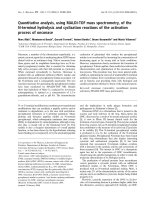
Fig. 1. Schematic illustrating the IVEC system used to identify and
isolate cDNAs from human adult brain library that encode Hrs
UIM-interacting proteins.
Hrs UIM-mediated protein interactions J. W. Pridgeon et al.
120 FEBS Journal 276 (2009) 118–131 ª 2008 The Authors Journal compilation ª 2008 FEBS
membrane cargo proteins and membrane cargo adap-
tor proteins (33%), consistent with the proposed func-
tion of Hrs in endosomal sorting and trafficking. The
other major functional groups to which the Hrs UIM-
interacting proteins belong include cell signaling
(17%), metabolism (17%), vesicular trafficking (13%),
and transport (10%), suggesting an interconnection
between Hrs UIM-mediated ubiquitin signaling and
these cellular processes.
Characterization of Munc18-1 and Hsc70 as Hrs
UIM-interacting ubiquitinated proteins
The ability of the Hrs UIM domain to bind ubiquitin
and ubiquitinated proteins has been well established
[9,11,14,17,19]. Thus, the direct interaction between
the Hrs UIM domain and each of the 48 proteins
identified from our IVEC screen (Table 1) raises the
possibility that these proteins are ubiquitinated in
cells. To test this possibility, we used a well-estab-
lished in vivo ubiquitination assay [35,36] to determine
the ubiquitination status of the identified Hrs UIM-
interacting protein Munc18-1, a key regulator of
Ca
2+
-dependent exocytosis [37], which has been
previously unrecognized as a ubiquitinated protein.
Lysates from HeLa cells expressing hemagglutinin
(HA)-tagged ubiquitin and Myc-tagged Munc18-1
were subjected to immunoprecipitation with anti-
bodies against Myc, followed by immunoblotting with
antibodies against HA to detect HA–ubiquitin-
conjugated Munc18-1 protein (Fig. 4A). We observed
a prominent band around 82 kDa that may represent
a diubiquitinated species of Munc18-1, as well as a
higher molecular mass smear that may represent poly-
ubiquitinated forms of Munc18-1. These results pro-
vide the first evidence that Munc18-1 is ubiquitinated
in vivo, and support the notion that Hrs UIM-binding
proteins isolated from our IVEC screen probably
represent ubiquitinated proteins.
Next, we sought to determine whether ubiquitinated
Munc18-1 is specifically recognized by Hrs UIM.
In vitro binding assays were performed by incubating
A
B
D
C
E
Fig. 2. IVEC screen for proteins that bind to the UIM domain of
Hrs. (A) Domain structure of full-length Hrs (top) and the GST-fused
Hrs UIM domain used in the IVEC screen (bottom). (B) Two cDNA
pools, IA1 and IB1, containing 100 independent cDNA clones per
pool from a human adult brain cDNA library, were in vitro tran-
scribed and translated in the presence of cold methionine with or
without ubiquitin. The control (CTL) reactions were carried out
under the same conditions with no cDNAs added. The synthesized
protein pools were analyzed by immunoblotting with antibody
against ubiquitin. (C) Primary screen for positive pools containing
Hrs UIM-binding proteins. Pools of cDNAs (100 independent cDNA
clones per pool) from a human adult brain cDNA library were
in vitro transcribed and translated in the presence of [
35
S]methio-
nine and ubiquitin and then subjected to a GST–Hrs UIM pull-down
assay. Bound proteins were analyzed by SDS ⁄ PAGE. Autoradiogra-
phy of gel samples was performed using a phosphoimager. Exam-
ple of positive pools (pools 1, 2, 4, 5, 7, and 8) selected for
secondary screen. The two bands labeled a and b in pool 4 repre-
sent distinct Hrs UIM-binding proteins, which would be individually
isolated by secondary screen. (D) Secondary screen for isolation of
individual positive cDNA clones encoding Hrs UIM-binding proteins.
In vitro translated products from individual cDNA clones isolated
from each of the positive pools were analyzed as described above
for their ability to bind GST–Hrs UIM. Example of eight single
clones isolated from pool 4, of which clones 3 and 5 are individual
positive cDNA clones encoding Hrs UIM-binding proteins a and b
indicated in (C). (E) Specificity of Hrs UIM domain binding. In vitro
translated products from three isolated individual cDNA clones
(Input) were incubated with immobilized GST–Hrs UIM fusion pro-
tein or GST control. Bound proteins were analyzed by SDS ⁄ PAGE
and autoradiography. Clone 2 encodes a protein that specifically
binds to GST–Hrs UIM but not to GST control, whereas clones 1
and 3 are negative interactors that bind neither to GST–Hrs UIM
nor to GST control.
J. W. Pridgeon et al. Hrs UIM-mediated protein interactions
FEBS Journal 276 (2009) 118–131 ª 2008 The Authors Journal compilation ª 2008 FEBS 121
immobilized GST–Hrs UIM, full-length GST–Hrs,
GST–HrsDUIM, or GST control proteins (Fig. 5A)
with soluble Myc-tagged Munc18-1 immunopurified
from transfected HeLa cells. Bound proteins were
probed with an antibody against ubiquitin and an anti-
body against Myc to detect ubiquitinated Munc18-1
and nonubiquitinated Munc18-1, respectively
(Fig. 5B). We found that both GST-fused Hrs UIM
domain and full-length Hrs selectively interacted with
ubiquitinated Munc18-1 but not with nonubiquitinated
Munc18-1 protein. The ability of Hrs to bind ubiquiti-
nated Munc18-1 was dramatically reduced by the dele-
tion of the UIM domain. Furthermore, the GST
control did not pull down any detectable level of ubiq-
uitinated or nonubiquitinated Munc18-1. Together,
these results indicate that the Hrs UIM domain is both
necessary and sufficient for binding Munc18-1 in a
ubiquitin-dependent manner, and support the validity
and specificity of our IVEC screen.
Our identification of 48 proteins as novel binding
partners for the Hrs UIM domain has led to a number
of interesting hypotheses. For example, previous stud-
ies have shown that Hrs is enriched with ubiquitinated
cargo proteins in flat clathrin-coated microdomains of
early endosomes [17,38,39]. These clathrin-coated
microdomains have been proposed to play a role in
endosomal sorting and retention of ubiquitinated cargo
proteins [17,39]. The flat clathrin coat has to be
dissociated prior to endosomal invagination and
budding of the MVB lumenal vesicles [17,39], but
the molecular machinery for the disassembly of the
endosomal clathrin coat remains unknown. Hsc70 is a
constitutively expressed member of the Hsp70 molecu-
lar chaperone family and has been shown to regulate
clathrin uncoating processes [40,41]. Although Hsc70 is
known to be ubiquitinated [30,31], it has been previ-
ously unrecognized as an Hrs-binding protein. Our
identification of Hsc70 as an Hrs UIM-interacting
protein raises an intriguing hypothesis that the
Hrs UIM-mediated interaction recruits Hsc70 to endo-
somes for clathrin uncoating prior to the budding of
MVB lumenal vesicles. As a first step to test this
hypothesis, we performed in vivo ubiquitination analy-
sis to confirm that Hsc70 is indeed ubiquitinated in
cells (Fig. 4B). Furthermore, we performed binding
experiments and found that ubiquitinated Hsc70
specifically bound to GST–Hrs UIM and GST–Hrs,
but not to GST–HrsDUIM or the GST control
(Fig. 5C), indicating that the Hrs UIM domain is both
necessary and sufficient for binding ubiquitinated
Hsc70. Our results showed that the Hrs UIM domain
is unable to bind Hsc70 in the absence of ubiquitina-
tion, as GST–Hrs UIM did not pull down any detect-
able level of nonubiquitinated Hsc70 (Fig. 5C).
Interestingly, our analysis revealed that the full-length
Hrs was capable of interacting with nonubiquitinated
Hsc70 and that this interaction was not affected by the
deletion of the UIM domain (Fig. 5C), suggesting that
the interaction of Hrs with nonubiquitinated Hsc70
is mediated by a binding site on Hrs that is located
outside of its UIM domain.
Hsc70 is essential for ligand-induced epidermal
growth factor receptor degradation
Next, we assessed the role of Hsc70 in the regulation
of Hrs-mediated endosomal trafficking by using the
epidermal growth factor (EGF) receptor (EGFR) as a
cargo protein. Previous studies have shown that bind-
ing of EGF to the EGFR at the plasma membrane
causes rapid internalization of the EGF–EGFR com-
plex and subsequent sorting at the early endosome for
delivery to the lysosome for degradation [42–44]. The
role of Hrs-mediated early endosomal sorting in the
regulation of EGF-induced EGFR degradation is well
established; both the overexpression and the depletion
of Hrs inhibit ligand-induced degradation of the
EGFR [13,45]. Our identification of the interaction
between Hsc70 and Hrs raises the possibility that
Hsc70 may participate in the regulation of ligand-
induced endocytic trafficking of the EGF–EGFR
A
B
Fig. 3. Classification of the identified proteins according to cellular
localization (A) and molecular function (B). The number of proteins
in each category is expressed as the percentage of the total num-
ber of different proteins identified from the screen.
Hrs UIM-mediated protein interactions J. W. Pridgeon et al.
122 FEBS Journal 276 (2009) 118–131 ª 2008 The Authors Journal compilation ª 2008 FEBS
Table 1. Hrs UIM-interacting proteins identified from the IVEC screen.
Gene Protein name (alternative name) Accession number
Membrane proteins
APP
a
Amyloid beta (A4) protein NP_958816
APLP2 Amyloid beta (A4) precursor-like protein 2 NP_001633
ATP1A1 Na
+
⁄ K
+
-ATPase alpha 1 subunit NP_000692
ATP2A2 ATPase, Ca
2+
transporting, slow twitch 2 NP_001672
ARL6IP1 ADP-ribosylation factor-like 6 interacting protein NP_055976
BSG Basigin NP_940991
C3orf1 Hypothetical protein LOC51300 NP_057673
FAM5C Family with sequence similarity 5, member C NP_950252
MEST Mesoderm specific transcript NP_002393
TMCC2 Transmembrane and coiled-coil domain family 2 NP_055673
TMEM49 Transmembrane protein 49 (VMP1) NP_112200
UNC84B Unc-84 homolog B (Rab5IP) NP_056189
Membrane protein-associated adaptor proteins
AHCYL1 S-adenosylhomocysteine hydrolase-like 1 (IRBIT) NP_006612
CASK
b
Calcium ⁄ calmodulin-dependent serine protein kinase NP_003679
TJP2 Tight junction protein 2 (ZO-2) NP_963923
TRAP1 TNF receptor-associated protein 1 NP_057376
Vesicular trafficking
GGA2
c
ADP-ribosylation factor binding protein 2 NP_055859
HSPA8
a
Heat shock 70 kDa protein 8 (Hsc70) NP_006588
MAP3K10
b
Mitogen-activated protein kinase 10 (MLK2) NP_002437
STXBP1 Syntaxin-binding protein 1 (Munc18-1) NP_003156
SCRN1 Secernin 1 NP_055581
Cytoskeleton and cytoskeleton-dependent transport
CRMP1 Collapsin response mediator protein 1 NP_001304
DCTN2 Dynactin 2 (dynamitin) NP_006391
GFAP
a
Glial fibrillary acidic protein NP_002046
MARK4
a
MAP ⁄ microtubule affinity-regulating kinase 4 NP_113605
PPP1R16A Protein phosphatase 1, regulator (inhibitor) subunit 16A (MYPT3) NP_116291
RHOBTB3 Rho-related BTB domain containing 3 NP_055714
TUBB
a
Tubulin, beta NP_821133
TUBB2A Tubulin, beta 2 NP_001060
Cell signaling
ESRRG Estrogen-related receptor gamma (ERR3) NP_996317
ILKAP Integrin-linked kinase-associated protein phosphatase 2C NP_789769
PPP1R7 Protein phosphatase 1 regulatory subunit 7 NP_002703
RSU1 Ras suppressor protein 1 NP_036557
UBA1 Ubiquitin-activating enzyme E1 NP_003325
Metabolism
ACLY ATP citrate lyase NP_942127
CBS Cystathionine-beta-synthase NP_000062
DECR 2,4-Dienoyl CoA reductase 1 NP_001350
MAT2A Methionine adenosyltransferase II alpha NP_005902
MTHFD1L C1 tetrahydrofolate synthase NP_056255
OSBPL5 Oxysterol-binding protein-like protein 5 NP_065947
PLD3 Phospholipase D family, member 3 NP_036400
PFKM Phosphofructokinase, muscle NP_000280
Ribonucleoprotein granules
HNRPDL Heterogeneous nuclear ribonucleoprotein D-like NP_112740
RPS3A Ribosomal protein S3a NP_000997
SF3B3 Splicing factor 3b, subunit 3 NP_036558
Novel proteins
LOC349114 Hypothetical protein LOC349114 Q8N836
PTCD3 Pentatricopeptide repeat domain 3 NP_060422
ZNF302 Zinc finger protein 302 NP_060913
a
Known to be ubiquitinated.
b
Interacts with an E2 or E3, but is not known to be ubiquitinated.
c
Thought to be ubiquitinated on the basis of
similarity.
J. W. Pridgeon et al. Hrs UIM-mediated protein interactions
FEBS Journal 276 (2009) 118–131 ª 2008 The Authors Journal compilation ª 2008 FEBS 123
complex to the lysosome for degradation. To test this
possibility, we examined the effect of depleting Hsc70
through small interfering RNA (siRNA) on EGF-
induced EGFR degradation. For selective depletion of
endogenous Hsc70, we used two distinct siRNA
duplexes, Hsc70 siRNA-1 and Hsc70 siRNA-2, which
specifically target different regions of the Hsc70
mRNA. Immunoblot analysis confirmed that Hsc70
siRNA-1 (data not shown) and Hsc70 siRNA-2
(Fig. 6A) both specifically inhibited the expression of
endogenous Hsc70, but not EEA1.
Next, we examined the effect of siRNA-mediated
knockdown of Hsc70 expression on the uptake and
degradation of [
125
I]EGF in HeLa cells. We found that
depletion of Hsc70 by Hsc70 siRNA-2 (Fig. 6B) had
no statistically significant effect on [
125
I]EGF internali-
zation. As shown in Fig. 6C, we observed a statisti-
cally significant (P < 0.05) decrease in [
125
I]EGF
degradation in Hsc70 siRNA-2 (41.9 ± 7.5%, n =4)
transfected HeLa cells as compared to the untransfected
controls (73.6 ± 2.2%, n = 4) and control siRNA
transfected cells (73.6 ± 7.3%, n = 4). Similar effects
were observed when using Hsc70 siRNA-1. Together,
these data provide strong evidence supporting a func-
tional role for Hsc70 in the regulation of the traffick-
ing of internalized EGF–EGFR complexes to the
lysosome for degradation.
Discussion
The present study represents the first large-scale unbi-
ased screen for candidate proteins that are specifically
recognized by the UIM domain of Hrs. Our screening
results demonstrate that the IVEC screen for identifica-
tion of Hrs UIM-interacting proteins is highly specific,
as out of 48 000 independent human brain cDNA
clones screened, we only isolated 64 positive clones
corresponding to 48 distinct proteins. Furthermore,
among the identified proteins, we did not find any pro-
teins that are exclusively localized to the extracellular
matrix. The validity of our IVEC screen is supported
by our in vivo ubiquitination assays showing that two
identified Hrs UIM-interacting proteins, Munc18-1
and Hsc70, are indeed ubiquitinated in cells. Further-
more, the results of our deletion mutagenesis and bind-
ing experiments clearly demonstrate that the Hrs UIM
domain is both necessary and sufficient for selective
interaction with the ubiquitinated forms of Munc18-1
and Hsc70 but not with the nonubiquitinated forms of
these proteins. Together, these data strongly suggest
that the Hrs UIM-interacting proteins identified in our
IVEC screen (Table 1) are likely to be ubiquitinated
proteins.
The current model for Hrs UIM domain function is
that the Hrs UIM domain binds ubiquitinated
membrane cargo proteins at early endosomes, thereby
facilitating the sorting of these proteins to the
lysosomal pathway [6,15,16]. In support of this model,
A
B
Fig. 4. Munc18-1 and Hsc70 are ubiquitinated in cell-based assays.
(A) HeLa cells were transfected with the indicated plasmids and
treated with proteasome inhibitor MG132 for 8 h before harvest.
Cell lysates were subjected to immunoprecipitation with antibody
against Myc, followed by immunoblotting with antibody against HA
to detect HA-tagged ubiquitin conjugated to Munc18-1 (upper
panel). The blot was then reprobed with antibody against Myc to
detect Myc-tagged Munc18-1 protein (lower panel). (B) In vivo ubiq-
uitination of Hsc70 was analyzed using the same assay as
described above. Data are representative of at least three indepen-
dent experiments.
Hrs UIM-mediated protein interactions J. W. Pridgeon et al.
124 FEBS Journal 276 (2009) 118–131 ª 2008 The Authors Journal compilation ª 2008 FEBS
we identified nine known and three novel membrane
proteins as Hrs UIM-interacting proteins (Table 1),
which probably represent endosomal cargo proteins
that undergo ubiquitination-dependent sorting by Hrs.
Among the Hrs UIM-interacting membrane proteins,
we identified amyloid beta A4 protein (APP) and the
related APP-like protein 2. Mutations in the APP gene
are associated with Alzheimer’s disease (AD) [46].
Previous studies have shown that APP localizes to
endosomes [47] and that APP is ubiquitinated [28].
Our finding that the Hrs UIM domain binds to APP is
of particular interest, given the increasing evidence that
endosomal abnormalities, specifically enlarged early
endosomes, precede the appearance of symptoms in
AD [48]. Our study provides the first report of an
interaction between a component of the endosomal
sorting machinery and APP and suggests that aberrant
Hrs-mediated endosomal sorting of APP may be
involved in AD pathogenesis.
Our IVEC screen results support an additional role
for the Hrs UIM domain in the sorting of nonubiquiti-
nated membrane cargo proteins to the lysosomal path-
way. Recent studies have revealed that not all
membrane cargo proteins require ubiquitination for
trafficking to lysosomes [49] and that ‘ubiquitination-
independent’ cargo trafficking also requires Hrs for
sorting to lysosomes [50]. The mechanism underlying
Hrs-dependent endosome-to-lysosome trafficking of
nonubiquitinated membrane cargo proteind is not
understood. Interestingly, our identification of four
membrane protein-associated adaptor proteins, CASK
[51], ZO-2 [52], IRBIT [53], and TRAP1 [54], as puta-
tive ubiquitinated proteins recognized by the Hrs UIM
domain raises an intriguing possibility that the ubiqui-
tination of adaptor proteins may act as a sorting signal
for targeting their associated membrane proteins to the
lysosomal pathway.
In addition to membrane cargo and adaptor
proteins, we identified five proteins that function in
vesicular trafficking (Table 1), including GGA2 and
MLK2. GGA2 belongs to a family of Arf-dependent
adaptors that bind clathrin and mediate the sorting of
cargo proteins at the trans-Golgi network for delivery
to endosomes [55]. Recent evidence indicates that
GGA proteins function not only at the trans-Golgi
network, but also at early endosomes to facilitate the
transport of endosomal cargo proteins into the MVB
[56]. MLK2 is a protein kinase that functions in the
A
BC
Fig. 5. Hrs directly binds ubiquitinated
Munc18-1 or ubiquitinated Hsc70 in a UIM-
dependent manner. (A) Domain structure of
GST–Hrs fusion proteins. (B) Soluble immu-
nopurified Myc-tagged Munc18-1 (input)
was incubated with similar amounts of
immobilized GST or GST–Hrs fusion proteins
(lower panel). Immunoblot analysis of bound
proteins with antibody against ubiquitin
(upper panel) and antibody against Myc
(middle panel) reveals a UIM-dependent
interaction of Hrs with ubiquitinated
Munc18-1 but not with nonubiquitinated
Munc18-1 protein. (C) Soluble immunopuri-
fied Myc-tagged Hsc70 (input) was incu-
bated with similar amounts of immobilized
GST or GST–Hrs fusion proteins (lower
panel). Immunoblot analysis of bound pro-
teins with antibody against ubiquitin (upper
panel) and antibody against Myc (middle
panel) reveals that Hrs binds ubiquitinated
Hsc70 and nonubiquitinated Hsc70 protein
through different domains. The immunopuri-
fied Myc-tagged, ubiquitinated and nonubiq-
uitinated forms of Munc18-1 or Hsc70 in
the input lane were detected by immuno-
blotting with antibody against ubiquitin and
antibody against Myc, respectively, but their
amounts were too low for detection by the
Coomassie stain.
J. W. Pridgeon et al. Hrs UIM-mediated protein interactions
FEBS Journal 276 (2009) 118–131 ª 2008 The Authors Journal compilation ª 2008 FEBS 125
stress-activated Jun N-terminal kinase signaling path-
way and has been shown to bind clathrin via its C-ter-
minal clathrin box motif and regulate clathrin-coated
vesicle trafficking [57]. Interestingly, Hrs also contains
a C-terminal clathrin box motif that binds clathrin,
and the ability of Hrs to bind clathrin is essential for
the formation of Hrs–clathrin sorting microdomains
on early endosomes [17,38,39,58]. The identification of
GGA2 and MLK2 as Hrs UIM-interacting ubiquiti-
nated proteins suggests that these two proteins may
work in concert with Hrs in the clathrin-dependent
endosomal sorting and retention process.
As clathrin is not incorporated into MVB lumenal
vesicles, the flat clathrin coat on the early endosome
has to be dissociated prior to the budding of the lume-
nal vesicles [17,39]. The molecular machinery for the
dissociation of the endosomal clathrin coat remains
undefined. In this study, we identified the clathrin-
uncoating ATPase Hsc70 as an Hrs UIM-interacting
ubiquitinated protein, and provided evidence that
Hsc70 is an essential component of the machinery that
regulates Hrs-mediated endosome-to-lysosome traffick-
ing of internalized EGF–EGFR complexes. Our find-
ings support the idea that Hsc70 is part of the
clathrin-uncoating machinery at early endosomes and
that loss of Hsc70 inhibits this uncoating process and
subsequent delivery of cargo proteins to the MVB
pathway for degradation in the lysosome.
The other two identified proteins in the vesicular
trafficking category are Munc18-1, an essential compo-
nent of the molecular machinery for synaptic vesicle
exocytosis [37,59], and secernin 1, a cytosolic protein
involved in the regulation of exocytosis from mast cells
[60]. Our identification of these two proteins as Hrs
UIM-binding partners suggests a role for Hrs in the
regulation of Ca
2+
-dependent exocytosis. Consistent
with this role, we and others have previously reported
a functional interaction between Hrs and SNAP-25, a
vesicular SNARE protein involved in synaptic vesicle
exocytosis [33,61–63]. Our results obtained from the
present study provide the first evidence that Munc18-1
is ubiquitinated in cells, and suggest that Munc18-1
ubiquitination and Hrs UIM-mediated ubiquitin
signaling may regulate the exocytosis process.
Our IVEC screen also resulted in the isolation of
eight proteins that function in the regulation of micro-
tubule, actin and intermediate filament cytoskeletal
networks and their associated motors (Table 1). Micro-
tubules are dynamic protein filaments that serve as
tracks for regulated movement and intracellular posi-
tioning of organelles, including endosomes [64]. The
identification of b-tubulins, MARK4 [32,65,66] and
dynactin 2 [67] as Hrs UIM-interacting proteins sug-
gests a previously unrecognized role of Hrs in regulat-
ing microtubule dynamics and microtubule-based
transport of endosomes. The interaction of the Hrs
UIM domain with dynactin 2 is of particular interest,
because it provides a mechanism for loading endo-
somes onto microtubules and converting them to a
motile pool. In addition to microtubules, the dynamics
of the actin cytoskeleton and intermediate filaments
have also been implicated in the regulation of endo-
somal trafficking [64,68,69]. Our identification of
C
B
A
Fig. 6. Hsc70 knockdown inhibits EGF-induced EGFR degradation.
(A) Equal amounts of proteins from HeLa cell lysates transfected
with the indicated siRNA were analyzed by immunoblotting with
antibodies against Hsc70 and EEA1. (B) HeLa cells transfected with
the indicated siRNAs were incubated with [
125
I]EGF for 10 min at
37 °C. The internalized [
125
I]EGF is expressed as a percentage of
the initially bound [
125
I]EGF. (C) HeLa cells transfected with the
indicated siRNAs were allowed to internalize [
125
I]EGF for 10 min,
and then chased for 1 h at 37 °C. The degraded [
125
I]EGF is
expressed as a percentage of the initially internalized [
125
I]EGF.
Data represent mean ± standard error of the mean from three inde-
pendent experiments. The asterisks indicate a statistically signifi-
cant difference (P < 0.05) from the control siRNA-transfected cells.
Hrs UIM-mediated protein interactions J. W. Pridgeon et al.
126 FEBS Journal 276 (2009) 118–131 ª 2008 The Authors Journal compilation ª 2008 FEBS
RhoBTB3 [70], CRMP-1 [71], MYPT3 [72,73] and
GFAP [74,75] as Hrs UIM-interacting proteins sug-
gests a role for Hrs in the coordinated regulation of
actin dynamics, intermediate filament dynamics, and
endosomal trafficking.
We and other laboratories have shown that Hrs
exists in both cytosolic and endosomal membrane-asso-
ciated pools [13,33,34]. Our screening results (Table 1)
raise the possibility that, in addition to endosome-asso-
ciated Hrs UIM-mediated ubiquitin signaling, the cyto-
solic Hrs UIM domain may play a role in the
regulation of multiple cellular processes, including exo-
cytosis, signal transduction, transport of ribonucleopro-
tein (RNP) granules, and various metabolic processes.
The wealth of data and interesting hypotheses gener-
ated from this study provide a basis for further studies
to elucidate the molecular mechanisms underlying Hrs
UIM-mediated ubiquitin signaling in cells.
Experimental procedures
Expression constructs and antibodies
Standard molecular biological techniques were used to
generate pGST–Hrs UIM, which directs the expression of an
N-terminal GST-tagged Hrs UIM domain corresponding to
amino acids 251–286 of rat Hrs [33]. The pGST–Hrs UIM
expression construct was sequenced to ensure that the fusion
was in the correct reading frame and there were no unwanted
changes in the codons. The pHA–ubiquitin [35], pGST–Hrs,
and pGST–HrsDUIM [76] constructs have been described
previously. The pMyc–Hsc70 and pMyc–Munc18-1 plasmids
were obtained as generous gifts from C. Patterson (Univer-
sity of North Carolina at Chapel Hill, NC, USA) and
T. Su
¨
dhof (University of Texas Southwestern, TX, USA),
respectively. Antibodies used in this study include the follow-
ing: anti-HA (3F10; Boehringer Mannheim, Mannheim,
Germany; HA.11, Covance, Princeton, NJ, USA), anti-
Hsc70 (Stressgen, Ann Arbor, MI, USA), anti-Myc (9E10.3;
Neomarkers, Fremont, CA, USA), anti-ubiquitin (P4G7 and
FL76; Covance), anti-EEA1 (BD Transduction Laborato-
ries, San Jose, CA, USA), and secondary antibodies conju-
gated to horseradish peroxidase (Jackson Immunoresearch
Labs, Inc., West Grove, PA, USA).
IVEC screen for Hrs UIM-interacting proteins
For identification of ubiquitinated proteins that bind to the
UIM domain of Hrs, an IVEC screen (Fig. 1) of a human
adult brain cDNA library was performed using the Proteo-
Link IVEC system (Promega Corporation, Madison, WI,
USA). The brain library cDNAs in a 96-well format with
100 cDNAs per well were in vitro transcribed and translated
in the Gold TNT Quick coupled transcription–translation
reticulocyte lysate system (Gold TNT SP6 Express 96-well
plate) in the presence of [
35
S]methionine and ubiquitin as
described previously [23]. The obtained protein pools were
incubated at 4 °C for 2 h in binding buffer with GST–Hrs
UIM fusion protein (Fig. 2A, bottom) or GST control
immobilized on glutathione–agarose beads. After extensive
washes with washing buffer, bound proteins were eluted by
boiling in the Laemmli sample buffer, and analyzed by
SDS ⁄ PAGE. Autoradiography of gel samples was per-
formed using a phosphoimager. For each positive protein
pool, the corresponding cDNA pool was progressively sub-
divided and re-examined in the same manner until individ-
ual positive cDNA clones were isolated [22]. Positive clones
were then analyzed by DNA sequencing and by blast
searches for sequence homology in the NCBI database.
Putative transmembrane proteins were identified using both
of the predictive hmmtop servers [77,78].
Classification of Hrs UIM-interacting proteins
The identified Hrs UIM-interacting proteins were classified
according to their subcellular localization and molecular
function as determined on the basis of the available litera-
ture, gene ontology, and homology searches. The percentage
of proteins in each category was calculated by normalizing
the number of proteins in each group to the total number of
different proteins identified from the IVEC screen.
In vivo ubiquitination assays
In vivo ubiquitination assays were performed as described
previously [35,36]. Briefly, HeLa cells were transfected with
pHA–ubiquitin in combination with pMyc–Munc18-1 or
pMyc–Hsc70, using Lipofectamine 2000 (Invitrogen, Carls-
bad, CA, USA) according to the manufacturer’s instruc-
tions. Twenty-four hours after transfection, the cells were
incubated for 8 h with proteasome inhibitor MG132
(20 lm in dimethylsulfoxide). The cells were then lysed, and
an equal amount of protein from each lysate was subjected
to denaturing immunoprecipitation using antibodies against
Myc. Immunoprecipitates were analyzed by SDS ⁄ PAGE,
followed by immunoblotting with an antibody against HA
to detect HA–ubiquitin conjugated to Munc18-1 or Hsc70.
Ubiquitin binding assays
GST–Hrs fusion proteins ( 200 pmol) or GST control
immobilized on glutathione–agarose beads were incubated
at 4 °C for 2 h in binding buffer (25 mm Tris, pH 7.5,
125 mm NaCl, 0.1% IGEPAL CA630) with ubiquitinated
Munc18-1 or Hsc70 immunopurified from transfected HeLa
cells [36,79]. After extensive washes, bound proteins were
eluted by boiling in the Laemmli sample buffer, and ana-
lyzed by SDS ⁄ PAGE and immunoblotting [80].
J. W. Pridgeon et al. Hrs UIM-mediated protein interactions
FEBS Journal 276 (2009) 118–131 ª 2008 The Authors Journal compilation ª 2008 FEBS 127
siRNA transfection
Two siRNAs (Dharmacon, Lafayette, CO, USA) were gen-
erated against the human Hsc70 mRNA sequences 3¢-GG
AGGUGUCUUCUAUGGUUUU-5¢ and 3 ¢-GAACAAG
AGAGCUGUAAGAUU-5¢, called Hsc70 siRNA-1 and
siRNA-2, respectively. In addition, a control siRNA with
no known mammalian homology (siCONTROL Non-
Targeting siRNA #1, Dharmacon) was used as a negative
control. HeLa cells were transfected with the indicated
siRNA (50 nm), using the TransIT siQUEST (Mirus, Madi-
son, WI, USA) reagent according to the manufacturer’s
instructions. At 72 h post-transfection, cells were lysed, and
an equal amount of protein from each lysate was subjected
to SDS ⁄ PAGE and immunoblotting with antibodies against
Hsc70 and EEA1.
[
125
I]EGF internalization and degradation assays
For measurement of [
125
I]EGF internalization, cells were
serum starved for 2 h, and then incubated on ice with
20 ngÆmL
)1
[
125
I]EGF (MP Biochemicals, Solon, OH,
USA) in binding buffer (1% BSA in serum-free DMEM).
Cells were then washed with cold binding buffer, and
either lysed immediately to measure the initially bound
[
125
I]EGF, or transferred to 37 °C for 10 min. After
washing of cells with acid wash (0.5 m NaCl, 0.2 m acetic
acid, pH 2.8) on ice, the internalized [
125
I]EGF was
measured as previously described [81,82] and expressed as
a percentage of the initially bound [
125
I]EGF. For
measurement of [
125
I]EGF degradation after internaliza-
tion, cells were chased in serum-free DMEM containing
1.5 lgÆmL
)1
EGF and 1% BSA at 37 °C for 60 min.
Degraded [
125
I]EGF was measured as previously described
[81,82] and expressed as a percentage of the initially
internalized [
125
I]EGF. Data are presented as the mean
(± SEM) and are representative of at least three indepe-
ndent experiments.
Acknowledgements
We thank T. Su
¨
dhof and C. Patterson for providing
the expression constructs for Munc18-1 and Hsc70,
respectively. J. W. Pridgeon was supported by
National Institute of Neurological Disorders and
Stroke Training Grant T32NS007480. This work was
supported by grants from the National Institutes of
Health (NS047575 and GM082828 to L. Li and
NS050650 to L S. Chin).
References
1 Hershko A & Ciechanover A (1998) The ubiquitin
system. Annu Rev Biochem 67, 425–479.
2 Pickart CM & Fushman D (2004) Polyubiquitin chains:
polymeric protein signals. Curr Opin Chem Biol 8 , 610–
616.
3 Weissman AM (2001) Themes and variations on ubiqui-
tylation. Nat Rev Mol Cell Biol 2, 169–178.
4 Hicke L (2001) Protein regulation by monoubiquitin.
Nat Rev Mol Cell Biol 2, 195–201.
5 Pickart CM (2001) Mechanisms underlying ubiquitina-
tion. Annu Rev Biochem 70, 503–533.
6 Katzmann DJ, Odorizzi G & Emr SD (2002) Receptor
downregulation and multivesicular-body sorting. Nat
Rev Mol Cell Biol 3, 893–905.
7 Ciechanover A & Schwartz AL (2004) The ubiquitin
system: pathogenesis of human diseases and drug tar-
geting. Biochim Biophys Acta 1695, 3–17.
8 Hofmann K & Falquet L (2001) A ubiquitin-interacting
motif conserved in components of the proteasomal and
lysosomal protein degradation systems. Trends Biochem
Sci 26, 347–350.
9 Polo S, Sigismund S, Faretta M, Guidi M, Capua MR,
Bossi G, Chen H, De Camilli P & Di Fiore PP (2002)
A single motif responsible for ubiquitin recognition and
monoubiquitination in endocytic proteins. Nature 416,
451–455.
10 Shih SC, Katzmann DJ, Schnell JD, Sutanto M, Emr
SD & Hicke L (2002) Epsins and Vps27p ⁄ Hrs contain
ubiquitin-binding domains that function in receptor
endocytosis. Nat Cell Biol 4, 389–393.
11 Fisher RD, Wang B, Alam SL, Higginson DS, Robin-
son H, Sundquist WI & Hill CP (2003) Structure and
ubiquitin binding of the ubiquitin-interacting motif.
J Biol Chem 278, 28976–28984.
12 Miller SL, Malotky E & O’Bryan JP (2004) Analysis of
the role of ubiquitin-interacting motifs in ubiquitin bind-
ing and ubiquitylation. J Biol Chem 279, 33528–33537.
13 Chin LS, Raynor MC, Wei X, Chen H & Li L (2001)
Hrs interacts with sorting nexin 1 and regulates degra-
dation of epidermal growth factor receptor. J Biol
Chem 276, 7069–7078.
14 Lloyd TE, Atkinson R, Wu MN, Zhou Y, Pennetta G
& Bellen HJ (2002) Hrs regulates endosome membrane
invagination and tyrosine kinase receptor signaling in
Drosophila. Cell 108, 261–269.
15 Clague MJ & Urbe S (2003) Hrs function: viruses pro-
vide the clue. Trends Cell Biol 13, 603–606.
16 Gruenberg J & Stenmark H (2004) The biogenesis of
multivesicular endosomes. Nat Rev Mol Cell Biol 5,
317–323.
17 Raiborg C, Bache KG, Gillooly DJ, Madshus IH,
Stang E & Stenmark H (2002) Hrs sorts ubiquitinated
proteins into clathrin-coated microdomains of early
endosomes. Nat Cell Biol 4, 394–398.
18 Bilodeau PS, Urbanowski JL, Winistorfer SC & Piper
RC (2002) The Vps27p Hse1p complex binds ubiquitin
Hrs UIM-mediated protein interactions J. W. Pridgeon et al.
128 FEBS Journal 276 (2009) 118–131 ª 2008 The Authors Journal compilation ª 2008 FEBS
and mediates endosomal protein sorting. Nat Cell Biol
4, 534–539.
19 Barriere H, Nemes C, Du K & Lukacs GL (2007) Plas-
ticity of poly-ubiquitin recognition as lysosomal target-
ing signals by the endosomal sorting machinery. Mol
Biol Cell 18, 3952–3965.
20 Urbe S, Sachse M, Row PE, Preisinger C, Barr FA,
Strous G, Klumperman J & Clague MJ (2003) The
UIM domain of Hrs couples receptor sorting to vesicle
formation. J Cell Sci 116, 4169–4179.
21 King RW, Lustig KD, Stukenberg PT, McGarry TJ &
Kirschner MW (1997) Expression cloning in the test
tube. Science 277, 973–974.
22 Lustig KD, Stukenberg PT, McGarry TJ, King RW,
Cryns VL, Mead PE, Zon LI, Yuan J & Kirschner MW
(1997) Small pool expression screening: identification of
genes involved in cell cycle control, apoptosis, and early
development. Methods Enzymol 283, 83–99.
23 Pridgeon JW, Geetha T & Wooten MW (2003) A
method to identify p62’s UBA domain interacting pro-
teins. Biol Proced Online 5, 228–237.
24 Peng J, Schwartz D, Elias JE, Thoreen CC, Cheng D,
Marsischky G, Roelofs J, Finley D & Gygi SP (2003) A
proteomics approach to understanding protein ubiquiti-
nation. Nat Biotechnol 21, 921–926.
25 Weekes J, Morrison K, Mullen A, Wait R, Barton P &
Dunn MJ (2003) Hyperubiquitination of proteins in
dilated cardiomyopathy. Proteomics 3, 208–216.
26 Sato S, Ward CL & Kopito RR (1998) Cotranslational
ubiquitination of cystic fibrosis transmembrane conduc-
tance regulator in vitro. J Biol Chem 273, 7189–7192.
27 Tibbles KW, Brierley I, Cavanagh D & Brown TD
(1995) A region of the coronavirus infectious bronchitis
virus 1a polyprotein encoding the 3C-like protease
domain is subject to rapid turnover when expressed in
rabbit reticulocyte lysate. J Gen Virol 76, 3059–3070.
28 d’Abramo C, Massone S, Zingg JM, Pizzuti A, Maram-
baud P, Dalla Piccola B, Azzi A, Marinari UM, Pronz-
ato MA & Ricciarelli R (2005) Role of peroxisome
proliferator-activated receptor gamma in amyloid pre-
cursor protein processing and amyloid beta-mediated
cell death. Biochem J 391 , 693–698.
29 Ren Y, Zhao J & Feng J (2003) Parkin binds to alpha ⁄
beta tubulin and increases their ubiquitination and
degradation. J Neurosci 23, 3316–3324.
30 Moore DJ, West AB, Dikeman DA, Dawson VL &
Dawson TM (2008) Parkin mediates the degradation-
independent ubiquitination of Hsp70. J Neurochem 105,
1806–1819.
31 Jiang J, Ballinger CA, Wu Y, Dai Q, Cyr DM, Hohfeld
J & Patterson C (2001) CHIP is a U-box-dependent E3
ubiquitin ligase: identification of Hsc70 as a target for
ubiquitylation. J Biol Chem 276, 42938–42944.
32 Al-Hakim AK, Zagorska A, Chapman L, Deak M,
Peggie M & Alessi DR (2008) Control of
AMPK-related kinases by USP9X and atypical
Lys(29) ⁄ Lys(33)-linked polyubiquitin chains. Biochem J
411, 249–260.
33 Kwong J, Roundabush FL, Moore PH, Montague M,
Oldham W, Li Y, Chin LS & Li L (2000) Hrs interacts
with SNAP-25 and regulates Ca(2+)-dependent exocy-
tosis. J Cell Sci 113, 2273–2284.
34 Urbe S, Mills IG, Stenmark H, Kitamura N & Clague
MJ (2000) Endosomal localization and receptor dynam-
ics determine tyrosine phosphorylation of hepatocyte
growth factor-regulated tyrosine kinase substrate. Mol
Cell Biol
20, 7685–7692.
35 Wheeler TC, Chin LS, Li Y, Roudabush FL & Li L
(2002) Regulation of synaptophysin degradation by
mammalian homologues of seven in absentia. J Biol
Chem 277, 10273–10282.
36 Chin LS, Vavalle JP & Li L (2002) Staring, a novel E3
ubiquitin-protein ligase that targets syntaxin 1 for deg-
radation. J Biol Chem 277, 35071–35079.
37 Li L & Chin LS (2003) The molecular machinery of
synaptic vesicle exocytosis. Cell Mol Life Sci 60, 942–
960.
38 Raiborg C, Bache KG, Mehlum A, Stang E & Sten-
mark H (2001) Hrs recruits clathrin to early endosomes.
EMBO J 20, 5008–5021.
39 Sachse M, Urbe S, Oorschot V, Strous GJ & Klumper-
man J (2002) Bilayered clathrin coats on endosomal
vacuoles are involved in protein sorting toward lyso-
somes. Mol Biol Cell 13, 1313–1328.
40 Newmyer SL & Schmid SL (2001) Dominant-interfering
Hsc70 mutants disrupt multiple stages of the clathrin-
coated vesicle cycle in vivo. J Cell Biol 152, 607–620.
41 Chang HC, Newmyer SL, Hull MJ, Ebersold M,
Schmid SL & Mellman I (2002) Hsc70 is required for
endocytosis and clathrin function in Drosophila. J Cell
Biol 159, 477–487.
42 Morino C, Kato M, Yamamoto A, Mizuno E, Haya-
kawa A, Komada M & Kitamura N (2004) A role for
Hrs in endosomal sorting of ligand-stimulated and
unstimulated epidermal growth factor receptor. Exp
Cell Res 297, 380–391.
43 Mellman I (1996) Endocytosis and molecular sorting.
Annu Rev Cell Dev Biol 12, 575–625.
44 Mellman I (1996) Membranes and sorting. Curr Opin
Cell Biol 8, 497–498.
45 Kanazawa C, Morita E, Yamada M, Ishii N, Miura S,
Asao H, Yoshimori T & Sugamura K (2003) Effects of
deficiencies of STAMs and Hrs, mammalian class E
Vps proteins, on receptor downregulation. Biochem
Biophys Res Commun 309, 848–856.
46 Wolfe MS & Guenette SY (2007) APP at a glance.
J Cell Sci 120, 3157–3161.
47 Nixon RA (2004) Niemann–Pick type C disease and
Alzheimer’s disease: the APP–endosome connection
fattens up. Am J Pathol 164, 757–761.
J. W. Pridgeon et al. Hrs UIM-mediated protein interactions
FEBS Journal 276 (2009) 118–131 ª 2008 The Authors Journal compilation ª 2008 FEBS 129
48 Cataldo AM, Peterhoff CM, Troncoso JC, Gomez-Isla
T, Hyman BT & Nixon RA (2000) Endocytic pathway
abnormalities precede amyloid beta deposition in spo-
radic Alzheimer’s disease and Down syndrome: differen-
tial effects of APOE genotype and presenilin mutations.
Am J Pathol 157, 277–286.
49 Tanowitz M & Von Zastrow M (2002) Ubiquitination-
independent trafficking of G protein-coupled receptors
to lysosomes. J Biol Chem 277, 50219–50222.
50 Hislop JN, Marley A & Von Zastrow M (2004) Role of
mammalian vacuolar protein-sorting proteins in endocy-
tic trafficking of a non-ubiquitinated G protein-coupled
receptor to lysosomes. J Biol Chem 279, 22522–22531.
51 Setou M, Nakagawa T, Seog DH & Hirokawa N
(2000) Kinesin superfamily motor protein KIF17 and
mLin-10 in NMDA receptor-containing vesicle trans-
port. Science 288, 1796–1802.
52 Kiener TK, Sleptsova-Friedrich I & Hunziker W (2007)
Identification, tissue distribution and developmental
expression of tjp1 ⁄ zo-1, tjp2 ⁄ zo-2 and tjp3 ⁄ zo-3 in the
zebrafish, Danio rerio. Gene Expr Patterns 7, 767–776.
53 Ando H, Mizutani A, Matsu-ura T & Mikoshiba K
(2003) IRBIT, a novel inositol 1,4,5-trisphosphate (IP3)
receptor-binding protein, is released from the IP3 recep-
tor upon IP3 binding to the receptor. J Biol Chem 278,
10602–10612.
54 Song HY, Dunbar JD, Zhang YX, Guo D & Donner
DB (1995) Identification of a protein with homology to
hsp90 that binds the type 1 tumor necrosis factor recep-
tor. J Biol Chem 270, 3574–3581.
55 Bonifacino JS (2004) The GGA proteins: adaptors on
the move. Nat Rev Mol Cell Biol 5, 23–32.
56 Puertollano R & Bonifacino JS (2004) Interactions of
GGA3 with the ubiquitin sorting machinery. Nat Cell
Biol 6, 244–251.
57 Akbarzadeh S, Ji H, Frecklington D, Marmy-Conus N,
Mok YF, Bowes L, Devereux L, Linsenmeyer M,
Simpson RJ & Dorow DS (2002) Mixed lineage
kinase 2 interacts with clathrin and influences
clathrin-coated vesicle trafficking. J Biol Chem 277,
36280–36287.
58 Raiborg C, Wesche J, Malerod L & Stenmark H (2006)
Flat clathrin coats on endosomes mediate degradative
protein sorting by scaffolding Hrs in dynamic microdo-
mains. J Cell Sci 119, 2414–2424.
59 Toonen RF & Verhage M (2007) Munc18-1 in secre-
tion: lonely Munc joins SNARE team and takes
control. Trends Neurosci 30, 564–572.
60 Way G, Morrice N, Smythe C & O’Sullivan AJ (2002)
Purification and identification of secernin, a novel cyto-
solic protein that regulates exocytosis in mast cells. Mol
Biol Cell 13, 3344–3354.
61 Komada M & Kitamura N (2001) Hrs and hbp:
possible regulators of endocytosis and exocytosis.
Biochem Biophys Res Commun 281, 1065–1069.
62 Tsujimoto S & Bean AJ (2000) Distinct protein
domains are responsible for the interaction of Hrs-2
with SNAP-25. The role of Hrs-2 in 7 S complex forma-
tion. J Biol Chem 275, 2938–2942.
63 Sun W, Yan Q, Vida TA & Bean AJ (2003) Hrs regu-
lates early endosome fusion by inhibiting formation of
an endosomal SNARE complex. J Cell Biol 162, 125–
137.
64 Murray JW & Wolkoff AW (2003) Roles of the cyto-
skeleton and motor proteins in endocytic sorting.
Adv
Drug Deliv Rev 55, 1385–1403.
65 Trinczek B, Brajenovic M, Ebneth A & Drewes G
(2004) MARK4 is a novel microtubule-associated pro-
teins ⁄ microtubule affinity-regulating kinase that binds
to the cellular microtubule network and to centrosomes.
J Biol Chem 279, 5915–5923.
66 Mandelkow EM, Thies E, Trinczek B, Biernat J &
Mandelkow E (2004) MARK ⁄ PAR1 kinase is a regula-
tor of microtubule-dependent transport in axons. J Cell
Biol 167, 99–110.
67 Valetti C, Wetzel DM, Schrader M, Hasbani MJ, Gill
SR, Kreis TE & Schroer TA (1999) Role of dynactin in
endocytic traffic: effects of dynamitin overexpression
and colocalization with CLIP-170. Mol Biol Cell 10,
4107–4120.
68 Qualmann B & Mellor H (2003) Regulation of endocy-
tic traffic by Rho GTPases. Biochem J 371, 233–241.
69 Styers ML, Kowalczyk AP & Faundez V (2005) Inter-
mediate filaments and vesicular membrane traffic: the
odd couple’s first dance? Traffic 6, 359–365.
70 Ramos S, Khademi F, Somesh BP & Rivero F (2002)
Genomic organization and expression profile of the
small GTPases of the RhoBTB family in human and
mouse. Gene 298, 147–157.
71 Leung T, Ng Y, Cheong A, Ng CH, Tan I, Hall C &
Lim L (2002) p80 ROKalpha binding protein is a novel
splice variant of CRMP-1 which associates with
CRMP-2 and modulates RhoA-induced neuronal mor-
phology. FEBS Lett 532, 445–449.
72 Skinner JA & Saltiel AR (2001) Cloning and identifica-
tion of MYPT3: a prenylatable myosin targetting sub-
unit of protein phosphatase 1. Biochem J 356, 257–267.
73 Vereshchagina N, Bennett D, Szoor B, Kirchner J,
Gross S, Vissi E, White-Cooper H & Alphey L (2004)
The essential role of PP1beta in Drosophila is to regu-
late nonmuscle myosin. Mol Biol Cell 15, 4395–4405.
74 Hughes EG, Maguire JL, McMinn MT, Scholz RE &
Sutherland ML (2004) Loss of glial fibrillary acidic pro-
tein results in decreased glutamate transport and inhibi-
tion of PKA-induced EAAT2 cell surface trafficking.
Brain Res Mol Brain Res 124, 114–123.
75 Tang G, Xu Z & Goldman JE (2006) Synergistic effects
of the SAPK ⁄ JNK and the proteasome pathway on
glial fibrillary acidic protein (GFAP) accumulation in
Alexander disease. J Biol Chem 281, 38634–38643.
Hrs UIM-mediated protein interactions J. W. Pridgeon et al.
130 FEBS Journal 276 (2009) 118–131 ª 2008 The Authors Journal compilation ª 2008 FEBS
76 Kirk E, Chin LS & Li L (2006) GRIF1 binds Hrs and
is a new regulator of endosomal trafficking. J Cell Sci
119, 4689–4701.
77 Tusnady GE & Simon I (2001) The HMMTOP trans-
membrane topology prediction server. Bioinformatics
(Oxford) 17, 849–850.
78 Tusnady GE & Simon I (1998) Principles governing
amino acid composition of integral membrane proteins:
application to topology prediction. J Mol Biol 283,
489–506.
79 Li Y, Chin LS, Weigel C & Li L (2001) Spring, a novel
RING finger protein that regulates synaptic vesicle exo-
cytosis. J Biol Chem 276, 40824–40833.
80 Chin LS, Nugent RD, Raynor MC, Vavalle JP & Li L
(2000) SNIP, a novel SNAP-25-interacting protein
implicated in regulated exocytosis. J Biol Chem 275,
1191–1200.
81 Valiathan RR & Resh MD (2004) Expression of human
immunodeficiency virus type 1 gag modulates ligand-
induced downregulation of EGF receptor. J Virol 78,
12386–12394.
82 Longva KE, Blystad FD, Stang E, Larsen AM, Johann-
essen LE & Madshus IH (2002) Ubiquitination and
proteasomal activity is required for transport of the
EGF receptor to inner membranes of multivesicular
bodies. J Cell Biol 156, 843–854.
J. W. Pridgeon et al. Hrs UIM-mediated protein interactions
FEBS Journal 276 (2009) 118–131 ª 2008 The Authors Journal compilation ª 2008 FEBS 131