Báo cáo khoa học: Arabidopsis thaliana CYP77A4 is the first cytochrome P450 able to catalyze the epoxidation of free fatty acids in plants potx
Bạn đang xem bản rút gọn của tài liệu. Xem và tải ngay bản đầy đủ của tài liệu tại đây (380.96 KB, 17 trang )
Arabidopsis thaliana CYP77A4 is the first cytochrome
P450 able to catalyze the epoxidation of free fatty
acids in plants
Vincent Sauveplane
1
, Sylvie Kandel
2
, Pierre-Edouard Kastner
1
,Ju
¨
rgen Ehlting
1
,
Vincent Compagnon
1
, Danie
`
le Werck-Reichhart
1
and Franck Pinot
1
1 Institut de Biologie Mole
´
culaire des Plantes, University of Strasbourg, France
2 Department of Pharmaceutical Chemistry, University of California, San Francisco, CA, USA
Fatty acid-oxidizing enzymes have been the subject of
an increasing number of studies in all organisms, as
the products of their reactions exhibit fundamental
biological activities [1–3]. Among these oxidases, cyto-
chromes P450 play a prominent role. For example, in
animals, arachidonic acid (C
20:4
) is oxidized through
the cytochrome P450 pathway, leading to the produc-
tion of hydroxylated and epoxidized derivatives [4–6].
The cytochrome P450 superfamily represents a highly
diversified set of heme-containing proteins found in
bacteria, fungi, animals and plants [7]. In animals,
members of the CYP4A gene subfamily mainly cata-
lyze the formation of x- and x-1-hydroxyl derivatives
of fatty acids. The regulation of some CYP4A enzymes
Keywords
cytochrome P450; defense; epoxide; fatty
acid; plant
Correspondence
F. Pinot, IBMP-CNRS UPR 2357, Institut de
Botanique, 28 rue Goethe, F-67083
Strasbourg Cedex, France
Fax: +33 3 90 24 19 21
Tel: +33 3 90 24 18 37
E-mail:
(Received 4 September 2008, revised 20
November 2008, accepted 26 November
2008)
doi:10.1111/j.1742-4658.2008.06819.x
An approach based on an in silico analysis predicted that CYP77A4, a
cytochrome P450 that so far has no identified function, might be a fatty
acid-metabolizing enzyme. CYP77A4 was heterologously expressed in a
Saccharomyces cerevisiae strain (WAT11) engineered for cytochrome P450
expression. Lauric acid (C
12:0
) was converted into a mixture of hydroxy-
lauric acids when incubated with microsomes from yeast expressing
CYP77A4. A variety of physiological C
18
fatty acids were tested as poten-
tial substrates. Oleic acid (cis-D
9
C
18:1
) was converted into a mixture of x-4-
to x-7-hydroxyoleic acids (75%) and 9,10-epoxystearic acid (25%). Linoleic
acid (cis,cis-D
9
,D
12
C
18:2
) was exclusively converted into 12,13-epoxyocta-
deca-9-enoic acid, which was then converted into diepoxide after epoxida-
tion of the D
9
unsaturation. Chiral analysis showed that 9,10-epoxystearic
acid was a mixture of 9S ⁄ 10R and 9R ⁄ 10S in the ratio 33 : 77, whereas
12,13-epoxyoctadeca-9-enoic acid presented a strong enantiomeric excess in
favor of 12S ⁄ 13R, which represented 90% of the epoxide. Neither stearic
acid (C
18:0
) nor linolelaidic acid (trans,trans- D
9
,D
12
C
18:2
) was metabolized,
showing that CYP77A4 requires a double bond, in the cis configuration, to
metabolize C
18
fatty acids. CYP77A4 was also able to catalyze the in vitro
formation of the three mono-epoxides of a-linolenic acid (cis,cis,cis-D
9
,
D
12
,D
15
C
18:3
), previously described as antifungal compounds. Epoxides gen-
erated by CYP77A4 are further metabolized to the corresponding diols by
epoxide hydrolases located in microsomal and cytosolic subcellular frac-
tions from Arabidopsis thaliana. The concerted action of CYP77A4 with
epoxide hydrolases and hydroxylases allows the production of compounds
involved in plant–pathogen interactions, suggesting a possible role for
CYP77A4 in plant defense.
Abbreviation
EET, epoxyeicosatrienoic acid.
FEBS Journal 276 (2009) 719–735 Journal compilation ª 2008 FEBS. No claim to original French government works 719
by peroxisome proliferator-activated receptors points
to a role in fatty acid catabolism [8]. After x-hydroxyl-
ation, fatty acids can be further oxidized to diacids,
which can then be eliminated by peroxisome b-oxida-
tion [9]. However, investigations describing the effect
of x-hydroxy fatty acids in different physiological pro-
cesses [10–13] have suggested that x-hydroxylation
cannot be considered only as a step leading to catabo-
lism. The epoxidation of polyunsaturated fatty acid
double bonds, particularly of arachidonic acid, has
generated much interest because of the biological activ-
ities of the resulting metabolites [14,15]. These epoxi-
dation reactions of C
20:4
are catalyzed by members of
the CYP2C subfamily and by the CYP2J2 isoform
[6,16,17]. Human CYP4F8 and CYP4F12 isoforms are
able to epoxidize docosahexaenoic acid (C
22:6
) [18].
In plants, fatty acids are also metabolized by cyto-
chrome P450-dependent oxygenases [19], and it is
possible to distinguish x-hydroxylases and in-chain
hydroxylases that attack the terminal and subtermi-
nal positions, respectively. So far, the majority of
work has addressed x-hydroxylases mainly repre-
sented in CYP86 and CYP94 families [19]. Their
involvement in the synthesis of cutin, a protective
biopolymer of fatty acids cross-linked by ester bonds
[20], has been established [21,22]. Studies of LCR
(LACERATA) and att1 (aberrant induction of type
three genes), the first Arabidopsis thaliana mutants
with alterations in the coding sequence of CYP86A8
and CYP86A2, respectively, have also shown that
x-hydroxylases have key roles to play in plant devel-
opment [21,22].
Despite the fact that the implication of a cyto-
chrome P450 in the epoxidation of a long-chain fatty
acid was first demonstrated in spinach leaves more
than three decades ago [20,23], a cytochrome P450 able
to epoxidize fatty acids is still poorly documented in
plants. Biochemical studies performed with unsatu-
rated analogues of lauric acid (C
12:0
) clearly demon-
strated the existence in plants of a cytochrome P450
able to epoxidize the double bonds of fatty acids. The
terminal olefin 11-dodecenoic acid is converted into
11,12-epoxylauric acid by a cytochrome P450 in
Vicia sativa microsomes [24]. The epoxidation of
unsaturated analogues of lauric acid by cytochrome
P450 was also reported in microsomes from Jerusalem
artichoke [25,26], as well as in microsomes from wheat
[27]. However, none of the enzymes implicated in these
reactions have been characterized and, to date, no
cytochrome P450 able to epoxidize free fatty acids has
been identified in plants. The epoxidation of physiolog-
ical substrates, such as oleic acid (cis-D
9
C
18:1
) and lino-
leic acid (cis,cis-D
9
,D
12
C
18:2
), has been reported in
Vicia faba [28] and Glycine max [29]. However, these
reactions were not catalyzed by cytochrome P450, but
rather by peroxygenases, which are hydroperoxide-
dependent fatty acid epoxidases. Recently, studies of a
peroxygenase purified from oat have demonstrated
that this enzyme is deeply buried in microsomes or in
lipid droplets [30]. Lee et al. [31] identified a non-heme
di-iron enzyme, a ‘desaturase-like’ protein, able to
transform linoleic acid into 12,13-epoxyoctadeca-cis-9-
enoic acid (vernolic acid). This compound can make
up 50–90% of total fatty acids in seed oil of certain
Euphorbiaceae, such as Euphorbia lagascae [32]. In this
plant, the enzyme involved in its production was
described recently [32]. This enzyme, classified as
CYP726A1, does not epoxidize free fatty acids, but
fatty acids bound to phosphatidylcholine [32].
A new approach, based on an in silico analysis of
publicly available transcriptome data, has been devel-
oped recently to map cytochrome P450 genes onto spe-
cific metabolic pathways [33]. This analysis identifies
metabolic genes that are co-expressed with a given bait
P450 during plant development, on stress and hormone
treatment, and in mutant wild-type comparisons.
Based on the functional annotation of co-expressed
genes, a metabolic pathway in which the bait P450
may act is predicted. This approach suggested that
CYP77A4 could be involved in fatty acid metabolism
as it is developmentally co-expressed across hundreds
of biological samples with several characterized
enzymes involved in lipid metabolism. The most simi-
larly expressed genes are CYP86A8 encoding a fatty acid
x-hydroxylase, a putative epoxide hydrolase, several
genes encoding enzymes involved in the synthesis of
fatty acids in plastids, including the stearoyl acyl carrier
protein desaturase SSI2, and the plastidic long-chain
acyl-CoA synthetase LACS9 (for a complete list of
co-expressed genes, see />~CYPedia/CYP77A4/CoExpr_CYP77A4_Organs.html).
In this work, we report the heterologous expression
and functional characterization of CYP77A4. Substrate
specificity and catalytic properties were explored using
recombinant CYP77A4 expressed in an engineered
yeast strain. Our study confirms that this enzyme is a
fatty acid-metabolizing enzyme. We show that
CYP77A4 is able to catalyze, in vitro, the epoxidation
of physiological unsaturated fatty acids. Our work also
shows that the epoxides generated can be further
hydrolyzed to the corresponding diols by epoxide
hydrolases present in subcellular fractions of A. thali-
ana. Thus, CYP77A4 from A. thaliana, described in
this work, is the first cytochrome P450 able to catalyze
free fatty acid epoxidation, identified in plants. Its
physiological significance remains to be established
CYP77A4, an epoxy fatty acid-forming enzyme V. Sauveplane et al.
720 FEBS Journal 276 (2009) 719–735 Journal compilation ª 2008 FEBS. No claim to original French government works
and will be assessed by future studies of A. thaliana
mutated in the coding sequence of CYP77A4.
Results
Selection, cloning and expression of CYP77A4
An approach based on an in silico analysis predicted
that CYP77A4 could be involved in fatty acid metabo-
lism [33]. The coding sequence of CYP77A4 was
amplified by PCR from a cDNA library of Arabidopsis
and subsequently cloned into a yeast expression vector.
The deduced protein (512 amino acids) has a calcu-
lated mass of 58 134 Da and a pI of 8.71. Enzymatic
characterization of CYP77A4 was carried out employ-
ing microsomes from the yeast strain WAT11 trans-
formed with the plasmid pYeDP60 [34] containing the
coding sequence of CYP77A4. WAT11 over-expresses
a plant P450 reductase in order to optimize electron
transfer during catalysis and probably to increase the
stability of the expressed P450. Furthermore, there are
only three cytochromes P450 encoded by the yeast gen-
ome. They are either not expressed or expressed at a
negligible level in the growth conditions used here, and
none is able to metabolize fatty acids, ensuring that
the metabolism described here results from enzymatic
reactions catalyzed by CYP77A4 [34]. After micro-
somal membrane isolation from the CYP77A4-trans-
formed yeasts, the level of expression of the enzyme
was evaluated on the basis of the differential absor-
bance of reduced CO-bound versus reduced micro-
somes at 450 nm [35]. The CYP77A4 content of the
microsomal preparation used in our experiments was
0.1 nmolÆmg
)1
protein (Fig. S1). No absorbance at
450 nm and no enzymatic activity with the substrates
tested were detected in microsomes from yeast trans-
formed with a void plasmid under the same growth
conditions.
Metabolism of lauric acid by CYP77A4
To validate the hypothesis of CYP77A4 being a fatty
acid-metabolizing enzyme, we incubated radiolabeled
lauric acid (C
12:0
) with microsomes from yeast express-
ing CYP77A4. The resolution of reaction products was
performed by directly loading the incubation medium
onto a TLC plate. Figure 1 shows the radiochromato-
grams obtained after incubation in the absence
(Fig. 1A) or presence (Fig. 1B–D) of NADPH. A large
peak of radioactivity was detected after 20 min of incu-
bation (peak 1, Fig. 1B). It was not formed in the
absence of NADPH (Fig. 1A), with microsomes from
yeast transformed with a void plasmid (Fig. 1C) or
with boiled microsomes (Fig. 1D). Taken together,
these results demonstrate the involvement of CYP77A4
in the formation of this radioactive peak. Metabolites
from this peak were purified, derivatized and subjected
to GC ⁄ MS analysis (Experimental procedures). The
mass spectrum of the derivatized metabolite 1 (Fig. S2)
showed ions at m ⁄ z (relative intensity, %) values of 73
(41%) [(CH
3
)
3
Si
+
], 75 (23%) [(CH
3
)
2
Si
+
=O], 117
(100%), 255 (15%) (M-47) [loss of methanol from the
(M-15) fragment], 271 (3%) (M-31) (loss of OCH
3
from
the methyl ester), 287 (6%) (M-15) (loss of a methyl
from the trimethylsilyl group). This fragmentation
pattern is characteristic of the derivative of 11-hydroxy-
lauric acid (x-1) (M = 302 gÆmol
)1
). The mass spec-
trum of derivatized metabolite 2 (Fig. S2) showed ions
at m ⁄ z (relative intensity, %) values of 73 (70%)
[(CH
3
)
3
Si
+
], 75 (30%) [(CH
3
)
2
Si
+
=O], 131 (100%),
255 (12%) (M-47) [loss of methanol from the (M-15)
fragment], 271 (4%) (M-31) (loss of OCH
3
from the
methyl ester), 273 (51%), 287 (2%) (M-15) (loss of a
methyl from the trimethylsilyl group). This fragmen-
tation pattern is characteristic of the derivative of
10-hydroxylauric acid ( x-2) (M = 302 gÆmol
)1
). The
mass spectrum of derivatized metabolite 3 (Fig. S2)
showed ions at m ⁄ z (relative intensity, %) values of 73
(75%) [(CH
3
)
3
Si
+
], 75 (31%) [(CH
3
)
2
Si
+
=O], 145
(100%), 255 (11%) (M-47) [loss of methanol from the
(M-15) fragment], 259 (59%), 271 (3%) (M-31) (loss of
OCH
3
from the methyl ester), 287 (2%) (M-15) (loss of
a methyl from the trimethylsilyl group). This fragmen-
tation pattern is characteristic of the derivative of
9-hydroxylauric acid (x-3) (M = 302 gÆmol
)1
). The
mass spectrum of derivatized metabolite 4 (Fig. S2)
showed ions at m ⁄ z (relative intensity, %) values of 73
(68%) [(CH
3
)
3
Si
+
], 75 (28%) [(CH
3
)
2
Si
+
=O], 159
(100%), 245 (68%), 255 (9%) (M-47) [loss of methanol
from the (M-15) fragment], 271 (4%) (M-31) (loss of
OCH
3
from the methyl ester), 287 (2%) (M-15) (loss of
a methyl from the trimethylsilyl group). This fragmen-
tation pattern is characteristic of the derivative of 8-hy-
droxylauric acid (x-4) (M = 302 gÆmol
)1
). The mass
spectrum of derivatized metabolite 5 (Fig. S2) showed
ions at m ⁄ z (relative intensity, %) values of 73 (97%)
[(CH
3
)
3
Si
+
], 75 (39%) [(CH
3
)
2
Si
+
=O], 173 (100%),
231 (71%) 255 (11%) (M-47) [loss of methanol from
the (M-15) fragment], 271 (4%) (M-31) (loss of OCH
3
from the methyl ester), 287 (5%) (M-15) (loss of a
methyl from the trimethylsilyl group). This fragment-
ation pattern is characteristic of the derivative of
7-hydroxylauric acid (x-5) (M = 302 gÆmol
)1
). Their
identification revealed that the reaction product is com-
posed of a mixture of five different in-chain hydroxyl-
ation products of lauric acid, which is predominantly
V. Sauveplane et al. CYP77A4, an epoxy fatty acid-forming enzyme
FEBS Journal 276 (2009) 719–735 Journal compilation ª 2008 FEBS. No claim to original French government works 721
hydroxylated on the x-1 position. When oxidizing lauric
acid, CYP77A4 exhibits the following regioselectivity:
x-1 (53%), x-2 (15%), x-3 (8%), x-4 (18%) and x-5
(6%). For substrate oxidation, we determined,
by kinetic studies, K
m,app
and V
max,app
values of
172 ± 13 lm and 117 ± 5 nmolÆmin
)1
Ænmol
)1
P450,
respectively (Fig. S3). The x-1 position of lauric acid
corresponds to carbon 11 of physiological C
18
fatty
acids, closely located near the double bonds of oleic,
linoleic and a-linolenic acids. We therefore tested these
different unsaturated fatty acids as potential substrates.
Metabolism of oleic acid by CYP77A4
We first incubated mono-unsaturated oleic acid
(C
18:1
). The radiochromatograms obtained after reso-
lution of the reaction products on TLC are presented
in Fig. 2. Incubation was carried out in the absence
(Fig. 2A) or presence (Fig. 2B–D) of NADPH. Two
new peaks of radioactivity (peak 1 and peak 2,
Fig. 2B) were detected; their formation required the
presence of NADPH in the incubation. They were
also not formed on incubation with microsomes from
yeast transformed with a void plasmid (Fig. 2C) or
with boiled microsomes (Fig. 2D). Metabolites from
peak 1 were purified, derivatized and subjected to
GC ⁄ MS analysis. The mass spectrum of derivatized
metabolite 1 (Fig. S4) showed ions at m ⁄ z (relative
intensity, %) values of 73 (100%) [(CH
3
)
3
Si
+
], 75
(58%) [(CH
3
)
2
Si
+
=O], 337 (9%) (M-47) [loss of
methanol from the (M-15) fragment], 353 (2%)
(M-31) (loss of OCH
3
from the methyl ester), 369
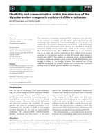
Fig. 1. Radiochromatographic resolution by TLC of metabolites generated in incubations of lauric acid with microsomes from yeast express-
ing CYP77A4. Microsomes were incubated with 100 l
M [1-
14
C]lauric acid in the absence (A) or presence (B) of NADPH. Incubations were
performed at 27 °C and contained 20 pmol of CYP77A4. They were stopped after 30 min by the addition of 20 lL of acetonitrile (containing
0.2% acetic acid) and directly spotted onto TLC. Peak S, lauric acid; peak 1, mixture of 11-, 10-, 9-, 8- and 7-hydroxylauric acids. Experiments
in (C) and (D) were performed as in (B), but with microsomes from yeast transformed with a void plasmid (C) or with boiled microsomes
(D). The structures of the metabolites are described in (E).
CYP77A4, an epoxy fatty acid-forming enzyme V. Sauveplane et al.
722 FEBS Journal 276 (2009) 719–735 Journal compilation ª 2008 FEBS. No claim to original French government works
(3%) (M-15) (loss of a methyl from the trimethylsilyl
group) and 384 (2%) (M). The mass spectrum also
showed ions at 159 (63%) and 327 (7%), resulting
from cleavage on both sides of the hydroxyl function
carrying the trimethylsilyl group. This fragmentation
pattern is characteristic of the derivative of 14-hy-
droxyoleic acid (x-4) (M = 384 gÆmol
)1
). The mass
spectrum of derivatized metabolite 2 (Fig. S4) showed
ions at m ⁄ z (relative intensity, %) values of 73
(100%) [(CH
3
)
3
Si
+
], 75 (50%) [(CH
3
)
2
Si
+
=O], 337
(11%) (M-47) [loss of methanol from the (M-15)
fragment], 369 (3%) (M-15) (loss of a methyl from
the trimethylsilyl group). The mass spectrum also
showed ions at 173 (34%) and 313 (18%), resulting
from cleavage on both sides of the hydroxyl function
carrying the trimethylsilyl group. This fragmentation
pattern is characteristic of the derivative of 13-hy-
droxyoleic acid (x-5) ( M = 384 gÆmol
)1
). The mass
spectrum of derivatized metabolite 3 (Fig. S4) showed
ions at m ⁄ z (relative intensity, %) values of 73 (48%)
[(CH
3
)
3
Si
+
], 75 (12%) [(CH
3
)
2
Si
+
=O], 337 (3%) (M-
47) [loss of methanol from the (M-15) fragment], 353
(1%) (M-31) (loss of OCH
3
from the methyl ester),
369 (0.5%) (M-15) (loss of a methyl from the trim-
ethylsilyl group). The mass spectrum also showed ions
at 187 (100%) and 299 (4%), resulting from cleavage
on both sides of the hydroxyl function carrying the
trimethylsilyl group. This fragmentation pattern is
characteristic of the derivative of 12-hydroxyoleic acid
(x-6) (M = 384 gÆmol
)1
). The mass spectrum of
derivatized metabolite 4 (Fig. S4) showed ions at m ⁄ z
(relative intensity, %) values of 73 (35%) [(CH
3
)
3
Si
+
],
75 (14%) [(CH
3
)
2
Si
+
=O], 337 (3%) (M-47) [loss
of methanol from the (M-15) fragment], 353 (1%)
(M-31) (loss of OCH
3
from the methyl ester), 369
(1%) (M-15) (loss of a methyl from the trimethylsilyl
group) and 384 (0.5%) (M). The mass spectrum also
showed ions at 201 (1%) and 285 (100%), resulting
Fig. 2. Radiochromatographic resolution by TLC of metabolites generated in incubations of oleic acid with microsomes from yeast express-
ing CYP77A4. Microsomes were incubated with 100 l
M [1-
14
C]oleic acid in the absence (A) or presence (B) of NADPH. Incubations were
performed at 27 °C and contained 20 pmol of CYP77A4. They were stopped after 30 min by the addition of 20 lL of acetonitrile (containing
0.2% acetic acid) and directly spotted onto TLC. Peak S, oleic acid; peak 1, mixture of 14-, 13-, 12- and 11-hydroxyoleic acids; peak 2, 9,
10-epoxystearic acid. Experiments in (C) and (D) were performed as in (B), but with microsomes from yeast transformed with a void plasmid
(C) or with boiled microsomes (D). The structures of the metabolites are described in (E).
V. Sauveplane et al. CYP77A4, an epoxy fatty acid-forming enzyme
FEBS Journal 276 (2009) 719–735 Journal compilation ª 2008 FEBS. No claim to original French government works 723
from cleavage on both sides of the hydroxyl function
carrying the trimethylsilyl group. This fragmentation
pattern is characteristic of the derivative of 11-hy-
droxyoleic acid (x-7) (M = 384 gÆmol
)1
). The identifi-
cation of metabolites from peak 1 by GC ⁄ MS after
purification and derivatization revealed that
CYP77A4 hydroxylates oleic acid with the following
regioselectivity: x-7 (58%), x-6 and x-5 (30%), x-4
(12%). The metabolite from peak 2 displayed the
TLC mobility expected for 9,10-epoxystearic acid,
and was indeed identified as 9,10-epoxystearic acid by
GC ⁄ MS analysis (Fig. S4). For substrate oxidation,
we determined, by kinetic studies, K
m,app
and V
max,app
values of 84 ± 23 lm and 26 ± 5 nmolÆmin
)1
Ænmol
)1
P450, respectively (Fig. S3). We determined the ste-
reochemistry of this epoxide after purification and
analysis by HPLC using a chiral column. The radio-
chromatogram of Fig. 3 shows that it is a mixture of
the two enantiomers, 9S ⁄ 10R and 9R ⁄ 10S, in the
ratio 33 : 77, respectively.
Metabolism of linoleic acid by CYP77A4
Figure 4 shows the radioactivity profiles obtained after
incubation of linoleic acid (C
18:2
) with microsomes
from yeast expressing CYP77A4. The addition of
NADPH to the incubation medium led to the forma-
Fig. 3. Radiochromatographic resolution by HPLC of the enantio-
mers of 9,10-epoxystearic acid produced by CYP77A4. (A) After
incubation of oleic acid with microsomes from yeast expressing
CYP77A4, the 9,10-epoxystearic produced (peak 2, Fig. 2B) was
purified and subjected to chiral HPLC analysis with hexane ⁄ propan-
2-ol ⁄ acetic acid (99.7 : 0.2 : 0.1, v ⁄ v ⁄ v) at a flow rate of 0.8
mLÆmin
)1
. (B) Structures of the enantiomers.
Fig. 4. Radiochromatographic resolution by TLC of metabolites gen-
erated in incubations of linoleic acid with microsomes from yeast
expressing CYP77A4. Microsomes were incubated with 100 l
M
[1-
14
C]linoleic acid in the absence (A) or presence (B) of NADPH.
Incubations were performed at 27 °C and contained 20 pmol of
CYP77A4. They were stopped after 30 min by the addition of 20 lL
of acetonitrile (containing 0.2% acetic acid) and directly spotted onto
TLC. Peak S, linoleic acid; peak 1, 9,10:12,13-diepoxyoctadecanoic
acid; peak 2, 12,13-epoxyoctadeca-9-enoic acid. Experiments in (C)
and (D) were performed as in (B), but with microsomes from yeast
transformed with a void plasmid (C) or with boiled microsomes (D).
The structures of the metabolites are described in (E).
CYP77A4, an epoxy fatty acid-forming enzyme V. Sauveplane et al.
724 FEBS Journal 276 (2009) 719–735 Journal compilation ª 2008 FEBS. No claim to original French government works
tion of a major radioactive peak (peak 2, Fig. 4B)
which was not present in the absence of NADPH
(Fig. 4A). It results from a reaction catalyzed by
CYP77A4, as it was not formed when the microsomes
were from yeast transformed with a void plasmid
(Fig. 4C) or were boiled (Fig. 4D). This peak contains
only one metabolite, which was identified by GC ⁄ MS
analysis (Fig. S5) after purification reaction in acidic
methanol and derivatization as 12,13-epoxyoctadeca-
9-enoic acid (vernolic acid), resulting from the epoxi-
dation of the D
12
double bond. The kinetic parameters
from the reaction of substrate oxidation are K
m,app
=
61±3 lm and V
max,app
= 13 ± 0.3 nmolÆmin
)1
Ænmol
)1
P450 (Fig. S3). Stereochemistry studies presented in
Fig. 5 show that CYP77A4 possesses a strong enantio-
specificity: the epoxide formed is a mixture of 12S ⁄ 13R
and 12R ⁄ 13S in the ratio 90 : 10, thus presenting a
strong enantiomeric excess in favor of the 12S ⁄ 13R
conformation. The metabolite from the minor peak
(peak 1, Fig. 4B) was identified by GC ⁄ MS (Fig. S5)
as 9,10:12,13-diepoxyoctadecanoic acid after puri-
fication and derivatization. CYP77A4 was also able
to catalyze its formation in incubations with purified
12,13-epoxyoctadeca-9-enoic acid (data not shown).
Metabolism of a-linolenic acid by CYP77A4
The incubation of a-linolenic acid (C
18:3
) with micro-
somes from yeast expressing CYP77A4 led to the
formation of one major radioactive peak, as shown on
the radiochromatogram in Fig. 6 (peak 2, Fig. 6B). It
results from a reaction catalyzed by CYP77A4, as it
requires the presence of NADPH and is not formed
with microsomes from yeast transformed with a void
plasmid (Fig. 6C) or on incubation with boiled micro-
somes (Fig. 6D). The shape of this peak suggests that
it contains more than one metabolite. After purifica-
tion, acidic treatment and derivatization, GC ⁄ MS
analysis showed that it was indeed a mixture of the
three epoxide derivatives of a-linolenic acid.
The mass spectrum of derivatized metabolite 1
(Fig. S6) showed ions at m ⁄ z (relative intensity, %)
values of 73 (100%) [(CH
3
)
3
Si
+
], 75 (14%)
[(CH
3
)
2
Si
+
=O], 439 (4%) (M-31) (loss of OCH
3
from
the methyl ester), 455 (1%) (M-15) (loss of a methyl
from the trimethylsilyl group), 470 (0.5%) (M). The
mass spectrum also showed ions at 171 (40%) and 299
(44%), resulting from the cleavage between two
hydroxyls carrying the trimethylsilyl group generated
by hydrolysis in perchloric acid. This fragmentation
pattern is characteristic of the derivative after acidic
hydrolysis of 12,13-epoxyoctadeca-9,15-dienoic acid
(M = 470 gÆmol
)1
) which represents 87% of the
metabolites. The mass spectrum of derivatized meta-
bolite 2 (Fig. S6) showed ions at m ⁄ z (relative inten-
sity, %) values of 73 (100%) [(CH
3
)
3
Si
+
], 75 (17%)
[(CH
3
)
2
Si
+
=O], 439 (2%) (M-31) (loss of OCH
3
from
the methyl ester), 455 (0.5%) (M-15) (loss of a methyl
from the trimethylsilyl group), 470 (1%) (M). The
mass spectrum also showed ions at 211 (11%) and 259
(81%), resulting from the cleavage between two
hydroxyls carrying the trimethylsilyl group generated
by hydrolysis in perchloric acid. This fragmentation
pattern is characteristic of the derivative after acidic
hydrolysis of 9,10-epoxyoctadeca-12,15-dienoic acid
(M = 470 gÆmol
)1
) which represents 7% of the meta-
bolites. The mass spectrum of derivatized metabolite 3
(Fig. S6) showed ions at m ⁄ z (relative intensity, %)
values of 73 (100%) [(CH
3
)
3
Si
+
], 75 (21%)
[(CH
3
)
2
Si
+
=O], 439 (3%) (M-31) (loss of OCH
3
from
the methyl ester), 455 (0.5%) (M-15) (loss of a methyl
from the trimethylsilyl group), 470 (4%) (M). The
mass spectrum also showed ions at 131 (67%) and 339
(20%), resulting from the cleavage between two
hydroxyls carrying the trimethylsilyl group generated
by hydrolysis in perchloric acid. This fragmentation
pattern is characteristic of the derivative after acidic
hydrolysis of 15,16-epoxyoctadeca-9,12-dienoic acid
Fig. 5. Radiochromatographic resolution by HPLC of the enantio-
mers of 12,13-epoxyoctadeca-9-enoic acid produced by CYP77A4.
(A) After incubation of linoleic acid with microsomes from yeast
expressing CYP77A4, the 12,13-epoxyoctadeca-9-enoic acid
produced (peak 2, Fig. 4B) was purified, methylated and subjected
to chiral HPLC analysis with 100% heptane at a flow rate of
0.5 mLÆmin
)1
. (B) Structures of the enantiomers.
V. Sauveplane et al. CYP77A4, an epoxy fatty acid-forming enzyme
FEBS Journal 276 (2009) 719–735 Journal compilation ª 2008 FEBS. No claim to original French government works 725
(M = 470 gÆmol
)1
) which represents 6% of the meta-
bolites. Kinetic parameters from the reaction of
substrate oxidation are K
m,app
=29±4lm and
V
max,app
= 38 ± 2 nmolÆmin
)1
Ænmol
)1
P450 (Fig. S3).
Metabolites from the minor peak (peak 1, Fig. 6B)
have not been identified.
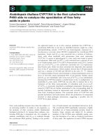
Fig. 7. Radiochromatographic resolution by TLC of metabolite gen-
erated in the incubation of 12,13-epoxyoctadeca-9-enoic acid with
microsomes or cytosol from A. thaliana. Microsomes (350 lg pro-
tein) or cytosol (600 lg protein) from A. thaliana was incubated
with 100 l
M of 12,13-epoxyoctadeca-9-enoic acid for 20 min at
27 °C. Incubation was stopped by the addition of 20 lL of acetoni-
trile (containing 0.2% acetic acid) and directly spotted onto TLC. (A)
Experiment performed with microsomes. (B) Experiment performed
with boiled microsomes. (C) Experiment performed with cytosol.
(D) Experiment performed with boiled cytosol. Peak S, 12,13-epoxy-
octadeca-9-enoic acid; peak 1, 12,13-dihydroxyoctadeca-9-enoic
acid. The structure of the metabolite is described in (E).
Fig. 6. Radiochromatographic resolution by TLC of metabolites gen-
erated in incubations of a-linolenic acid with microsomes from yeast
expressing CYP77A4. Microsomes were incubated with 100 lm
[1-
14
C]a-linolenic acid in the absence (A) or presence (B) of NADPH.
Incubations were performed at 27 °C and contained 20 pmol of
CYP77A4. They were stopped after 30 min by the addition of 20 lL
of acetonitrile (containing 0.2% acetic acid) and directly spotted onto
TLC. Peak S, a-linolenic acid; peak 1, non-identified; peak 2, mixture
of 12,13-epoxyoctadeca-9,15-dienoic, 9,10-epoxyoctadeca-12,15-die-
noic and 15,16-epoxyoctadeca-9,12-dienoic acids. Experiments in
(C) and (D) were performed as in (B), but with microsomes from
yeast transformed with a void plasmid (C) or with boiled micro-
somes (D). The structures of the metabolites are described in (E).
CYP77A4, an epoxy fatty acid-forming enzyme V. Sauveplane et al.
726 FEBS Journal 276 (2009) 719–735 Journal compilation ª 2008 FEBS. No claim to original French government works
Requirements for CYP77A4 activity
To check the importance of the double bond for
CYP77A4 activity, we tested as potential substrates
stearic acid (C
18:0
), which is saturated, and linolelaidic
acid, which is linoleic acid containing trans double
bonds. No metabolites were detected on TLC after
incubation of radiolabeled C
18:0
with microsomes from
yeast expressing CYP77A4 (data not shown). To test
linolelaidic acid, which is not available radiolabeled,
we performed two experiments. In the first experiment,
we incubated radiolabeled linoleic acid with micro-
somes in the presence of an increasing concentration
of unlabeled linolelaidic acid, and did not detect any
inhibition of epoxidation of linoleic acid (data not
shown). In a second experiment, we ran GC ⁄ MS anal-
ysis after the incubation of linolelaidic acid with yeast
microsomes expressing CYP77A4, and did not detect
any metabolite (data not shown). Together, this shows
that CYP77A4 requires the presence of unsaturation
to metabolize C
18
fatty acids; furthermore, unsatura-
tion must be in the cis configuration.
Hydrolysis of vernolic acid in microsomes and
cytosol from A. thaliana
In order to test whether the metabolites generated by
CYP77A4 were end products or could be substrates of
other enzymatic systems (i.e. epoxide hydrolase) from
A. thaliana, we purified vernolic acid that was produced
by CYP77A4 (peak 2, Fig. 4B). This epoxide was sub-
sequently incubated with microsomes isolated from
A. thaliana seedlings. The results are presented in Fig. 7.
A peak of radioactivity (peak 1, Fig. 7A) was detected
after resolving the products of reaction on TLC. No
metabolite was formed if the microsomes were boiled
before incubation (Fig. 7B). This demonstrates the enzy-
matic origin of the metabolite from peak 1. The mass
spectrum of this derivatized metabolite (Fig. S7) showed
ions at m⁄ z (relative intensity, %) values of 73 (100%)
[(CH
3
)
3
Si
+
], 75 (17%) [(CH
3
)
2
Si
+
=O], 457 (2%)
(M-15) (loss of a methyl from the trimethylsilyl group).
The mass spectrum also showed ions at 173 (40%) and
299 (8%), resulting from the cleavage between two
hydroxyls carrying the trimethylsilyl group. This frag-
mentation pattern is characteristic of the derivative of
12,13-dihydroxyoctadeca-9-enoic acid (M = 472 gÆ
mol
)1
). The same results were obtained when incubation
was carried out with the cytosolic fraction of A. thaliana
(Fig. 7C). Together, these experiments show that vernol-
ic acid produced by CYP77A4 can be converted to the
corresponding diol by microsomal and cytosolic epoxide
hydrolase (Fig. 8). These epoxide hydrolases can also
convert epoxides from C
18:3
into the corresponding diols
(data not shown).
Discussion
A new approach, based on an in silico analysis of pub-
licly available transcriptome data (http://www-ibmp.
u-strasbg.fr/~CYPedia/), has been developed recently
for the mapping of cytochromes P450 onto specific
metabolic pathways based on large-scale co-expression
analysis [33]. This approach showed that CYP77A4 was
co-regulated across 167 developmental samples (cover-
ing more than 400 publicly available Affymetrix micro-
array data sets) with a set of enzymes implicated in fatty
acid metabolism. Although co-expression correlations
were relatively low compared with other co-expressed
genes acting in a common pathway [33], with Pearson
correlation coefficients not exceeding 0.75, it was strik-
ing that the top eight co-expressed genes with CYP77A4
have been functionally characterized as being involved
in fatty acid metabolism ( />~CYPedia/CYP77A4/CoExpr_CYP77A4_Organs.html).
We thus found it worthwhile to test experimentally the
hypothesis generated by this bioinformatic approach
and to elucidate the physiological role of CYP77A4,
also because no function has been reported for members
belonging to this cytochrome P450 family to date.
Heterologous expression of CYP77A4 in an engineered
strain of yeast, and incubations of a diverse set of fatty
acids with yeast microsomes, allowed us to confirm the
capacity of this newly characterized P450 to metabolize
fatty acids, highlighting the predictive power of the
in silico co-expression analysis. Based on phylogenetic
reconstructions [36], CYP77A4 belongs to the CYP71
Fig. 8. Conversion of linoleic acid to 12,13-dihydroxyoctadeca-9-
enoic acid by CYP77A4 and epoxide hydrolases from A. thaliana.
(A) Linoleic acid. (B) 12,13-Epoxyoctadeca-9-enoic acid. (C) 12,13-
Dihydroxyoctadeca-9-enoic acid.
V. Sauveplane et al. CYP77A4, an epoxy fatty acid-forming enzyme
FEBS Journal 276 (2009) 719–735 Journal compilation ª 2008 FEBS. No claim to original French government works 727
clan and, within this clan, forms a basal clade with the
CYP89, CYP753 and CYP752 families, none of which
has been functionally characterized. In contrast, most
functionally characterized plant fatty acid hydroxylases
belong to the divergent CYP86 clan (including CYP86
and CYP94 families). Both the CYP71 and CYP86 clans
appear to have evolved independently within the green
plant lineage, as they are evolutionary separated by fam-
ilies that pre-date land plant evolution [36]. Thus, a
function of CYP77A4 as a fatty acid-metabolizing
enzyme would not have been predicted based on phylo-
genetic reconstructions, again highlighting the power of
the co-expression analysis approach, which is indepen-
dent of sequence or structural similarities.
On the model substrate lauric acid, CYP77A4
hydroxylated predominantly the x-1 carbon, which cor-
responds to a carbon in the environment of unsaturation
in oleic (C
18:1
), linoleic (C
18:2
) and a-linolenic (C
18:3
)
acids, the common physiological C
18
fatty acids in
plants. We therefore assayed these compounds as poten-
tial substrates and demonstrated that CYP77A4 was
able to produce, in vitro, epoxide derivatives of these
fatty acids. Investigations on the members of the CYP2
family in animals have previously demonstrated that the
regioselectivity and enantioselectivity of epoxidation are
cytochrome P450 dependent [37,38]. For CYP77A4, the
requirement of unsaturation, in the cis configuration,
together with the regioselectivity and enantioselectivity
observed, probably reflect steric constraints on the sub-
strate in the active site. The fact that C
18:2
is epoxidized
first exclusively on the D
12
unsaturated position, with
strong enantiomeric excess (the epoxide formed is a mix-
ture of 12S ⁄ 13R 12R ⁄ 13S in the ratio 90 : 10), shows
that it is probably hindered in the active site, suggesting
that it could be a physiological substrate.
In animals, epoxides of arachidonic acid (C
20:4
),
formed by epoxidases (mainly belonging to the CYP2
family), are well documented. This is mainly a result
of the large array of biological effects attributed to
epoxyeicosatrienoic acids (EETs). For example, activa-
tion of CYP epoxidases in endothelial cells is a key
step in vasodilatation events [14]. EETs also play a
major role in cell proliferation and angiogenesis via
the activation of an epidermal growth factor [39–41].
Over-expression of CYP2C9 and exogenous applica-
tion of EETs to cultured endothelial cells are associ-
ated with angiogenesis [41,42]. CYP2C and CYP2J2
have also been shown to be expressed in different
tumor tissues [43,44]. Epoxides of fatty acids are less
described in plants, and only a few biological activities
have been attributed to them. The discovery of such
activities in plants might help to understand the physi-
ological role of CYP77A4. This lack of data could
explain the small amount of information available
today concerning the ability of plant enzymes to gener-
ate epoxides of fatty acids, despite the fact that this
type of reaction was described for the first time more
than three decades ago [23]. Thus, the discovery of
CYP77A4 carrying such activity opens the door not
only for detailed biochemical characterizations, but
also for an understanding of the physiological role of
epoxides of fatty acids in plants.
In addition to cytochromes P450, two distinct types
of plant enzyme, unrelated to cytochrome P450, with
epoxidase activity, have been described. The first, a
peroxygenase, was reported in Vicia faba [28] and Gly-
cine max [29]. This type of enzyme uses hydroperox-
ides as cofactors to catalyze the epoxidation of fatty
acids. The second, described by Lee et al. [31], is a
non-heme di-iron enzyme, also named ‘desaturase-like’
enzyme. It thus appears that fatty acid epoxidation in
plants can be facilitated by evolutionarily divergent
sets of enzymes, further suggesting a pivotal role of
these epoxides or derivatives thereof.
CYP77A4, described in this work, catalyzed the oxy-
gen incorporation into double bonds of oleic (C
18:1
),
linoleic (C
18:2
) and a-linolenic (C
18:3
) acids, but did not
metabolize saturated stearic acid (C
18:0
). Furthermore,
it did not metabolize linolelaidic acid, which is the
homolog of linoleic acid possessing two trans double
bonds, not commonly found in natural fatty acids.
These observations suggest that the physiological func-
tion of CYP77A4 could be epoxidation of unsaturated
C
18
fatty acids. This hypothesis is supported by in silico
co-expression analysis, showing that CYP77A4 is
co-regulated with a stearoyl acyl carrier protein desat-
urase and a putative epoxide hydrolase [33]. Cahoon
et al. [32], in E. lagascae seed, identified a cytochrome
P450, classified as CYP726A1, able to convert linoleic
acid into 12,13-epoxyoctadeca-9-enoic acid (vernolic
acid). CYP77A4 differs from this enzyme; indeed, it
metabolizes free fatty acids, whereas CYP726A1 meta-
bolizes fatty acids incorporated into phosphatidylcho-
line [32,45]. The physiological role of CYP77A4 is
unlikely to be the production of fatty acid epoxides for
accumulation in seeds as, unlike E. lagascae and plants
belonging to the Aesteraceae genera, such as Crepis
palaestina, A. thaliana does not store fatty acid epox-
ides. Cytochrome P450-dependent fatty acid oxidases in
plants have been mainly investigated with regard to
cutin synthesis [19]. Cutin consists of a biopolymer of
fatty acids belonging to the protective envelope of
plants: the cuticle [20]. Epoxides of fatty acids may rep-
resent up to 60% of cutin monomers [46,47]. Cutin anal-
ysis of A. thaliana has been performed recently [48], and
18-hydroxy-9,10-epoxystearic acid was shown to be
CYP77A4, an epoxy fatty acid-forming enzyme V. Sauveplane et al.
728 FEBS Journal 276 (2009) 719–735 Journal compilation ª 2008 FEBS. No claim to original French government works
present in cutin. CYP77A4 could account for its forma-
tion by introducing the oxygen between carbon 9 and 10
of oleic acid before incorporation of the monomer into
the cutin. In this context, it is interesting to note that
inhibition studies allowed LeQueu et al. [49] to demon-
strate the involvement of a peroxygenase in the forma-
tion of cutin of corn.
The three epoxide derivatives from a-linolenic acid,
which are produced by CYP77A4, have been shown to
confer resistance of rice against rice blast disease [50].
This indicates a possible involvement of CYP77A4 in
plant defense events. As discussed below, diol deriva-
tives of fatty acids also participate in plant defense.
The presence of an epoxide hydrolase in A. thaliana
was first reported by Kiyosue et al. [51]. Furthermore,
a putative epoxide hydrolase is co-expressed with
CYP77A4 [33]. Therefore, it was interesting to
determine whether epoxides generated by CYP77A4
could be transformed to diols. By incubating these
compounds with subcellular fractions of A. thaliana,
we confirmed that epoxides were enzymatically
hydrolyzed into the corresponding diols. A recent
study from our laboratory [52] has shown that, in
A. thaliana microsomes, fatty acids can be specifically
hydroxylated in the x-1 position by a cytochrome
P450 which remains to be identified. The x-1 hydroxyl-
ation of the diol derivative from vernolic acid would
lead to the formation of 12,13,17-trihydroxyoctadeca-
9-enoic acid, which has been shown to exhibit strong
antifungal properties [53]. Distinct fatty acid x-hydrox-
ylases have been characterized in A. thaliana [21,52,54].
Hydroxylation of the terminal methyl of 9,10-dihydr-
oxystearic acid, resulting from the combined action of
CYP77A4 and an epoxide hydrolase, would generate
9,10,18-trihydroxystearic acid, which has also been
implicated in the elicitation of defense mechanisms
[55]. The antimicrobial activities of poly-hydroxy fatty
acids are well documented [56,57], and the interplay of
CYP77A4 with epoxide hydrolases and fatty acid
hydroxylases would thus allow the production of such
compounds involved in plant–pathogen interactions. It
is noteworthy that epoxide hydrolases from differ-
ent plants are induced at the transcriptional level by
stress, methyl jasmonate or pathogens [51,58,59]. As
reported in animals [60,61], plant epoxide hydrolases
could also be implicated in the control of epoxide levels,
and therefore in the control of their biological effects.
Plant oxylipins represent a vast family of com-
pounds derived from polyunsaturated fatty acids. They
originate either from chemical oxidation [62] or from
enzymatic reactions catalyzed by a-dioxygenase, lipox-
ygenases [2,63] and cytochrome P450 from the CYP74
family [62]. These oxylipins are major actors in plant
defense and they recruit signaling molecules as well as
molecules exhibiting antimicrobial and antifungal
properties [56]. They belong to different classes of
chemicals (i.e. aldehydes, divinyl ethers, ketones and
hydroperoxides) and some are cyclic compounds.
Extensively studied jasmonic acid and 12-oxo-phytodi-
enoic acids represent a good illustration of these cyclic
oxylipins [64,65]. They both originate from the cycliza-
tion of an allene oxide, which is an unstable epoxide
derived from a-linolenic acid. In this work, we have
shown that CYP77A4 can catalyze the in vitro
production of the di-epoxide derivative of linoleic acid.
Interestingly, biochemical studies performed with
mouse liver microsomes showed that this di-epoxide,
produced during the oxidation of linoleic acid by cyto-
chrome P450, was then converted to cyclic tetra-
hydrofurans after hydrolysis by epoxide hydrolases
[66]. In analogy, it would be very interesting to investi-
gate the cyclization of di-epoxide derivatives of linoleic
acid after hydrolysis by plant epoxide hydrolases,
because the resulting tetrahydrofurans could represent
a novel class of plant oxylipins.
In conclusion, we have described the first biochemi-
cal characterization of a member of the CYP77 family,
and have shown that CYP77A4 is capable of epoxidiz-
ing, in vitro, unsaturated C
18
fatty acids. This is also
the first report describing a cytochrome P450 which
can catalyze the epoxidation of free fatty acids in
plants. Plants are sessile organisms and therefore rely
on a battery of defense chemicals for survival. To pro-
duce these chemical defenses, they have developed a
complex metabolic network using the diversified cata-
lytic properties of cytochrome P450 enzymes [36]. Lipid
metabolism is a major player in the plant defense
network, and CYP77A4 could participate by producing
metabolites or precursors of metabolites with properties
similar to those described for fatty acid derivatives also
implicated in defense [62]. The biochemical character-
ization of CYP77A4 from A. thaliana means that
targeted mutant studies can be performed employing
the genomic toolbox available for this model plant.
This will help to elucidate, in planta, the physiological
role of CYP77A4 and, more generally, of epoxides
derived from free fatty acids in plants.
Experimental procedures
Chemicals
Radiolabeled [1-
14
C]lauric acid (1Æ6 MBqÆlmol
)1
) was
from CEA (Gif sur Yvette, France); [1-
14
C]oleic acid
(1Æ85 MBqÆlmol
)1
), [1-
14
C]linoleic acid (2Æ1 MBqÆlmol
)1
)
and [1-
14
C]a-linolenic acid (1Æ9 MBqÆlmol
)1
) were from
V. Sauveplane et al. CYP77A4, an epoxy fatty acid-forming enzyme
FEBS Journal 276 (2009) 719–735 Journal compilation ª 2008 FEBS. No claim to original French government works 729
Perkin Elmer (Courtaboeuf, France). Linolelaidic acid was
from Sigma (St Louis, MO, USA).
The silylating reagent N,O-bistrimethylsilyltrifluoroaceta-
mide, containing 1% of trimethylchlorosilane, was from
Pierce (Rockfold, IL, USA). NADPH was from Sigma.
Thin layer plates (Silica Gel G60 F254; 0.25 mm) were
from Merck (Darmstadt, Germany).
Cloning of CYP77A4
The coding sequence of CYP77A4 (AT5g04660) was cloned
by PCR from a DNA library of Arabidopsis ecotype
Columbia-0. Primers 5¢-CCCCAGATCTATGTTTCCTCT
AATCTC-3¢ and 5¢-GGGGGGTACCCTAAATCCTTGGT
TTG-3¢ were used as forward and reverse primers, respec-
tively. PCR was carried out with IsisÔ DNA polymerase
(Qbiogene, Illkirch, France) for 30 thermal cycles (1 min at
96 °C, 2 min at 54 °C, 2 min at 72 °C). After the addition
of adenine nucleotides on each side of the PCR product by
an additional step with Taq polymerase (10 min at 72 °C),
the purified PCR product was cloned into PCRII TOPO
vector (Invitrogen, Carlsbad, CA, USA), and transferred to
the pYeDP60 vector using the BamHI and KpnI restriction
sites. The sequence was verified by DNA sequencing after
the cloning step in the PCRII TOPO vector.
Heterologous expression of CYP77A4 in yeast
For functional expression of the full-length CYP77A4 clone,
we used a yeast expression system specifically developed for
the expression of P450 enzymes, and consisting of plasmid
pYeDP60 and Saccharomyces cerevisiae WAT11 strain [34].
Yeast cultures were grown and CYP77A4 expression was
induced as described in Pompon et al. [34] from one isolated
transformed colony. After growth, cells were harvested by
centrifugation and manually broken with glass beads
(0.45 mm in diameter) in 50 mm Tris ⁄ HCl buffer (pH 7.5)
containing 1 mm EDTA and 600 mm sorbitol. The homoge-
nate was centrifuged for 10 min at 10 000 g. The resulting
supernatant was centrifuged for 1 h at 100 000 g. The pellet
consisting of microsomal membranes was resuspended in
50 mm Tris ⁄ HCl (pH 7.4), 1 mm EDTA and 30% (v ⁄ v)
glycerol with a Potter–Elvehjem homogenizer and stored at
)30 °C. The volume of resuspension buffer was propor-
tional to the weight of the yeast pellet: microsomes extracted
from 6 g of yeast were resuspended in 3 mL of buffer. All
procedures for microsomal preparation were carried out at
0–4 °C. The cytochrome P450 content was measured by the
method of Omura and Sato [67].
Plant material and microsomal preparation
After sterilization, Arabidopsis (ecotype Columbia-0) seeds
were grown on Murashige and Skoog medium (MS med-
ium, 4.2 gÆL
)1
; sucrose, 10 gÆL
)1
; pastagar B, 8 gÆL
)1
; myo-
inositol, 100 mgÆL
)1
; thiamine, 10 mgÆL
)1
; nicotinic acid,
1mgÆL
)1
; pyridoxine, 1 mgÆL
)1
; final pH 5.7) for 5 weeks.
Arabidopsis plants (approximately 10 g) were homogenized
with a mortar and pestle in 50 mL of extraction buffer
(250 mm tricine, 50 mm NaHSO
3
,5gÆL
)1
BSA, 2 mm
EDTA, 100 mm ascorbic acid, 2 mm dithiothreitol, final
pH 8.2). The homogenate was filtered through 50 lm nylon
filtration cloth and centrifuged for 10 min at 10 000 g. The
resulting supernatant was centrifuged for 1 h at 100 000 g.
The supernatant (cytosol) was directly stored at )30 °C
and the microsomal pellet was resuspended in buffer at
pH 8.2 (50 mm NaCl, 100 mm tricine, 250 mm sucrose,
2mm EDTA, 2 mm dithiothreitol) with a Potter–Elvehjem
homogenizer, and stored at )30 °C. All procedures for
microsomal preparation were carried out at 0–4 °C.
Enzyme activities
All radiolabeled substrates were dissolved in ethanol which
was evaporated before the addition of microsomes into the
glass tube. Resolubilization of the substrates was confirmed
by measuring the radioactivity of the incubation media.
The enzymatic activities of CYP77A4 from transformed
yeast or Arabidopsis microsomes were determined by follow-
ing the formation rate of the metabolites. The standard assay
(0.1 mL) contained 20 mm sodium phosphate (pH 7.4),
1mm NADPH and radiolabeled substrate (100 lm). The
reaction was initiated by the addition of NADPH and was
stopped by the addition of 20 lL of acetonitrile (containing
0.2% acetic acid). The reaction products were resolved by
TLC or HPLC as described below.
For kinetic studies, we incubated 4.5 pmol of CYP77A4
for 5 min with various concentrations of substrate, ranging
from 10 to 200 lm for C
12:0
, 5 to 120 lm for C
18:1
,5to
80 lm for C
18:2
, and 5 to 120 lm for C
18:3
.
TLC methods
Incubation media were directly spotted onto TLC plates. For
the separation of metabolites from residual substrate, TLC
was developed with a mixture of diethyl ether ⁄ light petroleum
(boiling point, 40–60 °C) ⁄ formic acid (50 : 50 : 1, v ⁄ v ⁄ v).
The plates were scanned with a radioactivity detector (Rita
Star, Raytest, Straubenhardt, Germany). The areas corre-
sponding to the metabolites were scraped into counting vials
and quantified by liquid scintillation, or were eluted from the
silica with 10 mL of diethyl ether, which was removed by
evaporation. They were then subjected to GC⁄ MS analysis.
GC/MS analysis
GC ⁄ MS analysis was carried out on a gas chromatograph
(Agilent 6890 Series) equipped with a 30 m capillary
CYP77A4, an epoxy fatty acid-forming enzyme V. Sauveplane et al.
730 FEBS Journal 276 (2009) 719–735 Journal compilation ª 2008 FEBS. No claim to original French government works
column with an internal diameter of 0.25 mm and a film
thickness of 0.25 lm (HP-5MS). The gas chromatograph
was combined with a quadrupole mass-selective detector
(Agilent 5973N). Mass spectra were recorded at 70 eV and
analysed as in Eglinton et al. [68].
Metabolites of lauric acid
For the analysis of products generated by recombinant
CYP77A4 on incubation with lauric acid, metabolites of
peak 1 (Fig. 1B) were eluted from silica with 10 mL of
diethyl ether, methylated with diazomethane, trimethyl-
silylated with N,O-bistrimethylsilyltrifluoroacetamide con-
taining 1% (v ⁄ v) trimethylchlorosilane (1 : 1, v ⁄ v) and
subjected to GC ⁄ MS analysis, which revealed the presence
of five metabolites. The mass spectra are given in Fig. S2.
The regioselectivity of CYP77A4 was determined on the
basis of the peak area of each metabolite detected by GC.
Metabolites of oleic acid
For the analysis of the products generated by recombinant
CYP77A4 on incubation with oleic acid, the metabolite of
peak 2 (Fig. 2B) was eluted from silica with 10 mL of
diethyl ether, methylated with diazomethane and identified
as 9,10-epoxystearic acid as described previously [69].
Metabolites from peak 1 were eluted from silica with
10 mL of diethyl ether and subjected to GC ⁄ MS analysis
after methylation and silylation. GC ⁄ MS analysis showed
the presence of four metabolites. The mass spectra are
given in Fig. S4. The regioselectivity of CYP77A4 was
determined on the basis of the peak area of each metabolite
detected by GC.
Metabolites of linoleic acid
For the analysis of the products generated by recombinant
CYP77A4 on incubation with linoleic acid, the metabolite
from peak 2 (Fig. 4B) was eluted from silica with 10 mL of
diethyl ether, subjected to GC ⁄ MS analysis after reaction
in acidic methanol, methylation and silylation. It was iden-
tified as 12,13-epoxyoctadeca-9-enoic acid, as described in
[32] (mass spectrum in Fig. S5). The metabolite from peak
1 (Fig. 2B) was eluted from silica, methylated and identified
as 9,10:12,13-diepoxyoctadecanoic acid, as described in [29]
(mass spectrum in Fig. S5).
Metabolites of a-linolenic acid
For the analysis of the products generated by recombinant
CYP77A4 on incubation with a-linolenic acid, metabolites
from peak 2 (Fig. 6B) were eluted from silica with 10 mL
of diethyl ether and reacted in water ⁄ perchloric acid ⁄ aceto-
nitrile (47.5 : 2.5 : 50, v ⁄ v ⁄ v), as described in [70]. They
were then methylated and silylated before GC ⁄ MS analysis,
which revealed the presence of three metabolites (mass
spectra in Fig. S6).
Metabolites of vernolic acid
For the analysis of the product generated on incubation of
12,13-epoxyoctadeca-9-enoic acid with the microsomal frac-
tion and cytosol of A. thaliana, the metabolite from peak 1
(Fig. 7A,C) was eluted from silica with 10 mL of diethyl
ether, methylated, silylated and subjected to GC ⁄ MS analy-
sis (mass spectrum in Fig. S7).
Chiral analysis
Chiral analysis of 9,10-epoxystearic acid, produced by
CYP77A4 on incubation with oleic acid, was performed
using optically pure standards, as described previously [71].
The area corresponding to the epoxide (peak 2, Fig. 2B)
was scraped, and the epoxide was eluted from the silica
with 10 mL of diethyl ether. The residual epoxide was dis-
solved in hexane (40 lL) and analyzed by HPLC (Waters,
St Quentin en Yvelines, France) equipped with two 600
pumps and a Packard (Courtaboeuf, France) 500 TR series
radiodetector. Both enantiomers were resolved using a
chiral column (Chiracel OB; 4.6 mm · 250 mm; J.T. Baker
Chemical Co., Deventer, Netherlands) with an isocratic sol-
vent: hexane ⁄ propan-2-ol ⁄ acetic acid (99.7 : 0.2 : 0.1,
v ⁄ v ⁄ v) at a flow rate of 0.8 mLÆ min
)1
. Under the present
conditions of analysis, 9S,10R- and 9R,10S-epoxystearic
acids have retention times of 31 and 35 min, respectively.
Chiral analysis of 12,13-epoxyoctadeca-9-enoic acid,
produced by CYP77A4 on incubation with linoleic acid, was
performed as described previously [72]. The area
corresponding to the epoxide (peak 2, Fig. 4B) was scraped,
and the epoxide was eluted from the silica with 10 mL of
diethyl ether. The residual epoxide was dissolved in hexane
(40 lL), methylated with diazomethane and analyzed by
HPLC (Waters equipped with two 600 pumps and a Packard
500 TR series radiodetector). Both enantiomers were
resolved using a chiral column (Chiracel OB; 4.6 mm ·
250 mm; J.T. Baker Chemical Co.) with an isocratic solvent:
heptane 100% at a flow rate of 0.5 mLÆmin
)1
. Under the
present conditions of analysis, 12S ⁄ 13R and 12R ⁄ 13S enanti-
omers have retention times of 24 and 27 min, respectively.
Acknowledgements
Vincent Sauveplane was awarded a Bayer BioScience
and Association Nationale de la Recherche Technique
grant through a Convention Industrielle de Formation
par la Recherche contract. The authors thank
Dr I. Benveniste, Dr A. Olry and Dr J. N. Lampe for
critical reading of the manuscript, and Dr P. Denolf,
V. Sauveplane et al. CYP77A4, an epoxy fatty acid-forming enzyme
FEBS Journal 276 (2009) 719–735 Journal compilation ª 2008 FEBS. No claim to original French government works 731
Dr F. Meulewaeter (Bayer BioScience) and Dr E. Ble
´
e
for stimulating scientific discussions.
References
1 Funk CD (2001) Prostaglandins and leukotrienes:
advances in eicosanoid biology. Science 294, 1871–1875.
2 Blee E (2002) Impact of phyto-oxylipins in plant
defense. Trends Plant Sci 7 , 315–322.
3 Noverr MC, Erb-Downward JR & Huffnagle GB
(2003) Production of eicosanoids and other oxylipins by
pathogenic eukaryotic microbes. Clin Microbiol Rev 16,
517–533.
4 Oliw EH, Guengerich FP & Oates JA (1982) Oxygena-
tion of arachidonic acid by hepatic monooxygenases.
Isolation and metabolism of four epoxide intermediates.
J Biol Chem 257, 3771–3781.
5 Oliw EH (1994) Oxygenation of polyunsaturated fatty
acids by cytochrome P450 monooxygenases. Prog Lipid
Res 33, 329–354.
6 Daikh BE, Laethem RM & Koop DR (1994) Stereose-
lective epoxidation of arachidonic acid by cytochrome
P-450s 2CAA and 2C2. J Pharmacol Exp Ther 269,
1130–1135.
7 Werck-Reichhart D & Feyereisen R (2000) Cyto-
chromes P450: a success story. Genome Biol 1, doi:
10.1186/gb-2000-1-6-reviews3003.
8 Simpson AECM (1997) The cytochrome P450 4 (CYP4)
family. Gen Pharmacol 28, 351–359.
9 Ferdinandusse S, Denis S, Van Roermund CW, Wan-
ders RJ & Dacremont G (2004) Identification of the
peroxisomal beta-oxidation enzymes involved in the
degradation of long-chain dicarboxylic acids. J Lipid
Res 45, 1104–1111.
10 Alonso-Galicia M, Sun CW, Falck JR, Harder DR &
Roman RJ (1998) Contribution of 20-HETE to the
vasodilator actions of nitric oxide in renal arteries.
Am J Physiol 275, F370–F378.
11 Birks EK, Bousamra M, Presberg K, Marsh JA, Effros
RM & Jacobs ER (1997) Human pulmonary arteries
dilate to 20-HETE, an endogenous eicosanoid of lung
tissue. Am J Physiol 272, L823–L829.
12 Nowicki S, Chen SL, Aizman O, Cheng XJ, Li D,
Nowicki C, Nairn A, Greengard P & Aperia A
(1997) 20-Hydroxyeicosa-tetraenoic acid (20 HETE)
activates protein kinase C. Role in regulation of rat
renal Na+,K+-ATPase. J Clin Invest 99, 1224–
1230.
13 Messer-Letienne I, Bernard N, Roman RJ, Sassard J &
Benzoni D (1999) 20-Hydroxyeicosatetraenoic acid and
renal function in Lyon hypertensive rats. Eur J Pharma-
col 378, 291–297.
14 Fleming I (2007) Epoxyeicosatrienoic acids, cell signal-
ing and angiogenesis. Prostaglandins Other Lipid Mediat
82, 60–67.
15 Roman RJ (2002) P-450 metabolites of arachidonic acid
in the control of cardiovascular function. Physiol Rev
82, 131–185.
16 Zeldin DC, Moomaw CR, Jesse N, Tomer KB, Bee-
tham J, Hammock BD & Wu S (1996) Biochemical
characterization of the human liver cytochrome P450
arachidonic acid epoxygenase pathway. Arch Biochem
Biophys 330, 87–96.
17 King LM, Ma J, Srettabunjong S, Graves J, Bradbury
JA, Li L, Spiecker M, Liao JK, Mohrenweiser H & Zel-
din DC (2002) Cloning of CYP2J2 gene and identifica-
tion of functional polymorphisms. Mol Pharmacol 61,
840–852.
18 Stark K, Wongsud B, Burman R & Oliw EH (2005)
Oxygenation of polyunsaturated long chain fatty acids
by recombinant CYP4F8 and CYP4F12 and catalytic
importance of Tyr-125 and Gly-328 of CYP4F8. Arch
Biochem Biophys
441, 174–181.
19 Kandel S, Sauveplane V, Olry A, Diss L, Benveniste I
& Pinot F (2006) Cytochrome P450-dependent fatty
acid hydroxylases in plants. Phytochem Rev 5, 359–372.
20 Kolattukudy PE (1980) Biopolyester membranes of
plants: cutin and suberin. Science 208, 990–1000.
21 Wellesen K, Durst F, Pinot F, Benveniste I, Nettesheim
K, Wisman E, Steiner-Lange S, Saedler H & Yephre-
mov A (2001) Functional analysis of the LACERATA
gene of Arabidopsis provides evidence for different roles
of fatty acid omega-hydroxylation in development. Proc
Natl Acad Sci USA 98, 9694–9699.
22 Xiao F, Goodwin SM, Xiao Y, Sun Z, Baker D, Tang
X, Jenks MA & Zhou JM (2004) Arabidopsis
CYP86A2 represses Pseudomonas syringae type III
genes and is required for cuticle development. EMBO J
23, 2903–2913.
23 Croteau R & Kolattukudy PE (1975) Biosynthesis of
hydroxyfatty acid polymers. Enzymatic epoxidation of
18-hydroxyoleic acid to 18-hydroxy-cis-9,10-epoxystea-
ric acid by a particulate preparation from spinach
(Spinacia oleracea). Arch Biochem Biophys 170, 61–72.
24 Weissbart D, Salaun JP, Durst F, Pflieger P & Mios-
kowski C (1992) Regioselectivity of a plant lauric acid
omega hydroxylase. Omega hydroxylation of cis and
trans unsaturated lauric acid analogs and epoxygenation
of the terminal olefin by plant cytochrome P-450. Bio-
chim Biophys Acta 1124, 135–142.
25 Salaun JP, Weissbart D, Helvig C, Durst F & Mios-
kowski C (1993) Regioselective hydroxylation and epox-
idation of lauric acid and unsaturated analogues by
cytochrome P450 in Jerusalem artichoke microsomes.
Plant Physiol Biochem 31, 285–293.
26 Salaun JP, Weissbart D, Helvig C, Durst F, Pflieger P,
Bosch H & Mioskowski C (1992) Stereochemistry of
oxidized fatty acids generated during catalytic oxygena-
tion of lauric acid and unsaturated analogs by plant
microsomes. FEBS Lett 303, 109–112.
CYP77A4, an epoxy fatty acid-forming enzyme V. Sauveplane et al.
732 FEBS Journal 276 (2009) 719–735 Journal compilation ª 2008 FEBS. No claim to original French government works
27 Zimmerlin A, Salaun JP, Durst F & Mioskowski C
(1992) Cytochrome P-450-dependent hydroxylation of
lauric acid at the subterminal position and oxidation of
unsaturated analogs in wheat microsomes. Plant Physiol
100, 868–873.
28 Hamberg M & Hamberg G (1990) Hydroperoxide-
dependent epoxidation of unsaturated fatty acids in the
broad bean (Vicia faba L.). Arch Biochem Biophys 283,
409–416.
29 Blee E & Schuber F (1990) Efficient epoxidation of
unsaturated fatty acids by a hydroperoxide-dependent
oxygenase. J Biol Chem 265, 12887–12894.
30 Hanano A, Burcklen M, Flenet M, Ivancich A, Louwa-
gie M, Garin J & Blee E (2006) Plant seed peroxygenase
is an original heme-oxygenase with an EF-hand calcium
binding motif. J Biol Chem 281, 33140–33151.
31 Lee M, Lenman M, Banas A, Bafor M, Singh S, Schwe-
izer M, Nilsson R, Liljenberg C, Dahlqvist A & Gum-
meson PO (1998) Identification of non-heme diiron
proteins that catalyze triple bond and epoxy group
formation. Science 280, 915–918.
32 Cahoon EB, Ripp KG, Hall SE & McGonigle B (2002)
Transgenic production of epoxy fatty acids by expres-
sion of a cytochrome P450 enzyme from Euphorbia
lagascae seed. Plant Physiol 128, 615–624.
33 Ehlting J, Sauveplane V, Olry A, Ginglinger JF, Provart
NJ & Werck-Reichhart D (2008) An extensive (co-)
expression analysis tool for the cytochrome P450 super-
family in Arabidopsis thaliana. BMC Plant Biol 8, 47.
34 Pompon D, Louerat B, Bronine A & Urban P (1996)
Yeast expression of animal and plant P450s in optimized
redox environments. Methods Enzymol 272, 51–64.
35 Durst F & Nelson DR (1995) Diversity and evolution
of plant P450 and P450-reductases. Drug Metabol Drug
Interact 12, 189–206.
36 Nelson DR (2006) Plant cytochrome P450s from moss
to poplar. Phytochem Rev 5, 193–204.
37 Capdevila JH & Falck JR (2002) Biochemical and
molecular properties of the cytochrome P450 arachi-
donic acid monooxygenases. Prostaglandins Other Lipid
Mediat 68, 325–344.
38 Capdevila JH, Karara A, Waxman DJ, Martin MV,
Falck JR & Guenguerich FP (1990) Cytochrome P-450
enzyme-specific control of the regio- and enantiofacial
selectivity of the microsomal arachidonic acid epoxygen-
ase. J Biol Chem 265, 10865–10871.
39 Chen JK, Capdevila J & Harris RC (2002) Heparin-
binding EGF-like growth factor mediates the biological
effects of P450 arachidonate epoxygenase metabolites in
epithelial cells. Proc Natl Acad Sci USA 99, 6029–6034.
40 Chen JK, Wang DW, Falck JR, Capdevila J & Harris
RC (1999) Transfection of an active cytochrome P450
arachidonic acid epoxygenase indicates that 14,15-
epoxyeicosatrienoic acid functions as an intracellular
second messenger in response to epidermal growth
factor. J Biol Chem 274, 4764–4769.
41 Michaelis UR, Fisslthaler B, Medhora M, Harder D,
Fleming I & Busse R (2003) Cytochrome P450 2C9-
derived epoxyeicosatrienoic acids induce angiogenesis
via cross-talk with the epidermal growth factor receptor
(EGFR). FASEB J 17, 770–772.
42 Medhora M, Daniels J, Mundey K, Fisslthaler B, Busse
R, Jacobs ER & Harder DR (2003) Epoxygenase-driven
angiogenesis in human lung microvascular endothelial
cells. Am J Physiol 284, H215–H224.
43 Yokose T, Doy M, Taniguchi T, Shimada T, Kakiki
M, Horie T, Matsuzaki Y & Mukai K (1999) Immuno-
histochemical study of cytochrome P450 2C and 3A in
human non-neoplastic and neoplastic tissues. Virchows
Arch 434
, 401–411.
44 Jiang JG, Chen CL, Card JW, Yang S, Chen JX, Fu
XN, Ning YG, Xiao X, Zeldin DC & Wang DW (2005)
Cytochrome P450 2J2 promotes the neoplastic pheno-
type of carcinoma cells and is up-regulated in human
tumors. Cancer Res 65, 4707–4715.
45 Bafor M, Smith MA, Jonsson L, Stobart K & Stymne
S (1993) Biosynthesis of vernoleate (cis-12-epoxyocta-
deca-cis-9-enoate) in microsomal preparations from
developing endosperm of Euphorbia lagascae. Arch
Biochem Biophys 303, 145–151.
46 Holloway PJ & Deas AHB (1973) Epoxyoctadecanoic
acids in plant cutins and suberins. Phytochemistry 12,
1721–1735.
47 Matzke K & Riederer M (1990) The composition of the
cutin of the caryopses and leaves of Triticum aestivum
L. Planta 182, 461–466.
48 Bonaventure G, Beisson F, Ohlrogge J & Pollard M
(2004) Analysis of the aliphatic monomer composition
of polyesters associated with Arabidopsis epidermis:
occurrence of octadeca-cis-6,cis-9-diene-1,18-dioate as
the major component. Plant J 40, 920–930.
49 Lequeu J, Fauconnier ML, Chammai A, Bronner R &
Blee E (2003) Formation of plant cuticle: evidence for
the occurrence of the peroxygenase pathway. Plant J
36, 155–164.
50 Kato T, Yamaguchi Y, Namai T & Hirukawa T (1993)
Oxygenated fatty acids with anti-rice blast fungus
activity in rice plants. Biosci Biotechnol Biochem 57,
283–287.
51 Kiyosue T, Beetham JK, Pinot F, Hammock BD, Yam-
aguchi-Shinozaki K & Shinozaki K (1994) Characteriza-
tion of an Arabidopsis cDNA for a soluble epoxide
hydrolase gene that is inducible by auxin and water
stress. Plant J 6, 259–269.
52 Kandel S, Sauveplane V, Compagnon V, Franke R,
Millet Y, Schreiber L, Werck-Reichhart D & Pinot F
(2007) Characterization of a methyl jasmonate and
wounding-responsive cytochrome P450 of Arabidopsis
V. Sauveplane et al. CYP77A4, an epoxy fatty acid-forming enzyme
FEBS Journal 276 (2009) 719–735 Journal compilation ª 2008 FEBS. No claim to original French government works 733
thaliana catalyzing dicarboxylic fatty acid formation
in vitro. FEBS J 274, 5116–5127.
53 Hou CT & Forman RJ (2000) Growth inhibition of
plant pathogenic fungi by hydroxy fatty acids. J Ind
Microbiol Biotechnol 24, 275–276.
54 Benveniste I, Tijet N, Adas F, Philipps G, Salaun JP &
Durst F (1998) CYP86A1 from Arabidopsis thaliana
encodes a cytochrome P450-dependent fatty acid
omega-hydroxylase. Biochem Biophys Res Commun 243,
688–693.
55 Fauth M, Schweizer P, Buchala A, Markstadter C,
Riederer M, Kato T & Kauss H (1998) Cutin mono-
mers and surface wax constituents elicit H
2
O
2
in
conditioned cucumber hypocotyl segments and enhance
the activity of other H
2
O
2
elicitors. Plant Physiol 117,
1373–1380.
56 Prost I, Dhondt S, Rothe G, Vicente J, Rodriguez MJ,
Kift N, Carbonne F, Griffiths G, Esquerre-Tugaye MT
& Rosahl S (2005) Evaluation of the antimicrobial
activities of plant oxylipins supports their involvement
in defense against pathogens. Plant Physiol 139 , 1902–
1913.
57 Cowley T & Walters D (2005) Local and systemic
effects of oxylipins on powdery mildew infection in
barley. Pest Manag Sci 61, 572–576.
58 Stapleton A, Beetham JK, Pinot F, Garbarino JE,
Rockhold DR, Friedman M, Hammock BD & Belknap
WR (1994) Cloning and expression of soluble epoxide
hydrolase from potato. Plant J 6, 251–258.
59 Gomi K, Yamamato H & Akimitsu K (2003) Epoxide
hydrolase: a mRNA induced by the fungal pathogen
Alternaria alternata on rough lemon (Citrus jambhiri
Lush). Plant Mol Biol 53, 189–199.
60 Yu Z, Xu F, Huse LM, Morisseau C, Draper AJ, New-
man JW, Parker C, Graham L, Engler MM, Hammock
BD et al. (2000) Soluble epoxide hydrolase regulates
hydrolysis of vasoactive epoxyeicosatrienoic acids. Circ
Res 87, 992–998.
61 Sinal CJ, Miyata M, Tohkin M, Nagata K, Bend JR &
Gonzalez FJ (2000) Targeted disruption of soluble
epoxide hydrolase reveals a role in blood pressure regu-
lation. J Biol Chem 275, 40504–40510.
62 Stumpe M & Feussner I (2006) Formation of oxylipins
by CYP74 enzymes. Phytochem Rev 5, 347–357.
63 Feussner I & Wasternack C (2002) The lipoxygenase
pathway. Annu Rev Plant Biol 53, 275–297.
64 Mueller S, Hilbert B, Dueckershoff K, Roitsch T, Kris-
chke M, Mueller MJ & Berger S (2008) General detoxi-
fication and stress responses are mediated by oxidized
lipids through TGA transcription factors in Arabidop-
sis. Plant Cell 20, 768–785.
65 Browse J & Howe GA (2008) New weapons and a rapid
response against insect attack. Plant Physiol 146, 832–
838.
66 Moghaddam M, Motoba K, Borhan B, Pinot F &
Hammock BD (1996) Novel metabolic pathways for
linoleic and arachidonic acid metabolism. Biochim
Biophys Acta 1290, 327–339.
67 Omura T & Sato R (1964) The carbon monoxide-
binding pigment of liver microsomes. I. Evidence
for its hemoprotein nature. J Biol Chem 239, 2370–
2378.
68 Eglinton G, Hunneman D & McCormick A (1968) Gas
chromatography-mass spectrometry studies of long-
chain hydroxy acids III. The mass spectra of the methyl
esters and TMS ethers of aliphatic hydroxy acids. A
facile method of double bond location. Org Mass Spec-
trom 1, 593–611.
69 Kandel S, Morant M, Benveniste I, Blee E, Werck-
Reichhart D & Pinot F (2005) Cloning, functional
expression, and characterization of CYP709C1, the first
sub-terminal hydroxylase of long chain fatty acid in
plants – induction by chemicals and methyl jasmonate.
J Biol Chem 280, 35881–35889.
70 Jude AR, Little JM, Freeman JP, Evans JE, Rado-
minska-Pandya A & Grant DF (2000) Linoleic acid
diols are novel substrates for human UDP-glu-
curonosyltransferases. Arch Biochem Biophys 380, 294–
302.
71 Pinot F, Bosch H, Salaun JP, Durst F, Mioskowski C
& Hammock BD (1997) Epoxide hydrolase activities in
the microsomes and the soluble fraction from Vicia
sativa seedlings. Plant Physiol Biochem 35, 103–110.
72 Blee E, Stahl U, Schuber F & Stymne S (1993)
Regio- and stereoselectivity of cytochrome P-450
and peroxygenase-dependent formation of cis-12,
13-epoxy-9(Z)-octadecenoic acid (vernolic acid) in
Euphorbia lagascae. Biochem Biophys Res Commun
197, 778–784.
Supporting information
The following supplementary material is available:
Fig. S1. Differential absorbance at 450 nm of reduced
CO-bound versus reduced yeast microsomes expressing
CYP77A4.
Fig. S2. Fragmentation patterns of the derivatives of
hydroxylauric acids produced by CYP77A4.
Fig. S3. Lineweaver–Burk reciprocal plot of oxidation
of fatty acids by CYP77A4.
Fig. S4. Fragmentation patterns of the derivatives of
hydroxyoleic and epoxystearic acids produced by
CYP77A4.
Fig. S5. Fragmentation patterns of the derivatives of
the epoxides of linoleic acid produced by CYP77A4.
Fig. S6. Fragmentation patterns of the derivatives of
the epoxides of linolenic acid produced by CYP77A4.
CYP77A4, an epoxy fatty acid-forming enzyme V. Sauveplane et al.
734 FEBS Journal 276 (2009) 719–735 Journal compilation ª 2008 FEBS. No claim to original French government works
Fig. S7. Fragmentation pattern of derivatives of 12,
13-dihydroxyoctadeca-9-enoic acid produced by hydro-
lysis of vernolic acid by epoxide hydrolases from
A. thaliana.
This supplementary material can be found in the
online version of this article.
Please note: Wiley-Blackwell is not responsible for
the content or functionality of any supplementary
materials supplied by the authors. Any queries (other
than missing material) should be directed to the corre-
sponding author for the article.
V. Sauveplane et al. CYP77A4, an epoxy fatty acid-forming enzyme
FEBS Journal 276 (2009) 719–735 Journal compilation ª 2008 FEBS. No claim to original French government works 735