Báo cáo khoa học: Interactions between coenzyme B12 analogs and adenosylcobalamin-dependent glutamate mutase from Clostridium tetanomorphum pot
Bạn đang xem bản rút gọn của tài liệu. Xem và tải ngay bản đầy đủ của tài liệu tại đây (384.1 KB, 9 trang )
Interactions between coenzyme B
12
analogs and
adenosylcobalamin-dependent glutamate mutase
from Clostridium tetanomorphum
Hao-Ping Chen
1
, Huei-Ju Hsu
1
, Fang-Ciao Hsu
1
, Chien-Chen Lai
2
and Chung-Hua Hsu
3
1 Institute of Biotechnology, National Taipei University of Technology, Taiwan
2 Institute of Molecular Biology, National Chung-Hsing University, Taichung, Taiwan
3 Department of Agricultural Chemistry, National Taiwan University, Taipei, Taiwan
Glutamate mutase from Clostridium tetanomorphum is
one of a group of adenosylcobalamin (AdoCbl)-depen-
dent mutases that catalyzes the inter-conversion of
l-glutamate and threo-b-methyl-l-aspartate. It com-
prises two weakly-associating subunits, MutS and
MutE, which combine with AdoCbl to form the active
holo-enzyme [1]. The coenzyme is known to be bound
by glutamate mutase in ‘base-off ⁄ His-on’ mode [2]. As
shown in Fig. 1A, the lower axial ligand of the cobalt
atom, 5,6-dimethylbenzimidazole, is replaced by a his-
tidine residue within a conserved B
12
-binding motif,
DXHXXG(14–19). Model studies have shown that the
cobalt–carbon bond dissociation energy of the cofactor
is sensitive to changes in the pK
a
of the lower axial
base [3]. This has led to speculation that proteins
might modulate the pK
a
of the histidine via the
hydrogen bond between the His–Asp pair and so ‘fine
tune’ the reactivity of AdoCbl. Mutations of either
residue result in significant impairment of the protein’s
coenzyme-binding ability, as well as its catalytic
ability [4].
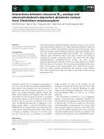
The biosynthesis of AdoCbl is a very complicated
process. 5¢-deoxyadenosyl- cobinamide (AdoCbi) and
AdoCbi-GDP are intermediates during the biosynthesis
of AdoCbl (Fig. 2A). Previous studies have shown that
AdoCbl-dependent methylmalonyl CoA mutase binds
both coenzyme analogs in ‘base-off’ mode, which indi-
cates that the histidine residue located on the conserved
cobalamin-binding motif is unable to coordinate to the
cobalt atom [5,6]. However, the AdoCbi-GDP-reconsti-
Keywords
adenosylcobalamin; adenosylcobinamide;
AdoCbi-GDP; B
12
; glutamate mutase
Correspondence
H P. Chen, Institute of Biotechnology,
National Taipei University of Technology 1,
Sec 3, Chung-Hsiao East Road, Taipei 106,
Taiwan
Fax: +886 2 27317117
Tel: +886 2 27712171 ext. 2528
E-mail:
(Received 14 August 2008, revised 30
September 2008, accepted 2 October
2008)
doi:10.1111/j.1742-4658.2008.06724.x
Adenosylcobalamin (AdoCbl)-dependent glutamate mutase from Clostrid-
ium tetanomorphum comprises two weakly-associating subunits, MutS and
MutE, which combine with AdoCbl to form the active holo-enzyme. Three
coenzyme analogs, methylcobinamide (MeCbi), adenosylcobinamide (Ado-
Cbi) and adeosylcobinamide-GDP (AdoCbi-GDP), were synthesized at
milligram scale. Equilibrium dialysis was used to measure the binding of
coenzyme B
12
analogs to glutamate mutase. Our results show that, unlike
AdoCbl-dependent methylmalonyl CoA mutase, the ratio k
cat
⁄ K
m
decreased approximately 10
4
-fold in both cases when AdoCbi or AdoCbi-
GDP was used as the cofactor. The coenzyme analog-binding studies show
that, in the absence of the ribonucleotide tail of AdoCbl, the enzyme’s
active site cannot correctly accommodate the coenzyme analog AdoCbi.
The results presented here shed some light on the cobalt–carbon cleavage
mechanism of B
12
.
Abbreviations
AdoCbi, adenosylcobinamide; AdoCbl, adenosylcobalamin; Ado-PCC, (Cob-5¢-Deoxyadenosin-5¢-yl)-(p-cresyl)cobamide; (Bza)AdoCba,
(benzimidazolribofuranosyl)-adenosylcobinamide; CobU, adenosyl-cobinamide kinase ⁄ adenosyl-cobinamide-phosphate guanylyltransferase;
MeCbi, methylcobinamide.
5960 FEBS Journal 275 (2008) 5960–5968 ª 2008 The Authors Journal compilation ª 2008 FEBS
tuted enzyme is catalytically active. More importantly,
the k
cat
⁄ K
m
of methylmalonyl CoA mutase is only
four-fold lower when AdoCbi-GDP is used as cofactor
[5,6]. This unexpected result suggests that coordination
by the lower axial ligand is not essential in the case of
methylmalonyl CoA mutase. To study the reactivity of
glutamate mutase toward these coenzyme analogs, a
chemo-enzymatic method was developed to synthesize
AdoCbi-GDP at the milligram scale. Our results show
that, in contrast to methylmalonyl CoA mutase, neither
AdoCbi nor AdoCbi-GDP can efficiently act as cofac-
tor for glutamate mutase [5]. The binding of AdoCbl
and three coenzyme analogs, methylcobinamide (MeC-
bi), AdoCbi and AdoCbi-GDP, to glutamate mutase
was measured by equilibrium dialysis. Kinetic proper-
ties towards AdoCbi and AdoCbi-GDP were also
investigated. Here, we report the results of coenzyme-
binding and kinetic studies of AdoCbl analogs with
glutamate mutase.
Results
Synthesis of MeCbi, AdoCbi and AdoCbi-GDP
MeCbi and AdoCbi were successfully separated from
unreacted MeCbl and AdoCbl and the dealkylated side
products using an SP–Sepharose ion-exchange column.
The relative molecular masses of MeCbi and AdoCbi
determined by ESI-MS were 1004.5 and 1240, which
compare favorably with calculated relative molecular
masses for MeCbi and AdoCbi of 1004.1 and 1239.6,
respectively. The bifunctional enzyme CobU (adenosyl-
cobinamide kinase ⁄ adenosyl-cobinamide-phosphate
guanylyltransferase) is involved in biosynthesis and
assembly of the nucleotide loop of cobalamin [7,8]
(Fig. 2A,B) Using chemically synthesized AdoCbi as the
CobU substrate, AdoCbi-GDP was enzymatically pre-
pared in large quantities. The yield of AdoCbi-GDP
could be significantly enhanced by using phenol ⁄ dichlo-
romethane extraction to remove the salt component of
the AdoCbi solution. The recovery of AdoCbi-GDP by
reverse-phase HPLC was very reproducible (Fig. 3). The
relative molecular mass of AdoCbi-GDP determined by
ESI-MS was 1664.4, and the calculated relative molecu-
lar mass of AdoCbi-GDP is 1664.6. The HPLC method
that we developed in this study is quite straightforward,
separating AdoCbi and AdoCbi-GDP directly without
further modification. In contrast, the reactant and prod-
uct, AdoCbi and AdoCbi-GDP, were analyzed in the
form of (CN)
2
Cbi and (CN)
2
Cbi-GDP, respectively, in
previous reports [7,8].
1
H-NMR spectra for MeCbi and
AdoCbi have been published previously [9,10]. The
600 MHz NMR spectrum of AdoCbi-GDP in
D
2
O ⁄ H
2
O was analyzed using two-dimensional COSY
and NOESY experiments. The results are summarized
in Table 1 and Fig. 2B.
D14
C15
H16
G120
T121
S61
V60
L59
G92
G91
Y117
I22
L23
A118
I334
E330
T94
R66
A67
G68
E subunit
(53.7 kDa)
S subuniT
(14 kDa)
H610
D608
G609
G686
G685
G613
G653
V654
S655
Y705
T709
T706
I617
E370
E247
Y243
Y89
Q330
L374
AB
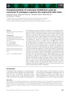
Fig. 1. (A) Model of glutamate mutase showing AdoCbl bound between the MutS and MutE subunits. The coenzyme-binding domain is on
the MutS subunit. (B) Model of methylmalonyl CoA mutase. The AdoCbl molecule is shown in grey and protein residues are shown in black.
H P. Chen et al. Adenosylcobalamin-dependent glutamate mutase
FEBS Journal 275 (2008) 5960–5968 ª 2008 The Authors Journal compilation ª 2008 FEBS 5961
O
NH
2
CONH
2
CONH
2
H
2
NOC
H
2
NOC
CONH
2
O
OH
NH
H
N
N
N
N
NH
2
O
OH
HO
HH
Co
N
N
N
N
AdoCbi
O
NH
2
CONH
2
CONH
2
H
2
NOC
H
2
NOC
CONH
2
O
O
-
P
O
HN
H
O
O
N
N
N
N
NH
2
O
OH
HO
HH
Co
N
N
N
N
N
N
N
N
NH
2
O
OH
O
O
-
P
O
O
AdoCbi-GDP
O
NH
2
CONH
2
CONH
2
H
2
NOC
H
2
NOC
CONH
2
O
NH
H
3
C
H
N
N
N
N
NH
2
O
OH
HO
HH
Co
N
N
N
N
AdoCbi-P
P
O
O
O
-
O
ATP
ADP
GTP
PP
i
Cobinamide
kinase
Cobinamide
kinase
O
NH
2
CONH
2
CONH
2
H
2
NOC
H
2
NOC
CONH
2
O
N
N
O
OH
HO
O
P
O
NH
H
O
O
-
N
N
N
N
NH
2
O
OH
HO
HH
Co
N
N
N
N
AdoCbl
α-ribazole
GMP
Cobalamin
synthase
A
Pr
AdoCbi-GDP
N
N
NN
Co
NH
2
O
H
2
N
H
2
N
O
NH
2
O
O
NH
2
O
NH
2
O
NH
O
P
O
P
O
O
O
N
OH
N
N
NH
O
NH
2
R
5
4
6
3
2
1
7
8
9
10
11
12
13
14
15
16
17
18
19
20
25
26
27
30
31
32
35
37
36
38
41
42
46
47
48
49
50
53
54
55
56
57
60
61
Pr1
Pr2
3
R2
R3
R4
R5
R=
N
N
N
N
O
OH
OH
H
2
C
NH
2
A15
A14
A13
A12
A11
O
-
O
-
O
O
R1
A2
A8
a
b
c
d
43
e
f
g
B2
A4
A5
B
Fig. 2. (A) Schematic representation of the
final steps of the de novo AdoCbl biosyn-
thetic pathway. (B) The chemical structure
of AdoCbi-GDP.
Adenosylcobalamin-dependent glutamate mutase H P. Chen et al.
5962 FEBS Journal 275 (2008) 5960–5968 ª 2008 The Authors Journal compilation ª 2008 FEBS
Determination of dissociation constants for
cofactors by equilibrium dialysis
The binding of AdoCbl, MeCbi, AdoCbi and AdoCbi-
GDP to glutamate mutase was investigated by equilib-
rium dialysis. Figure 4 shows the analog binding
curves with a fixed concentration of glutamate mutase.
AdoCbl, MeCbi, AdoCbi and AdoCbi-GDP were
bound with apparent K
d
values of 3.7 ± 0.5,
6.0 ± 0.9, 18 ± 3 and 14 ± 3 lm, respectively
(Fig. 4A–D).
UV–visible spectra of protein-bound MeCbi,
AdoCbi and AdoCbi-GDP complexes
The UV–visible spectra of cobalamins provide a
useful tool to examine the coordination state of
cobalt. The UV–visible absorption spectra of the
MeCbi-glutamate mutase, AdoCbi-glutamate mutase
and AdoCbi-GDP-glutamate mutase complexes were
measured. A red shift was observed in the spectra of
protein-bound MeCbi, AdoCbi and AdoCbi-GDP.
The 522 nm absorption maximum suggests that the
histidine residue occupies the lower axial ligand posi-
tion of the cobalt atom. However, we estimate that
approximately 55–60% of the AdoCbi–glutamate
mutase complex binds the cofactor in the ‘His-on’
form (Fig. 5).
150
A
B
AdoCbi
100
50
0
150
100
50
0
0 5 10 15 20
Time
25 30 35 40 45
AdoCbi-GDP
300
200
100
0
300
200
100
0
0 5 10 15 20
Tim
e
25 30 35 40 45
Fig. 3. Purification of AdoCbi-GDP from the reaction mixture
by reverse-phase HPLC. (A) Before the CobU enzymatic reaction.
(B) After the CobU enzymatic reaction.
Table 1. 600 MHz
1
H-NMR data for AdoCbi-GDP. d, doublet; q,
quadruplet; s, singlet; t, triplet; td, triplet of doublets; dd, doublet of
doublets.
Assignment
Signal
type
Chemical shifts
AdoCbi-GDP
(pH 7.0, 25 °C)
(p.p.m.)
J couplings
(AdoCbi-GDP)
(Hz)
Corrin
methyl
C20 s 0.77
C25 s 1.38
C35 s 2.38
C36 s 1.79
C46 s 0.83
C47 s 1.57
C53 s 2.36
C54 s 1.12
Corrin CH C3 m 4.19
C8 m 3.76
C10 s 6.92
C13 dd 3.35 5.77, 3.23
C18 td 2.8 10.4, 3.58
C19 d 4.62 3.58
Corrin CH
2
side chain
C26 d 2.62, 2.3 14.6
C30 m 1.96, 1.85
C31 m 2.44
C37 d 2.22, 1.74 14.8
C41 m 1.93, 1.81
C42 m 2.32, 2.24
C48 m 1.92, 1.77
C49 m 2.16
C55 m 1.75
C56 m 2.27
C60 d 2.63, 2.40 10.4
Aminopropan-2-
ol side chain
Pr1(CH
2
) t 3.30, 3.185 5.5
Pr2(CH) m 4.38
Pr3(CH
3
) d 1.21 6.5
Loop ribose R1 d 5.85 6.81
R2 m 4.68
R3 dd 4.46 4.87, 3.47
R4 m 4.27
R5 m 4.15
Adenosyl A2 s 8.20
A8 s 8.02
A11 d 5.60 3.54
A12 dd 4.40 5.54, 5.75
A13 dd 3.75 6.54, 5.75
A14 dd 1.91 9.3, 6.54
A15 d, dd 0.50, 0.32 8.6;8.6, 9.3
Base B2 s 8.01
NH s 8.20
s 7.94
s 7.84
s 7.6
s 7.57
s 7.3
s 7.27
s 7.09
s 7.02
s 6.88
s 6.86
s 6.60
s 6.57
s 6.36
H P. Chen et al. Adenosylcobalamin-dependent glutamate mutase
FEBS Journal 275 (2008) 5960–5968 ª 2008 The Authors Journal compilation ª 2008 FEBS 5963
Enzyme assay
In order to investigate the role of the ribonucleotide tail
of AdoCbl in catalysis, the coenzyme analogs were used
to examine the enzymatic activity. Our results indicate
that, perhaps not surprisingly, MeCbi is a totally inac-
tive coenzyme. The K
m
values for AdoCbi and Ado-
Cbi-GDP were 26 ± 8 and 75 ± 28 lm, respectively,
and the k
cat
values were (9.8 ± 1.0) · 10
)3
Æs
)1
and
(4.5 ± 0.8) · 10
)3
Æs
)1
, respectively. In both cases, the
k
cat
⁄ K
m
was decreased by approximately 10
4
-fold
compared with that of AdoCbl.
Discussion
Both methylmalonyl CoA mutase and glutamate
mutase belong to the subfamily of B
12
-dependent car-
bon-skeleton mutases, but their 1,2-rearrangement
mechanisms are obviously different [11]. Previous
studies have shown that (a) AdoCbi does not support
the turnover of methylmalonyl CoA mutase, but Ado-
Cbi-GDP does, and (b) the enzyme binds both AdoCbi
and AdoCbi-GDP in ‘base-off ⁄ His-off’ mode. The
results presented here indicate that, in contrast to
methylmalonyl CoA mutase, the k
cat
⁄ K
m
of glutamate
mutase for both analogs decreased by approximately
10
4
-fold. These results suggest that the ribonucleotide
tail of AdoCbl plays an important role in catalysis in
the case of glutamate mutase. In addition, both cofac-
tor analogs tested are bound by glutamate mutase in
‘base-off ⁄ His-on’ mode. Histidine–cobalt ligation
therefore cannot efficiently facilitate turnover of the
enzyme in the absence of the ribonucleotide tail of
AdoCbl. It is apparent that glutamate mutase is mech-
anistically different from methylmalonyl CoA mutase.
Significant differences in the affinity for AdoCbl
between these two enzymes appear to exit. Methylmal-
onyl CoA mutase binds AdoCbl very tightly with a K
d
of 0.17 lm, while glutamate mutase binds AdoCbl
relatively weakly with a K
d
between 1.8 and 6.8 lm
[1]. Moreover, glutamate mutase is very sensitive to
perturbation of the cofactor’s nucleotide tail, while
methylmalonyl CoA mutase is not. (Benzimidazolribo-
furanosyl)-adenosylcobinamide [(Bza)AdoCba] is a
coenzyme B
12
analog in which the dimethylbenzimi-
dazole moiety of AdoCbl is replaced by benzimidazole.
Previous studies have shown that the apparent K
m
of
glutamate mutase for (Bza)AdoCba is 0.5 lm, while
that for AdoCbl is 18 lm under similar conditions
[12]. However, the only difference between AdoCbl
and (Bza)AdoCba is two methyl groups. In contrast,
(Co-b-5¢-Deoxyadenosin-5¢-yl)-(p-cresyl)cobamide (Ado-
PCC) is another ‘base-off’ coenzyme B
12
analog in
which the dimethylbenzimidazole moiety of AdoCbl is
replaced by a p-cresolyl group. It fully supports the
turnover of methylmalonyl CoA mutase. The apparent
K
m
values of methylmalonyl CoA mutase for Ado-
PCC and AdoCbl are 354 and 64 nm, respectively [13].
A structural comparison of the protein–AdoCbl com-
plexes for these two enzymes is shown in Fig. 1A,B.
The glutamate mutase-bound nucleotide tail is located
in a more crowded environment, where the space is
more restricted. In particular, a bulkier residue, Leu59,
is situated at the bottom of the nucleotide tail-binding
pocket of glutamate mutase, but a small residue,
Gly653, is located in the same position of methylmalo-
nyl CoA mutase. The relatively restricted space in the
nucleotide tail-binding pocket might account for the
low activity and affinity of glutamate mutase towards
AdoCbi-GDP. Our unpublished results also show that
0
0.02
0.04
0.06
0.08
0.1
0.12
A B
C D
0 2 4 6 8 10 12 14
A
AdoCbl (µM)
0
0.02
0.04
0.06
0.08
0.1
0 20 40 60 80 100
A
MeCbi (µM)
0
0.01
0.02
0.03
0.04
0.05
0.06
0.07
0.08
0 20 40 60 80 100
A
AdoCbi (µM)
0
0.01
0.02
0.03
0.04
0.05
0.06
0 20 40 60 80 100
A
AdoCbi-GDP (µM)
Fig. 4. Binding of AdoCbl and its analogs to
glutamate mutase by equilibrium dialysis.
(A) AdoCbl, (B) MeCbi, (C) AdoCbi, and (D)
AdoCbi-GDP. The proteins, 20 l
M MutE and
100 l
M MutS in 0.1 mL buffer (50 mM
Tris ⁄ HCl, pH 8.5, 2 mM dithiothreitol), were
dialyzed against 1 mL buffer containing
50 m
M Tris ⁄ HCl, pH 8.5, 2 mM dithiothreitol
and cofactors. The data obtained were fitted
using
KALEIDA GRAPH software.
Adenosylcobalamin-dependent glutamate mutase H P. Chen et al.
5964 FEBS Journal 275 (2008) 5960–5968 ª 2008 The Authors Journal compilation ª 2008 FEBS
AdoCbl-dependent lysine aminomutase binds AdoCbl
with a K
d
of 18 ± 4 lm. Neither AdoCbi nor Ado-
Cbi-GDP efficiently support the catalysis of AdoCbl-
dependent l-lysine or d-ornithine aminomutase [14,15].
In short, the manipulation of coenzyme B
12
by methyl-
malonyl CoA mutase is quite different to that by glu-
tamate mutase, l-lysine and d-ornithine aminomutase.
Two mechanisms, electronic effect and steric effect,
have been postulated to explain the enzyme-accelerated
cobalt–carbon cleavage of AdoCbl [3,16]. AdoCbi-GDP
is bound by methylmalonyl CoA mutase in ‘base-off’
form, and is capable of supporting the enzyme’s cataly-
sis, suggesting that the electronic effect plays a minor
role in cleavage of the cobalt–carbon bond. However, as
far as we know, no experimental results from the studies
of coenzyme–protein interactions have previously been
provided to support the steric effect to explain the
cobalt–carbon cleavage mechanism.
The binding energy for AdoCbl comes from inter-
actions between proteins and the cofactor. From the
viewpoint of coenzyme molecule itself, these interac-
tions can be divided into three parts: the ribonucleo-
tide tail, corrin ring ⁄ cobalt–histidine ligation, and the
adenosyl group (Fig. 6). As shown in Table 2, the
apparent K
d
values of glutamate mutase for MeCbi
and AdoCbi are 6.0 ± 0.9 and 18 ± 3, respectively.
As shown in Table 2, the binding energy difference
between MeCbi and AdoCbi is approximately
2.5 kJÆmol
)1
. This result suggests that, in the absence
of the ribonucleotide tail of AdoCbl, the enzyme’s
active site cannot correctly accommodate the coen-
zyme analog AdoCbi. In accordance with this result,
the histidine residue on the conserved cobalamin-
binding motif can coordinate to the cobalt atom
when MeCbi is used as the cofactor (Fig. 5A). How-
ever, only approximately 60% of the glutamate
mutase-bound AdoCbi is in the ‘base-off ⁄ His-on’
form (Fig. 5B). Although AdoCbi-GDP cannot effi-
ciently support catalysis, its modified ribonucleotide
tail helps the histidine residue coordinate to the
cobalt atom (Fig. 5C). Previous studies have shown
that glutamate mutase binds AdoCbl, methylcobal-
amin (MeCbl) and cob(II)alamin with similar affinity
[17]. These results indicate that the ribonucleotide
tail of AdoCbl is important in coenzyme binding.
We hereby propose that the role of the ribonucleo-
tide tail of AdoCbl is to distort the adenosyl group
to fit into the enzyme’s active site during the coen-
zyme-binding process. However, recent spectroscopic
studies have indicated that the Co–C bond of gluta-
mate mutase-bound AdoCbl is not weakened within
the enzyme active site [18,19]. The correlation
between the distortion of the adenosyl group and
cleavage of the cobalt–carbon bond is still not clear.
Although the precise mechanism remains obscure,
the results presented here do shed some light on the
cobalt–carbon cleavage mechanism of B
12
.
Experimental procedures
Materials
AdoCbl and methylcobalamin (MeCbl) were obtained from
Sigma (St Louis, MO, USA). SP–Sepharose Fast Flow cat-
ion-exchange gel medium was purchased from GE Health-
care (Uppsala, Sweden). The production and purification of
glutamate mutase from C. tetanomorphum have been
0
0.1
0.2
0.3
0.4
0.5
0.6
A
B
C
350 400 450 500 550 600 650 700
Free MeCbi
Protein-bound MeCbi
A
Wavelength (nm)
0
0.2
0.4
0.6
0.8
1
350 400 450 500 550 600 650 700
Free AdoCbi
Protein-bound AdoCbi
A
Wavelength (nm)
0
0.05
0.1
0.15
0.2
0.25
0.3
350 400 450 500 550 600 650 700
Free AdoCbi-GDP
Protein-bound AdoCbi-GDP
A
Wavelength (nm)
Fig. 5. UV–visible spectra of free and glutamate mutase-bound
MeCbi (A), AdoCbi (B) and AdoCbi-GDP (C).
H P. Chen et al. Adenosylcobalamin-dependent glutamate mutase
FEBS Journal 275 (2008) 5960–5968 ª 2008 The Authors Journal compilation ª 2008 FEBS 5965
described previously [1]. All chemicals used were of analyti-
cal grade or higher.
Preparation of MeCbi and AdoCbi
Because the cobalt–carbon bond of cobalamin is light-
sensitive, the following procedure was carried out in a dark
environment. The chemical synthesis of AdoCbi and MeCbi
was slightly modified from that described previously [20].
For this reaction, 0.5 g of AdoCbl or MeCbl was used. The
products, AdoCbi or MeCbi, were separated from the
reaction mixture using a SP–Sepharose Fast Flow cation-
exchange column (2.6 · 40 cm). The column was equili-
brated in 10 mm potassium phosphate buffer, pH 7.0.
AdoCbi or MeCbi were eluted with a 500 mL gradient from
0 to 0.5 m KCl. The flow rate was 3 mLÆmin
)1
; 4 mL frac-
tions were collected. Fractions containing AdoCbi or MeCbi
were pooled separately. The yield was approximately 30%.
Chemo-enzymatic preparation of AdoCbi-GDP
The cobU gene from Salmonella typhimurium ATCC 19585
has been successfully cloned and over-expressed in Escheri-
chia coli [21]. CobU protein, in 50 mm Tris ⁄ HCl, pH 8.5,
and other solutions used for the reaction were made
anaerobic and equilibrated using alternate cycles of vacuum
and hydrated argon gas for 15 min. The 1.5 mL reaction
mixture containing 1.5 mm GTP, 1.5 mm MgCl
2
,1mm
b-mercaptoethanol, 10 lm CobU and 250 lm AdoCbi was
buffered in 100 mm Tris ⁄ HCl, pH 8.5. Each solution was
N
N
H
N
N
Co
+3
N
N
H
Co
+3
CH
3
N
N
N
N
H
2
N
O
OH
OH
H
H
H
H
OH
N
N
H
Co
+3
OH
N
N
N
N
H
2
N
O
OH
OH
H
H
H
H
Corrin ring and His ligation
Nucleotide tail
Adenosyl group
AdoCbl
MeCbi AdoCbi
No contribution
Distortion
Free energy change
contributed by:
Fig. 6. Illustrations of the binding free energy change contributed by each fragment in coenzyme B
12
.
Table 2. Comparison of the k
cat
⁄ K
m
value, dissociation constants and binding free energies of various coenzyme analogs. The k
cat
⁄ K
m
value
for AdoCbl is calculated from the results in [1].
Coenzyme analogs Upper ligand of cobalt k
cat
⁄ K
m
(s
)1
ÆlM
)1
) K
d
(lM) DG (kJÆmol
)1
)
AdoCbi Adenosyl group (4.3 ± 1.7) · 10
)4
18 ± 3 25.19 ± 0.39
MeCbi Methyl group N ⁄ A 6.0 ± 0.9 27.72 ± 0.35
AdoCbi-GDP Adenosyl group (7.4 ± 3.9) · 10
)5
14 ± 3 25.79 ± 0.50
AdoCbl Adenosyl group 1.12 ± 0.09 3.7 ± 0.5 28.83 ± 0.31
Adenosylcobalamin-dependent glutamate mutase H P. Chen et al.
5966 FEBS Journal 275 (2008) 5960–5968 ª 2008 The Authors Journal compilation ª 2008 FEBS
injected separately into a rubber-sealed 2 mL vial that had
been flushed with argon for 10 min prior to use. The reac-
tion was incubated at room temperature overnight and was
terminated by incubation at 95 °C for 10 min.
AdoCbi-GDP was isolated from the reaction mixture by
reverse-phase HPLC on a 5lm, 25 cm · 4.6 mm, Supelco
AscentisÔ C
18
column (Bellefonte, PA, USA). The eluents
used were as follows: eluent A, 100 mm potassium phos-
phate buffer, pH 6.5; eluent B, 100 mm potassium phos-
phate buffer, pH 8.0 containing 50% CH
3
CN. The flow
rate was 1 mLÆmin
)1
. The following profile was used for
separation: 2 min isocratic development with 98% A; 5 min
linear gradient from 98% A to 75% A; 15 min linear gradi-
ent from 75% A to 65% A; 3 min linear gradient from
65% A to 0% A; 10 min isocratic development with 100%
B. Both analogs, AdoCbi and AdoCbi-GDP, were charac-
terized by ESI-MS.
NMR spectroscopy
NMR spectra of AdoCbi-GDP were recorded on a Bruker
AVANCE 600 AV system (Bruker BioSpin GmbH; Rhein-
stetten, Germany) at 25 °C. Approximately 2 mg of AdoCbi-
GDP dissolved in 0.25 mL H
2
O containing 10% D
2
O was
used for the NMR experiment. Two-dimensional homo-
nuclear (TOCSY and ROESY) and heteronuclear (HMQC
and HMBC) spectra of AdoCbi-GDP were collected for the
chemical shift assignment. The ROESY spectra were
obtained with mixing times of 50 and 150 ms, to classify the
relative strengths of the observed NOEs. All spectra were pro-
cessed and analyzed by using topspin 2.1 software (Bruker
BioSpin GmbH; Rheinstetten, Germany).
Measurement of the binding of coenzyme
analogs to proteins
The binding of coenzyme analogs to glutamate mutase was
measured by equilibrium dialysis. About 100 lLof20lm
E component and 100 lm S component were loaded into
microdialysis tubes. The protein solutions were dialyzed
against 1 mL of 50 mm Tris buffer, pH 8.5, in the presence
of various concentrations of coenzyme B
12
or its analogs at
4 °C overnight. The absorbance was recorded at 522 nm
using an Amersham Bioscience Ultrospec 2100 spectropho-
tometer; a sample of the corresponding dialysis buffer was
used to subtract the contribution of unbound coenzyme
analogs from the absorbance of the enzyme. The kaleida
graph program (Synergy Software, Reading, PA, USA)
was used to fit data to estimate the dissociation constant.
Protein UV–visible spectra
To determine the coordination state of the cobalt atom of
enzyme-bound coenzyme analogs, 100 lL of protein solu-
tion containing 400 lm S component, 100 lm E compo-
nent, and 50 or 100 lm coenzyme analog was dialyzed
against 1 mL 50 mm Tris buffer, pH 8.5, at 4 °Cinthe
dark overnight, by which time equilibrium had been
reached. Spectra were recorded using an Amersham Bio-
science Ultrospec 2100 Pro spectrophotometer (Uppsala,
Sweden); a sample of the dialysis buffer was used to sub-
tract the contribution of unbound coenzyme analog from
the spectra of the holoenzymes.
Enzyme assay
An HPLC-based method was used to assay glutamate
mutase activity [22]. The assay was made irreversible by
coupling the formation of 3-methylaspartate to the pro-
duction of mesaconate through deamination by methylas-
partase. In a typical reaction, 10 lm E component and
50 lm S component proteins were used in a total volume
of 100 lL containing 2 mm MgCl
2
,40mml-glutamate
and 50 mm Tris buffer, pH 8.5. The K
m
and k
cat
for Ado-
Cbi were determined in the presence of 10, 25, 50, 75 and
120 lm cofactor, and the K
m
and k
cat
for AdoCbi-GDP
were determined in the presence of 20, 70, 100, 150 and
200 lm cofactor. The reaction was initiated by adding
l-glutamate and incubating at room temperature for
15 min. The formation of mesaconate was then analyzed
by reverse-phase HPLC on a C
18
column (4.6 · 250 mm)
as described previously [22].
Acknowledgements
This work was supported by grants NSC-94-2320-B-
027-002 and NSC-95-2113-M-027-005-MY2 from the
National Scientific Council, Taiwan, Republic of
China, to H P.C.
References
1 Holloway DE & Marsh ENG (1994) Adenosylcobala-
min-dependent glutamate mutase from Clostridium
tetanomorphum. J Biol Chem 269, 20425–20430.
2 Zelder O, Beatrix B, Kroll F & Buckel W (1995)
Coordination of a histidine residue of the protein-com-
ponent S to the cobalt atom in coenzyme B
12
-dependent
glutamate mutase from Clostridium cochlearium. FEBS
Lett 369, 252–254.
3 Halpern J (1985) Mechanisms of coenzyme B
12
-depen-
dent rearrangements. Science 227, 869–875.
4 Chen HP & Marsh ENG (1997) How enzymes control
the reactivity of adenosylcobalamin: effect on coenzyme
binding and catalysis of mutations in the conserved his-
tidine-aspartate pair of glutamate mutase. Biochemistry
36, 7884–7889.
H P. Chen et al. Adenosylcobalamin-dependent glutamate mutase
FEBS Journal 275 (2008) 5960–5968 ª 2008 The Authors Journal compilation ª 2008 FEBS 5967
5 Chowdhury S & Banerjee R (1999) Role of the dimeth-
ylbenzimidazole tail in the reaction catalyzed by coen-
zyme B
12
-dependent methylmalonyl-CoA mutase.
Biochemistry 38, 15287–15294.
6 Chowdhury S, Thomas MG, Escalante-Semerena JC &
Banerjee R (2001) The coenzyme B
12
analog 5’-deoxy-
adenosylcobinamide-GDP supports catalysis by methyl-
malonyl-CoA mutase in the absence of trans-ligand
coordination. J Biol Chem 276 , 1015–1019.
7 O’Toole GA & Escalante-Semerena JC (1995) Purifica-
tion and characterization of the bifunctional CobU
enzyme of Salmonella typhimurium LT2. Evidence for a
CobU-GMP intermediate. J Biol Chem 270, 23560–
23569.
8 Thomas MG, Thompson TB, Rayment I & Escalante-
Semerena JC (2000) Analysis of the adenosylcobinamide
kinase ⁄ adenosylcobinamide-phosphate guanylyltransfer-
ase (CobU) enzyme of Salmonella typhimurium LT2.
Identification of residue His-46 as the site of guanylyla-
tion. J Biol Chem 275, 27576–27586.
9 Brown KL, Zou X & Salmin L (1991) Facile a ⁄ b dia-
stereomerism in organocobalt corrins. Generality of the
phenomenon and characterization of additional a-dia-
stereomers. Inorg Chem 30, 1949–1953.
10 Pagano TG, Yohannes PG, Hay BP, Scott JR, Finke
RG & Marzilli LG (1989) Solution behavior and com-
plete proton and carbon-13 NMR assignments of the
coenzyme B
12
derivative (5’-deoxyadenosyl)cobinamide
using modern 2D NMR experiments, including
600 MHz proton NMR data. J Am Chem Soc 111,
1484–1491.
11 Banerjee R & Rasdale SW (2003) The many faces of
vitamin B12: catalysis by cobalamin-dependent
enzymes. Annu Rev Biochem 72, 209–247.
12 Holloway DE, Harding SE & Marsh ENG (1996)
Adenosylcobalamin-dependent glutamate mutase:
properties of a fusion protein in which the cobalamin-
binding subunit is linked to the catalytic subunit.
Biochem J 320, 825–830.
13 Poppe L, Stupperich E, Hull WE, Buckel T & Retey J
(1997) A base-off analogue of coenzyme-B
12
with a
modified nucleotide loop
1
H-NMR structure analysis
and kinetic studies with (R)-methylmalonyl-CoA
mutase, glycerol dehydratase, and diol dehydratase. Eur
J Biochem 250, 303–307.
14 Chang CH & Frey PA (2000) Cloning, sequencing,
heterologous expression, purification, and characteriza-
tion of adenosylcobalamin-dependent d-lysine 5,6-ami-
nomutase from Clostridium sticklandii. J Biol Chem 275,
106–114.
15 Chen HP, Wu SH, Lin YL, Chen CM & Tsay SS
(2001) Cloning, sequencing, heterologous expression,
purification and characterization of adenosylcobalamin-
dependent d-ornithine aminomutase from Clostridium
sticklandii. J Biol Chem 276, 44744–44750.
16 Pratt JM (1985) The B12-dependent isomerase enzymes;
how the protein controls the active site. Chem Soc Rev
14, 161–170.
17 Chen HP & Marsh ENG (1997) Adenosylcobalamin-
dependent glutamate mutase: examination of substrate
and coenzyme binding in an engineered fusion protein
possessing simplified subunit structure and kinetic prop-
erties. Biochemistry 36, 14939–14945.
18 Brooks AJ, Fox CC, Marsh ENG, Vlasie M, Banerjee
R & Brunold TC (2005) Electronic structure studies of
the adenosylcobalamin cofactor in glutamate mutase.
Biochemistry 44, 15167–15181.
19 Sension RJ, Cole AG, Harris AD, Fox CC, Woodbury
NW, Lin S & Marsh ENG (2004) Photolysis and
recombination of adenosylcobalamin bound to
glutamate mutase. J Am Chem Soc 126, 1598–1599.
20 Hay BP & Finke RG (1987) Thermolysis of the Co–C
bond in adenosylcorrins. 3. Quantification of the axial
base effecting adenosylcobalamin by the synthesis and
thermolysis of axial base-free adenosylcobinamide.
Insights into the energetics of enzyme-assisted cobalt–
carbon bond homolysis. J Am Chem Soc 109, 8012–8018.
21 Hsu FC, Ho TJ, Lai CC, Lin CF & Chen HP (2005)
Cloning, sequencing, expression, and single-step purifi-
cation of the adenosylcobinamide kinase ⁄ adenosylcobi-
namide-phosphate guanylyltransferase (CobU) from
Salmonella typhimurium ATCC 19585. Protein Expr
Purif 42, 178–181.
22 Marsh ENG (1995) Tritium isotope effects in adenosyl-
cobalamin-dependent glutamate mutase: implications
for the mechanism. Biochemistry 34, 7542–7547.
Adenosylcobalamin-dependent glutamate mutase H P. Chen et al.
5968 FEBS Journal 275 (2008) 5960–5968 ª 2008 The Authors Journal compilation ª 2008 FEBS