Báo cáo khoa học: Comprehensive interaction of dicalcin with annexins in frog olfactory and respiratory cilia pdf
Bạn đang xem bản rút gọn của tài liệu. Xem và tải ngay bản đầy đủ của tài liệu tại đây (1.08 MB, 14 trang )
Comprehensive interaction of dicalcin with annexins
in frog olfactory and respiratory cilia
Tatsuya Uebi
1
, Naofumi Miwa
1,2,
* and Satoru Kawamura
1,2
1 Department of Biology, Graduate School of Science, Osaka University, Japan
2 Graduate School of Frontier Biosciences, Osaka University, Japan
Calcium ions are known to modulate signal transduc-
tion in various cells. This effect is usually mediated
by Ca
2+
-binding proteins. For example, in olfactory
receptor cells, odorant stimuli induce Ca
2+
influx
through a cyclic nucleotide gated channel [1]. The
increase in the Ca
2+
concentration is detected by
calmodulin, a well-known Ca
2+
-binding protein. The
Ca
2+
-bound form of calmodulin has essential roles in
olfactory adaptation [2,3]. In photoreceptor cells, sev-
eral Ca
2+
-binding proteins are known to be present
and to modulate phototransduction signals [4].
We previously found a Ca
2+
-binding protein, dical-
cin (renamed from p26olf [5]), in frog olfactory epithe-
lium, and reported that dicalcin is expressed in the
olfactory epithelium, lung, and spleen [6,7]. In the
olfactory epithelium and lung, dicalcin localizes in the
cilia. Dicalcin has partial homology to S100 proteins, a
family of EF-hand Ca
2+
-binding proteins, and consists
of two S100A11-like regions aligned in sequence. The
amino acid sequences in the N-terminal and the C-ter-
minal halves show 58% and 45% identity, respectively,
to chick S100A11 [7]. The predicted structure of dical-
cin is similar to that of an S100 dimer [8].
S100 proteins are known to be involved in various
cellular functions, such as cell cycle progression and
cell survival [9–11]. S100 proteins show no enzymatic
activities by themselves and, instead, modulate the
function of other proteins through direct binding to
Keywords
annexin; dicalcin; olfactory cilia; respiratory
cilia; S100
Correspondence
S. Kawamura, Graduate School of Frontier
Biosciences, Osaka University, Yamada-oka
1–3, Suita, Osaka 565-0871, Japan
Fax: +81 6 6879 4614
Tel: +81 6 6879 4610
E-mail:
*Present address
Department of Physiology, School of
Medicine, Toho University, Tokyo, Japan
Database
Amino acid sequences have been submitted
to DDBJ under the following accession
numbers: frog annexin A1, AB286845; frog
annexin A2, AB286846; frog annexin A4,
AB286848; frog annexin A5, AB286847
(Received 8 May 2007, revised 20 July
2007, accepted 24 July 2007)
doi:10.1111/j.1742-4658.2007.06007.x
Dicalcin (renamed from p26olf) is a dimer form of S100 proteins found
in frog olfactory epithelium. S100 proteins form a group of EF-hand
Ca
2+
-binding proteins, and are known to interact with many kinds of tar-
get protein to modify their activities. To determine the role of dicalcin in
the olfactory epithelium, we identified its binding proteins. Several proteins
in frog olfactory epithelium were found to bind to dicalcin in a
Ca
2+
-dependent manner. Among them, 38 kDa and 35 kDa proteins were
most abundant. Our analysis showed that these were a mixture of annex-
in A1, annexin A2 and annexin A5. Immunohistochemical analysis showed
that dicalcin and all of these three subtypes of annexin colocalize in the
olfactory cilia. Dicalcin was found to be present in a quantity almost suffi-
cient to bind all of these annexins. Colocalization of dicalcin and the three
subtypes of annexin was also observed in the frog respiratory cilia. Dicalcin
facilitated Ca
2+
-dependent liposome aggregation caused by annexin A1 or
annexin A2, and this facilitation was additive when both annexin A1 and
annexin A2 were present. In this facilitation effect, the effective Ca
2+
con-
centrations were different between annexin A1 and annexin A2, and there-
fore the dicalcin–annexin system in frog olfactory and respiratory cilia can
cover a wide range of Ca
2+
concentrations. These results suggested that
this system is associated with abnormal increases in the Ca
2+
concentration
in the olfactory and other motile cilia.
FEBS Journal 274 (2007) 4863–4876 ª 2007 The Authors Journal compilation ª 2007 FEBS 4863
these proteins. p53, RAGE and annexins are known to
be binding proteins of S100 proteins. S100 proteins are
known to form dimers, and the dimer form binds to
the binding protein to exert the effect. Because dicalcin
consists of two S100-like domains aligned in sequence,
the function of dicalcin is probably similar to that of
an S100 dimer.
Although the Ca
2+
-binding property has been inves-
tigated in detail in dicalcin [12], little is known about
its physiologic function. To investigate this, in the
present study we first tried to determine the binding
proteins of dicalcin. We found that several of the pro-
teins in frog olfactory epithelium bind to dicalcin in a
Ca
2+
-dependent manner. Among them, 38 kDa and
35 kDa proteins were the major proteins. We identified
them as annexin A1, annexin A2 and annexin A5. We
further examined their localizations and the effect of
dicalcin on the activities of these annexins by measur-
ing liposome aggregation.
Results
Purification of binding proteins of dicalcin
Binding proteins of dicalcin were searched for among
the soluble and membrane-associated proteins of frog
olfactory cilia. Because dicalcin is an S100-like
EF-hand Ca
2+
-binding protein, we expected that the
binding proteins would bind to dicalcin in a Ca
2+
-
dependent manner. The Chaps-solubilized fraction
(see Experimental procedures) containing the mem-
brane-associated proteins in frog olfactory cilia
(Fig. 1, cilia) was loaded onto a dicalcin-Sepharose
column at 1 mm Ca
2+
. Most of the proteins were
found in the pass-through fraction (Fig. 1, elution
peak A and lane A), but some of the proteins were
retained, and eluted by reducing the Ca
2+
concentra-
tion (Fig. 1, elution peak B and lane B). Several pro-
teins were found in lane B, but the major proteins
were 38 kDa and 35 kDa proteins. The latter could
be one of the binding proteins detected in our previ-
ous dicalcin-overlay analysis [13]. In control studies,
we did not see the binding of these proteins when
dicalcin was not attached to the Sepharose beads
(Fig. 1C). Although the amount of each of the eluted
proteins varied among preparations, 38 kDa and
35 kDa proteins were always the major constituents.
We therefore focused on these proteins in the follow-
ing study. Essentially similar binding proteins were
detected when we used the soluble protein fraction,
but the amounts of the proteins were greater in the
Chaps-solubilized fraction. For this reason, we used
this fraction in the following studies.
Amino acid sequence analysis of 38 kDa and
35 kDa proteins
During the course of this study, we realized that
35 kDa proteins contained proteolytic fragments of
38 kDa proteins: in the presence of protease inhibi-
tors, the amount of 38 kDa proteins was larger than
that in the absence of the inhibitors. However, we
could not inhibit the proteolysis completely: even in
the presence of a cocktail of inhibitors, our immuno-
logic study detected signals of 38 kDa proteins at the
35 kDa position (see Fig. 3A below). In addition, the
degree of inhibition was variable, depending on each
preparation. Nevertheless, the binding proteins of di-
calcin, mainly the 38 kDa and the 35 kDa proteins,
were fragmented by a protease. The resultant proteo-
lytic fragments were isolated by RP-HPLC, and their
amino acid sequences were determined. The result
suggested that the 38 kDa and the 35 kDa proteins
are the annexin family proteins. The result, however,
was complex: the amino acid sequences of the frag-
ments did not match the sequence of a single annexin
family protein. Instead, the sequence of a fragment
showed some similarity to the sequence of annex-
in A1, annexin A2, annexin A4 or annexin A5 of
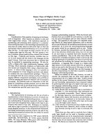
Fig. 1. Purification of binding proteins of dicalcin by affinity column
chromatography. The Chaps-solubilized protein fraction of the cilia
of frog olfactory epithelium was loaded to a dicalcin-Sepharose col-
umn at 1 m
M Ca
2+
. Most of the proteins passed through the col-
umn (A in the elution profile) at a high (1 m
M)Ca
2+
concentration,
but some proteins remained in the column and came out only after
addition of 5 m
M EGTA (B in the profile). Inset: SDS ⁄ PAGE patterns
of the Chaps-solubilized cilia protein fraction (cilia), the pass-through
fraction (A) and the eluate in the presence of 5 m
M EGTA (B). As a
control, an eluate was obtained similarly as in (B), but with the use
of Sepharose beads without dicalcin conjugated (C). Proteins were
stained with silver.
Role of dicalcin in frog olfactory cilia T. Uebi et al.
4864 FEBS Journal 274 (2007) 4863–4876 ª 2007 The Authors Journal compilation ª 2007 FEBS
other animal species, which suggested that the
38 kDa and the 35 kDa proteins were a mixture of
these annexins. We therefore tried to isolate cDNAs
of annexin A1, annexin A2, annexin A4 and annex-
in A5 to identify which annexins were in the fraction
of the 38 kDa and the 35 kDa proteins.
Cloning of annexin cDNAs
On the basis of the partial amino acid sequences of the
proteolytic fragments as determined above, we synthe-
sized oligonucleotide degenerate primers and used
them to search for the cDNA fragments of the corres-
ponding annexins. Partial cDNA fragments of annex-
in A1, annexin A2, annexin A4 and annexin A5 were
amplified, and the frog olfactory cDNA library
was screened with these fragments. The full-length
sequences of frog annexin cDNAs were obtained, and
the amino acid sequences were deduced (supplemen-
tary Fig. S1). The amino acid sequences detected in
the proteolytic fragments were found in the deduced
amino acid sequences of frog annexin A1, annexin A2,
and annexin A5, but not in the sequence of frog an-
nexin A4. This result indicated that annexin A4 was
not present, or the content of annexin A4 was small in
the fraction of the 38 kDa and 35 kDa proteins.
Among our recombinant annexins (see below), the
apparent molecular mass of annexin A4 was slightly
lower than 35 kDa on our SDS ⁄ PAGE gel. Because
the density of the corresponding position on the
SDS ⁄ PAGE gel of the binding proteins of dicalcin was
faint, this result also suggested that the content of
annexin A4 in the 35 kDa proteins was small even if
it was present. For these reasons, we did not study
annexin A4 further.
Identification of annexin A1, annexin A2 and
annexin A5 as the binding proteins of dicalcin
Our results were so far consistent with the notion that
the 38 kDa and the 35 kDa proteins are annexin A1,
annexin A2, and annexin A5. However, we were not
totally sure of this at this stage. Therefore, we first
tried to confirm that annexin A1, annexin A2 and an-
nexin A5 show Ca
2+
-dependent binding to dicalcin, as
the 38 kDa and the 35 kDa proteins do. For this, we
obtained recombinant annexin A1, annexin A2 and
annexin A5 expressed in Escherichia coli. The apparent
molecular masses of recombinant annexin A1 and ann-
exin A2 were both 38 kDa, and that of annexin A5
was 35 kDa (Fig. 2), and all of them bound to the di-
calcin-Sepharose beads in a Ca
2+
-dependent manner
(Fig. 2), as the native 38 kDa and 35 kDa proteins do.
Second, we identified the 38 kDa and the 35 kDa pro-
teins as annexin A1, annexin A2 and annexin A5 immu-
nologically. We raised specific antiserum against
annexin A1, annexin A2 or annexin A5 in mouse and
rabbit using recombinant annexins (supplementary
Fig. S2). Antiserum against annexin A1 recognized both
the 38 kDa and the 35 kDa proteins (Fig. 3A, low Ca
2+
eluate), and antiserum against annexin A2 also detected
the 38 kDa and the 35 kDa proteins. Antiserum against
annexin A5 detected only the 35 kDa proteins.
From the above results, it became evident that the
38 kDa proteins contained both full-length annexin A1
and annexin A2, and the 35 kDa proteins contained
full-length annexin A5 together with proteolytic
fragments of annexin A1 and annexin A2. Our two-
dimensional electrophoresis confirmed this (Fig. 3B).
This two-dimensional analysis also indicated that pro-
teins other than annexin A1, annexin A2 and annex-
in A5 were not present in significant amounts in the
38 kDa and 35 kDa proteins (Fig. 3B). In Fig. 3B,
there are weak signals of annexin A1 at around
pH 5.1. They are probably the signals of annexin A1
that was not focused in our two-dimensional electro-
phoresis.
Fig. 2. Ca
2+
-dependent binding of recombinant annexins to dicalcin.
The cell lysate of E. coli (lysate) expressing recombinant annex-
in A1, annexin A2 or annexin A5 was mixed with dicalcin-Sepha-
rose beads at 1 m
M Ca
2+
. The beads were washed 10 times by
centrifugation with K-gluc buffer supplemented with 1 m
M Ca
2+
,
and the 1st and the 10th extracts were subjected to SDS ⁄ PAGE
(high-Ca
2+
wash 1 and high-Ca
2+
wash 10). The beads were finally
washed with K-gluc buffer supplemented with 5 m
M EGTA, and
the extract was subjected to SDS ⁄ PAGE (low-Ca
2+
wash).
T. Uebi et al. Role of dicalcin in frog olfactory cilia
FEBS Journal 274 (2007) 4863–4876 ª 2007 The Authors Journal compilation ª 2007 FEBS 4865
Colocalization of annexins and dicalcin in frog
olfactory and respiratory epithelium
Dicalcin has been reported to localize in the cilia of
frog olfactory and respiratory epithelium [7]. To
understand the possible association of annexin A1,
annexin A2 and annexin A5 with the function of dical-
cin, we examined the colocalization of each annexin
with dicalcin, using specific antisera (supplementary
Fig. S2). In addition, we also examined whether differ-
ent subtypes of the annexins colocalize in the same
cilia. Figures 4 and 5 show the immunohistochemical
studies of dicalcin and annexin A1, annexin A2 and
annexin A5. In Fig. 4, the olfactory cilia, which were
identified immunohistochemically with olfactory cilia-
specific G
olf
antibody (Fig. 4M), were found to be
reactive to antiserum against dicalcin (Fig. 4A,D,G).
The cilia were also positively stained with antiserum
against annexin A1 (Fig. 4B), annexin A2 (Fig. 4E),
and annexin A5 (Fig. 4H). The merged image clearly
showed colocalization of dicalcin with each of the an-
nexins (Fig. 4C,F,I). In this study, the conditions for
obtaining immunofluorescence were kept constant in
each of the observations with rabbit antiserum (Texas
Red) or mouse antiserum (fluorescein isothiocyanate),
and therefore the color in the merged picture was
dependent on the relative intensities of red and green
fluorescence, namely, the titers of antisera against di-
calcin and annexins. Preabsorption of the specific anti-
bodies by recombinant proteins significantly reduced
the signals (Miwa et al. [13] for anti-dicalcin serum
and Fig. 4N for anti-annexin A2 serum).
Because all the annexins examined in this study co-
localized with dicalcin, we then examined whether ann-
exin A1, annexin A2 and annexin A5 all colocalize in
the same cell. Figure 5 shows the immunohistochemi-
cal study of colocalization of annexin A1, annexin A2,
and annexin A5. For any combination of these three
subtypes of annexin, colocalization was demonstrated
(Fig. 5). Therefore, it was evident that all three sub-
types of annexin are present in the same olfactory
cilium. From the results in Figs 4 and 5, it became
evident that dicalcin, annexin A1, annexin A2 and
annexin A5 all colocalize in the same olfactory cilium.
In the respiratory epithelium, similar colocalization
was observed (supplementary Fig. S3), although the
signal of G
olf
, a marker protein of olfactory cells, was
not seen.
Estimation of the relative molecular abundance
of dicalcin and annexins in frog olfactory cilia
The above immunohistochemical study showed that all
subtypes of the annexins studied here colocalize with
AB
Fig. 3. Identification of annexin A1, annexin A2 and annexin A5 by western blot analysis. (A) Determination that the 38 kDa proteins are a
mixture of annexin A1 and A2 and that the 35 kDa proteins are a mixture of annexin A5 and proteolytic fragments of annexin A1 and annex-
in A2. Purified recombinant annexin A1, annexin A2 and annexin A5 (A1, A2 and A5), together with the binding proteins of dicalcin (low-Ca
2+
eluate), were electrophoresed on an SDS ⁄ PAGE gel, and the proteins were stained with silver (silver stain). The proteins were probed with
specific antisera against annexins (anti-A1, anti-A2 and anti-A5) by western blot. The 38 kDa proteins contained both annexin A1 and annex-
in A2, and the 35 kDa proteins contained annexin A5 together with annexin A1 and annexin A2, possibly fragmented by proteolysis during
preparation. (B) Two-dimensional electrophoretic identification of the 38 kDa and the 35 kDa proteins as annexin A1, annexin A2, and annex-
in A5. A similar analysis as in (A) was performed by two-dimensional electrophoresis. Annexins were identified at the apparent molecular
mass of 38 kDa with pI values of 6.2–7.1 (annexin A1), and of c. 8 (annexin A2), and a single spot at 35 kDa with pI ¼ 5.6 (annexin A5).
Each annexin subtype is indicated by a circle.
Role of dicalcin in frog olfactory cilia T. Uebi et al.
4866 FEBS Journal 274 (2007) 4863–4876 ª 2007 The Authors Journal compilation ª 2007 FEBS
dicalcin in frog olfactory cilia. To understand the sig-
nificance of this colocalization, we tried to estimate the
relative molecular abundance of dicalcin and annexins.
In this quantification, we used both the soluble and
the membrane fraction after detachment of the cilia
(see Experimental procedures). They were solubilized
with the SDS ⁄ PAGE sample buffer, and were directly
electrophoresed with known amounts of recombinant
dicalcin and annexins. The contents of annexins and
dicalcin in the cilia were estimated by western blot,
and their ratio determined in three frogs was annex-
in A1 ⁄ annexin A2 ⁄ annexin A5 ⁄ dicalcin ¼ 1.0 : 0.42 ±
0.09 : 0.54 ± 0.15 : 1.9 ± 0.6. Dicalcin is a soluble
protein, and annexins were mostly present in the
Chaps-solubilized fraction. Dicalcin might have been
lost during isolation of the olfactory epithelium, and
therefore the content of dicalcin could be higher than
the value determined above. Because the number and
the volume of the cilia in the sample were not known,
it was not possible to determine the actual concentra-
tions of these proteins.
Effect of dicalcin on the activity of annexins
As has been reported previously, annexins are known
to induce membrane aggregation in a Ca
2+
-dependent
manner [14], and it is also known that this activity of
annexins is enhanced by binding of S100 proteins [15].
We therefore examined the effect of dicalcin on
the membrane aggregation activity of annexins. The
ABC
DEF
GHI
JKL
MNO
Fig. 4. Colocalization of dicalcin with annexin A1, annexin A2 or annexin A5 in frog olfactory epithelium. (A–I) Immunofluorescence double-
staining of dicalcin and annexins. A section was treated with rabbit anti-dicalcin serum (red; A, D and G) and mouse antiserum raised against
one subtype of annexin (green: B, annexin A1; E, annexin A2; H, annexin A5). The corresponding images were merged (merged; C, F and I).
(J–L) Controls. A section for controls was treated with normal serum of rabbit (J) and mouse (K), and the images were merged (L). (M) A
representative section treated with antibody to G
olf
. All positive signals against dicalcin, annexins and G
olf
were observed in the cilia layer
(arrowheads). (N) A control. Antiserum against annexin A2 was preabsorbed with recombinant annexin A2. (O) Frog olfactory epithelium
stained with toluidine blue. Bars indicate 20 lm in (L) (applicable to A–N) and 50 lm in (O).
T. Uebi et al. Role of dicalcin in frog olfactory cilia
FEBS Journal 274 (2007) 4863–4876 ª 2007 The Authors Journal compilation ª 2007 FEBS 4867
activity was measured as the increase in the absorbance
due to aggregation of phosphatidylserine liposomes
(see inset in Fig. 6E, for example). The dose effect of
each of the annexins in the presence or absence of di-
calcin was examined (Fig. 6A). Annexin A1 and annex-
in A2 alone increased liposome aggregation similarly in
a dose-dependent manner (filled rectangles and filled
circles, respectively). Dicalcin increased their activities,
and the effect was higher on annexin A2 (open circles)
than on annexin A1 (open rectangles). Annexin A5 did
not show liposome aggregation activity (open and filled
triangles). Although the effect of dicalcin was obvious
at annexin concentrations above 40 nm, the increase in
the absorbance was often too rapid for reliable data to
be obtained. For this reason, we used annexins at low
concentrations. The concentrations of annexins were
kept at 12.5 nm (annexin A1), 5 nm (annexin A2) and
7.5 nm (annexin A5) throughout the measurement,
based on the relative molecular abundance of annex-
ins in the cilia, i.e. annexin A1 : annexin A2 : annex-
in A5 ¼ 1.0 : 0.42 : 0.54 (see above). Dicalcin was
added in excess.
The effect of dicalcin on liposome aggregation
induced by annexins was measured at various Ca
2+
concentrations, and the initial rate of increase was
plotted as a function of Ca
2+
concentrations. As
shown in Fig. 6B, no significant aggregation was
observed in the absence of annexins (filled triangles) or
dicalcin (open triangles). In the absence of liposomes,
no significant increase in absorbance was detected (not
shown). In the presence of annexins alone, slight
aggregation was observed, but the effect was not so
large (filled circles in Fig. 6B–E) at the annexin con-
centrations used (see above). When dicalcin was
present (open circles), the liposome aggregation activi-
ties of annexin A1 or annexin A2 were facilitated
ABC
DEF
GHI
JKL
Fig. 5. Colocalization of annexin A1, annexin A2 and annexin A5 in frog olfactory epithelium. (A–I) Immunofluorescence double-staining of
one subtype of annexin with the other subtype of annexin. A section was treated with rabbit antiserum raised against one subtype of annex-
in (red: A, annexin A1; D, annexin A5; G, annexin A5) and mouse antiserum raised against the other subtype of annexin (green: B, annex-
in A2; E, annexin A1; H, annexin A2). The corresponding images were merged (C, F, I). (J–L) Controls. A section for controls was treated
with normal serum of rabbit (J) and mouse (K), and the images were merged (L). Positive signals were observed only in the cilia layer
(arrowhead).
Role of dicalcin in frog olfactory cilia T. Uebi et al.
4868 FEBS Journal 274 (2007) 4863–4876 ª 2007 The Authors Journal compilation ª 2007 FEBS
greatly when the Ca
2+
concentration was increased
(Fig. 6B,C). Essentially, the effect of dicalcin was not
seen with annexin A5 (Fig. 6D).
The effective Ca
2+
concentrations depended on the
subtype of annexin: annexin A2 was more sensitive to
Ca
2+
than annexin A1. The half-maximal dicalcin
effect was observed at < 5 lm Ca
2+
with annexin A2,
but at about 30 lm with annexin A1. Although the ini-
tial rate of aggregation increased to a similar level for
both annexin A1 and annexin A2 at high Ca
2+
con-
centrations (Fig. 6B,C), this was partly because of the
difference in the concentrations used (12.5 nm annex-
in A1 vs. 5 nm annexin A2; see above). When the con-
centration of annexin A2 was increased to the same
level as that of annexin A1, the effect of dicalcin was
at least two times larger for annexin A2 than for
annexin A1 (Fig. 6A).
To simulate the effect of dicalcin in a cell, dicalcin
was added to the mixture of annexin A1, annexin A2
and annexin A5 according to their ratios of the con-
centrations in the cilia (see above). The observed acti-
vity (Fig. 6E, filled lines) was equal to the calculated
sum of each of the activities of annexin A1, annex-
in A2 and annexin A5 (Fig. 6E, thick dotted lines).
Binding of truncated forms of annexins
to dicalcin
In the present study, we found that dicalcin binds to
annexin A1, annexin A2 and annexin A5, and that it
facilitates the membrane aggregation activities of ann-
exin A1 and annexin A2. In mammal S100 proteins
and annexins, an S100–annexin complex is formed in
a subtype-specific manner: S100A10 binds to annex-
in A2 [16], and S100A11 binds to annexin A1 [17]. In
the case of mammal annexin A1 and annexin A2, the
specificity has been reported to arise in part at their
N-terminal 1–13 amino acids [18,19]. Because dicalcin
binds to both annexin A1 and annexin A2, in addi-
tion to annexin A5, as shown in this study, the bind-
ing sites of dicalcin and those of frog annexins could
be different from those known previously. To test this
possibility, we examined the binding to dicalcin of
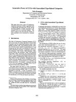
Fig. 6. Effect of dicalcin on liposome aggregation induced by an-
nexins. Time courses of annexin-induced liposome aggregation
were measured as the increase in the absorbance at 350 nm [see
inset in (E)]. In (A), the time course was measured at various con-
centrations of annexin in the presence (open symbols) and absence
(filled symbols) of 200 n
M dicalcin at 100 lM Ca
2+
. The initial rate of
the absorbance increase was plotted against the annexin concen-
tration. In (B)–(E), liposome aggregation was measured at various
Ca
2+
concentrations in the presence (open circles) and absence
(filled circles) of dicalcin (DC). The initial rate of the absorbance
increase was plotted against the Ca
2+
concentration [annexin A1 in
(B), annexin A2 in (C), annexin A5 in (D), annexin A1 + annex-
in A2 + annexin A5 in (E)]. Data points represent mean ± standard
error determined in two different preparations (n ¼ 3 in each prepa-
ration). For controls, the result with dicalcin but no annexins pres-
ent (open triangles) and that with neither dicalcin nor annexins
(filled triangles) are shown in (B). These two controls are shown as
thin dotted lines in (C) and (D). The result obtained in the presence
of dicalcin and all of the annexins (E) was compared with the calcu-
lated sum of each of the initial rates obtained in (B)–(D) (thick dot-
ted lines).
T. Uebi et al. Role of dicalcin in frog olfactory cilia
FEBS Journal 274 (2007) 4863–4876 ª 2007 The Authors Journal compilation ª 2007 FEBS 4869
N-terminal-truncated forms of frog annexin A1 and
annexin A2. The result showed that, indeed, dicalcin
binds to these truncated forms (Fig. 7A), which indi-
cated that the N-terminal region is not essential for
the interaction of frog annexin A1 and annexin A2
with dicalcin. Consistently, we observed that the
35 kDa forms of annexin A1 and annexin A2 found
in the fraction of the binding proteins of dicalcin
(Fig. 1) were the N-terminal-truncated annexins
(Fig. 7B).
Discussion
In the present study, we showed that the major bind-
ing proteins of dicalcin in frog olfactory epithelium are
annexin A1, annexin A2 and annexin A5 (Figs 1–3
and supplementary Fig. S1). The binding does not
require the N-terminal region of annexins (Fig. 7). Di-
calcin and all these annexins colocalize in the olfactory
and respiratory cilia (Figs 4 and 5 and supplementary
Fig. S3). Dicalcin was found to increase the rate of
liposome aggregation caused by annexins (Fig. 6).
Specificity of the binding between dicalcin and
annexins
In the present study, we identified the 38 kDa and the
35 kDa proteins as annexin A1, annexin A2 and ann-
exin A5. Annexins are known to bind to a dimer
form of S100 proteins. In mammals, the binding
between annexins and S100 dimer proteins has been
shown to be subtype-specific. S100A11 binds to annex-
in A1 [17] (but see [20] also), and S100A10 binds to
annexin A2 [16]. Because dicalcin in frogs shows the
highest amino acid sequence homology to S100A11
(45–58%), the binding of dicalcin to annexin A1 is not
surprising. However, binding to all of annexin A1,
annexin A2 and annexin A5 is a rather unique charac-
teristic of dicalcin, although similar comprehensive
binding has been suggested for some of the S100 pro-
teins [11]. The comprehensive binding of dicalcin to
various subtypes of annexin could be due to the char-
acteristics of frog annexins and ⁄ or dicalcin (see below).
Annexin consists of two domains, the N-terminal
region and the C-terminal protein core. Although the
N-terminal region has been suggested to be responsible
for the binding to S100 proteins [21], the N-terminal
truncated forms of annexin A1 and annexin A2 bind
to dicalcin (Fig. 7). The binding of these forms sug-
gests that these annexins bind to dicalcin not with the
N-terminal regions but with the sites that have not yet
been identified in their core domains.
In S100A10 and S100A11, the amino acid residues
contacting the corresponding annexins are known [22–
24]. In dicalcin, several of them are conserved (supple-
mentary Fig. S4). The amino acids thought to give the
subtype-specificity of S100 binding to annexin are also
known [25]. However, these residues in dicalcin are dif-
ferent from those in S100A10 or S100A11 (supplemen-
tary Fig. S4), which suggests that the specificity of
binding of dicalcin to annexins is not so strict.
From the above considerations, we speculate that
the binding between annexins and dicalcin occurs via
the interaction between the conserved amino acids in
dicalcin and the still unknown site in the core domain
of annexin. Because annexin A5 lacks the correspond-
ing N-terminal region of annexin A1 or annexin A2
(supplementary Fig. S1), it would not be surprising if
frog annexin A5 bound to dicalcin. Recombinant frog
annexin A4, which also lacks the corresponding N-ter-
minal region, also showed Ca
2+
-dependent binding to
dicalcin (data not shown). Similarly, as in the present
study, it was reported recently that the N-terminus of
A
B
Fig. 7. Ca
2+
-dependent binding to dicalcin of N-terminal region-trun-
cated annexin A1 and annexin A2. (A) Recombinant annexin A1 and
annexin A2 were truncated at their N-termini with elastase and chy-
motrypsin, respectively, and mixed with dicalcin-Sepharose beads.
The truncated annexin A1 and annexin A2 bound to the beads at a
high Ca
2+
concentration, but they were eluted by reducing the Ca
2+
concentration (low-Ca
2+
wash). (B) Amino acid sequence analysis
showed that the proteolytic fragments used in (A) lacked the N-ter-
minal regions. Arrowheads show the sites cleaved and the mole-
cular masses of the rest of the cleaved peptides. Arrows show the
N-termini of the 35 kDa forms of annexin A1 and annexin A2.
Role of dicalcin in frog olfactory cilia T. Uebi et al.
4870 FEBS Journal 274 (2007) 4863–4876 ª 2007 The Authors Journal compilation ª 2007 FEBS
annexin 6 is not required for the interaction of annexin
6 with S100A11 [26].
Colocalization of dicalcin and annexins
in the cilia
We previously reported that dicalcin is present in the
olfactory and the respiratory cilia [7]. Expression of
S100 proteins has been reported in the olfactory epi-
thelium in teleosts and rodents [27,28], and in the cilia
of human bronchial epithelial cells [29]. Annexins have
been detected in the tissues containing ciliated cells:
the respiratory epithelium [30,31] and bronchial epithe-
lial cells [29]. So far, however, localization of annexins
in the olfactory cilia has not been reported, and there-
fore, this is the first report that annexin A1, annex-
in A2 and annexin A5 are expressed in the cilia of
olfactory cells. In the present study, we showed that
dicalcin, annexin A1, annexin A2 and annexin A5 co-
localize in the olfactory cilia. Because ciliated cells
seem to express both S100 proteins and annexins, our
result could apply to cells that contain motile cilia in
general.
Annexin A1, annexin A2, annexin A5 and dicalcin
are present in the olfactory cilia at a ratio of
1 : 0.42 ± 0.09 : 0.54 ± 0.15 : 1.9 ± 0.6, and dicalcin
may be present in greater amounts (see Results). A
molecular modeling study showed that the structure of
dicalcin is similar to that of an S100 dimer [8]. Because
a dimer form of S100 protein binds two annexin mole-
cules [21], one dicalcin molecule would bind to two
molecules of annexins. If it is the case, the amount of
dicalcin is stoichiometrically sufficient to form com-
plexes with annexin A1, annexin A2 and annexin A5.
Facilitation by dicalcin of membrane aggregation
induced by annexins
The half-maximal dicalcin effects were observed at
<5 lm Ca
2+
with annexin A2 and at about 30 lm
with annexin A1 (Fig. 6). These Ca
2+
concentrations
are the effective ranges of annexin A2 and annexin A1
of other animal species [32]. The dissociation constant
of Ca
2+
binding to dicalcin has been reported to be
10–20 lm [12]. A simple expectation, therefore, was
that the Ca
2+
concentration effective for liposome
aggregation in the presence of annexin A2 and dicalcin
would be determined by dicalcin, which shows lower
affinity for Ca
2+
than does annexin A2. Similarly, one
could expect that the effective Ca
2+
concentration in
the presence of annexin A1 and dicalcin would be
determined by annexin A1. However, the results were
different from what we expected. The effective Ca
2+
concentrations did not change significantly in the
presence or absence of dicalcin. The results indicated
that the Ca
2+
dependency of liposome aggregation in
the presence of dicalcin is determined by annexins, not
by dicalcin. The result therefore suggested that there is
cooperative regulation of Ca
2+
binding to dicalcin by
annexins. The increase in the degree of Ca
2+
binding
in the presence of binding proteins is known for
S100A4 [33] and has been suggested for S100A11 [34].
We measured liposome aggregation in a mixture of
dicalcin, annexin A1, annexin A2 and annexin A5
(Fig. 6D). The observed liposome aggregation profile
could be explained by the sum of each of the constitu-
ents in the mixture. In this study, we mixed all of these
proteins at once. If, as we assumed, dicalcin binds to
two molecules of annexin, a dicalcin molecule would
be able to bind two annexin molecules of different sub-
types, such as annexin A1 plus annexin A2, and ann-
exin A1 plus annexin A5. However, the aggregation
profile obtained in the mixture could be explained by
the sum of the results obtained independently using
single species of annexin. This result suggests that even
when all of the annexins are present in the mixture,
annexins of a homomeric pair, not a heteromeric one,
tend to bind to dicalcin to form a complex.
Possible physiologic functions of dicalcin
and annexins in the cilia
It has been estimated that the intracellular Ca
2+
con-
centration in the olfactory cilia is about 40 nm at the
resting level, and increases to higher levels after
odorant stimulation [35]. In respiratory cilia, the intra-
cellular Ca
2+
concentration increases up to a sub-
micromolar level at the maximum [36]. The range of
Ca
2+
concentration where the dicalcin–annexin com-
plex has an effect seems to be higher than these ‘physi-
ologic’ Ca
2+
concentrations. Therefore, we believe that
the dicalcin–annexin complex exerts its effect when the
Ca
2+
concentration is abnormally increased. The cell
membranes of motile cilia are subject to mechanical
stress and are often disrupted [37]. In addition to this,
the olfactory cilia are exposed to environmental chemi-
cals, microorganisms and viruses, etc., so that the cil-
ium membrane is likely to be damaged. In these cases,
the cytoplasmic Ca
2+
concentration at the disrupted
site could possibly be quite high. Because (a) the effec-
tive Ca
2+
concentrations are different between annex-
in A1 and annexin A2 (Fig. 6), (b) dicalcin is present
in a quantity sufficient to bind all of the annexins (see
Results), and (c) all these molecules colocalize in the
same cilia (Figs 4 and 5), it is possible that the dical-
cin–annexin system could cover a wide range of Ca
2+
T. Uebi et al. Role of dicalcin in frog olfactory cilia
FEBS Journal 274 (2007) 4863–4876 ª 2007 The Authors Journal compilation ª 2007 FEBS 4871
concentrations inside the cell to reseal the disrupted
membranes. It has been reported that annexin A1 [38]
and annexin A1 and annexin A2 [39] have important
roles in membrane repair.
Annexin A5 did not show liposome aggregation
activity, in agreement with the findings of a previous
study [14], even in the presence of dicalcin (Fig. 6D).
Because antibody against annexin A5 has been reported
to inhibit the survival of oxidation-damaged cells [40],
the dicalcin–annexin A5 complex may possibly contri-
bute to a recovery process after chemical damage.
Dicalcin in other species
So far, we have found dicalcin in Rana catesbeiana [6]
and Xenopus laevis [5]. In addition, the sequence of
dicalcin mRNA of X. tropicalis has been registered in
a database (NM_001016706). Thus, dicalcin has been
found only in the three species of frogs. The Mexican
salamander, Ambystoma mexicanum, has an S100A11-
like protein with an insertion of four amino acid resi-
dues in its C-terminal half EF hand (supplementary
Fig. S3), and this insertion is characteristically observed
in dicalcin. Nevertheless, this S100A11-like protein is a
monomer form of an S100 protein and is not like dical-
cin. Therefore, dicalcin might be derived from a unique
S100 protein of ancestral amphibia, and could be a
frog-specific protein. Members of the Caudata, includ-
ing the Mexican salamander, have a tendency to stay
either in an aquatic or a terrestrial environment. In
contrast, most frogs are more biphasic, and actively
move between land and water. Because the olfactory
motile cilia in these frogs could be exposed to vigorous
mechanical stress very often, they might have needed to
have a very effective membrane repair system. Dicalcin,
a homodimer form of S100 proteins, could be the form
of S100 protein that exerts this effect most efficiently.
Experimental procedures
Solutions
The standard buffer solution contained 115 mm potassium
gluconate, 2.5 mm KCl, and 10 mm Hepes (pH 7.5) (K-gluc
buffer). Low-salt K-gluc buffer (LS-K-gluc buffer) con-
tained 50 mm potassium gluconate and 20 mm Hepes
(pH 7.5). Either 1 mm CaCl
2
or 5 mm EGTA was added to
the LS-K-gluc buffer. Ringer’s solution contained 115 mm
NaCl, 3 mm KCl, 2 mm MgCl
2
,2mm CaCl
2
,10mm glu-
cose, and 5 mm Tris ⁄ HCl (pH 7.5). Tris-buffered saline
(NaCl ⁄ Tris) contained 0.9% NaCl and 100 mm Tris ⁄ HCl
(pH 7.5). NaCl ⁄ P
i
contained 137 mm NaCl, 2.7 mm KCl,
8.1 mm Na
2
HPO
4
, and 1.5 mm NaH
2
PO
4
(pH 7.4).
Preparation of Chaps-solubilized proteins of the
olfactory cilia
Animal care was carried out in accordance with the institu-
tional guidelines of Osaka University.
Partially purified cilia from frog olfactory epithelium
were obtained as described previously [13]. Briefly, olfactory
cilia were detached from the epithelia by abruptly raising
the Ca
2+
concentration to 10 mm. The deciliated epithelia
were removed by brief centrifugation (1500 g, 5 min;
TOMY MRX-150, TMA-11 rotor, TOMY SEIKO, Tokyo,
Japan), and the supernatant containing the cilia was further
centrifuged at 12 000 g for 15 min (TOMY MRX-150,
TMA-11 rotor). The supernatant was removed and used as
the soluble protein fraction of frog olfactory epithelium.
The resulting pellet containing the isolated cilia was washed
twice with K-gluc buffer, resuspended in LS-K-gluc buffer
containing 4% Chaps, and kept at 4 °C overnight to solubi-
lize the membrane-associated proteins of the isolated cilia.
The Chaps-solubilized proteins were then obtained in the
supernatant after centrifugation at 440 000 g for 5 min
(Hitachi CS100, RP100AT4 rotor, Hitachi Koki, Tokyo,
Japan). The supernatant was diluted with LS-K-gluc buffer
containing 1 mm Ca
2+
so that the concentration of Chaps
was reduced to 0.05%. The diluted fraction was centrifuged
at 12 000 g for 30 min (TOMY MRX-150, TMA-11 rotor)
to remove any precipitates before subjecting it to affinity
column chromatography as described below. A cocktail
of protease inhibitors (leupeptin, 5 lgÆmL
)1
; phenyl-
methanesulfonyl fluoride, 5 lgÆmL
)1
; aprotinin, 5 lgÆmL
)1
;
bestatin, 40 lgÆmL
)1
) was present at the indicated final con-
centrations during the preparation of the above fraction.
Affinity purification of binding proteins of
dicalcin
A dicalcin-Sepharose column was prepared as previously
described [13]. Chaps-solubilized proteins of the isolated
cilia were loaded on the dicalcin-Sepharose column pre-
equilibrated with LS-K-gluc buffer containing 1 m m CaCl
2
and 0.05% Chaps. After elution of unbound proteins, pro-
teins that were bound to the column at 1 mm Ca
2+
were
eluted by reducing the Ca
2+
concentration with LS-K-gluc
buffer containing 5 mm EGTA and 0.05% Chaps. In some
studies, K-gluc buffer was used instead of LS-K-gluc buffer
to isolate the binding proteins, but no significant differences
were observed in the detected proteins.
Determination of partial amino acid sequences
of binding proteins of dicalcin
Purified binding proteins of dicalcin were digested with
lysyl endopeptidase (Wako, Osaka, Japan) at an
enzyme ⁄ substrate ratio of 1 : 100 in 1 mL of a Tris buffer
solution (100 mm Tris, pH 9.2) overnight at 37 °C. The
Role of dicalcin in frog olfactory cilia T. Uebi et al.
4872 FEBS Journal 274 (2007) 4863–4876 ª 2007 The Authors Journal compilation ª 2007 FEBS
peptide fragments were isolated by RP-HPLC (SMART
System; GE Healthcare, Piscataway, NJ, USA). Major peak
fractions were collected, and the amino acid sequences of
the peptides in these fractions were analyzed with a protein
sequencer (G1000A; Hewlett-Packard, Palo Alto, CA,
USA).
Isolation of annexin cDNA clones
Screening in the frog olfactory epithelium cDNA library to
isolate annexin cDNAs was carried out in a similar way as
described previously [6]. On the basis of either the partial
amino acid sequences of annexins determined in the amino
acid sequence analysis shown above (VDEATIT, KDITSD,
and KDIVSD), or the conserved sequences in vertebrate
annexin A4 and annexin A5 (AMKGAG, YEAGEKKW,
and RKVFDKYM), we synthesized the primers to isolate
cDNA fragments of annexins. The fragments were ampli-
fied by RT-PCR, and nucleotide sequences were determined
with a DNA sequencer (Prism 377; Applied Biosystems,
Foster City, CA, USA). The isolated fragments were used
as the authentic cDNA probes. The frog olfactory epithe-
lium cDNA library was screened with the cDNA probes to
isolate the full-length cDNAs of annexins.
Expression of annexins in E. coli
The coding region of each annexin was amplified by PCR
with a pair of primers. The sequences of the primers were:
5¢-GGAATTCCATATGTCATTCATTTCCGAG-3¢ (for-
ward) and 5¢-CGGGATCCTTAAGCTCCTCCAATAAGT
G-3¢ (reverse) for annexin A1; 5¢-CATATGGCTACTATT
CATGAAAT-3¢ (forward) and 5¢-GGTTCCTCAGTCA
TCTCCAGCACATAG-3¢ (reverse) for annexin A2; and
5¢-GGAATTCCATATGGCAACGACAAAAAG-3¢ (for-
ward) and 5¢-CGGGATCCTTACTCATCATCCCCA-3¢
(reverse) for annexin A5. The NdeI- and BamHI-digested
cDNA was ligated with pET-3a (Novagen, Darmstadt, Ger-
many). The recombinant plasmids were introduced into
E. coli BL21 pLysS (Novagen), and the proteins were
expressed as described previously [6]. Recombinant annexins
were affinity-purified with a dicalcin-Sepharose column in a
similar way as used for the isolation of native annexins.
Binding of recombinant annexins to dicalcin
Transformed E. coli cells expressing each of the annexins
were suspended and sonicated in K-gluc buffer. The lysate
was centrifuged at 27 000 g for 15 min (Hitachi CR21,
R20A2 rotor), and 1 mm CaCl
2
was then added to the
supernatant. The supernatant was then centrifuged again
under the same conditions to remove aggregated proteins,
and a portion of the supernatant was mixed with dicalcin-
Sepharose beads in K-gluc buffer containing 1 mm CaCl
2
for 30 min at 4 °C. After the mixture had been centrifuged
(7000 g, 1 min; TOMY MRX-150, TMA-11 rotor), the
supernatant was discarded. The dicalcin-Sepharose beads
were then washed 10 times with K-gluc buffer containing
1mm CaCl
2
. Proteins bound to dicalcin-Sepharose beads
at a high Ca
2+
concentration were then eluted with
K-gluc buffer containing 5 mm EGTA. When truncated
annexins were used, annexin A1 and annexin A2 were
digested with elastase and chymotrypsin, respectively.
These enzymes are known to cleave the N-terminal regions
of annexin A1 and annexin A2 [18,41], respectively. The
cleaved sites in these annexins were determined with a
protein sequencer.
Two-dimensional electrophoresis
For the first dimension of isoelectric focusing, a protein
sample was mixed with isoelectric focusing sample buffer
containing 2% immobilized pH gradient buffer (IPG buffer,
GE Healthcare), 1.2% DeStreak Reagent (GE Healthcare),
8 m urea, and 2% Chaps, and then the mixture was applied
to an immobilized pH gradient strip (Immobiline DryStrip,
7 cm, pH 3–10; GE Healthcare) by rehydration overnight.
Proteins were focused by applying an increasing voltage
from 500 V to 3500 V linearly during the first 2 h, and then
keeping the voltage at 3500 V for another 1 h. The second
dimension of electrophoresis was performed on a 12%
SDS ⁄ PAGE gel.
Western blot analysis
Proteins were electrophoresed onto a poly(vinylidene
difluoride) membrane (Immobilon P; Millipore, Billerica,
MA, USA). After blocking with 10% skimmed milk in
NaCl ⁄ Tris, the membrane was incubated with anti-annexin
or anti-dicalcin serum overnight at 4 °C. Antiserum was
used after dilution with a solution of Can Get Signal
(Nacalai Tesque, Kyoto, Japan) to either 1 : 500 for
SDS ⁄ PAGE or 1 : 100 for two-dimensional electrophoresis.
After washing with NaCl ⁄ Tris, the membrane was reacted
with secondary and tertiary reagents using the Vectastain
ABC Elite kit (Vector Laboratories, Burlingame, CA,
USA). Immunoreactive proteins were visualized with a
Chemi-Lumi One reagent (Nacalai Tesque) and detected
with LAS-1000 (Fuji Film, Tokyo, Japan).
Immunofluorescence staining
Frog olfactory or respiratory epithelium was quickly
removed after decapitation and fixed with 4% paraformal-
dehyde in NaCl ⁄ P
i
for 2 days at 4 °C. The fixed tissues
were embedded in 33% OCT compound diluted with
NaCl ⁄ P
i
containing 20% sucrose, and cryosectioned at
8 lm thickness. The sections were preincubated in 10%
T. Uebi et al. Role of dicalcin in frog olfactory cilia
FEBS Journal 274 (2007) 4863–4876 ª 2007 The Authors Journal compilation ª 2007 FEBS 4873
normal goat serum in NaCl ⁄ P
i
for 6 h at room tempera-
ture, and then reacted with various antisera and antibody.
Antisera against annexin A1, annexin A2 and annexin A5
were raised in both rabbit and mouse, and antiserum
against dicalcin was raised in rabbit. G
olf
antibody raised
in rabbit was purchased from Santa Cruz Biotechnology
(Santa Cruz, CA, USA). For double staining, sections were
reacted first with antiserum or antibody raised in rabbit
overnight at 4 °C, and then were further reacted with anti-
serum raised in mouse overnight at 4 °C. Antisera and
antibody were used after dilution with NaCl ⁄ P
i
: 1 : 100 for
both antiserum against annexin A1 and that against annex-
in A5, 1 : 50 for antiserum against annexin A2, 1 : 500 for
dicalcin antiserum, and 1 : 500 for G
olf
antibody. Immu-
noreactivities were detected by secondary antibody labeled
with fluorescein isothiocyanate (anti-mouse) (Vector Labo-
ratories) or Texas Red (anti-rabbit) (Vector Laboratories).
The reactivity against dicalcin was detected by cyanine 3
using the tyramide signal amplification method (TSA Plus
Fluorescence System; PerkinElmer, Wellesley, MA, USA).
The conditions used to obtain immunofluorescence were
kept constant in each of the observations with rabbit
antiserum (Texas Red) or mouse antiserum (fluorescein
isothiocyanate), and all of the images obtained with anti-
sera from one type of animal (rabbit or mouse, i.e. red or
green) were adjusted in the same way. For these reasons,
the color of the merged picture was dependent on the rela-
tive intensities of red and green fluorescence in each pair
of antisera.
Liposome aggregation assay
The membrane aggregation activity of annexin and dicalcin
was measured with the liposome aggregation assay method
[42]. Briefly, 10 mg of phosphatidylserine dissolved in
CHCl
3
was dried under an N
2
stream for 30 min. Then,
4 mL of a solution containing 10 mm Hepes, 100 mm NaCl
and 10 lm EDTA (pH 7.2) was added, and the sample was
mixed vigorously to obtain liposomes. The resulting suspen-
sion of liposomes was filtered first through a 0.2 lm poly-
carbonate filter (Whatman, Brentford, UK) five times, and
then through a 0.1 lm polycarbonate filter 10 times under
an N
2
atmosphere. The final concentration of liposomes
(60 lm) was determined as the lipid concentration by mea-
suring the phosphate content [43].
The liposome aggregation reaction was initiated by addi-
tion of liposomes to K-gluc buffer (500 lL) containing
2mm MgCl
2
in the presence or absence of dicalcin and ⁄ or
annexins. The absorbance increase due to the increase in
the light scattering caused by aggregation of liposomes
was measured at 350 nm and at 25 °C. Data were continu-
ously collected for 2.5 min. Because the signal was dis-
turbed by mixing solutions for the first 20 s or so, the
initial rate of the absorbance increase was determined after
the signal had been stabilized. The Ca
2+
concentration of
the solution was varied from 5 to 500 lm. In the Ca
2+
-
free solution, 1 mm EGTA was added. Final concentra-
tions of proteins were 12.5 nm (annexin A1), 5 nm (annex-
in A2), 7.5 nm (annexin A1), and 40 nm (dicalcin), so that
the ratio of the concentration of annexins was similar to
that in the olfactory cilia (see Results section), but dicalcin
was added in excess.
Acknowledgements
We thank Dr H. Matsumoto at the University of
Oklahoma and Dr H. Kurono at Kurume University
for MS analysis of the binding proteins at the initial
stage of this study. This research was supported by
grants from the JSPS to S. Kawamura and N. Miwa,
and from the Human Frontier Science Program to
S. Kawamura.
References
1 Menini A (1999) Calcium signalling & regulation in
olfactory neurons. Curr Opin Neurobiol 9, 419–426.
2 Kurahashi T & Shibuya T (1990) Ca
2+
-dependent adap-
tive properties in the solitary olfactory receptor cell of
the newt. Brain Res 515, 261–268.
3 Chen TY & Yau KW (1994) Direct modulation by
Ca
2+
-calmodulin of cyclic nucleotide-activated channel
of rat olfactory receptor neurons. Nature 368, 545–548.
4 Kawamura S (1999) Modulation and adaptation. In The
Retinal Basis of Vision (Toyoda J-I, Murakami M,
Kaneko A & Saito T, eds), pp. 53–63. Elsevier, Amster-
dam.
5 Miwa N, Sinmyo Y & Kawamura S (2007) Cloning and
characterization of Xenopus dicalcin, a novel S100-like
calcium-binding protein in Xenopus eggs. DNA
Sequence 18, 400–404.
6 Miwa N, Kobayashi M, Takamatsu K & Kawamura S
(1998) Purification and molecular cloning of a novel cal-
cium-binding protein, p26olf, in the frog olfactory epi-
thelium. Biochem Biophys Res Commun 251, 860–867.
7 Miwa N & Kawamura S (2003) Frog p26olf, a molecule
with two S100-like regions in a single peptide. Microsc
Res Tech 60, 593–599.
8 Tanaka T, Miwa N, Kawamura S, Sohma H, Nitta K
& Matsushima N (1999) Molecular modeling of single
polypeptide chain of calcium-binding protein p26olf
from dimeric S100B (bb). Protein Eng 12, 395–405.
9 Heizmann CW (2002) The multifunctional S100 protein
family. Methods Mol Biol 172, 69–80.
10 Donato R (2003) Intracellular and extracellular roles of
S100 proteins. Microsc Res Tech 60, 540–551.
11 Santamaria-Kisiel L, Rintala-Dempsey AC & Shaw GS
(2006) Calcium-dependent and -independent interactions
of the S100 protein family. Biochem J 396, 201–214.
Role of dicalcin in frog olfactory cilia T. Uebi et al.
4874 FEBS Journal 274 (2007) 4863–4876 ª 2007 The Authors Journal compilation ª 2007 FEBS
12 Miwa N, Shinmyo Y & Kawamura S (2001) Calcium-
binding by p26olf, an S100-like protein in the frog
olfactory epithelium. Eur J Biochem 268, 6029–6036.
13 Miwa N, Uebi T & Kawamura S (2000) Characteriza-
tion of p26olf, a novel calcium-binding protein in the
frog olfactory epithelium. J Biol Chem 275, 27245–
27249.
14 Blackwood RA & Ernst JD (1990) Characterization of
Ca
2+
-dependent phospholipid binding, vesicle aggrega-
tion and membrane fusion by annexins. Biochem J 266,
195–200.
15 Drust DS & Creutz CE (1988) Aggregation of chromaf-
fin granules by calpactin at micromolar levels of cal-
cium. Nature 331, 88–91.
16 Gerke V & Weber K (1985) The regulatory chain in the
p36-kd substrate complex of viral tyrosine-specific pro-
tein kinases is related in sequence to the S-100 protein
of glial cells. EMBO J 4, 2917–2920.
17 Naka M, Qing ZX, Sasaki T, Kise H, Tawara I, Ham-
aguchi S & Tanaka T (1994) Purification and character-
ization of a novel calcium-binding protein, S100C, from
porcine heart. Biochim Biophys Acta 1223, 348–353.
18 Seemann J, Weber K & Gerke V (1996) Structural
requirements for annexin I–S100C complex-formation.
Biochem J 319, 123–129.
19 Johnsson N, Marriott G & Weber K (1988) p36, the
major cytoplasmic substrate of src tyrosine protein
kinase, binds to its p11 regulatory subunit via a short
amino-terminal amphiphatic helix. EMBO J 7, 2435–
2442.
20 Rintala-Dempsey AC, Santamaria-Kisiel L, Liao Y,
Lajoie G & Shaw GS (2006) Insights into S100 target
specificity examined by a new interaction between
S100A11 and annexin A2. Biochemistry 45, 14695–
14705.
21 Gerke V & Moss SE (2002) Annexins: from structure to
function. Physiol Rev 82, 331–371.
22 Rety S, Sopkova J, Renouard M, Osterloh D, Gerke V,
Tabaries S, Russo-Marie F & Lewit-Bentley A (1999)
The crystal structure of a complex of p11 with the an-
nexin II N-terminal peptide. Nat Struct Biol 6, 89–95.
23 Rety S, Osterloh D, Arie JP, Tabaries S, Seeman J,
Russo-Marie F, Gerke V & Lewit-Bentley A (2000)
Structural basis of the Ca
2+
-dependent association
between S100C (S100A11) and its target, the N-terminal
part of annexin I. Structure 8, 175–184.
24 Sopkova J, Oling FK, Rety S, Brisson A, Smith JC &
Lewit-Bentley A (2000) S100 protein–annexin interac-
tions: a model of the (Anx2-p11)(2) heterotetramer
complex. Biochim Biophys Acta 1498, 181–191.
25 Dempsey AC, Walsh MP & Shaw GS (2003) Unmask-
ing the annexin I interaction from the structure of Apo-
S100A11. Structure 11, 887–897.
26 Chang N, Sutherland C, Hesse E, Winkfein R,
Wiehler WB, Pho M, Veillette C, Li S, Wilson DP,
Kiss E et al. Identification of a novel interaction
between the Ca
2+
-binding protein S, 100A, 11 and the
Ca
2+
- and phospholipid-binding protein annexin A6.
Am J Physiol Cell Physiol 292, C1417–C1430.
27 Yamashita N, Ilg EC, Scha
¨
fer BW, Heizmann CW &
Kosaka T (1999) Distribution of a specific calcium-bind-
ing protein of the S100 protein family, S100A6 (calcy-
clin), in subpopulations of neurons and glial cells of the
adult rat nervous system. J Comp Neurol 404, 2235–2257.
28 Germana A, Montalbano G, Laura R, Ciriaco E, del
Valle ME & Vega JA (2004) S100 protein-like immuno-
reactivity in the crypt olfactory neurons of the adult
zebrafish. Neurosci Lett 371, 196–198.
29 Ostrowski LE, Blackburn K, Radde KM, Moyer MB,
Schlatzer DM, Moseley A & Boucher RC (2002) A pro-
teomic analysis of human cilia: identification of novel
components. Mol Cell Proteomics 1, 451–465.
30 Mayran N, Traverso V, Maroux S & Massey-Harroche D
(1996) Cellular and subcellular localizations of annexins
I, IV, and VI in lung epithelia. Am J Physiol 270, L863–
L871.
31 Rodrigo JP, Garcia-Pedrero JM, Gonzalez MV,
Fernez MP, Suarez C & Herrero A (2004) Expression of
annexin A1 in normal and chronically inflamed nasal
mucosa. Arch Otolaryngol Head Neck Surg 130,
211–215.
32 Raynal P & Pollard HB (1994) Annexins: the problem
of assessing the biological role for a gene family of mul-
tifunctional calcium- and phospholipid-binding proteins.
Biochim Biophys Acta 1197, 63–93.
33 Dukhanina EA, Dukhanin AS, Lomonosov MY,
Lukanidin EM & Georgiev GP (1997) Spectral studies
on the calcium-binding properties of Mts1 protein and
its interaction with target protein. FEBS Lett 410,
403–406.
34 Allen BG, Durussel I, Walsh MP & Cox JA (1996)
Characterization of the Ca
2+
-binding properties of cal-
gizzarin (S100C) isolated from chicken gizzard smooth
muscle. Biochem Cell Biol 74, 687–694.
35 Leinders-Zufall T, Greer CA, Shepherd GM & Zufall F
(1998) Imaging odor-induced calcium transients in single
olfactory cilia: specificity of activation and role in trans-
duction. J Neurosci 18, 5630–5639.
36 Zhang L & Sanderson MJ (2003) The role of cGMP in
the regulation of rabbit airway ciliary beat frequency.
J Physiol 551, 765–776.
37 McNeil PL & Steinhardt RA (2003) Plasma membrane
disruption: repair, prevention, adaptation. Annu Rev
Cell Dev Biol 19, 697–731.
38 McNeil AK, Rescher U, Gerke V & McNeil PL (2006)
Requirement for annexin A1 in plasma membrane
repair. J Biol Chem 281, 35202–35207.
39 Cagliani R, Magri F, Toscano A, Merlini L,
Fortunato F, Lamperti C, Rodolico C, Prelle A, Sironi
M, Aguennouz M et al. (2005) Mutation finding in
T. Uebi et al. Role of dicalcin in frog olfactory cilia
FEBS Journal 274 (2007) 4863–4876 ª 2007 The Authors Journal compilation ª 2007 FEBS 4875
patients with dysferlin deficiency and role of the dysfer-
lin interacting proteins annexin A1 and A2 in muscular
dystrophies. Hum Mutat 26, 283.
40 Han S, Zhang KH, Lu PH & Xu XM (2004) Effects of
annexins II and V on survival of neurons and astrocytes
in vitro. Acta Pharmacol Sin 25, 602–610.
41 Huang K-S, McGray P, Mattaliano RJ, Burne CE,
Chow P, Sinclair LK & Pepinsky RB (1987) Purification
and characterization of proteolytic fragments of
lipocortin I that inhibit phospholipase A2. J Biol Chem
262, 7639–7645.
42 Lee G & Pollard HB (1997) Highly sensitive and stable
phosphatidylserine liposome aggregation assay for an-
nexins. Anal Biochem 252, 160–164.
43 Morrison WR (1964) A fast, simple and reliable method
for the microdetermination of phosphorus in biological
materials. Anal Biochem 7, 218–224.
Supplementary material
The following supplementary material is available
online:
Fig. S1. Alignment of amino acid sequences of Rana
catesbeiana annexin A1, annexin A2 and annexin A5
with those of mammalian orthologs.
Fig. S2. Specificity of antisera against dicalcin and an-
nexins.
Fig. S3. Colocalization of dicalcin with annexin A1,
annexin A2 and annexin A5 in frog respiratory epithe-
lium.
Fig. S4. Alignment of amino acid sequences of Rana
catesbeiana dicalcin with those of S100 proteins.
This material is available as part of the online article
from
Please note: Blackwell Publishing is not responsible
for the content or functionality of any supplementary
materials supplied by the authors. Any queries (other
than missing material) should be directed to the corres-
ponding author for the article.
Role of dicalcin in frog olfactory cilia T. Uebi et al.
4876 FEBS Journal 274 (2007) 4863–4876 ª 2007 The Authors Journal compilation ª 2007 FEBS