Báo cáo khoa học: Molecular mechanisms of the phospho-dependent prolyl cis ⁄ trans isomerase Pin1 docx
Bạn đang xem bản rút gọn của tài liệu. Xem và tải ngay bản đầy đủ của tài liệu tại đây (555.41 KB, 12 trang )
MINIREVIEW
Molecular mechanisms of the phospho-dependent prolyl
cis
⁄
trans isomerase Pin1
G. Lippens
1
, I. Landrieu
1
and C. Smet
2
1 CNRS UMR 8576 Unite
´
de Glycobiologie Structurale et Fonctionnelle, Universite
´
des Sciences et Technologies de Lille 1-59655,
Villeneuve d’Ascq, France
2 Institut de Recherche Interdisciplinaire, CNRS, Lille, France
Introduction
Prolyl cis ⁄ trans isomerases form a special class of
enzymes in many respects. Most other enzymes change
the covalent chemistry of their substrates (be it through
cleavage, or addition ⁄ removal of a phosphate, acetyl
or methyl group), leading to a product that is distin-
guishable by mass spectrometry or immunochemistry
from the incoming molecular entity. The modification
imposed by prolyl cis ⁄ trans isomerases is not covalent,
but merely conformational. One therefore encounters
many technical difficulties when attempting to observe
their molecular action in vivo and even in vitro. Experi-
ments with isolated proteins have indicated the involve-
ment of two major classes of isomerases, the
cyclophilins (Cyps) and the FK506-binding proteins
(FKBPs), in the protein-folding process, where the con-
formational state of a prolyl peptide bond can be a
rate-limiting step [1]. The importance of ‘binding ver-
sus catalysis’ remains an open issue in many cases [2],
partly because tinkering with the enzymatic activity
through mutations also leads to changes in interaction
parameters [3]. Assessing the role of the isomerization
function in vivo is even harder. In one of the most
intensively studied cases, the interaction between CypA
and the Gag protein derived from the HIV virus, the
Keywords
cell cycle; CKS subunit; dynamics; enzyme;
interaction module; phosphorylation; Pin1;
prolyl cis ⁄ trans isomerase; protein
degradation; protein structure
Correspondence
G. Lippens, CNRS UMR 8576 Unite
´
de
Glycobiologie Structurale et Fonctionnelle,
Universite
´
des Sciences et Technologies de
Lille 1-59655, Villeneuve d’Ascq Cedex,
France
Fax: +33 3 20 43 65 55
Tel: +33 3 20 33 42 71
E-mail:
(Received 5 June 2007, revised 3 August
2007, accepted 17 August 2007)
doi:10.1111/j.1742-4658.2007.06057.x
Since its discovery 10 years ago, Pin1, a prolyl cis ⁄ trans isomerase essential
for cell cycle progression, has been implicated in a large number of molecu-
lar processes related to human diseases, including cancer and Alzheimer’s
disease. Pin1 is made up of a WW interaction domain and a C-terminal
catalytic subunit, and several high-resolution structures are available that
have helped define its function. The enzymatic activity of Pin1 towards
short peptides containing the pSer ⁄ Thr-Pro motif has been well docu-
mented, and we discuss the available evidence for the molecular mecha-
nisms of its isomerase activity. We further focus on those studies that
examine its cis ⁄ trans isomerase function using full-length protein substrates.
The interpretation of this research has been further complicated by the
observation that many of its pSer ⁄ Thr-Pro substrate motifs are located in
natively unstructured regions of polypeptides, and are characterized by
minor populations of the cis conformer. Finally, we review the data on the
possibility of alternative modes of substrate binding and the complex role
that Pin1 plays in the degradation of its substrates. After considering the
available work, it seems that further analysis is required to determine
whether binding or catalysis is the primary mechanism through which Pin1
affects cell cycle progression.
Abbreviations
APP, amyloid precursor protein; CDK, cyclin-dependent kinase; CKS, cyclin-dependent kinase subunit; CTD, C-terminal domain; Cyp,
cyclophilin; FKBP, FK506-binding protein; IRF, interferon regulatory factor; Pol II, polymerase II.
FEBS Journal 274 (2007) 5211–5222 ª 2007 The Authors Journal compilation ª 2007 FEBS 5211
mere interaction of CypA with Gag might protect the
virion from an as yet poorly identified cellular integra-
tion factor [4]. Whether the prolyl cis ⁄ trans isomerase
activity detected in vitro of CypA on Pro90 in the full-
length Gag protein [5] plays a role in the viral
restriction process in vivo remains an open question. In
the case of the Itk kinase, the cis ⁄ trans isomerization
catalyzed by CypA was shown to generate novel inter-
action surfaces for two distinct molecular partners [6],
and the conformation of a proline in the linker region
between two SH3 domains of the Crp adaptor protein
determines the autoinhibition of the domains [7]. These
latter examples provide a structural basis for the
manner in which prolyl cis ⁄ trans isomerization could
act as a conformational switch in biological processes.
Beyond the difficulties of characterizing this confor-
mational switch in a cellular context and even more in
a living organism, another problem with the molecular
characterization of the prolyl-isomerase enzymes is
their redundancy in the cell. Indeed, a model organism
such as yeast has as many as eight Cyps and four
FKBPs, and even knocking down all of them does not
lead to a clear phenotype under normal conditions [8].
The discovery of Ess1, a novel Saccharomyces cerevisiae
prolyl cis⁄ trans isomerase [9] that proved essential for
cell division, was therefore highly relevant. Its human
homolog was identified as a protein interacting with
the NIMA kinase during the G
1
⁄ S cell cycle stage, and
was called Pin1 for this reason [10]. Since its initial
identification, Pin1 has attracted a great deal of
attention, as the enzyme seems to be implicated in
various human diseases, ranging from cancer and
neurodegenerative diseases to inflammation. Rather
than adding to the list of excellent general reviews on
Pin1 [11–14] or to those reviews that emphasize its role
in cellular processes related to diseases [15–17], we
focus here on what is known about the molecular
mechanisms of the enzyme action. Most importantly,
we want to critically review the evidence that Pin1
would or would not act as a prolyl cis ⁄ trans isomerase.
Molecular mechanisms of Pin1 action
The initial X-ray structure of the catalytic domain
complexed with an Ala-Pro dipeptide and a sulfate ion
[18] suggested an important role for two structural ele-
ments in catalysis. First, the loop between residues 66
and 77 (human Pin1 numbering) is involved in binding
the phosphate moiety of the substrate (Fig. 1). This
loop is flexible, as suggested by its different conforma-
tion in a second crystallographic structure, where the
catalytic domain was not complexed to a substrate
peptide [19]. Heteronuclear NOE data on human Pin1,
however, indicated only a limited decrease in the flexi-
bility of this loop on peptide binding [20] or even a
closed conformation in the absence of substrate [21].
Our NMR data on the Arabidopsis thaliana analog,
Pin1At, showed severe line broadening in the equiva-
lent stretch, giving weight to the dynamic character of
this loop on the 100 ls to 1 ms time scale [22], but the
crystal structure of the Candida albicans Ess1 revealed
a closed loop even in the absence of substrate [23]. The
dynamic character of this loop was recently shown by
NMR relaxation dispersion measurements on the
human enzyme during substrate binding [24] and
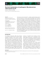
Fig. 1. Top: Molecular structure of Pin1 (Protein Data Bank code:
1Pin [4]), showing the catalytic domain (green), the WW domain
(red), and the linker region (yellow). The positively charged residues
of the active site loop (Lys63, Arg68, and Arg69) are in blue,
whereas the presumed catalytic Cys113 is in pink. The Ala-Pro pep-
tide in the active site is indicated by sticks, and is complemented
by a sulfate ion (light blue). A poly(ethylene glycol) molecule (not
shown) is sequestered between the catalytic domain and the WW
domain. Bottom: The complex between Pin1 and a Pol II CTD
phospho-peptide (Protein Data Bank code: 1F8A [5]). The active site
loop adopts a more extended conformation, and the peptide inter-
acts with the WW domain mainly through the phospho-Thr ⁄ Pro
moiety.
Molecular mechanisms of Pin1 G. Lippens et al.
5212 FEBS Journal 274 (2007) 5211–5222 ª 2007 The Authors Journal compilation ª 2007 FEBS
catalysis [25]. Interestingly, in the case of CypA, the
dynamic character of its active site was found to be
essential for its catalytic function [3]. The fact that
Pin1 can compensate for the lack of Ess1 in yeast was
exploited to further investigate the functional role of
the different Pin1 residues. Behrsin et al. [26] applied
unigenic evolution, where the capacity of a randomly
mutated pin1 gene to compensate for Ess1 in a yeast
strain devoid of the wild-type ess1 gene is screened.
They showed that, in particular, Lys63 is essential for
the anchoring function of the phosphorylated sub-
strate, whereby the K63A mutant affects the catalytic
efficiency because of weaker binding. The two argi-
nines in the loop, Arg68 and Arg69, would provide no
more than a single positive charge [26].
A second important structural feature of the initial
X-ray structure of Pin1 was the spatial proximity of
Cys113 to the Ala-Pro bond (Fig. 1) [18]. Although
this initially suggested a catalytic mechanism through a
nucleophilic attack on the substrate carbonyl carbon
by the S
c
of Cys113, the fact that the C113D mutant
remains functional calls this mechanism into question
[26]. The Cys113 residue, through its unusually low
pK
a
value, might indeed maintain an overall electro-
negative environment that is crucial for destabilizing
the double bond character of the pThr-Pro bond [26].
Further evidence is available for the catalytic mecha-
nism of Pin1 resembling more closely that of the other
prolyl cis ⁄ trans isomerases, with only its loop region
selecting for Pro residues preceded by a negative
charge. In the A. thaliana Pin1 homolog, we did not
observe significant chemical shift changes for the
equivalent Cys70 upon saturation of the catalytic
domain with several phosphopeptides [22]. In yeast,
the equivalent C120R mutant of Ess1 only prevented
growth at the higher temperature of 37 °C [27].
Finally, the chemically and structurally similar binding
pockets of Pin1 and FKBP and the structural resem-
blance between their respective high-affinity inhibitors
[28] further underscore the similarity between Pin1 and
the other peptide prolyl isomerases (PPIases).
A second crystallographic structure (Fig. 1) with the
Pin1 WW domain complexed to a phospho-peptide
derived from the C-terminal domain of polymerase II
(Pol II CTD) indicated the trans conformation of the
prolyl bond following the phosphorylated Ser [19].
NMR spectroscopy confirmed that the cis conformer
in a Cdc25-derived peptide could not interact with the
WW domain [29]. Despite these findings, recently pre-
sented data hint at other potential binding modes.
First, when the role of Pin1 in amyloid precursor pro-
tein (APP) processing and ensuing b-amyloid produc-
tion was studied, the interaction between the WW
domain and a phosphorylated peptide (V-pT
668
-P-E-E)
derived from the APP cytoplasmic domain was probed
by NMR spectroscopy [30]. For this latter phospho-
peptide, the
15
N-labeled Glu670 was the primary probe
of the interaction. Interestingly, the correlation peaks
corresponding to both the trans and cis conformers of
the pThr668-Pro prolyl bond were found to shift upon
interaction with the single WW domain, with the larg-
est shift for the cis form. In an earlier study, the same
group examined in great detail the structure of both
cis and trans conformers of the same peptide by NMR
spectroscopy [31]. The local structure of the cis con-
former of this peptide is characterized by a hydrogen
bond between the amide proton of Val667 and the
Glu671 side chain carboxyl group. This particular
motif might be recognized by the Pin1 WW domain,
or, alternatively, the presence of two glutamate resi-
dues downstream of the proline could lead the WW
domain to read the cis form in a reversed manner. The
initial screen for Pin1 substrates, which identified it as
a phospho-dependent prolyl cis ⁄ trans isomerase,
indicated that, at least for some peptides, using a
glutamate (but not aspartate) rather than a phospho-
Ser ⁄ Thr group before the critical proline did not
decrease Pin1 enzymatic efficiency [32]. However, we
found that a Tau mutant carrying multiple Glu-Pro
motifs did not significantly interact with the Pin1
WW domain (G. Lippens, I. Landrieu, C. Smet and
R. Brandt, unpublished results). Further structural
characterization of the complex between the Pin1 WW
domain and the amyloid peptide will be necessary, and
might form a novel starting point for the development
of WW domain inhibitors.
Even more surprising is the recent finding that Pin1
could recognize cyclin E via a noncanonical pThr384-
Gly385 motif [33] rather than the pThr380-Pro381
motif. The main argument was that the latter
pThr380-Pro381 motif is buried in the yeast Cdc4
molecular surface that was determined by X-ray crys-
tallography [34]. In this structure of the peptide–CDC4
complex, however, Pin1 is missing, whereas in the
study on cyclin E degradation, Pin1 was brought in by
the phospho-cyclin E and not by the CDC4a compo-
nent of the final complex (Fig. 2) [35]. Therefore, the
outcome of the molecular competition between Pin1
and CDC4 for the same phosphorylated motif is not
clear, and still leaves open the possibility that Pin1
could interfere with the cyclin E–CDC4 interface. Pin1
was proposed to isomerize the peptide bond between
Pro381 and Pro382. The concomittant structural rear-
rangement would cause cyclin E to approach the
distant E2 ligase of a different SKP ⁄ Cullin ⁄ F-box
protein (SCF)
Cdc4c
complex. During our work on the
G. Lippens et al. Molecular mechanisms of Pin1
FEBS Journal 274 (2007) 5211–5222 ª 2007 The Authors Journal compilation ª 2007 FEBS 5213
neuronal Tau protein, we studied by NMR spectroscopy
a peptide containing the sequence (LP) pT
217
(PPT),
which matches the optimal L ⁄ I-L ⁄ I ⁄ P-pT-P CDC4
phospho-degron sequence [36], and moreover strongly
resembles the cyclin E peptide (LL)pT380(PPQ) that
was crystallized at the CDC4 interface [34]. In the Tau
peptide, despite significant proportions of cis conform-
ers for the three proline residues preceded by a phos-
pho-Thr [37], the Pro218-Pro219 peptide bond did not
show detectable levels of cis conformer (Fig. 3), and this
dipeptide did not interact with Pin1. Moreover, whereas
a catalytic amount of Pin1 greatly enhanced the isomeri-
zation rate for the pThr212-Pro213 bond, we did not
detect any Pin1-catalyzed exchange peak for the neigh-
boring pThr217-Pro218 bond in the same peptide
(Fig. 3). The mechanistic details of how CDC4a could
overrule the strict phospho-Ser ⁄ Thr dependence of Pin1,
be it for binding or for prolyl cis⁄ trans isomerization,
therefore await further structural elucidation.
Structural features of Pin1 substrates
Concerning the function of Pin1, a first intriguing
observation is that many of its substrates meet the cri-
teria for the recently identified class of intrinsically
unfolded proteins. Although unstructured regions in
proteins or fully unstructured proteins have been
known since the beginning of structural biology, only
recently have they been identified as a true class of
proteins that challenge the sequence–structure–function
paradigm [38]. Phosphorylation in such regions is a
recurring theme, and transforms them into effective
anchoring points for novel components in the multi-
protein complexes that govern the fate of the cell [39].
Unfortunately, for many of these complexes, we do
not yet know how these intrinsically unstructured
domains exert their molecular function. For Cdc25, for
example, one of Pin1’s most extensively studied sub-
strates, it is not clear how phosphorylation at the
Thr48 ⁄ Thr67 sites regulates the phosphatase activity;
the same is true for Tau, a neuronal protein involved
in tubulin polymerization. Tau loses its ability to poly-
merize tubulin after phosphorylation at the Thr231
position, and Pin1 can restore this function [40].
Understanding the binding of Tau to tubulin and its
modulation by phosphorylation will be necessary
before we can evaluate the role of Pin1 in this complex
process. Structural studies on peptides derived from
the two proteins, Cdc25 and Tau, have shown that
only a low percentage of the prolines downstream of
the phospho-Thr ⁄ Ser residues adopt the cis confor-
mation, typically 3–10% [41,42], and at least for the
Fig. 2. Schematic view of the parallel
between Pin1 and CKS in protein degrada-
tion. Top: Model of the SCF
CDC4
E3 ligase
and the role of Pin1. Pin1 is brought in with
the cyclin E substrate, through interaction
with the pSer384-Pro motif. When the com-
plex contains the CDC4a isoform, the iso-
merase activity of Pin1 leads to cyclin E
dissociation, and allows association with a
novel CDC4c complex, where ubiquitin addi-
tion would occur (adapted from Brazin et al.
[6]). Bottom: Model of the SCF
Skp2
E3 ligase, where CKS1 associates with the
Skp2 protein and hence forms an integral
part of the E3 ligase that recognizes its
phosphorylated p27
kip1
substrate (adapted
from Sarkar et al. [7] and Dolinski [8]).
Molecular mechanisms of Pin1 G. Lippens et al.
5214 FEBS Journal 274 (2007) 5211–5222 ª 2007 The Authors Journal compilation ª 2007 FEBS
full-length Tau protein, our preliminary NMR data
indicate that the same low population of cis conform-
ers is found in the full-length protein. For the cis con-
former to be the predominant one, it needs to be
stabilized by the surrounding (folded) structure. It was
suggested that multiple phosphorylations might create
a locally strained conformation [43], favoring the
cis conformation of one or more prolines, but NMR
studies have as yet not detected a prevalent cis con-
former in peptides carrying two or more of these
motifs [37,44]. Because many of the Pin1-recognized
phosphorylation motifs are in unstructured regions, we
thus can reasonably expect conformational heterogene-
ity at the level of its substrate pSer ⁄ Thr-Pro prolyl
bonds, with the cis form being the less common one.
The predominant trans conformation at the pThr ⁄
Ser-Pro bonds combined with the Pin1-mediated
increase in immunoreactivity of the MPM-2 antibody
[45] towards its substrates suggests that the antibody
could recognize the cis conformer of the pThr-Pro
prolyl bond in substrates such as phospho-Cdc25 or
phospho-Tau. Indeed, Pin1 as an isolated enzyme
would merely lower the energetic barrier separating
both conformations without changing their relative
populations. However, when coupled to another
molecular process that is conformer dependent (such
as protease sensitivity or antibody recognition),
isomerization could catalyze changes in the relative
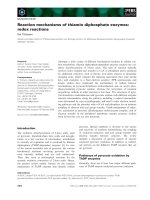
tPro213
Fig. 3. NOESY spectrum of the triply phosphorylated SRSRpT
212
PpS
214
LPpT
217
PPTR peptide of Tau. The cis conformation for the Pro219 is
below the limit of detection. Upon addition of a catalytic amount of Pin1, enhanced cis ⁄ trans isomerization of the pThr212-Pro213 peptide
bond leads to an additional red peak (red box, peak connecting the trans Pro213 Ha resonance at 4.94 p.p.m. and the cis Pro213 Ha at
4.42 p.p.m.), whereas in the same spectrum, the equivalent exchange peak for the pThr217-Pro218 bond (which should be in the green box
at 4.72 p.p.m., trans Pro218 Ha, and 5.20 p.p.m., cis Pro218 Ha) was not detected.
G. Lippens et al. Molecular mechanisms of Pin1
FEBS Journal 274 (2007) 5211–5222 ª 2007 The Authors Journal compilation ª 2007 FEBS 5215
populations, as one of the forms would continuously
disappear from the pool of free peptides. Structural
characterization of the MPM-2 antibody with a
phospho-peptide substrate might be very informative
in this aspect, and shed further light on this issue.
Another well-studied process for which Pin1’s cata-
lytic activity has been put forward is dephosphoryla-
tion by the PP2A phosphatase [46]. The crystal
structure of a cyclin-dependent kinase (CDK)2 ⁄
cyclin A kinase [47] revealed the existence of the trans
conformation of the Ser-Pro bond in the peptide sub-
strate. Similarly, the major proline-directed phospha-
tase PP2A recognizes and dephosphorylates only the
trans conformer of its phosphorylated peptide sub-
strate [46]. Pin1 could thus enhance the molecular
function of this phosphatase by speeding up the
cis–trans interconversion rate, as was proposed for
both Tau and Cdc25 [46]. Indeed, for the small phos-
pho-peptides that are used in most studies, a pool of
mainly cis conformers can be obtained by lithium ⁄
trifluoroethanol stabilization and⁄ or selective proteo-
lytic cleavage of the major pool of trans conformers
[46,48]. The dephosphorylation of this pool of mainly
cis conformers takes less time in the presence of Pin1
[46], and this unambiguously involves Pin1’s prolyl
cis ⁄ trans isomerase activity. For the in vitro phosphor-
ylated full-length Tau and CDC25, however, no effort
was made to prepare a similar large pool of cis con-
formers. We found at the peptide level that the
pThr231-Pro bond of Tau is mainly in the trans form
[37]. Extending our combined in vitro phosphorylation
and NMR spectroscopy of full-length Tau from
protein kinase A [49] to a CDK kinase, we have
preliminary data that the pThr231-Pro bond in this
CDK-phosphorylated full-length Tau also adopts the
trans conformation to a major extent (I. Landrieu,
L. Amniai and G. Lippens, unpublished results).
Isomerization from cis to trans hence cannot be
invoked any more as the sole mechanism promoting
the accelerated dephosphorylation by PP2A. Pin1
might in an as yet unidentified manner favor the inter-
action between PP2A and its phosphorylated sub-
strates, and hence stimulate their dephosphorylation
without necessarily requiring its catalytic prolyl
cis ⁄ trans isomerase activity.
Role of Pin1 in protein stability
Regulation of protein degradation seems to be an all-
important role for Pin1, and as such, a remarkable
parallel with the CKS (CDK subunit) family can be
established. CKS targets the activated CDK complex
towards phosphorylated substrates such as CDC25,
and is as such essential for the entry of Xenopus laevis
egg extracts into mitosis [50]. Well characterized struc-
turally, CKS proteins can be found in different confor-
mations with regard to their last b-strand. Folded
back on itself in a monomeric compact form [51], the
C-terminal b-strand in the swapped dimer is locked in
a second monomer [52]. The structural differences
between compact monomer and swapped dimer are
mainly limited to the conformation of the Glu-Pro
dipeptide in the hinge region between the last b-strand
and the core of CKS. The crystal structure of the com-
plex of CKS with CDK2 ⁄ cyclin A clearly showed that
only the monomeric, compact CKS could bind to the
CDK subunit [53], the swapped dimer giving rise to
important steric clashes preventing the interaction.
Pin1 antagonizes the stimulatory role of CKS in mito-
sis entry [50], and we initially assumed that this was
through a direct interaction with the Glu-Pro hinge
motif and subsequent conformational transition
between both structural forms. Experiments proved the
hypothesis wrong, as we did not obtain any evidence
of an interaction between Pin1 and this Glu-Pro dipep-
tide of CKS. We did, however, show in vitro competi-
tion for the same Cdc25-derived phosphorylated
peptide between the Pin1 WW domain and the CKS
binding module [54], and showed that the interaction
surfaces were quite similar in terms of amino acid
composition and structure (Fig. 4).
As well as a comparable role in regulating the phos-
phorylation state of CDC25 or other substrates, the
parallel between the WW and CKS interaction
domains can be drawn further when considering their
respective implications for the ubiquitination process
directing proteins towards degradation. Indeed, human
Cks1 was identified as an important factor in the
SCF
Skp2
ubiquitin E3 ligase. SCF
Skp2
plays a role in
the degradation of the CDK inhibitor p27
kip1
in late
G
1
phase after phosphorylation on its Thr187 residue
[55,56]. CKS1 interacts both with Skp2 and with a
p27
kip1
-derived pThr187 peptide [57], and hence plays
the role of an adaptor protein that is an integral part
of the E3 ligase (Fig. 2). Similarly, Schizosaccharo-
myces pombe p13
suc1
binds to the activated anaphase-
promoting complex (APC) ⁄ cyclosome [58].
Pin1 intervenes in a complex manner with the degra-
dation of cyclin E through regulation of the interaction
of cyclin E with the SCF
Cdc4
complex. It stimulates
ubiquitin addition to cyclin E by the SCF
Cdc4c
E3 ligase, after releasing the same cyclin E from the
complex with SCF
Cdc4a
[33]. Pin1 would, however, not
be part of the initial SCF
Cdc4a ⁄ c
complex, but would
be brought in by the substrate itself (Fig. 2). The
enhanced cyclin E degradation by Pin1 contrasts with
Molecular mechanisms of Pin1 G. Lippens et al.
5216 FEBS Journal 274 (2007) 5211–5222 ª 2007 The Authors Journal compilation ª 2007 FEBS
its role in cyclin D stability. Pin1 overexpression was
found to stabilize this cyclin at both the protein and
mRNA levels [59]. A similar stabilizing effect was
described for Emi1, an inhibitor of the APC complex
required to induce S-phase and M-phase entry by driv-
ing cyclin A and cyclin B accumulation. Pin1 prevents
its association with SCF
bTrcp
, and hence stabilizes
Emi1 when this latter is phosphorylated on its Ser10-
Pro motif [60]. In yet another case, the Drosophila
homolog Dodo was shown to facilitate the degradation
of the transcription factor CF2 [61]. Finally, the neuro-
nal Tau protein also contains the aforementioned
phospho-degron sequence (LPT
217
PPLSP). Although
Pin1 has as yet not been implicated directly in its deg-
radation, phosphorylated Tau is targeted for proteaso-
mal degradation through the E3 ubiquitin ligase
CHIP, complexed to an Hsc70 moiety [62], where
CHIP would selectively ubiquitinate (natively)
unfolded proteins by collaborating with the molecular
chaperone [63]. If this CDC4 phospho-degron sequence
on Tau is physiologically phosphorylated and recog-
nized by the ubiquitin ligase, this would close the circle
of Pin1’s preference for unfolded substrates. In any
case, the WW domain of Pin1 is an excellent example
of the complex roles played by protein–protein inter-
action modules [64], and whether the catalytic domain
is a second interaction domain or rather a genuine
enzyme awaits further elucidation.
Functional overlap of Pin1 with other
prolyl cis
⁄
trans isomerases?
Regulation of the transcription machinery was early
described as an essential function of Ess1 [65,66]. Its
interaction with the numerous YSPTSPS heptapeptide
repeats of the Pol II CTD [67,68], although of weak
R12
W29
S13
R99
R30
Q78
W82
Fig. 4. Molecular surface (left) and ribbon
diagrams (right) of the Pin1 WW domain
(top) or CKS (p13
Suc1
) interaction domain.
Color coding is according to the chemical
shift changes observed with a CDC25-
derived phosphopeptide. The residues
whose chemical shift is most affected upon
peptide binding (red) are Arg12 and Trp29
for the WW domain, and Arg30, Gln78 and
Trp82 for p13
Suc1
. Blue color indicates the
absence of chemical shift changes.
G. Lippens et al. Molecular mechanisms of Pin1
FEBS Journal 274 (2007) 5211–5222 ª 2007 The Authors Journal compilation ª 2007 FEBS 5217
affinity when these are tested as isolated peptides
in vitro [69], could lead to the assembly of different
molecular complexes when mRNA is transcribed. Ess1
would regulate the phosphorylation state of the CTD,
and the yeast Fcp1 phosphatase plays a similar role as
PP2A in this process. However, conflicting results have
been reported, with Ess1 either stimulating CTD
dephosphorylation by Fcp1 [70] or inhibiting this same
process [71,72]. Differences in the exact nature of the
substrate (full-length Pol II or the isolated CTD
domain) might explain this discrepancy [72]. Fcp1 was
found to more effectively dephosphorylated the pSer5
position [70], whereas the WW domain of Pin1 binds
with slightly better affinity the pSer2 position [69]. This
suggests a sequential mechanism, with preliminary
binding of the WW domain to the pSer2-Pro motif
and subsequent isomerization by the catalytic domain
at the Ser5 position. The yeast Ess1 protein, however,
prefers both to bind and isomerize pSer5-Pro over
pSer2-Pro [73]. A role of Pin1 in transcriptional regu-
lation through the (de)stabilization of complexes
formed at the CTD of Pol II was suggested by the
observation that Ess1 and the yeast Nedd4 ubiquitin
ligase Rsp5 compete for the largest subunit of the
RNA Pol II, possibly through their respective WW
domains [74].
Using a number of ess1 temperature-sensitive
mutants, two groups unexpectedly discovered that
CypA can functionally replace Ess1 [66,75]. Both
prolyl cis ⁄ trans isomerases could catalyze protein con-
formational changes essential for the assembly and ⁄ or
activity of the Sin3–Rpd3 histone deacetylase complex,
but not through binding and ⁄ or catalytic action
towards the same peptide motifs [76]. Ess1 interacts
directly with the Sin3 component, and downregulates
in this manner the deacetylase activity of Rpd3,
whereas CypA would drive the equilibrium towards
the formation of a Sin3–Rpd3–Sap30 complex.
Whereas this provides the first evidence of crosstalk
among different PPIase families, the observation of a
basal enzymatic activity towards phosphorylated sub-
strates in cell lysates from Pin1
– ⁄ –
knockout mice [77]
hints at a direct functional overlap. An intriguing com-
plementarity is further found in the inflammatory
response towards antiviral double-stranded RNA. Pin1
interacts with the pSer339-Pro340 motif on interferon
regulatory factor (IRF)-3, leading ultimately to its deg-
radation and ensuing impaired production of inter-
feron-b [78]. In the homologous IRF-4, the Ser-Pro
motif of IRF-3 is interrupted by a Leucine, despite
being in one of the best conserved regions between
both transcription factors. Pin1 no longer recognizes
this motif, and no IRF-4 regulation by Pin1 has been
reported. However, IRF-4 is regulated by FKBP52, a
member of the FK5060-binding prolyl cis ⁄ trans isome-
rases [79]. The tetratricopeptide repeats of FKBP52
mediate the interaction with IRF-4 and hence might be
the equivalent of Pin1’s WW domain, whereas its
catalytic domain could induce structural changes in
the N-terminal proline-rich domain of IRF-4. In the
same field of immunology, Pin1 also regulates the
production of such proinflammatory cytokines as
granulocyte–macrophage colony-stimulating factor
[80], interleukin-2 and interferon-c [81]. The ARE-con-
taining cytokine mRNAs interact with AUF1 factors,
and this interaction targets them for degradation. Pin1
interferes with this interaction, and hence stimulates
cytokine production. In the resting eosinophils, how-
ever, Pin1’s activity is suppressed through phosphory-
lation on one or more of its own Ser ⁄ Thr residues.
Regulation of Pin1 function through phosphoryla-
tion is indeed an important topic that has not been
extensively explored. Phosphorylation at the Ser16 resi-
due in the WW domain prevents its interaction with
phosphorylated substrates [82], and thereby partially
inactivates the function of Pin1. Polo-like kinase-1-
mediated phosphorylation, on the other hand, stabi-
lizes Pin1 by inhibiting its ubiquitination [83]. Pin1
stability and regulated activity itself hence intervene in
its complex relationship with phosphorylation.
Conclusions and perspectives
The list of potential substrates of Pin1 seems never-
ending, and one wonders how one single protein could
be involved in such a variety of cellular processes. We
can only propose some possibilities. First, the WW
domain is clearly not very selective with regard to its
molecular targets. Its binding pocket mostly sequesters
the phosphate moiety and the proline side chain
(Figs 1 and 4), whereas other amino acids around this
motif only marginally contribute to the binding affin-
ity. When studying phosphorylated peptides derived
from Tau, we found that the best binder was actually
the dipeptide pThr-Pro, with a K
D
of 100 lm [84]. Par-
allel studies with Pol II CTD-derived peptides have
shown similar results, with only a two-fold better affin-
ity for the pSer5-Pro motive over the pSer2-Pro motif
[66]. We thus believe that the WW domain will recog-
nize in vitro basically any pThr ⁄ pSer-Pro pattern, as
long as it is in a rather unstructured region. Second,
the weak affinity precludes the formation of stable
complexes, and leaves room for the Pin1 molecule to
sample a large number of potential substrates during
its half-life. Finally, the group of S. Hanes, who was
the first to describe the Sacch. cerevisiae parvulin Ess1
Molecular mechanisms of Pin1 G. Lippens et al.
5218 FEBS Journal 274 (2007) 5211–5222 ª 2007 The Authors Journal compilation ª 2007 FEBS
[9], has described a large redundancy of protein copy
numbers in the cell, at least under normal growth con-
ditions. Indeed, they found that although wild-type
yeast cells contain on the order of 200 000 molecules
of Ess1 per cell, a level lower than 400 molecules per
cell is sufficient for growth, leaving plenty of Ess1
molecules for many substrates [73]. Only under certain
conditions of stress does the large pool of Ess1 seem
essential for growth, which probably brings us proba-
bly to the situation existing in human diseases.
Whereas the detrimental role of Pin1 in human dis-
eases, and especially cancer, seemed at first to be evi-
dent, recent findings suggest that the picture in certain
cases might be more complex [85]. Certainly, Pin1
overexpression correlates strongly with poor prognosis
in a variety of cancers, and these clinical data cannot
be overlooked [86]. Nonetheless, Pin1 also stabilizes
p53 and increases its transcriptional activity, which is
essential to counteract oncogenesis [87,88]. At the cel-
lular level, its role in cyclin E and c-Myc degradation
or Emi1 stabilization would equally point to a protec-
tive role as a conditional tumor suppressor. Yeh et al.
have pointed out that the genetic background of the
mouse lines might lead to different outcomes for the
same mutation, making the construction of a single
coherent framework more problematic [89]. As is the
case for p53, where the relative levels of protein and
its inhibitors ⁄ activators can lead to subtle but signifi-
cant differences between results in cell and animal
models [90], careful analysis of in vivo models will be
needed to validate all data acquired in vitro or in cell
models before drawing conclusions on Pin1’s role in
cancer. Finally, in the context of Alzheimer’s disease,
Pin1 was shown to have a beneficial role, as it restores
the capacity of Cdc2-phosphorylated Tau to polymer-
ize tubulin into microtubules [40]. However, the tan-
gles of Tau and other amyloid species, although
characteristic in Alzheimer’s disease and correlating
well with cognitive decline, are now seen in a new
light by the scientific community. Over a period of
10 years, they have shifted from being an important
cause of the disease towards consituting a cellular
defense against the toxic oligomeric but soluble spe-
cies, although these latter still await clear identifica-
tion [91]. Could Pin1 be intended primarily as a
protective mechanism, recognizing aberrant phosphor-
ylated Ser ⁄ Thr-Pro motifs and targeting them through
interaction or conformational change towards dephos-
porylation, degradation, or aggregation? Is prolyl
cis ⁄ trans isomerization required for this function? and
could this mechanism go awry in certain diseases such
as cancer? Further research will be necessary to deter-
mine the exact role of Pin1 in human disease, and
thereby its potential as a molecular target for novel
drugs.
Acknowledgements
We thank two anonymous reviewers and Dr
E. Appella (NIH, Bethesda, USA) for careful reading
and constructive suggestions, and Dr X. Hanoulle
(Lille, France) for help with the figures. The NMR
facility used in this work was sponsored by the Re
´
gion
Nord-Pas de Calais, the CNRS, the Pasteur Institute
of Lille, and the University of Lille I.
References
1 Lang K, Schmid FX & Fischer G (1987) Catalysis of
protein folding by prolyl isomerase. Nature 329, 268–
270.
2 Fischer G, Tradler T & Zarnt T (1998) The mode of
action of peptidyl prolyl cis ⁄ trans isomerases in vivo:
binding vs. catalysis. FEBS Lett 426, 17–20.
3 Eisenmesser EZ, Millet O, Labeikovsky W, Korzhnev
DM, Wolf-Watz M, Bosco DA, Skalicky JJ, Kay LE &
Kern D (2005) Intrinsic dynamics of an enzyme under-
lies catalysis. Nature 438, 117–121.
4 Sokolskaja E & Luban J (2006) Cyclophilin, TRIM5,
and innate immunity to HIV-1. Curr Opin Microbiol 9,
404–408.
5 Bosco DA, Eisenmesser EZ, Pochapsky S, Sundquist
WI & Kern D (2002) Catalysis of cis ⁄ trans isomeriza-
tion in native HIV-1 capsid by human cyclophilin A.
Proc Natl Acad Sci USA 99, 5247–5252.
6 Brazin KN, Mallis RJ, Fulton DB & Andreotti AH
(2002) Regulation of the tyrosine kinase Itk by the pept-
idyl–prolyl isomerase cyclophilin A. Proc Natl Acad Sci
USA 99, 1899–1904.
7 Sarkar P, Reichman C, Saleh T, Birge RB & Kalodimos
CG (2007) Proline cis–trans isomerization controls
autoinhibition of a signaling protein. Mol Cell 25,
413–426.
8 Dolinski K, Muir S, Cardenas M & Heitman J (1997)
All cyclophilins and FK506 binding proteins are,
individually and collectively, dispensable for viability in
Saccharomyces cerevisiae. Proc Natl Acad Sci USA 94,
13093–13098.
9 Hanes SD, Shank PR & Bostian KA (1989) Sequence
and mutational analysis of ESS1, a gene essential for
growth in Saccharomyces cerevisiae. Yeast 5, 55–72.
10 Lu KP, Hanes SD & Hunter T (1996) A human pept-
idyl–prolyl isomerase essential for regulation of mitosis.
Nature 380, 544–547.
11 Lu KP (2000) Phosphorylation-dependent prolyl isomer-
ization: a novel cell cycle regulatory mechanism. Prog
Cell Cycle Res 4, 83–96.
G. Lippens et al. Molecular mechanisms of Pin1
FEBS Journal 274 (2007) 5211–5222 ª 2007 The Authors Journal compilation ª 2007 FEBS 5219
12 Joseph JD, Yeh ES, Swenson KI, Means AR & Win-
kler. (2003) The peptidyl–prolyl isomerase Pin1. Prog
Cell Cycle Res 5, 477–487.
13 Landrieu I, Smet C, Wieruszeski JM, Sambo AV,
Wintjens R, Buee L & Lippens G (2006) Exploring the
molecular function of PIN1 by nuclear magnetic reso-
nance. Curr Protein Pept Sci 7, 179–194.
14 Wulf G, Finn G, Suizu F & Lu KP (2005) Phosphoryla-
tion-specific prolyl isomerization: is there an underlying
theme? Nat Cell Biol 7, 435–441.
15 Wulf G, Ryo A, Liou YC & Lu KP (2003) The prolyl
isomerase Pin1 in breast development and cancer.
Breast Cancer Res 5, 76–82.
16 Lu KP (2004) Pinning down cell signaling, cancer
and Alzheimer’s disease. Trends Biochem Sci 29,
200–209.
17 Butterfield DA, Abdul HM, Opii W, Newman SF, Joshi
G, Ansari MA & Sultana R (2006) Pin1 in Alzheimer’s
disease. J Neurochem 98, 1697–1706.
18 Ranganathan R, Lu KP, Hunter T & Noel JP (1997)
Structural and functional analysis of the mitotic rotam-
ase Pin1 suggests substrate recognition is phosphoryla-
tion dependent. Cell 89, 875–886.
19 Verdecia MA, Bowman ME, Lu KP, Hunter T & Noel
JP (2000) Structural basis for phosphoserine–proline
recognition by group IV WW domains. Nat Struct Biol
7, 639–643.
20 Jacobs DM, Saxena K, Vogtherr M, Bernado P, Pons
M & Fiebig KM (2003) Peptide binding induces large
scale changes in inter-domain mobility in human Pin1.
J Biol Chem 278, 26174–26182.
21 Bayer E, Goettsch S, Mueller JW, Griewel B, Guiber-
man E, Mayr LM & Bayer P (2003) Structural analysis
of the mitotic regulator hPin1 in solution: insights into
domain architecture and substrate binding. J Biol Chem
278, 26183–26193.
22 Landrieu I, Wieruszeski JM, Wintjens R, Inze D & Lip-
pens G (2002) Solution structure of the single-domain
prolyl cis ⁄ trans isomerase PIN1At from Arabidopsis
thaliana. J Mol Biol 320, 321–332.
23 Li Z, Li H, Devasahayam G, Gemmill T, Chaturvedi V,
Hanes SD & Van Roey P (2003) Structural analysis of
the mitotic regulator hPin1 in solution: insights into
domain architecture and substrate binding. J Biol Chem
278, 26183–26193.
24 Namanja AT, Peng T, Zintsmaster JS, Elson AC,
Shakour MG & Peng JW (2007) Substrate recognition
reduces side-chain flexibility for conserved hydrophobic
residues in human Pin1. Structure 15, 313–327.
25 Labeikovsky W, Eisenmesser EZ, Bosco DA & Kern D
(2007) Structure and dynamics of Pin1 during catalysis
by NMR. J Mol Biol 367, 1370–1381.
26 Behrsin CD, Bailey ML, Bateman KS, Hamilton KS,
Wahl LM, Brandl CJ, Shilton BH & Litchfield DW
(2007) Functionally important residues in the peptidyl–
prolyl isomerase Pin1 revealed by unigenic evolution.
J Mol Biol 365, 1143–1162.
27 Wu X, Wilcox CB, Devasahayam G, Hackett RL,
Arevalo-Rodriguez M, Cardenas ME, Heitman J &
Hanes SD (2000) The Ess1 prolyl isomerase is linked to
chromatin remodeling complexes and the general
transcription machinery. EMBO J 19, 3727–3738.
28 Zhang Y, Daum S, Wildemann D, Zhou XZ, Verdecia
MA, Bowman ME, Lu
¨
cke C, Hunter T, Lu KP, Fischer
G et al. (2007) Structural analysis of the mitotic regula-
tor hPin1 in solution: insights into domain architecture
and substrate binding. ACS Chem Biol 2, 320–328.
29 Wintjens R, Wieruszeski JM, Drobecq H, Rousselot-
Pailley P, Buee L, Lippens G & Landrieu I (2001) 1H
NMR study on the binding of Pin1 Trp-Trp domain
with phosphothreonine peptides. J Biol Chem 276,
25150–25156.
30 Pastorino L, Sun A, Lu PJ, Zhou XZ, Balastik M, Finn
G, Wulf G, Lim J, Li SH, Li X et al. (2006) The prolyl
isomerase Pin1 regulates amyloid precursor protein pro-
cessing and amyloid-beta production. Nature 440, 528–
534.
31 Ramelot TA & Nicholson LK (2001) Phosphorylation-
induced structural changes in the amyloid precursor
protein cytoplasmic tail detected by NMR. J Mol Biol
307, 871–884.
32 Yaffe MB, Schutkowski M, Shen M, Zhou XZ, Stu-
kenberg PT, Rahfeld JU, Xu J, Kuang J, Kirschner
MW, Fischer G et al. (1997) Sequence specific and
phosphorylation-dependent proline isomerization: a
potential mitotic regulatory mechanism. Science 278,
1957–1960.
33 Yeh ES, Lew BO & Means AR (2006) The loss of
PIN1 deregulates cyclin E and sensitizes mouse
embryo fibroblasts to genomic instability. J Biol Chem
281, 241–251.
34 Orlicky S, Tang X, Willems A, Tyers M & Sicheri F
(2003) Structural basis for phosphodependent substrate
selection and orientation by the SCFCdc4 ubiquitin
ligase. Cell 112, 243–256.
35 van Drogen F, Sangfelt O, Malyukova A, Matskova L,
Yeh E, Means AR & Reed SI (2006) Ubiquitylation of
cyclin E requires the sequential function of SCF com-
plexes containing distinct hCdc4 isoforms. Mol Cell 23,
37–48.
36 Nash P, Tang X, Orlicky S, Chen Q, Gertler FB, Men-
denhall MD, Sicheri F, Pawson T & Tyers M (2001)
Multisite phosphorylation of a CDK inhibitor sets a
threshold for the onset of DNA replication. Nature 414,
514–521.
37 Smet C, Wieruszeski JM, Buee L, Landrieu I & Lippens
G (2005) Regulation of Pin1 peptidyl–prolyl cis ⁄ trans
isomerase activity by its WW binding module on a
multi-phosphorylated peptide of Tau protein. FEBS
Lett 579, 4159–4164.
Molecular mechanisms of Pin1 G. Lippens et al.
5220 FEBS Journal 274 (2007) 5211–5222 ª 2007 The Authors Journal compilation ª 2007 FEBS
38 Dunker AK, Brown CJ, Lawson JD, Iakoucheva LM &
Obradovic Z (2002) Intrinsic disorder and protein func-
tion. Biochemistry 41, 6573–6582.
39 Iakoucheva LM, Radivojac P, Brown CJ, O’Connor
TR, Sikes JG, Obradovic Z & Dunker AK (2004) The
importance of intrinsic disorder for protein phosphory-
lation. Nucleic Acids Res 32, 1037–1049.
40 Lu PJ, Wulf G, Zhou XZ, Davies P & Lu KP (1999)
The prolyl isomerase Pin1 restores the function of Alz-
heimer-associated phosphorylated tau protein. Nature
399, 784–788.
41 Daly NL, Hoffmann R, Otvos L Jr & Craik DJ (2000)
Role of phosphorylation in the conformation of tau
peptides implicated in Alzheimer’s disease. Biochemistry
39, 9039–9046.
42 Lippens G, Sillen A, Smet C, Wieruszeski JM, Leroy A,
Buee L & Landrieu I (2006) Studying the natively
unfolded neuronal Tau protein by solution NMR spec-
troscopy. Protein Pept Lett 13, 235–246.
43 Stukenberg PT & Kirschner MW (2001) Pin1 acts cata-
lytically to promote a conformational change in Cdc25.
Mol Cell 7, 1071–1083.
44 Bielska AA & Zondlo NJ (2006) Hyperphosphorylation
of tau induces local polyproline II helix. Biochemistry
45, 5527–5537.
45 Davis FM, Tsao TY, Fowler SK & Rao PN (1983)
Monoclonal antibodies to mitotic cells. Proc Natl Acad
Sci USA 80, 2926–2930.
46 Zhou XZ, Kops O, Werner A, Lu PJ, Shen M, Stoller
G, Kullertz G, Stark M, Fischer G & Lu KP (2000)
Pin1-dependent prolyl isomerization regulates dephos-
phorylation of Cdc25C and tau proteins. Mol Cell 6,
873–883.
47 Brown NR, Noble ME, Endicott JA & Johnson LN
(1999) The structural basis for specificity of substrate
and recruitment peptides for cyclin-dependent kinases.
Nat Cell Biol 1, 438–443.
48 Kofron JL, Kuzmic P, Kishore V, Colon-Bonuilla E
& Rich D (1991) Determination of kinetic constants
for petidyl prolyl cis–trans isomerases by an
improved spectrophotometric assay. Biochemistry 30,
6127–6134.
49 Landrieu I, Lacosse L, Leroy A, Wieruszeski JM,
Trivelli X, Sillen A, Sibille N, Schwalbe H, Saxena K,
Langer T et al. (2006) NMR analysis of a Tau
phosphorylation pattern. J Am Chem Soc 128,
3575–3583.
50 Patra D, Wang SX, Kumagai A & Dunphy WG (1999)
The xenopus Suc1 ⁄ Cks protein promotes the phosphor-
ylation of G(2) ⁄ M regulators. J Biol Chem 274, 36839–
36842.
51 Arvai AS, Bourne Y, Hickey MJ & Tainer JA (1995)
Crystal structure of the human cell cycle protein
CksHs1: single domain fold with similarity to kinase
N-lobe domain. J Mol Biol 249, 835–842.
52 Bourne Y, Arvai AS, Bernstein SL, Watson MH, Reed
SI, Endicott JE, Noble ME, Johnson LN & Tainer JA
(1995) Crystal structure of the cell cycle-regulatory pro-
tein suc1 reveals a beta-hinge conformational switch.
Proc Natl Acad Sci USA 92, 10232–10236.
53 Bourne Y, Watson MH, Hickey MJ, Holmes W, Roc-
que W, Reed SI & Tainer JA (1996) Crystal structure
and mutational analysis of the human CDK2 kinase
complex with cell cycle-regulatory protein CksHs1. Cell
84, 863–874.
54 Landrieu I, Odaert B, Wieruszeski JM, Drobecq H,
Rousselot-Pailly P, Inze
´
D & Lippens G (2001) p13
Suc1
and the WW domain of Pin1 bind to the same phos-
pho-threonine ⁄ proline epitope. J Biol Chem 276, 1434–
1438.
55 Spruck C, Strohmaier H, Watson M, Smith AP, Ryan
A, Krek TW & Reed SI (2001) A CDK-independent
function of mammalian Cks1: targeting of SCFSkp2 to
the CDK inhibitor p27Kip1. Mol Cell 7, 639–650.
56 Ganoth D, Bornstein G, Ko TK, Larsen B, Tyers M,
Pagano M & Hershko A (2001) The cell-cycle regula-
tory protein Cks1 is required for SCF (Skp2)-mediated
ubiquitinylation of p27. Nat Cell Biol 3, 321–324.
57 Hao B, Zheng N, Schulman BA, Wu G, Miller JJ, Pag-
ano M & Pavletich NP (2005) Structural basis of the
Cks1-dependent recognition of p27(Kip1) by the
SCF(Skp2) ubiquitin ligase. Mol Cell 20, 9–19.
58 Sudakin V, Shteinberg M, Ganoth D, Hershko J &
Hershko A (1997) Binding of activated cyclosome to
p13(suc1). Use for affinity purification. J Biol Chem
272, 18051–18059.
59 Wulf GM, Ryo A, Wulf GG, Lee SW, Niu T, Petkova
V & Lu KP (2001) Pin1 is overexpressed in breast can-
cer and cooperates with Ras signaling in increasing the
transcriptional activity of c-Jun towards cyclin D1.
EMBO J 20, 3459–3472.
60 Bernis C, Vigneron S, Burgess A, Labbe JC, Fesquet D,
Castro A & Lorca T (2007) Pin1 stabilizes Emi1
during G2 phase by preventing its association with
SCF(bTrcp). EMBO Rep 8, 91–98.
61 Hsu T, McRackan D, Vincent TS & Gert de Couet H
(2001) Drosophila Pin1 prolyl isomerase Dodo is a
MAP kinase signal responder during oogenesis. Nat Cell
Biol 3, 538–543.
62 Shimura H, Schwartz D, Gygi SP & Kosik KS (2004)
CHIP–Hsc70 complex ubiquitinates phosphorylated tau
and enhances cell survival. J Biol Chem 279, 4869–4876.
63 Murata S, Minami Y, Chiba T & Tanaka K (2001)
CHIP is a chaperone-dependent E3 ligase that ubiquity-
lates unfolded protein. EMBO Rep 2, 1133–1138.
64 Seet BT, Dikic I, Zhou MM & Pawson T (2006) Read-
ing protein modifications with interaction domains. Nat
Rev Mol Cell Biol 7, 473–483.
65 Hani J, Schelbert B, Bernhardt A, Domdey H, Fischer
G, Wiebauer K & Rahfeld JU (1999) Mutations in a
G. Lippens et al. Molecular mechanisms of Pin1
FEBS Journal 274 (2007) 5211–5222 ª 2007 The Authors Journal compilation ª 2007 FEBS 5221
peptidylprolyl-cis ⁄ trans-isomerase gene lead to a defect
in 3¢-end formation of a pre-mRNA in Saccharomyces
cerevisiae. J Biol Chem 274, 108–116.
66 Wu X, Wilcox CB, Devasahayam G, Hackett RL, Are-
valo-Rodriguez M, Cardenas ME, Heitman J & Hanes
SD (2000) The Ess1 prolyl isomerase is linked to chro-
matin remodeling complexes and the general transcrip-
tion machinery. EMBO J 19, 3727–3738.
67 Morris DP, Phatnani HP & Greenleaf AL (1999) Phos-
pho-carboxyl-terminal domain binding and the role of a
prolyl isomerase in pre-mRNA-3¢-end formation. J Biol
Chem 274, 31583–31587.
68 Albert A, Lavoie S & Vincent M (1999) A hyper-
phosphorylated form of RNA polymerase II is the
major interphase antigen of the phosphoprotein anti-
body MPM-2 and interacts with the peptidyl–prolyl
isomerase Pin1. J Cell Sci 112, 2493–2500.
69 Myers JK, Morris DP, Greenleaf AL & Oas TG (2001)
Phosphorylation of RNA polymerase II CTD fragments
results in tight binding to the WW domain from the
yeast prolyl isomerase Ess1. Biochemistry 40, 8479–
8486.
70 Kops O, Zhou XZ & Lu KP (2002) Pin1 modulates the
dephosphorylation of the RNA polymerase II C-termi-
nal domain by yeast Fcp1. Febs Lett 513, 305–311.
71 Xu YX, Hirose Y, Zhou XZ, Lu KP & Manley JL
(2003) Pin1 modulates the structure and function of
human RNA polymerase II. Genes Dev 17, 2765–2776.
72 Palancade B, Marshall NF, Tremeau-Bravard A, Bensa-
ude O, Dahmus ME & Dubois MF (2004) Dephosphor-
ylation of RNA polymerase II by CTD-phosphatase
FCP1 is inhibited by phospho-CTD associating pro-
teins. J Mol Biol 335, 415–424.
73 Gemmill TR, Wu X & Hanes SD (2005) Vanishingly
low levels of Ess1 prolyl-isomerase activity are sufficient
for growth in Saccharomyces cerevisiae. J Biol Chem
280, 15510–15517.
74 Wu X, Chang A, Sudol M & Hanes SD (2001) Genetic
interactions between the ESS1 prolyl-isomerase and the
RSP5 ubiquitin ligase reveal opposing effects on RNA
polymerase II function. Curr Genet 40, 234–242.
75 Fujimori F, Gunji W, Kikuchi J, Mogi T, Ohashi Y,
Makino T, Oyama A, Okuhara K, Uchida T &
Murakami Y (2001) Crosstalk of prolyl isomerases,
Pin1 ⁄ Ess1, and cyclophilin A. Biochem Biophys Res
Commun 289, 181–190.
76 Arevalo-Rodriguez M, Cardenas ME, Wu X, Hanes SD
& Heitman J (2000) Cyclophilin A and Ess1 interact
with and regulate silencing by the Sin3–Rpd3 histone
deacetylase. EMBO J 19, 3739–3749.
77 Fanghanel J, Akiyama H, Uchida C & Uchida T (2006)
Comparative analysis of enzyme activities and mRNA
levels of peptidyl prolyl cis ⁄ trans isomerases in various
organs of wild type and Pin1– ⁄ – mice. FEBS Lett 580,
3237–3245.
78 Saitoh T, Tun-Kyi A, Ryo A, Yamamoto M, Finn G,
Fujita T, Akira S, Yamamoto N, Lu KP & Yamaoka S
(2006) Negative regulation of interferon-regulatory fac-
tor 3-dependent innate antiviral response by the prolyl
isomerase Pin1. Nat Immunol 7, 598–605.
79 Mamane Y, Sharma S, Petropoulos L, Lin R & Hiscott
J (2000) Posttranslational regulation of IRF-4 activity
by the immunophilin FKBP52. Immunity 12, 129–140.
80 Shen ZJ, Esnault S & Malter JS (2005) The peptidyl–
prolyl isomerase Pin1 regulates the stability of granulo-
cyte–macrophage colony-stimulating factor mRNA in
activated eosinophils. Nat Immunol
6, 1280–1287.
81 Esnault S, Braun RK, Shen ZJ, Xiang Z, Heninger E,
Love RB, Sandor M & Malter JS (2007) Pin1 modulates
the type 1 immune response. PLoS ONE 2, e226
doi: 10.1371/journal.pone.0000226.
82 Lu PJ, Zhou XZ, Liou YC, Noel JP & Lu KP (2001)
Critical role of WW domain phosphorylation in regulat-
ing phosphoserine binding activity and Pin1 function.
J Biol Chem 277, 2381–2384.
83 Eckerdt F, Yuan J, Saxena K, Martin B, Kappel S,
Lindenau C, Kramer A, Naumann S, Daum S, Fischer
G et al. (2005) Polo-like kinase 1-mediated phosphoryla-
tion stabilizes Pin1 by inhibiting its ubiquitination in
human cells. J Biol Chem 280, 36575–36583.
84 Smet C, Sambo AV, Wieruszeski JM, Leroy A, Land-
rieu I, Buee L & Lippens G (2004) The peptidyl prolyl
cis ⁄ trans-isomerase Pin1 recognizes the phospho-
Thr212-Pro213 site on Tau. Biochemistry 43, 2032–2040.
85 Yeh ES & Means AR (2007) PIN1, the cell cycle and
cancer. Nat Rev Cancer 7, 381–388.
86 Bao L, Kimzey A, Sauter G, Sowadski JM, Lu KP &
Wang DG (2004) Prevalent overexpression of prolyl
isomerase Pin1 in human cancers. Am J Pathol 164,
1727–1737.
87 Zheng H, You H, Zhou XZ, Murray SA, Uchida T,
Wulf G, Gu L, Tang X, Lu KP & Xiao ZX (2002) The
prolyl isomerase Pin1 is a regulator of p53 in genotoxic
response. Nature 419, 849–853.
88 Zacchi P, Gostissa M, Uchida T, Salvagno C, Avolio F,
Volinia S, Ronai Z, Blandino G, Schneider C & Del Sal
G (2002) The prolyl isomerase Pin1 reveals a mecha-
nism to control p53 functions after genotoxic insults.
Nature 419, 853–857.
89 Yeh E, Cunningham M, Arnold H, Chasse D, Monteith
T, Ivaldi G, Hahn WC, Stukenberg PT, Shenolikar S,
Uchida T et al. (2004) A signalling pathway controlling
c-Myc degradation that impacts oncogenic transforma-
tion of human cells. Nat Cell Biol 6 , 308–318.
90 Toledo F & Wahl GM (2006) Regulating the p53 path-
way: in vitro hypotheses, in vivo veritas. Nat Rev Cancer
6, 909–923.
91 Ross CA & Poirier MA (2005) What is the role of pro-
tein aggregation in neurodegeneration? Nat Rev Mol
Cell Biol 6, 891–898.
Molecular mechanisms of Pin1 G. Lippens et al.
5222 FEBS Journal 274 (2007) 5211–5222 ª 2007 The Authors Journal compilation ª 2007 FEBS