Báo cáo khoa học: High resolution structure and catalysis of O-acetylserine sulfhydrylase isozyme B from Escherichia coli pot
Bạn đang xem bản rút gọn của tài liệu. Xem và tải ngay bản đầy đủ của tài liệu tại đây (2.26 MB, 8 trang )
High resolution structure and catalysis of O-acetylserine
sulfhydrylase isozyme B from Escherichia coli
Georg Zocher, Ulrich Wiesand and Georg E. Schulz
Institut fu
¨
r Organische Chemie und Biochemie, Albert-Ludwigs-Universita
¨
t, Freiburg im Breisgau, Germany
In bacteria, archaea and plants, the biosynthesis of
l-cysteine involves l-serine and inorganic sulfur com-
pounds [1–5]. In higher animals, however, l-cysteine
is derived from l-methionine [1]. The bacterial path-
way starts with a transferase that uses acetyl-CoA to
modify serine. The resulting O-acetylserine (OAS) is
then converted to cysteine by a sulfhydrylase (OASS,
EC 2.5.1.47), which in general uses hydrogen sulfide.
In a number of bacteria, the second step of synthesis
is performed by the two isozymes A and B, named
CysK and CysM, respectively. CysK uses mostly
hydrogen sulfide, which is produced in a reduction
pathway that begins with sulfate and requires dioxy-
gen. In contrast, CysM has a characteristic main
chain variation around position 210 that opens the
active center for larger thiol-carrying compounds, in
particular for thiosulfate [2,6]. The reaction with thio-
sulfate results in S-sulfo-cysteine, which can be easily
converted to cysteine and sulfate. Consequently, the
use of thiosulfate is of particular importance in an
anaerobic environment, because it does not require
dioxygen for the reduction of sulfate to hydrogen
sulfide. The isozyme CysM is of technical interest
because it processes compounds much larger than
hydrogen sulfide, and is therefore a promising candi-
date for the production of novel b-substituted
l-amino acids as building blocks for the synthesis of
pharmaceuticals and agrochemicals [7–9].
Keywords
biosynthesis of
L-cysteine; enzymatic assay;
homodimer asymmetry; nonstandard
L-amino acids; X-ray diffraction
Correspondence
G. E. Schulz, Institut fu
¨
r Organische Chemie
und Biochemie, Albert-Ludwigs-Universita
¨
t,
Albertstr. 21, 79104 Freiburg im Breisgau,
Germany
Fax: +49 761 203 6161
Tel: +49 761 203 6058
E-mail:
Website: uctbio.
uni-freiburg.de
(Received 24 July 2007, revised 22 August
2007, accepted 23 August 2007)
doi:10.1111/j.1742-4658.2007.06063.x
The crystal structure of the dimeric O-acetylserine sulfhydrylase isozyme B
from Escherichia coli (CysM), complexed with the substrate analog citrate,
has been determined at 1.33 A
˚
resolution by X-ray diffraction analysis.
The C1-carboxylate of citrate was bound at the carboxylate position of
O-acetylserine, whereas the C6-carboxylate adopted two conformations.
The activity of the enzyme and of several active center mutants was deter-
mined using an assay based on O-acetylserine and thio-nitrobenzoate
(TNB). The unnatural substrate TNB was modeled into the reported struc-
ture. The substrate model and the observed mutant activities may facilitate
future protein engineering attempts designed to broaden the substrate spec-
trum of the enzyme. A comparison of the reported structure with previ-
ously published CysM structures revealed large conformational changes.
One of the crystal forms contained two dimers, each of which comprised
one subunit in a closed and one in an open conformation. Although the
homodimer asymmetry was most probably caused by crystal packing, it
indicates that the enzyme can adopt such a state in solution, which may be
relevant for the catalytic reaction.
Abbreviations
CysK, O-acetylserine sulfhydrylase (EC 2.5.1.47) isozyme A; CysM, O-acetylserine sulfhydrylase (EC 2.5.1.47) isozyme B from Escherichia
coli; CysM(K268A), surface mutant K268A of CysM; CysM(RKE), triple surface mutant E57R-Y148K-R184E of CysM; CysM(salmo),
isozyme B from Salmonella typhimurium; DTNB, S,S¢-bis(5-thio-2-nitrobenzoate); TNB, thio-nitrobenzoate; OAS, O-acetylserine; OASS,
O-acetylserine sulfhydrylase; PLP, pyridoxal 5¢-phosphate.
5382 FEBS Journal 274 (2007) 5382–5389 ª 2007 The Authors Journal compilation ª 2007 FEBS
Five structures of CysK-type enzymes from bacteria
[10–14], archaea [15] and plants [16,17], and two struc-
tures of bacterial CysM [6,18], have been published.
The differences between the isozymes CysK and CysM
have been described [6,18]. In this article, we present
the structure of CysM complexed with the substrate
analog citrate at high resolution, together with enzy-
matic activity data of several mutants. Moreover, we
provide a model of the substrate thio-nitrobenzoate
(TNB) bound at the active center, which may be a
guide for future enzyme engineering studies.
Results and Discussion
CysM structures
In solution, CysM from E. coli is a dimer of
2 · 32 893 Da consisting of 303 amino acid residues
per subunit. An earlier study [6] yielded a medium
quality structure of the wild-type enzyme in crystal
form I at 2.7 A
˚
resolution [P6
5
22, four subunits per
asymmetric unit; reservoir: 0.1 m ammonium sulfate,
0.1 m citrate pH 5.6 with poly(ethyleneglycol)]. An
improved structure was derived from crystal form II of
the triple surface mutant CysM(RKE) that diffracted
to 2.1 A
˚
resolution, but was completely twinned,
decreasing the effective resolution [I4
1
, four subun-
its per asymmetric unit; reservoir: 0.15 m CaCl
2
, 0.1 m
Hepes pH 7.6 with poly(ethyleneglycol)] [6]. In this
article, we report the structure of the surface mutant
CysM(K268A) at 1.33 A
˚
resolution in crystal form III
(Table 1). Crystal form III was grown essentially under
the same conditions as form I, except for the absence
of ammonium sulfate. The surface mutation K268A
was at the rim of a packing contact and was not
required for crystallization, but was essential for the
superior packing order and for reproducible crystal
growth.
The structure of crystal form III was determined by
the molecular replacement method. In contrast with
the other crystal forms, form III contained only one
subunit per asymmetric unit and a lower solvent
content, both of which are typical prerequisites for
high resolution X-ray diffraction (Table 1). Although
crystal forms I and III were grown from the same
citrate buffer, only form III showed a citrate molecule
bound to the active center. Apparently, the high ionic
strength of ammonium sulfate prevented citrate
binding in form I. The structure of CysM in crystal
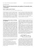
form III is shown in Fig. 1. Citrate was bound in
two conformations with occupancies of 60% and
40%, as revealed by the electron density depicted in
Fig. 2. Binding in multiple conformations indicates
low affinity, which, in turn, agrees with our observa-
tion that citrate does not inhibit the enzyme (see
below).
In order to identify established structures of related
enzymes, we searched the Protein Data Bank for
sequence homologs and detected 11 entries with
sequence identity above 30%, all of which were
OASSs. Lowering the threshold, the next entries were
two cystathione b-synthases with 29% and 24% iden-
tity. Ten of the entries were CysK-type enzymes, which
showed around 40% sequence identity with isozyme
CysM and are not considered in the following analysis.
One entry was CysM from Salmonella typhimurium
[CysM(salmo)] [18], which has 94% sequence identity
and is closely related to the enzyme CysM from E. coli
presented here.
Enzymatic activity and reaction geometry
In order to obtain data on enzyme engineering for the
synthesis of novel compounds [7–9], we produced
active center mutants and determined their catalytic
activity using TNB as the nucleophile. TNB seems to
be most appropriate for guiding enzyme engineering
intended for the synthesis of compounds of similar
size. The activities of wild-type CysM and of the crys-
tallized mutant K268A were identical, and only the
wild-type value is given in Table 2. This agreement
Table 1. Structure analysis. Values in parentheses are for the high-
est resolution shell. The data were collected at 0.9050 A
˚
wave-
length at beamline PX-II of the Swiss Light Source (SLS, Villigen,
Switzerland). The crystal belonged to space group P6
5
22 with unit
cell axes a ¼ b ¼ 76.6 A
˚
and c ¼ 209.8 A
˚
containing one CysM
subunit per asymmetric unit and 55% solvent.
Data collection
Resolution (A
˚
) 63–1.33 (1.37–1.33)
Unique reflections 83156 (6220)
Completeness (%) 98.6 (87.9)
Multiplicity 7.4 (7.9)
R
sym-I
(%) 5.9 (35)
Average I ⁄ r
I
21.4 (3.5)
Refinement
Number of atoms, protein
(residues 1–294)
2290
Number of atoms, glycerol ⁄ citrate 12 ⁄ 26
Number of water molecules 329
R
cryst
⁄ R
free
(2% test set) 0.158 ⁄ 0.172
Average isotropic B-factors (A
˚
2
)
main chain ⁄ side chains 16.6 ⁄ 20.4
glycerol ⁄ citrate ⁄ water 24.2 ⁄ 16.6 ⁄ 33.0
R
msd
bond lengths (A
˚
) ⁄ angles (°) 0.016 ⁄ 1.68
Ramachandran: most
favorable ⁄ allowed (%)
98.0 ⁄ 2.0
G. Zocher et al. Structure of the O-acetylserine sulfhydrylase CysM
FEBS Journal 274 (2007) 5382–5389 ª 2007 The Authors Journal compilation ª 2007 FEBS 5383
was expected because position 268 is at the surface dis-
tant from the active center and from the dimer inter-
face (Fig. 1).
The reported CysM structure contains a substrate-
like active center ligand, which is the bound citrate
molecule depicted in Fig. 2. A comparison with the
four known external aldimine complexes of CysK-type
enzymes [11,13,14,16] showed clearly that the C1-car-
boxylate of citrate occupies the binding site of the car-
boxylate of OAS. Whereas the C1-carboxylate is well
fixed at loop 69 (residues 68–72), the distal C6-carbox-
ylate of citrate adopts two conformations. The hydro-
xyl group of citrate points towards the internal
aldimine, as is expected for the amino group of OAS
(Fig. 2). In view of the bound citrate molecule, we
determined the enzyme activity in the presence of up
to 25 mm citrate, but observed no change. Therefore,
citrate is not an inhibitor. This agrees with the two
observed citrate conformations, because multiple bind-
ing is usually weak.
The observed kinetic parameters of wild-type CysM
from E. coli are in general agreement with those of the
homolog CysM(salmo) [18,19]. Of the active center
mutants produced, the deletion of a methyl group near
Fig. 1. Stereo ribbon plot of the high resolu-
tion structure of the CysM dimer, including
the molecular twofold axis (black), which is
crystallographic. The position of the surface
mutation K268A is shown as a yellow
sphere 25 A
˚
away from the active center.
The cofactor PLP covalently linked to Lys41,
the bound citrate molecule in its major con-
formation and the mutated residues Thr68,
Gln140 and Arg210 in the active center are
depicted as ball-and-stick models. The sub-
units have different colors. The mobile loops
defined in Fig. 5 are labeled using gray
spheres. The active center pocket opening
is indicated by a yellow stick.
Fig. 2. Detailed stereoview of the active
center of CysM. The covalently bound PLP
and the associated citrate are shown in
orange. Citrate was bound with 100% occu-
pancy. The minor conformation of citrate is
gray. The (F
o
) F
c
) electron density map of
citrate is outlined at the 3.0 r contour level.
The mutated residues are cyan. Hydrogen
bonds to the citrate molecule are indicated
by broken lines. Chain cuts are marked by
halos.
Table 2. Enzymatic activity of CysM from Escherichia coli. The esti-
mated relative errors are about 20%. The OAS concentration was
always 10 m
M; the TNB concentration varied from 10 to 1000 lM.
The temperature was 37 °C. The values in parentheses were mea-
sured at 25 °C.
k
cat
(s
)1
)
K
M
(TNB)
(m
M)
k
cat
⁄ K
M
(TNB)
(%)
Temperature
dependence
a
Wild-type 24 0.7 100
b
(41)
c
2.4
T68S 11 0.6 55 (26) 2.1
R210A – – 2 (0.8) 2.5
Q140A – – 0.4 (0.1) 4
T68A – – 0.1 (0.01) 10
Q140E – – Inactive –
a
The temperature dependence is defined here as k
cat
⁄ K
M
(TNB)
measured at 37 °C relative to the value measured at 25 °C.
b
The
absolute k
cat
⁄ K
M
(TNB) value at 37 °C was 3.5 · 10
4
M
)1
Æs
)1
. This
value was set to 100%.
c
The absolute k
cat
⁄ K
M
(TNB) value at 25 °C
was 1.4 · 10
4
M
)1
Æs
)1
.
Structure of the O-acetylserine sulfhydrylase CysM G. Zocher et al.
5384 FEBS Journal 274 (2007) 5382–5389 ª 2007 The Authors Journal compilation ª 2007 FEBS
pyridoxal 5¢-phosphate (PLP) in mutant T68S caused
the smallest disturbance (Table 2). Given the high
activity of this mutant, we determined the K
M
(TNB)
value, which was essentially identical to that of the
wild-type (Table 2). We conclude that the missing
methyl group of T68S decreases the activity only
slightly and does not affect TNB binding. A decisive
decrease to merely 2% catalytic efficiency was
observed with mutant R210A. Even stronger decreases
were caused by the removal of a carboxamide in
mutant Q140A, and by the deletion of a hydroxyl
group in mutant T68A. The enzyme was inactive when
a carboxylate was introduced at position 140 (Q140E).
The moderate activity reduction of T68S and the
strong effects of mutations Q140A, T68A and Q140E
agree well with the data derived for the corresponding
mutants of the CysK-type enzyme from Arabidopsis
thaliana [16].
In a second series of experiments, we determined the
k
cat
⁄ K
M
(TNB) values at 25 °C. The results were similar
to those at 37 °C, except for a 2.3-fold decrease for the
wild-type and for mutants T68S and R210A (Table 2).
The 2.3-fold decrease relates well to the decrease in k
cat
expected from the ‘rule-of-thumb’ factor of two for a
10 K temperature drop [20], showing that the activa-
tion energy of the catalyzed reaction lies in the usual
range and does not change for T68S and R210A. In
contrast, mutants Q140A and T68A showed much
higher temperature dependence factors, corresponding
to an appreciable increase in the activation energy [20].
We conclude that Q140A and T68A, which are close
to PLP, directly affect the reaction. In contrast, the
activity decrease of R210A, which is rather distant from
PLP, is probably a result of inefficient TNB binding,
causing a large increase in K
M
(TNB). The proposed
binding deficiency agrees with our TNB model (see
below) and also with an earlier thiosulfate model [6].
The mutants were also checked with respect to their
A
280
⁄ A
412
ratio. A photometric measurement of
CysM(K268A) yielded a ratio of 4.3, which agrees well
with the ratio of 4.0–4.2 established for the closely
homologous CysM(salmo) [18]. It also agrees with the
theoretical value calculated from the absorption spec-
tra of the tryptophans, tyrosines and PLP. The
mutants showed A
280
⁄ A
412
ratios in the range 4.3–4.5,
except for mutant Q140E with a ratio of 5.5. This
deviation was significant. It corresponds to a PLP
occupancy of about 75%. Mutant Q140E showed no
enzymatic activity (Table 2). It is conceivable that the
newly introduced glutamate adjacent to PLP made a
salt bridge to Lys41, prohibiting the formation of the
internal aldimine (see Fig. 2).
In order to model the reaction geometry, we used
the established external aldimine structure of a related
CysK structure [11] and transferred it to CysM, where
it could be accommodated without steric collision
(Fig. 3). The expected reaction geometry at the exter-
nal aldimine intermediate [11] defines the thiol position
of TNB to a small region above the plane of the acry-
late double bond. As a result of this constraint and of
the spacious active center pocket of CysM, TNB was
placed rather easily. In our model, the carboxylate of
TNB is fixed by Arg210 and the nitro group points to
the solvent (Fig. 3). The thiolate is located above the
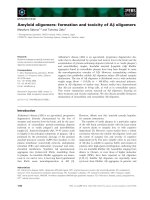
Fig. 3. Stereoview of the reaction geometry based on the structure of CysM(K268A). The observed internal aldimine with Lys41 is given in a
transparent mode (gray). The external aldimine structure has been transferred from a CysK-type enzyme [11]. It is shown together with a
manually placed model of the bound substrate TNB, the carboxylate of which is fastened to Arg210. The thiolate of TNB is approximately at
the same position as the attacking sulfur of thiosulfate in a previous model [6], which is well suited for the nucleophilic attack (red dotted
line) on the amino acrylate double bond (green spheres). Hydrogen bonds are given as black broken lines. All van der Waals distances
between TNB and its environment are above 3.0 A
˚
. The two shortest contacts are marked by green dotted lines.
G. Zocher et al. Structure of the O-acetylserine sulfhydrylase CysM
FEBS Journal 274 (2007) 5382–5389 ª 2007 The Authors Journal compilation ª 2007 FEBS 5385
acrylate plane forming an S–Cb–Ca angle of about
90°. This is an ideal position for attacking the double
bond. In summary, our negative experience with muta-
tions close to PLP suggests that this region should not
be touched when trying to produce novel l-amino
acids [7–9]. Rather, such engineering attempts should
follow the TNB model, which suggests residues
Met119, Phe141, Thr175, Pro207 and Arg210 as the
main targets.
Induced fit
A comparison between the E. coli CysM structures in
the three crystal forms revealed several characteristic
features, which are also valid for the crystal structure
of CysM(salmo) [18]. In order to establish the intrinsic
mechanical properties of CysM, we superimposed the
observed chain folds in Fig. 4. Deviations occurred at
the N- and C-termini and at the four surface loops at
positions 21, 60, 190 and 271, far away from the active
center and also from the dimer interface. These dif-
ferences are of low significance, because they are at
positions that are usually mobile. More interesting
variations occurred near the active center.
As shown in Fig. 1, the opening of the active center
pocket to the solvent is rather distant from the dimer
interface. The opening can be considered as a mouth
with two lips. One lip consists of loops 69, 94 and
helix a4(118–132), and the other is formed by
loops 202 and 215 (Fig. 4). The lip positions vary
greatly between the structures. Similar changes have
been reported for the other isozyme, CysK, for which
the extreme lip positions have been named ‘open’ and
‘closed’ [11]. The chain fold of CysM(K268A) with the
bound citrate is ‘half-closed’ (Fig. 4). Although the
variations in Fig. 4 are probably caused by more or
less random crystal contacts, they still outline the
available conformational space and, most probably,
the induced fit motions during the reaction.
The conformational changes are also reflected in the
B-factor distributions that report the polypeptide chain
mobility. As the B-factor level is strongly dependent
on the quality of the crystal order, the B-factor distri-
butions have been normalized by referring them to the
average B-factors of the respective chains. They are
displayed in Fig. 5. The distribution of CysM(K268A)
shows nine characteristic mobility peaks. Of these, the
loops at peak positions 94, 116, 132, 202 and 215 form
the lips of the mouth of the active center pocket
(Fig. 1) and are therefore important for catalysis. The
other peaks correspond to loops at the surface that are
usually mobile (Fig. 1). Interestingly, loop 69 is close
to PLP and not mobile (Fig. 5), although it partici-
pates in the induced fit (Fig. 4).
The mobility distributions of CysM(K268A), wild-
type CysM and CysM(salmo), and those of subunits B
and D of CysM(RKE), resemble each other closely
(Fig. 5). However, a most surprising deviation of the
B-factor distribution occurs in subunits A and C of
CysM(RKE) [6]. The CysM(RKE) crystal contains
dimers A–B and C–D, providing four independent sub-
unit structures. Dimer A–B is asymmetric with respect
to mobility and also with respect to structure. The
B-factor distribution of subunit A is exceptional, as it
shows almost no mobility peak. In contrast, the
respective distribution of subunit B shows the common
mobility peaks, including those of the active center lips
(Fig. 5). The same asymmetry is observed with sub-
units C and D of the other dimer. As the three muta-
tions of CysM(RKE) are all at the surface distant
from the active center, they are unlikely to affect the
internal stability of the protein. Consequently, the
Fig. 4. Stereoview of a superposition of five
distinct CysM chain folds showing wild-type
CysM in blue [6], CysM(RKE) subunit A in
green, CysM(RKE) subunit D in orange [6],
CysM(K268A) in red and CysM(salmo) in
gray [18]. The highly mobile regions are
labeled using gray spheres (see Fig. 5).
Structure of the O-acetylserine sulfhydrylase CysM G. Zocher et al.
5386 FEBS Journal 274 (2007) 5382–5389 ª 2007 The Authors Journal compilation ª 2007 FEBS
observed asymmetry should reflect a general property
of CysM.
The asymmetry of CysM(RKE) is probably caused
by crystal packing contacts. Such contacts are usually
weak, so that they can only switch between conforma-
tions that are connected via low energy barriers. As a
consequence, the observation of two independent
asymmetric homodimers in a crystal indicates that this
asymmetric state can be easily adopted in solution.
Therefore, it is conceivable that the ‘closed’ conforma-
tion of subunit A corresponds to the CysM conforma-
tion after substrate binding, whereas the ‘open’
conformation of subunit B shows CysM when releas-
ing the products after the reaction has taken place.
Such a see-saw system is discussed as ‘half-site reactiv-
ity’ [21]. We conclude that the observed asymmetry
suggests that CysM is a suitable candidate for explor-
ing the half-site reactivity hypothesis.
Experimental procedures
Mutagenesis and activity assay
The mutants were produced with the QuikChange method
(Stratagene, Heidelberg, Germany), verified by DNA
sequencing (SeqLab, Go
¨
ttingen and GATC, Konstanz,
Germany) and expressed and purified as described previ-
ously [6]. They were stored at )20 °Cina12mgÆmL
)1
solution containing 10 mm Tris ⁄ HCl pH 8.0. For the
assay, we incubated 950 lL of buffer A (100 mm Hepes
pH 7.0, 10 mm OAS, 10–1000 lm TNB) at 37 °C (or
25 °C) for 3 min, and started the reaction by adding
50 lL of a solution containing 0.5–80 lg of the enzyme.
The enzyme solution was always freshly prepared from
stored protein so that the exposure time to 37 °C (or
25 °C) was minimized. This was important for the low
activity mutants at positions 68 and 140 near PLP. TNB
was always freshly prepared in 50 mm Hepes pH 7.0
by adding 2 mm dithiothreitol and 0.5 mm S,S¢-bis(5-thio-
2-nitrobenzoate) (DTNB) to yield 1 mm TNB. The
absorption of TNB was monitored at 412 nm using e
412
¼
13 600 m
)1
Æcm
)1
[19], as well as at 500 nm using e
500
¼
970 m
)1
Æcm
)1
, which was established in a separate experi-
ment. The measurement at 500 nm was necessary in order
to reach TNB concentrations beyond the Michaelis con-
stant of 0.7 mm. The cysteine-nitrobenzoate produced has
its absorption maximum at 312 nm and does not absorb
light at 412 nm. The values for k
cat
and K
M
(TNB) were
obtained from reciprocal plots; the values for
k
cat
⁄ K
M
(TNB) were derived from linear plots.
Crystallization, structure determination,
refinement and modeling
The surface mutant K268A was produced and purified as
described previously [6] and then crystallized using the
hanging drop method. The drops contained 2 lLofan
8mgÆmL
)1
enzyme solution mixed with 2 lL of reservoir
buffer [100 mm sodium citrate pH 5.4, 18% (w ⁄ v) poly(eth-
yleneglycol) 3000]. Crystals of CysM(K268A) grew within
about 10 days at 20 °C to sizes of up to 1000 lm ·
400 lm · 400 lm. The crystals were transferred in four
steps to 28% (v ⁄ v) glycerol in reservoir buffer and flash-
frozen in a 100 K nitrogen gas stream.
Fig. 5. Relative B-factor distributions of CysM subunits in four dif-
ferent crystal forms. The B-factors were referred to the respective
subunit averages in order to eliminate differences arising from crys-
tal packing quality variations. All distributions were smoothed by
sliding a three-residue-averaging window along the chain. The top
diagram K268A refers to the reported high resolution structure with
labels at nine high mobility peaks (see Figs 1 and 4). Distribution
WT is an average of the four closely related subunit chains of the
wild-type structure [6]. The distribution of CysM(salmo) is from sub-
unit A, which is virtually the same as those of the other seven sub-
units [18]. The two distributions at the bottom are from dimers
A–B and C–D of CysM(RKE) [6] which, however, were split into an
average of the closely related subunits B and D and the equally
well-related subunits A and C.
G. Zocher et al. Structure of the O-acetylserine sulfhydrylase CysM
FEBS Journal 274 (2007) 5382–5389 ª 2007 The Authors Journal compilation ª 2007 FEBS 5387
The X-ray data were collected at the Swiss Light Source
(Villigen, Switzerland) (Table 1) and processed with pro-
grams xds and xscale [22]. Using phaser [23] and the
wild-type CysM structure [6], the phases were established
by molecular replacement. To avoid model bias, the CysM
structure and the water structure were completely rebuilt
using arp ⁄ warp [24]. The structure was manually com-
pleted using coot [25] and then refined with refmac5 [26].
Finally, we performed a translation libration screw refine-
ment with refmac5 using the 12 translation libration screw
groups (1–22, 23–65, 66–84, 85–98, 99–114, 115–131, 132–
164, 165–188, 189–208, 209–221, 222–249, 250–294) pro-
posed by the program tlsmd [27]. The CysM structure was
validated with rampage [28]. The rigid TNB molecule was
positioned manually into the active center. Numerous
options were checked visually using coot [25], and inter-
preted with respect to the quality of all contacts. The short-
est distance to the adjacent residues was maximized in
order to avoid steric hindrance as much as possible. Figures
were drawn using povscript+ [29] and povray (http://
www.povray.org). The coordinates and structure factors
have been deposited in the Protein Data Bank under acces-
sion code 2v03.
Acknowledgements
We thank the team of beamline PX-II at the Swiss
Light Source (Villigen, Switzerland) for their help with
data collection, and Wacker-Chemie (Munich, Ger-
many) for support of the project.
References
1 Cooper AJL (1983) Biochemistry of sulfur-containing
amino acids. Annu Rev Biochem 52, 187–222.
2 Kredich NM (1996) Biosynthesis of cysteine. In Escheri-
chia coli and Salmonella typhimurium: Cellular and
Molecular Biology (Neidhard FC, ed.), pp. 514–527.
ASM Press, Washington DC.
3 Borup B & Ferry JG (2000) Cysteine biosynthesis in the
Archaea: Methanosarcina thermophila utilizes O-acetyl-
serine sulfhydrylase. FEMS Microbiol Lett 189, 205–
210.
4 Mino K & Ishikawa K (2003) Characterization of a
novel thermostable O-acetylserine sulfhydrylase from
Aeropyrum pernix K1. J Bacteriol 185, 2277–2284.
5 Wirtz M, Droux M & Hell R (2004) O-acetylserine
(thiol) lyase: an enigmatic enzyme of plant cysteine bio-
synthesis revisited in Arabidopsis thaliana. J Exp Bot 55,
1785–1798.
6 Claus MT, Zocher GE, Maier THP & Schulz GE (2005)
Structure of the O-acetylserine sulfhydrylase isoenzyme
CysM from Escherichia coli. Biochemistry 44, 8620–
8626.
7 Maier THP (2003) Semisynthetic production of unnatu-
ral 1-a-amino acids by metabolic engineering of the cys-
teine biosynthetic pathway. Nat Biotechnol 21, 422–427.
8 Watkins KJ (2001) Peptides: a boom in the making.
Chem Eng News January 8, 11–15.
9 Kaldor SW, Kalish VJ, Davies IIJF, Shetty BV,
Fritz JE, Appelt K, Burgess JA, Campanale KM, Chir-
gadze NY, Clawson DK et al. (1997) Viracept (Nelfina-
vir Mesylate, AG1343): a potent, orally bioavailable
inhibitor of HIV-1 protease. J Med Chem 40, 3979–3985.
10 Burkhard P, Rao GSJ, Hohenester E, Schnackerz KD,
Cook PF & Jansonius JN (1998) Three-dimensional
structure of O-acetylserine sulfhydrylase from Salmo-
nella typhimurium. J Mol Biol 283, 121–133.
11 Burkhard P, Tai CH, Ristroph CM, Cook PF & Janso-
nius JN (1999) Ligand binding induces a large confor-
mational change in O-acetylserine sulfhydrylase from
Salmonella typhimurium. J Mol Biol 291 , 941–953.
12 Burkhard P, Tai CH, Jansonius JN & Cook PF (2000)
Identification of an allosteric anion-binding site on
O-acetylserine sulfhydrylase: structure of the enzyme
with chloride bound. J Mol Biol 303, 279–286.
13 Huang B, Vetting MW & Roderick SL (2005) The
active site of O-acetylserine sulfhydrylase is the anchor
point
for bienzyme complex formation with serine ace-
tyltransferase. J Bacteriol 187, 3201–3205.
14 Schnell R, Oehlmann W, Singh M & Schneider G
(2007) Structural insights into catalysis and inhibition of
O-acetylserine sulfhydrylase from Mycobacterium tuber-
culosis: crystal structures of the enzyme-a-aminoacrylate
intermediate and an enzyme–inhibitor complex. J Biol
Chem 282, 23 473–23 481.
15 Heine A, Canaves JM, von Delft F, Brinen LS, Dai X,
Deacon AM, Elsliger MA, Eshaghi S, Floyd R,
Godzik A et al. (2004) Crystal structure of O-acetylser-
ine sulfhydrylase (TM0665) from Thermotoga maritima
at 1.8 A
˚
resolution. Proteins 56, 387–391.
16 Bonner ER, Cahoon RE, Knapke SM & Jez JM (2005)
Molecular basis of cysteine biosynthesis in plants. Struc-
tural and functional analysis of O-acetylserine sulfhydr-
ylase from Arabidopsis thaliana. J Biol Chem 280,
38 803–38 813.
17 Francois JA, Kumaran S & Jez JM (2006) Structural
basis for interaction of O-acetylserine sulfhydrylase and
serine acetyltransferase in the Arabidopsis cysteine syn-
thase complex. Plant Cell 18, 3647–3655.
18 Chattopadhyay A, Meier M, Ivaninskii S & Burkhard P
(2007) Structure, mechanism, and conformational
dynamics of O-acetylserine sulfhydrylase from Salmo-
nella typhimurium: comparison of A and B isozymes.
Biochemistry 46, 8315–8330.
19 Tai CH, Nalabolu SR, Jacobson TM, Minter DE &
Cook PF (1993) Kinetic mechanisms of the A and B
isozymes of O-acetylserine sulfhydrylase from
Structure of the O-acetylserine sulfhydrylase CysM G. Zocher et al.
5388 FEBS Journal 274 (2007) 5382–5389 ª 2007 The Authors Journal compilation ª 2007 FEBS
Salmonella typhimurium LT-2 using the natural and
alternative reactants. Biochemistry 32, 6433–6442.
20 Price NC & Stevens L (1984) Fundamentals of Enzymol-
ogy, p. 136. Oxford University Press, Oxford.
21 Brzovic PS, Choi WE, Borchardt D, Kaarsholm NC &
Dunn MF (1994) Structural asymmetry and half-site
reactivity in the T to R allosteric transition of the insu-
lin hexamer. Biochemistry 33, 13 057–13 069.
22 Kabsch W (1993) Automatic processing of rotation dif-
fraction data from crystals of initially unknown symme-
try and cell constants. J Appl Crystallogr 26, 795–800.
23 Storoni LC, McCoy AJ & Read RJ (2004) Likelihood-
enhanced fast rotation functions. Acta Crystallogr D60,
432–438.
24 Perrakis A, Morris RM & Lamzin VS (1999) Auto-
mated protein model building combined with iterative
structure refinement. Nat Struct Biol 6, 458–463.
25 Emsley P & Cowtan K (2004) COOT: model-building
tools for molecular graphics. Acta Crystallogr D60,
2126–2132.
26 Murshudov GN, Vagin AA & Dodson EJ (1997)
Refinement of macromolecular structures by the maxi-
mum likelihood method. Acta Crystallogr D53, 240–255.
27 Painter J & Merritt EA (2006) Optimal description of a
protein structure in terms of multiple groups undergoing
TLS motion. Acta Crystallogr D62, 439–450.
28 Lovell SC, Davis IW, Arendall WB III, de Bakker PIW,
Word JM, Prisant MG, Richardson JS &
Richardson DC (2003) Structure validation by Ca
geometry: /, w, and Cb deviation. Proteins 50, 437–450.
29 Fenn TD, Ringe D & Petsko GA (2003) POVScript+:
a program for model and data visualization using per-
sistence of vision ray-tracing. J Appl Crystallogr 36,
944–947.
G. Zocher et al. Structure of the O-acetylserine sulfhydrylase CysM
FEBS Journal 274 (2007) 5382–5389 ª 2007 The Authors Journal compilation ª 2007 FEBS 5389