Báo cáo khoa học: Dynamics driving function ) new insights from electron transferring flavoproteins and partner complexes pdf
Bạn đang xem bản rút gọn của tài liệu. Xem và tải ngay bản đầy đủ của tài liệu tại đây (2.98 MB, 24 trang )
REVIEW ARTICLE
Dynamics driving function ) new insights from electron
transferring flavoproteins and partner complexes
Helen S. Toogood, David Leys and Nigel S. Scrutton
Manchester Interdisciplinary Biocentre, Faculty of Life Sciences, University of Manchester, UK
Introduction
Electron transferring flavoprotein (ETF) is positioned
at a key metabolic branch point, and is responsible for
transferring electrons from up to 10 primary dehydro-
genases to the membrane-bound respiratory chain, the
nature and diversity of which vary between organisms
[1]. ETFs are highly dynamic and engage in novel
mechanisms of interprotein electron transfer, which is
dependent on large-scale conformational sampling to
explore optimal configurations to maximize electronic
coupling. Sampling mechanisms enable efficient com-
munication with structurally distinct redox partners
[2], but require additional mechanisms for complex
assembly to impart specificity in the protein–protein
interaction.
ETFs are soluble heterodimeric FAD-containing
proteins that are found in all kingdoms of life. They
contain a second nucleotide-binding site which is
usually occupied by an AMP molecule [1]. In bacteria
and eukaryotes, ETFs function primarily as solu-
ble one- or two-electron carriers between various
Keywords
acyl-CoA dehydrogenase; conformational
sampling; electron transferring flavoprotein;
imprinting; trimethylamine dehydrogenase
Correspondence
N. Scrutton, Faculty of Life Sciences,
University of Manchester, 131 Princess
Street, Manchester M1 7DN, UK
Fax: + 44 1613065201
Tel: + 44 1613065152
E-mail:
Website:
(Received 10 July 2007, revised 24 August
2007, accepted 14 September 2007)
doi:10.1111/j.1742-4658.2007.06107.x
Electron transferring flavoproteins (ETFs) are soluble heterodimeric FAD-
containing proteins that function primarily as soluble electron carriers
between various flavoprotein dehydrogenases. ETF is positioned at a key
metabolic branch point, responsible for transferring electrons from up to
10 primary dehydrogenases to the membrane-bound respiratory chain.
Clinical mutations of ETF result in the often fatal disease glutaric aciduria
type II. Structural and biophysical studies of ETF in complex with partner
proteins have shown that ETF partitions the functions of partner binding
and electron transfer between (a) a ‘recognition loop’, which acts as a static
anchor at the ETF–partner interface, and (b) a highly mobile redox-active
FAD domain. Together, this enables the FAD domain of ETF to sample a
range of conformations, some compatible with fast interprotein electron
transfer. This ‘conformational sampling’ enables ETF to recognize structur-
ally distinct partners, whilst also maintaining a degree of specificity. Com-
plex formation triggers mobility of the FAD domain, an ‘induced disorder’
mechanism contrasting with the more generally accepted models of pro-
tein–protein interaction by induced fit mechanisms. We discuss the implica-
tions of the highly dynamic nature of ETFs in biological interprotein
electron transfer. ETF complexes point to mechanisms of electron transfer
in which ‘dynamics drive function’, a feature that is probably widespread
in biology given the modular assembly and flexible nature of biological
electron transfer systems.
Abbreviations
ACAD, acyl-CoA dehydrogenase; DMButA, n-butyldimethylamine; ETF, electron transferring flavoprotein; ETFQO, electron transferring
flavoprotein ubiquinone oxidoreductase; Fc
+
, ferricenium ion (oxidized); GAII, glutaric acidaemia ⁄ aciduria type II; MCAD, medium-chain acyl-
CoA dehydrogenase; SAXS, small-angle X-ray solution scattering; TMA, trimethylamine; TMADH, trimethylamine dehydrogenase.
FEBS Journal 274 (2007) 5481–5504 ª 2007 The Authors Journal compilation ª 2007 FEBS 5481
flavoprotein-containing dehydrogenases. Electrons are
accepted or donated to ETF via the formation of
transient complexes with their partners [3]. Almost all
ETFs are mobile carriers containing a flexible domain
essential for function [4]. ETFs need to balance pro-
miscuity with specificity in their interactions with pro-
tein donors and acceptors, in keeping with their
function in respiratory pathways. In this review, we
discuss new aspects of the structure and mechanism
of ‘typical’ ETFs, and explore the diversity in func-
tion and structure of ETFs across kingdoms. Finally,
we analyse, in the context of new structural informa-
tion, the role of clinical mutations in human ETFs
and their partner proteins that give rise to severe
metabolic diseases.
ETF families
ETFs across kingdoms interact with a variety of elec-
tron donors ⁄ acceptors that are involved in diverse met-
abolic pathways. ETFs belong to the same families
of a ⁄ b-heterodimeric FAD-containing proteins [5–7].
Members of these families can be divided roughly into
three groups based on sequence homology and func-
tional types.
Group I ETFs are a well-studied group of electron
carriers, typically found in mammals and a few bacte-
ria. Mammalian ETFs are physiological electron
acceptors for at least nine mitochondrial matrix flavo-
protein dehydrogenases [4,8]. These dehydrogenases
include the chain length-specific acyl-CoA dehydrogen-
ases (e.g. medium-chain acyl-CoA dehydrogenase,
MCAD) involved in fatty acid b-oxidation, isovaleryl-
CoA dehydrogenase, 2-methyl branched-chain acyl-
CoA dehydrogenase, glutaryl-CoA dehydrogenase
involved in amino acid oxidation, as well as dimethyl-
glycine and sarcosine dehydrogenases involved in cho-
line metabolism [4,8]. Electrons are passed from these
primary dehydrogenases through ETF to membrane-
bound ETF ubiquinone oxidoreductase (ETFQO)
[9,10].
Another well-studied group I ETF is from the bacte-
rium Paracoccus denitrificans [11–13]. It is capable of
accepting electrons from P. denitrificans glutaryl-CoA
dehydrogenase, in addition to the butyryl-CoA and
octanoyl-CoA dehydrogenases from pig liver. The
physiological electron acceptor for ETF has been
found to be ETFQO [12].
Group II ETFs are homologous to the proteins
FixB and FixA, equivalent to a-ETF and b-ETF,
respectively, which are found in nitrogen-fixing and
diazotrophic bacteria [14]. These ETFs are often
electron donors to enzymes such as butyryl-CoA
dehydrogenase, and may also accept electrons from
donors such as ferredoxin and NADH [15]. No ETF-
dependent activity has been observed with the mem-
brane-bound respiratory enzymes in nitrogen-fixing
bacteria, and so it is thought that the electron transfer
pathway from ETF to dinitrogen is via the enzymes
ETF:ferredoxin oxidoreductase, ferredoxin, nitrogenase
reductase and nitrogenase [14].
A well-studied group II ETF is from the bacterium
Methylophilus methylotrophus strain W3A1, which con-
tains only one known dehydrogenase partner, namely
trimethylamine dehydrogenase (TMADH) [3,16]. FixB ⁄
FixA proteins have been characterized from the micro-
aerobic Azorhizobium caulinodans, which is known to
accept electrons from pyruvate dehydrogenase under
aerobic conditions [14]. The nitrogen-fixing organism
Bradyrhizobium japonicum contains two sets of ETF-
like genes: one with high homology to group I ETFs
(etfSL), and the other very similar to group II FixB ⁄
FixA proteins [17]. Under aerobic conditions, only the
etfSL genes are expressed, whereas the reverse is true
for anaerobic growth, as nitrogen fixation only occurs
anaerobically [17].
One ETF from the anaerobe Megasphaera elsdenii
(formerly Peptostreptococcus elsdenii) is unusual, as it
contains two FAD-binding sites per ETF molecule,
and so does not bind AMP [6,15,18,19]. This ETF
serves as an electron donor to butyryl-CoA dehydro-
genase via its NADH dehydrogenase activity [6], and
is an electron acceptor for d-lactate dehydrogenase
[15]. It has also been shown to contain a low percent-
age of the modified flavins 6-OH-FAD and 8-OH-
FAD [6].
Group III ETFs include a pair of putative proteins,
YaaQ and YaaR, located adjacent to the cai operon,
which encodes carnitine-inducible proteins in Escheri-
chia coli [7]. Group III members will not be discussed
further in this review.
An examination of the databases of genomic
sequences shows organisms containing multiple ETF-
like genes as well as ETFs fused with other proteins
(Pedant; ). The genome of the
eubacterium Fusobacterium nucleatum ssp. nucleatum
(ATCC 25586) suggests the presence of two complete
ETF molecules, each positioned upstream of an acyl-
CoA dehydrogenase. The genome also contains a large
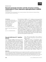
ORF (GI:19704756; Pedant; ) con-
taining a fusion of three proteins comprising an N-ter-
minal short-chain acyl-CoA dehydrogenase, followed
by the a-subunit only of ETF and a C-terminal rubre-
doxin (Fig. 1). As no functional studies of this enzyme
have been published, it is presumed that the absence of
the b-ETF subunit is a result of its role as a ‘fixed’
ETF and partners – structure, function and dynamics H. S. Toogood et al.
5482 FEBS Journal 274 (2007) 5481–5504 ª 2007 The Authors Journal compilation ª 2007 FEBS
electron carrier, although flexibility within the multi-
domain complex may be possible.
Another example of an organism with multiple ETF
content is the iron-reducing, nitrogen-fixing bacterium
Geobacter metallireducens (Pedant; http://pedant.
gsf.de). At least three of the sets of ETF genes are
unusual (e.g. ORF4) as the N-terminal portion of the
a-ETF subunit contains the gene sequence encoding a
[4Fe)4S]
2+ ⁄ +
ferredoxin domain (Fig. 1). These ETFs
are found upstream of genes such as putative Fe–S
oxidoreductases (Pedant; ). At least
nine other putative [4Fe)4S]
2+ ⁄ +
ferredoxin-contain-
ing ETFs have been identified (NCBI blast; http://
www.ncbi.nlm.nih.gov/BLAST).
Many archaea contain ETF- or FixB⁄ A-like
sequences, such as Archaeoglobus fulgidus DSM 4304,
Pyrobaculum aerophilum st. IM2, Aeropyrum pernix
and Thermoplasma volcanium st. GSS1, but these are
absent in methanogens (Pedant; ).
Several genera, such as Thermoplasma and Sulfolobus,
contain multiple ETF genes, including a fusion protein
of the two subunits, with the b-subunit at the N-termi-
nus (ba-ETF). In Sulfolobus solfataricus, ba-ETF is
found in an operon-like cluster of genes containing the
primary dehydrogenase 2-oxoacid ferredoxin oxido-
reductase, a putative ferredoxin-like protein and a
FixC-like protein, homologous to the membrane-
bound ETF ferredoxin oxidoreductase in nitrogen-
fixing organisms [14].
A blast search of the structurally equivalent N-ter-
minal (non-FAD-binding) a-ETF and b-ETF
sequences against known ORFs showed homology
with a variety of adenosine nucleotide-binding enzymes
(NCBI blast; ). Such
enzymes include members of the adenosine nucleotide
a-hydrolase superfamily from Oryza sativa, which con-
tains an ATP-binding fold [20]. The thiamine bio-
synthesis-like protein from three Leishmania species
contains b-ETF and aminotransferase components at
the N- and C-termini, respectively [21]. This class of
enzyme is known to bind ATP. Other ATP-binding
enzymes with homology to b-ETF in the database
(NCBI blast; ) include
adenylyl-sulfate kinase from Anaeromyxobacter sp.
Fw109-5 (GI:121539501), the predicted glutamate-
dependent NAD(+) synthase from Strongylocentrotus
purpuratus (GI:115971088) and the asparagine synthase
from Desulfovibrio vulgaris ssp. vulgaris DP9
(GI:120564303). As b-ETF typically binds AMP,
homology to domains of other enzymes known to bind
adenosine nucleotides is not surprising.
Sequence homology of ETFs
An alignment of a- and b-ETFs from all kingdoms of
life (Fig. 2) shows that, within the a-ETF family, the
overall sequence homology is low, although high
sequence homology is found in the C-terminal region.
By contrast, in the b-ETF family, there is a similar
degree of sequence similarity throughout the length of
the protein. Group I ETFs align better than group II
ETFs, although both groups contain significant
sequence similarity in conserved regions.
The C-terminal portion of a-ETF contains a highly
conserved region, known as the b
1
ab
2
region of FAD
enzymes, which binds the adenosine pyrophosphoryl
moiety of FAD [22]. Within this region is the a-ETF
consensus sequence of PX[L,I,V]Y[L,I,V]AXGIS-
GX[L,I,V]QHX
2
G [7], similar to the consensus
sequence for FAD-binding dehydrogenases of
GXGXXGX
15
[E ⁄ D] [22]. The b-ETF family contains a
conserved signature sequence of VXRX
2
[E,D]-
X
3
[E,Q]X[L,I,V]X
3
LP[C,A][L,I,V]
2
which is used to
identify members of the b-ETF family [7]. Adjacent to
this signature sequence, group I b-ETFs also show
the highly conserved region of DLRLNEPR-
YA[S ⁄ T]LPNIMKAKKK (residues 184–204; human
numbering), containing the recognition loop and the
highly conserved L195 necessary for partner binding in
Fusobacterium nucleatum
Butyryl-CoA
dehydrogenase
α-ETF
Rubredoxin
β-ETF
Fusion protein
Probable Fe-S
oxidoreductase
Geobacter metallireducens
Ferredoxin
α-ETF
Rubredoxin
oxidoreductase
Fusion protein
Fig. 1. Schematic diagram of the ‘operon-like’ arrangement of
genes and fusion proteins from Fusobacterium nucleatum ssp.
nucleatum (ATCC 25586) and Geobacter metallireducens (ORF4;
Pedant; ).
H. S. Toogood et al. ETF and partners – structure, function and dynamics
FEBS Journal 274 (2007) 5481–5504 ª 2007 The Authors Journal compilation ª 2007 FEBS 5483
humans [23]. The group II b-ETF from M. methylotro-
phus also contains a recognition loop and the highly
conserved L193 partner binding to TMADH [3]. Other
group II members appear not to contain a significant
group I-like recognition loop, suggesting a different
mode of partner binding.
ETF and partners – structure, function and dynamics H. S. Toogood et al.
5484 FEBS Journal 274 (2007) 5481–5504 ª 2007 The Authors Journal compilation ª 2007 FEBS
Structure of ETF
Domains of ETF
The three-dimensional structures of group I ETFs have
been solved from humans (Fig. 3A) [1] and P. denitrifi-
cans [13], and group II ETF from M. methylotrophus
(W3A1; Fig. 3B) [3]. The structure of the P. denitrifi-
cans ETF is nearly identical to human ETF, with the
major difference being a random loop between residues
b90–96 which is an a-helix in humans [13]. All three
structures can be divided into three distinct domains.
Domain I is composed of mostly the a-subunit,
whereas domain III is made up entirely of the b-sub-
unit [1]. These domains share nearly identical polypep-
tide folds related by a pseudo-twofold axis, in spite of
a lack of sequence similarity. Both domains I and III
are composed of a core of a seven-stranded parallel
b-sheet, flanked by solvent-exposed a-helices. These
domains also contain a three-stranded antiparallel
b-sheet with a fourth strand coming from the opposite
domain. Together these two domains form a shallow
bowl shape, and make up the ‘rigid’ or more static
part of the molecule upon which domain II rests.
Domain III contains a deeply buried AMP molecule
which plays a purely structural role [1].
Domain II is the FAD-binding domain, and is
attached to domains I and III by flexible linker regions
(Fig. 3) [1]. Domain II can be subdivided into two
domains, II a and IIb, which are composed of the
C-terminal portions of the a- and b-subunits, respec-
tively. Domain IIa is the larger of the two, folds in a
manner similar to bacterial flavodoxins [24] and forms
most of the region that binds FAD. This is the region
of high sequence similarity within the a-subunit. This
fold consists of a core of a five-stranded parallel
b-sheet surrounded by alternating a-helices [1]. A sixth
strand of the b-sheet is provided by the b-subunit.
FAD is bound in an orientation in which the isoallox-
azine ring is situated in a crevice between domains II
and III, with the xylene portion pointed towards the
b-subunit. By contrast, domain IIb does not interact
with FAD, but instead wraps around the lower portion
of domain IIa near domains I and III [1].
Despite the low sequence similarity between the
two groups of ETF, the overall folding of the struc-
tures is very similar, with the exception of the orien-
tation of the flavin-binding domain. Domain II of
W3A1 ETF is rotated by about 40° relative to the
human and P. denitrificans flavin domains, with
Va190 and Pb235 (W3A1 numbering) serving as
hinge points [3]. In human ETF, the conserved
Eb165 of domain III interacts with Na259, which is
located near the conserved Ra249 (Ra237 in W3A1)
and FAD (Fig. 4A). There are also hydrophobic
interactions between the C7- and C8-methyl groups
II II
FAD
AB
FAD
III I III
I
Human W3A
1
Fig. 3. Overall structures of the ETFs from
humans (A) and Methylophilus methylotro-
phus W3A1 (B). PDB codes: human, 1EFV
[1]; W3A1, 1O96 [3]. a- and b-ETF chains
are shown as magenta and blue cartoons.
FAD and AMP are shown as yellow and
orange sticks, respectively. Conserved
Leub195 ⁄ 194 for human and W3A1 ETFs,
respectively, are shown as red spheres.
Fig. 2. Alignment of a-ETFs (A) and b-ETFs (B) across kingdoms. Organisms: BRADI, Bradyrhizobium japonicum etfSL genes
(P53573 ⁄ P53575); BRADII, Bradyrhizobium japonicum FixB ⁄ A genes (P10449 ⁄ P53577); HUMAN, mature human sequence
(P13804 ⁄ P38117); METH, Methylophilus methylotrophus (P53571 ⁄ P53570); PARA, Paracoccus denitrificans (P38974 ⁄ P38975); SULF, Sulfol-
obus solfataricus (Q97V72 ⁄ Q97V71). Sequences were obtained from the Swiss-Prot database () with accession num-
bers in parentheses. The numbering for W3A1 and P. denitrificans a-ETF residues in the text are for the cloned forms of the protein in
which a methionine (in bold typeface) has been inserted at the beginning of each gene. Residue colours: orange, FAD binding; blue, AMP
binding; red, interaction with partners; green, interaction between domain III and flexible domain II; violet, b-ETF signature sequence; yellow,
hinge points. The dotted red line refers to the recognition loop.
H. S. Toogood et al. ETF and partners – structure, function and dynamics
FEBS Journal 274 (2007) 5481–5504 ª 2007 The Authors Journal compilation ª 2007 FEBS 5485
of the isoalloxazine ring of FAD and residues Fb41
and Yb16, respectively, of domain III [1]. These
interactions are likely to transiently stabilize the fla-
vin domain in this position [25]. Sequence alignments
show that Eb165 (human numbering, Fig. 1) is
highly conserved amongst mostly group I ETFs,
including P. denitrificans ETF (Eb162), which also
contains the flavin domain in the same position as
humans. This suggests that this may be a common
orientation of the flavin domain amongst group I
members.
As a result of the change in orientation of the flavin
domain in W3A1 ETF, Eb163 (equivalent to human
Eb165) interacts instead with the conserved Ra237 via
a bifurcated salt bridge (Fig. 4B) [3]. This arginine resi-
due also forms a single salt bridge with Da241 of
domain II. A second interaction between these two
domains is seen in the low-resolution W3A1 ETF
structure [3], between residues Ra211 and Eb37. In
humans, the equivalent arginine residue, Ra223, inter-
acts directly with the flavin and is over 8 A
˚
from
domain III [3].
R 211
E
37
L
184
D
241
W
38
R
237
FAD
E
163
F 41
FAD
R
249
E 165
N
259
AB
CD
3 structures
Multiple positions of
the flavin domain
Low resolution solution
structure
II
II
III IIIII
Fig. 4. Interactions between domains II and III in human (A) and Methylophilus methylotrophus W3A1 (B) ETFs. PDB codes: human,
1EFV [1]; W3A1, 1O96 [3]. a- and b-ETF chains are shown as magenta and blue cartoons and sticks. FAD is shown as yellow sticks and a
water molecule is shown as a red sphere. Hydrogen bonds and hydrophobic interactions are shown as dotted and broken lines, respectively.
(C) Small-angle X-ray scattering solvent envelope of W3A1 ETF, with a superimposition of the crystal structures of free ETF within it [4].
a- and b-ETF chains are shown as blue and magenta cartoons, respectively. Domains are labelled with Roman numerals. Adapted from [3].
(D) Superimposition of three free ETF structures showing the two positions of the flavin domain. Adapted from [4]. a- and b-ETF chains are
shown as green and red cartoons, respectively. Domains are labelled with Roman numerals.
ETF and partners – structure, function and dynamics H. S. Toogood et al.
5486 FEBS Journal 274 (2007) 5481–5504 ª 2007 The Authors Journal compilation ª 2007 FEBS
Solution structure of free ETF
Small-angle X-ray solution scattering (SAXS) studies
carried out on human, P. denitrificans and W3A1
ETFs have shown that the solvent envelopes of each
ETF are almost identical, in spite of the different con-
formations of domain II [4]. A superimposition of the
solvent envelope of W3A1 ETF onto the structure of
its free ETF shows that, although domains I and III fit
well, the envelope around domain II shows the exis-
tence of multiple conformations in solution (Fig. 4C)
[3]. These conformations appear to arise from domain
II rotating about 30–50° with respect to domains I and
III via two flexible hinge regions. This corresponds to
a shift in position of domain II from the W3A1 posi-
tion to the human ⁄ P. denitrificans position. The lack
of an appropriate shoulder in the intermediate angle
range, which can be associated with the static lobed
domain structures, suggests that all three ETFs possess
similar domain arrangements in solution, with the fla-
vin domain sampling a range of conformational states.
These states are likely to include multiple discrete, but
transient states. A superimposition of W3A1 ETFs
with different flavin domain positions, modelled by
weighted masses molecular dynamics, has shown that
these conformations are consistent with the solvent
envelope of ETF [3]. The solvent envelopes of both
oxidized and reduced W3A1 ETF are essentially identi-
cal, suggesting that no large conformational change
occurs as a result of changing the redox state [4]. The
conformations seen crystallographically may have
arisen from the trapping of a particular discrete state
as a result of crystal packing constraints, but may also
reflect differences in the proportions of the discrete
states between the different ETFs [25].
Cofactor binding
The isoalloxazine rings of FAD from human and
W3A1 ETFs are sandwiched between several conserved
residues that make distinct, but structurally equivalent,
interactions (Fig. 5A) [1,3]. A key characteristic of
ETF FAD-binding domains is the ‘bent’ conformation
of the ribityl chain of FAD as a result of 4¢OH hydro-
gen bonding with N1 of the isoalloxazine ring [1]. It is
thought that the 4¢OH group helps to stabilize the
semiquinone ⁄ dihydroquinone couple, and may be
involved in electron transfer to ETFQO. Another char-
acteristic feature is the absence of aromatic residues
that stack parallel to the ring. One or two aromatic
residues (Yb16 and Fb41 in humans) are within hydro-
phobic interaction distance, but the rings are not ori-
ented towards FAD. In its place the guanidinium
portion of the side chain of the conserved Ra249 is
perpendicular to the xylene portion of the isoalloxazine
ring, which may function by stabilizing the anionic
reduced FAD [13], and also by conferring a kinetic
block on full reduction to the dihydroquinone [3].
Other key interactions include the N1 residue of
Ha268 with O2 of the isoalloxazine ring, which may
also function in stabilizing the anionic semiquinone [1].
The hydroxyl group of Ta266 interacts with N5 of
FAD, which may aid in modulating the redox poten-
tial. The ADP moiety of FAD is solvent exposed,
more so in W3A1 ETF [3]. Stabilization of the nega-
tive charge imposed by the phosphates is achieved
through interactions with residues such as Sa248 and
Sa281 [1].
A
B
Fig. 5. (A) Schematic representation of the FAD-binding region of
human ETF. PDB code, 1EFV [1]. FAD residues and water are
shown as atom-coloured sticks and red circles, respectively.
(B) AMP-binding region of human ETF. Residues and FAD are
shown as atom-coloured sticks and water molecules are shown as
red spheres. Potential interactions are shown as dotted lines.
H. S. Toogood et al. ETF and partners – structure, function and dynamics
FEBS Journal 274 (2007) 5481–5504 ª 2007 The Authors Journal compilation ª 2007 FEBS 5487
The AMP-binding sites of all three ETF structures
are very similar, both in terms of the position and
types of interaction between AMP and b-ETF. AMP
is buried deeply within domain III and is thought to
play a purely structural role (Fig. 5B) [1]. These inter-
actions are mostly backbone interactions; thus,
although there is a high degree of conservation of posi-
tion of the interacting residues, there is often a low
sequence conservation (Fig. 2; blue residues). The
phosphate moiety of AMP from humans forms hydro-
gen bonds with the residues Ab126, Db29, Nb32,
Qb33 and Tb34, as well as a water molecule. A few
hydrogen bonds are found to anchor the rest of the
AMP molecule, including backbone interactions with
Cb66 and Ab9 and two water molecules [1]. It is
thought that AMP binding may be a structural rem-
nant of a NADP-binding site, which is a known elec-
tron donor of the group II ETF from Megasphaera
elsdenii, which does not bind AMP [6].
Structure of ETF–partner complexes
Methylophilus methylotrophus TMADH:ETF
The first structure of an ETF in complex with its part-
ner protein was solved between TMADH and ETF
from M. methylotrophus W3A1 [3]. The structure of
the free TMADH dimer had been solved previously,
and was shown to contain the redox-active cofactors
6-S-cysteinyl FMN and [4Fe)4S]
2+ ⁄ +
(electron donor
to ETF), as well as a purely structural ADP molecule
(Fig. 6A) [26,27]. Two crystal forms were obtained for
the wild-type complexes, which were found to be virtu-
ally identical, suggesting that the structure is largely
independent of crystal packing contacts. The total bur-
ied interfacial surface visible in the structures was elon-
gated in shape and covered 1750 A
˚
2
, with 10% and
8% of the surface contributed by ETF and TMADH,
respectively [3]. Surprisingly, there was a complete
absence of density for the mobile flavin domain
of ETF, in spite of SDS-PAGE analysis of the
TMADH:ETF crystals showing its presence [3].
The structures showed that there was an interaction
site between the two proteins, which was distinct from
the predicted location of the flavin-binding domain of
ETF [3]. This consists of a hydrophobic interaction
between a surface patch in the ADP-binding domain
of TMADH and a loop in ETF domain III (residues
Pb189–Ib197), termed the ‘recognition loop’ (Fig. 6B).
This loop consists of the N-terminal portion of an
a-helix and part of the preceding loop. A residue key
to this interaction is the ETF residue Lb194 (red
sphere in Fig. 3), which is buried within this hydro-
phobic patch of TMADH. Other hydrophobic residues
of ETF interacting with TMADH are Yb191, Ib197
and Sb193, the latter of which stabilizes the initial turn
of the a-helix in the recognition loop. These residues
are highly conserved, in particular within group I
ETFs (Fig. 1). Several residues preceding Yb191 which
do not contact TMADH are also conserved, including
Lb186, Nb187, P b189 and Rb190. The recognition
loop is stabilized by both the close packing of these
residues and a bifurcating salt bridge between Rb190
and residues Eb44 and Eb51. Several other residues
involved in complex formation include a salt bridge
between the N-terminus of TMADH and Db16 of
ETF, and a number of direct or water-mediated hydro-
gen bonds. This relatively small number of interactions
helps to explain why the dissociation constant
($ 5 lm) of TMADH:ETF is weak [3,28].
In free ETF, the recognition loop is more flexible
and is oriented slightly differently, with Pb189 and
Pb204 serving as hinge points [3]. Limited trypsin pro-
teolysis, which removed the recognition loop, produced
an ETF whose structure and redox capabilities with
dithionite were virtually identical to native ETF, yet it
had lost its ability to accept electrons from TMADH.
This shows the pivotal role of the recognition loop in
complex formation, and serves as an ‘anchor’ distant
to the redox centres [3]. This anchor may serve as a
means of recognizing specific redox partners, as all
that would be required would be a suitably placed
hydrophobic patch to interact with the recognition
loop [3].
The absence of density for the flavin domain of ETF
occurs after residues Va190 and Pb235, which serve as
hinge points [3]. This total lack of density was initially
surprising, as the free ETF structure showed clear den-
sity for the flavin domain, in spite of the known flexi-
bility of the molecule in solution from SAXS studies
[4]. This suggests that either the flavin domain has an
increased mobility within the complex, or packing con-
straints with the free ETF structure lock the domain in
one position. This mobility of the flavin domain within
the complex lends support to the transient nature of
the electron transfer-competent state, as predicted from
kinetics and other studies [4,25].
Several mutant TMADH:ETF complexes were
designed which altered the interactions between the
flavin domain and domain III of ETF, as well as its
interaction with TMADH (see ‘Human MCAD:ETF’
section below). At least two of each of the mutant com-
plex structures were determined, TMADH WT:ETF
Eb37Q and TMADH Y442F:ETF WT, including
two structures in a new space group (H. S. Toogood,
D. Leys & N. S. Scrutton, unpublished results). All
ETF and partners – structure, function and dynamics H. S. Toogood et al.
5488 FEBS Journal 274 (2007) 5481–5504 ª 2007 The Authors Journal compilation ª 2007 FEBS
structures were virtually identical to the wild-type
complex, including the absence of the flavin domain,
highlighting the rapid mobility of this domain.
Modelling studies in which the flavin domain of
ETF was docked into the TMADH:ETF complex,
based on its position in free ETF, showed that the
flavin domain had to undergo a significant conforma-
tional change to prevent clashes with TMADH [3,4].
This is supported by the detection of structural
changes on complex formation by observing spectral
changes during difference spectroscopy studies of
TMADH:ETF [29]. Shifting the domain into a human-
like conformation would allow the domain to fit within
the allowable space. The ‘empty volume’ observed
gp
FMN
9
[4Fe-4S]
2+/+
R
37
L 194
Reco nition loo
Y442
AMP
6-S-cysteinyl
L14
ADP
A
BC
TMADH
(monomer)
ETF
FAD
2
Y442
V344
FAD
G479
A480
S391
L393
T414
Q462
H416
Y478
A464
R 195
S 193
A
192
Y 191
Fig. 6. (A) Structure of the TMADH:ETF complex. Only one TMADH and ETF are shown for clarity. PDB code for all, 1O94 [3]. a- and b-ETF
chains and TMADH are shown as magenta, blue and green cartoons, respectively. The TMADH cofactor 6-S-cysteinyl FMN is shown as yel-
low sticks, and the [4Fe)4S]
2+ ⁄ +
centre is shown as red and yellow spheres. TMADH ADP and ETF AMP are shown as orange sticks. Resi-
dues Y442 and V344 are shown as blue sticks. The recognition loop of ETF is shown as a red cartoon with the conserved Lb194 residue
shown as red sticks. The dotted circle refers to the approximate position of the missing flavin domain. (B) Structure of the recognition loop
in TMADH:ETF. Residues are shown as atom-coloured sticks with green and blue carbons for TMADH and ETF, respectively. (C) Model of
ETF domain II in the TMADH:ETF complex. a-ETF and TMADH are shown as magenta and green cartoons, respectively. The two FAD mole-
cules are shown as yellow sticks. Highlighted residues are shown as atom-coloured sticks with green and magenta carbons for TMADH and
ETF, respectively.
H. S. Toogood et al. ETF and partners – structure, function and dynamics
FEBS Journal 274 (2007) 5481–5504 ª 2007 The Authors Journal compilation ª 2007 FEBS 5489
between TMADH and ETF is of sufficient size and
shape to allow the flavin domain of ETF to undergo a
‘ball-in-socket’ type of motion [3], suggesting that mul-
tiple (> 2) conformations are possible. This suggests
an ‘induced fit’ model for partner association, with
electron transfer likely to be possible from an ensemble
of thermodynamically metastable complexes rather
than one discrete species [3].
Kinetics studies have shown that, in the electron
transfer-competent state, the flavin of ETF is likely to
be close to a surface groove of TMADH close to resi-
dues V344 and Y442 [30]. Molecular dynamics calcula-
tions were performed on the flavin domain of free ETF
superimposed onto the complex to determine potential
electron transfer-competent states [3]. A model of one
of the putative ‘active’ conformations between the
[4Fe)4S]
2+ ⁄ +
centre of TMADH and the flavin
domain of ETF gives an intercofactor distance of less
than 14 A
˚
(Fig. 6C) [3]. In this state, the guanidinium
ion of the conserved Ra237 is located close to the aro-
matic ring and hydroxyl group of Y442 of TMADH.
Cross-linking studies using bismaleimidohexane with
TMADH Y442C and ETF Ra237C mutants led to the
rapid formation of a cross-linked complex, establishing
the close contact of these residues in the complex. Also,
difference spectroscopy studies with TMADH and the
ETF mutant Ra237A showed that electron transfer
was severely compromised as a result of a change in the
rate of rearrangement of ETF to form the electron
transfer-competent state, rather than a change in the
intrinsic rate of electron transfer [29]. However, any
interactions between TMADH and the flavin domain
of ETF are likely to be fleeting, and simply increase the
half-life of the electron transfer-competent states to
allow fast electron transfer [3].
Human MCAD:ETF
To investigate the way in which ETF can interact with
its structurally distinct partners, the structure of
human ETF with its partner MCAD was determined
[23]. The structure of free MCAD had been solved pre-
viously, and was shown to be a homotetramer of
43 kDa monomers (dimer of dimers) containing one
FAD per monomer [31]. The first structure of the com-
plex between MCAD and ETF was found to contain a
tetramer of MCAD with one ETF molecule [23]. The
total buried interfacial surface visible in the structures
(excluding the ETF flavin domain) was elongated in
shape and covered 536 A
˚
2
, with 3.2% and 4.3% of the
surface contributed by ETF and MCAD, respectively.
In this structure, the flavin domain of ETF was barely
visible in the density [23].
Four mutant MCAD:ETF complexes were designed
which altered the interactions between the flavin
domain and domain III of ETF (MCAD:ETF
Eb165A), as well as its interaction with
MCAD (MCA D:ETF Ra249A; MCAD E212A:ETF;
MCAD E359A:ETF) [25]. The aim was to alter the
ratio of the different conformational states sufficiently
to trap discrete flavin domain positions. Kinetic studies
of these complexes showed a reduction in electron
transfer rates [when using 2,6-dichloroindophenol as
the terminal electron acceptor], except for the MCAD:
ETF Eb165A complex, which showed both a dramatic
increase in rate and decrease in the apparent K
m
value.
Crystal structures of all four mutant complexes were
obtained (Fig. 7A; last three: H. Toogood, A. van
Thiel, D. Leys & N. S. Scrutton, unpublished work),
which showed an increase in density for the flavin
domain to about 70% occupancy (except for MCAD:
ETF Ra249A), with the flavin domain in the same
position as in the wild-type structure. In these struc-
tures, ETF is interacting with a dimer of MCAD [25].
As with the TMADH:ETF structures, human ETF
contains a recognition loop (Pb190–Ib198), including
the highly conserved residue Lb195, which interacts
with a hydrophobic pocket on MCAD (Fig. 7B) [23].
The recognition loop interacts with the MCAD surface
in such a way that causes an extension of a-helix C of
MCAD [31], with a nearly perfect alignment of the
axes and corresponding dipoles of both helices [23].
The side chain of Lb195 is buried within a hydropho-
bic pocket formed by a-helices A, C and D of MCAD,
and is lined by residues such as F23, L61, L73 and
I83. ETF residues which also interact with this pocket
include Yb192, Pb197, Ib198 and Mb199 [23].
A comparison of the free and complex crystal struc-
tures reveals that, although MCAD adopts a nearly
identical conformation in both structures, ETF adopts a
slightly different backbone conformation with more
extensive side chain rearrangements, including Lb195
[23]. The structure of the free ETF mutant Lb195A does
not show any significant rearrangements of the recogni-
tion loop, yet kinetic studies with both MCAD, isovale-
ryl-CoA dehydrogenase and the structurally distinct
partner dimethylglycine dehydrogenase show a severe
decrease in electron transfer rates (A. van Thiel,
H. Toogood, H. L. Messiha, D. Leys & N. S. Scrutton,
unpublished work). Mutations of MCAD, such as
L61M, L73W and L75Y, which were designed to ‘fill in’
the binding pocket, were all severely impaired in elec-
tron transfer rates with ETF [25]. Microelectrospray
ionization mass spectrometry and surface plasma reso-
nance studies showed competitive binding of ETF to
acyl-CoA dehydrogenases and dimethylglycine dehydro-
ETF and partners – structure, function and dynamics H. S. Toogood et al.
5490 FEBS Journal 274 (2007) 5481–5504 ª 2007 The Authors Journal compilation ª 2007 FEBS
genase, suggesting similar or closely overlapping bind-
ing sites for each [32]. Cross-linking experiments with
ETFQO showed that it preferentially interacts with the
b-subunit of ETF [33]. These results suggest a similar
mode of interaction between ETF and its structurally
distinct partners [23].
An alignment of MCAD-like partners shows very
little sequence conservation of the residues interacting
with the recognition loop [23]. However, the amino acid
substitutions tend to retain their hydrophobic or hydro-
gen-bonding ability, suggesting that ETF does not have
to recognize an exact binding pocket, but a structurally
equivalent one. The high conservation of the recogni-
tion loop, particularly in group I ETFs, suggests that
ETFs across kingdoms may also interact with their part-
ners in a similar manner via a recognition loop [23].
ETF
MCAD
A
AMP
E 165
Recognition loop
L
195
R
24
FAD
E212
Y360
Q163
FAD
Q 265
E212
W166
N354
E359
R
249
Q
285
FAD
T26
L73
L75
G60
L59
L61
F23
I83
F30
E34
P 196
N
197
T
194
A
193
I
198 M 199
L 195
Y 192
BC
Fig. 7. (A) Structure of the MCAD:ETF Eb165A complex. Only one dimer of MCAD and ETF are shown for clarity. PDB code for all, 2A1T
[25]. a- and b-ETF chains and MCAD are shown as magenta, blue and green cartoons, respectively. The cofactors FAD and AMP are shown
as yellow and orange sticks, respectively. Highlighted side chains of MCAD and ETF are shown as blue sticks. The recognition loop of ETF
is shown as a red cartoon with the conserved Lb194 residue shown as red sticks. ETF Eb165 is shown as a red sphere. (B) Structure of the
recognition loop in MCAD:ETF. Residues are shown as atom-coloured sticks with green and blue carbons for MCAD and ETF, respectively.
(C) Structure of the electron transfer interaction site. a-ETF and MCAD are shown as magenta and green cartoons, respectively. The two
FAD molecules are shown as yellow sticks. Highlighted residues are shown as atom-coloured sticks with green and magenta carbons for
MCAD and ETF, respectively.
H. S. Toogood et al. ETF and partners – structure, function and dynamics
FEBS Journal 274 (2007) 5481–5504 ª 2007 The Authors Journal compilation ª 2007 FEBS 5491
The orientation of the flavin domain within the
MCAD:ETF complex is dramatically different from its
position in any of the free ETF structures (Fig. 7C)
[25]. The contact surface between MCAD and the fla-
vin domain is about 330 A
˚
2
, with a shape complemen-
tarity value of 0.56, suggesting that the interaction is
weak and of a transient nature [25]. Within this inter-
face, Ra249 of the flavin domain forms a salt bridge
with E212 of MCAD, as well as interacting with E359
via a bridging water molecule. This is in agreement
with chemical modification studies, which show that an
arginine residue in ETF and carboxylates on MCAD
are involved in complex formation [34]. Other interac-
tions between ETF and MCAD include direct hydro-
gen bonds between Qa285 ⁄ N354, Qa265 ⁄ E359 and a
phosphate of ETF FAD ⁄ Q163, respectively [25]. The
smallest distance between the isoalloxazine rings of the
two FAD molecules is 9.7 A
˚
, suggesting that this is an
electron transfer-competent state. The indole group of
MCAD W166 is positioned between the isoalloxazine
rings, and is within van der Waals’ contact with both
the C7 and C8 methyl groups of ETF FAD [25].
The complex structure shows that electrostatic inter-
actions are essentially absent from the interface, yet it
is known that the electron transfer rate decreases with
increasing ionic strength [25]. These observations could
be a result of the destabilization of the protein–protein
interaction between E212 and Arga249. Alternatively,
these results may arise from enhanced hydrophobic
interaction at high ionic strength involving the hydro-
phobic patch ⁄ recognition loop. The concomitant
decrease in the rate of complex dissociation following
electron transfer might lead to the observed reduction
in steady-state turnover [25].
Although there are no structural similarities between
TMADH and MCAD, ETF interacts in a similar man-
ner with both proteins [23]. This is a result of the rec-
ognition loop interacting with distinct, but structurally
equivalent, hydrophobic patches on the partners,
which creates a near-identical volume and shape of the
space occupied by the flavin domain of ETF. The rela-
tive positions of the docking sites for the leucine
anchoring residue within the recognition loop between
the two complexes are very similar. However, the two
partner proteins interact with ETF via different redox
cofactors, with the electron-donating cofactors in dif-
ferent relative positions within the two complex struc-
tures. This highlights the need for the flavin domain to
sample the available conformational space to find an
electron transfer-competent state, as seen by the lack
of density for the flavin domain in both wild-type
structures. These conformations are transiently stabi-
lized through key interactions between conserved resi-
dues specific to each dehydrogenase type [23]. As both
the [4Fe)4S]
2+ ⁄ +
and FAD cofactors of TMADH
and MCAD, respectively, are located within a 10 A
˚
radius of the ETF FAD, this suggests that a similar
conformation of ETF in both complexes is possible for
fast interprotein electron transfer.
Kinetics of electron transfer between
ETF and partners
Methylophilus methylotrophus TMADH:ETF
TMADH is a 166 kDa homodimeric iron–sulfur flavo-
protein which catalyses the oxidative demethylation of
trimethylamine (TMA) to form dimethylamine and
formaldehyde (Eqn 1) [35]. Substrate oxidation is
accompanied by the transfer of reducing equivalents,
first to the covalently bound cofactor 6-S-cysteinyl
FMN [27], followed by reduction of a ferredoxin-like
[4Fe)4S]
2+ ⁄ +
located approximately 4–6 A
˚
from the
8-a-methyl group of FMN [36]. The physiological ter-
minal electron acceptor of TMADH from M. methy-
lotrophus is ETF, with electron transfer from the
[4Fe)4S]
2+ ⁄ +
centre occurring via quantum electron
tunnelling [37,38]. Stopped-flow kinetics studies of the
reductive half-reaction shows that it occurs in three
kinetic phases. The fast phase represents the two-elec-
tron reduction of 6-S-cysteinyl FMN, followed by
intermediate and slow phases which reflect the transfer
of one electron from the dihydroquinone of flavin to
the [4Fe)4S]
2+ ⁄ +
centre, and the formation of a spin-
interacting state between the flavin semiquinone and
the reduced [4Fe)4S]
2+ ⁄ +
[39]. This latter state is
formed after the binding of a second substrate mole-
cule, which induces the ionization of Y169 located
close to the pyrimidine ring of 6-S-cysteinyl FMN [36].
This state is distinguished by a complex EPR signal
centred near g $ 2 with an unusually intense half-field
g $ 4 signal [39]. However, the kinetics are further
complicated as the extent of the biphasic nature
changes with both substrate concentration and pH
[39]. Detailed kinetic and mechanistic analyses of the
reductive half-reaction have been studied extensively,
and readers are referred to papers such as Scrutton
et al. [40], Scrutton and Sutcliffe [35], Roberts et al.
[41], Basran et al. [42–46], and references cited therein.
ðCH
3
Þ
3
N+H
2
O !ðCH
3
Þ
2
NH þ CH
2
O þ 2H
þ
þ 2e
À
ð1Þ
TMADH:ETF oxidative half-reaction
The oxidative half-reaction of TMADH involves the
transfer of two electrons through [4Fe)4S]
2+ ⁄ +
to the
ETF and partners – structure, function and dynamics H. S. Toogood et al.
5492 FEBS Journal 274 (2007) 5481–5504 ª 2007 The Authors Journal compilation ª 2007 FEBS
ETF FAD in two single electron transfer steps. The
midpoint reduction potential of the oxidized flavin ⁄
semiquinone couple of ETF (E
ox ⁄ sq
) is unusually posi-
tive (+ 153 mV) [29], but is consistent with the need
to accept electrons from the [4Fe)4S]
2+ ⁄ +
centre of
TMADH, which has a 4Fe)4S
2+
⁄ 4Fe)4S
+
potential
of + 102 mV [47]. This highly positive redox potential
of ETF suggests exceptional stabilization of the anio-
nic semiquinone, most probably because of the loca-
tion of the guanidinium group of the conserved Ra237
over the si face of the flavin isoalloxazine ring.
Conversion of FAD to the dihydroquinone form is
incomplete, with a midpoint potential of the
semiquinone ⁄ dihydroquinone couple of less than
) 250 mV, as a result of the presence of both kinetic
and thermodynamic blocks on full reduction of FAD
[29].
Recent mutagenic studies have shown the impor-
tance of Sa254 as a hydrogen bond donor to the N(5)
atom in the oxidized state of the flavin [48,49]. Muta-
tion of Sa254 to threonine and cysteine abolished the
kinetic barrier to dihydroquinone formation, as well as
significantly decreasing the E
ox ⁄ sq
midpoint potential.
Changes in the observed K
d
values showed that,
although the mutations destabilized the oxidized state
of the flavin, the anionic semiquinone state was also
destabilized to a much greater extent. Thus, Sa254
plays a key role in establishing the high E
ox ⁄ sq
value
and the unusually high stability of the anionic semiqui-
none of ETF [48].
Further studies have suggested that the redox prop-
erties of E
ox ⁄ sq
of ETF may be perturbed on complex
formation with TMADH [50]. In the presence of
TMADH, ETF can be fully reduced to the dihydroqui-
none species by dithionite, with estimated redox poten-
tials of + 196 mV and 0 mV for the E
ox ⁄ sq
and E
sq ⁄ hq
couples, respectively. This change in redox potentials
of ETF is thought to be a result of a conformational
change in the flavin-binding domain on complex for-
mation [50].
Steady-state kinetic parameters for the electron
transfer between the [4Fe)4S]
2+ ⁄ +
centre of TMADH
and ETF flavin give k
cat
and K
m
values of
16.8 ± 0.5 s
)1
and 14.8 ± 1.2 l m, respectively, at
25 °C [30]. Modelling studies of the complex between
TMADH and ETF, as well as kinetics studies of
TMADH mutants, revealed the existence of a small
surface groove on TMADH which may accommodate
the isoalloxazine ring of FAD bound to ETF [30].
These studies revealed the existence of two possible
routes of electron transfer from the [4Fe)4S]
2+ ⁄ +
cen-
tre to an external electron acceptor. The shortest path-
way extends from C345, a ligand on the [4Fe)4S]
2+ ⁄ +
centre, to V344, which is located at the bottom of a
small groove on the surface of TMADH. The second
pathway extends from C345 to E439 and finally to
Y442, the latter of which forms one side of the groove
on the surface of the enzyme [30].
The steady-state kinetic parameters for five muta-
tions of V344 and four mutations of Y442 of
TMADH with ETF were determined [30]. The
Michaelis constant K
m
was largely unaffected by the
mutations, except for Y442G, which showed a five-
fold increase, and the effect on k
cat
was minimal for
V344 mutants (at most twofold for V344I). By con-
trast, Y442 substitutions had a more noticeable effect,
particularly with smaller and less bulky Y442C and
Y442G mutations, which resulted in 19- and 31-fold
reductions in turnover number, respectively. The
steady-state kinetic parameters of these TMADH
mutations were also determined with ferricenium ion
(Fc
+
) as the electron acceptor. In this case, mutations
of V344 had quite dramatic effects, with substitutions
of V344 to cysteine, alanine or glycine causing a sig-
nificant reduction in the apparent K
m
value and
increase in k
cat
⁄ K
m
, but only a modest overall
increase in k
cat
. The kinetics of the reductive half-
reaction of these mutants showed only small changes
in the rate of intramolecular electron transfer from
6-S-cysteinyl FMN to the [4Fe)4S]
2+ ⁄ +
centre,
which may reflect minor structural changes around
the [4Fe)4S]
2+ ⁄ +
centre [30].
Stopped-flow studies on the kinetics of transfer of
electrons from two-electron-reduced TMADH to oxi-
dized ETF revealed complex multiphasic kinetics [51].
To simplify these studies, the 6-S-cysteinyl FMN co-
factor of TMADH was inactivated by phenylhydr-
azine, rendering it inert to reduction ⁄ oxidation. This
allows TMADH to be reduced anaerobically to the
one-electron state, via titration with dithionite, with
the electron located on the [4Fe)4S]
2+ ⁄ +
centre [52].
Fast reaction studies of this one-electron-reduced mod-
ified TMADH with ETF eliminates the complications
arising from internal electron transfer in TMADH
[30]. Initial studies of the kinetics of the oxidative half-
reaction with ETF were carried out at 25 °C, and
showed a hyperbolic dependence on ETF concen-
tration, which exhibited saturation behaviour [53].
However, more recent rigorous studies of the reaction
kinetics carried out at 5 °C, slowing the reaction
sufficiently to obtain more precise data, demonstrated
the biphasic nature of the transients and a linear
dependence of ETF concentration on the rate [30]. A
recent attempt was made to reinstate the saturation
behaviour of the electron transfer rate on ETF concen-
tration. However, this study omitted to show data for
H. S. Toogood et al. ETF and partners – structure, function and dynamics
FEBS Journal 274 (2007) 5481–5504 ª 2007 The Authors Journal compilation ª 2007 FEBS 5493
reactions carried out at 5 °C, and there was no
rigorous attempt to analyse changes in the nature of
reaction transients at different temperatures [54].
Both wild-type TMADH and mutants of V344
showed a linear dependence on ETF concentration
at 5 °C, with a second-order rate constant of
1.44 · 10
6
M
)1
Æs
)1
for the native enzyme [30]. By con-
trast, mutants of Y442 displayed saturation behaviour
on ETF concentration, with saturation behaviour with
respect to ETF detected with mutants Y442F, Y442L
and Y442G [53]. The dissociation constants calculated
for these Y442 mutant complexes varied from 6 to
46 lm, compared with 3–7 lm for the native complex,
the latter determined by analytical ultracentrifugation
[28]. By contrast, the reactions of the mutants and
native TMADH with Fc
+
all showed a linear depen-
dence on Fc
+
concentration at 25 °C. Val344 substitu-
tions to cysteine, alanine or glycine showed a moderate
increase in second-order rate constants, whereas the
opposite was true for the bulkier substitutions tyrosine
and isoleucine, with the latter showing a nearly 20-fold
reduction. This could be the result of a change in the
length of the electron transfer distance and ⁄ or changes
in packing density. These mutations were found to
have little or no effect on the binding or limiting rate
constant (k
lim
) for oxidation of the substrate. These
stopped-flow and steady-state results suggest that elec-
tron transfer to ETF proceeds via the longer pathway
through Y442, whereas electron transfer to Fc
+
is via
the shorter route through V344, as Fc
+
is likely to
penetrate the groove more fully than the ETF flavin.
Substitutions at Y344 showed that shortening the
length of the side chain increased the electron transfer
rates to Fc
+
, presumably by reducing the pathway
length. Mutations at Y442 possibly disrupt electron
transfer by perturbing the interaction geometry of
TMADH and ETF in the complex, leading to less effi-
cient coupling between the [4Fe)4S]
2+ ⁄ +
centre and
FAD [30,53].
Intrinsic rate of electron transfer to ETF
Given the highly dynamic nature of ETF, a kinetic
model of intermolecular electron transfer incorporating
intermediate state(s) representing flavin domain motion
is needed. These intermediate states include the large
change in position of domain II on complex formation
[3] and small-scale conformational changes in the for-
mation of electron transfer-competent state(s). A sim-
plified kinetic scheme for such a system, where A is
one-electron-reduced TMADH (4Fe)4S
+
) and B is
oxidized ETF, is shown in Scheme 1 [30]. In this
scheme, k
r
(and k
–r
) refer to the reversible rate of reor-
ganization of the flavin domain to form an electron
transfer-competent state:
A+BÐ
k
a
k
Àa
ðABÞ
1
Ð
k
r
k
Àr
ðABÞ
2
!
k
eT
ðA
þ
B
À
Þ!A
þ
þ B
À
ðScheme 1)
As native and V344 mutants of TMADH do not
display saturation kinetics with ETF at 5 °C, this
model predicts that complex formation is rate limiting
[30]. Thus, both k
eT
and any possible conformational
reorganization of the ETF flavin domain following
complex formation are predicted to be fast. As the
Y442 mutants display saturation kinetic behaviour,
this suggests that either k
eT
or the rate of conforma-
tional reorganization has been dramatically reduced.
Given that the observed limiting rates of electron
transfer for Y442 mutants are relatively slow, the latter
is possibly more likely [30].
Simulations have shown that, for k
eT
values above
10
3
s
)1
, there is essentially a linear relationship
between k
obs
and ETF concentration [30]. The values
of k
obs
obtained from the simulations were very similar
to those obtained experimentally for native and Y442
mutant enzymes. The switch to saturation behaviour
was seen only when k
eT
was less than $ 10
3
s
)1
. Such
low predicted k
eT
values for Y442 mutants are not
likely to correspond to intrinsic electron transfer rates
for transfers occurring over 11–12 A
˚
[55], and so rate
limitation is likely to be the result of impaired struc-
tural reorganization during complex assembly. In
native TMADH, Y442 may enhance the rates of reor-
ganization of the electron transfer complex by a direct
interaction with ETF via its phenolic hydroxyl group.
Disruption of favourable interactions by Y442 mutants
could thereby alter the nature of the electron transfer-
competent state, such as a change in the [4Fe)4S]
2+ ⁄ +
to ETF FAD distance, leading to a dramatically
reduced k
eT
value [30].
Kinetic scheme of intra- and interprotein
electron transfer
A branching kinetic steady-state scheme has been pro-
posed for intra- and interprotein electron transfer of
TMADH (Fig. 8) [41]. TMADH is unusual as it shows
substrate inhibition at high TMA concentrations. Fig-
ure 8 presents a branching kinetic scheme in which
TMADH can utilize two alternative catalytic cycles: a
0 ⁄ 2 cycle, in which it cycles between an oxidized and
two-electron-reduced enzyme, and a 1⁄ 3 cycle, in
which it cycles between a one- and three-electron-
reduced enzyme [30]. This situation exists because,
although the substrate can donate two electrons at a
ETF and partners – structure, function and dynamics H. S. Toogood et al.
5494 FEBS Journal 274 (2007) 5481–5504 ª 2007 The Authors Journal compilation ª 2007 FEBS
time, the terminal electron acceptor ETF (or phenazine
methosulfate) can only accept one electron at a time,
and as a result of the presence of both a 6-S-cysteinyl
FMN and a [4Fe)4S]
2+ ⁄ +
centre, TMADH can take
up as many as three electrons [41].
Stopped-flow studies of the reaction of TMADH
with TMA using diode array detection showed four
characteristic spectra of the reductive half-reaction [41].
The states distinguishable are the oxidized enzyme,
two-electron-reduced flavin dihydroquinone, two-elec-
tron-reduced anionic flavin semiquinone plus reduced
[4Fe)4S]
2+ ⁄ +
and, finally, the so-called spin-inter-
acting state. The one- and three-electron-reduced forms
are not observed in single turnover studies, although
they are likely to be present in steady-state reactions.
Direct evidence for the presence of alternative redox
cycles was detected with enzyme-monitored turnover
experiments with TMA concentrations of 20 lm to
2mm. At high TMA concentrations, the spectrum
indicates that the predominant species at steady state is
the one-electron-reduced flavin semiquinone ⁄ oxidized
[4Fe)4S]
2+ ⁄ +
, consistent with the species predicted to
accumulate in the 1 ⁄ 3 cycle. At low TMA concentra-
tions, the predominant species is oxidized TMADH,
with only a small quantity of flavin semiquinone. Thus,
at low substrate concentrations, the 0⁄ 2 cycle is
predominant and substrate inhibition does not occur.
Interestingly, at low Fc
+
concentrations (oxidizing
substrate), the switch between the 1 ⁄ 3 and 0 ⁄ 2 cycles
with a decrease in TMA concentration does not occur.
Thus, as the ratio of reducing to oxidizing substrate
increases, the level of steady-state enzyme reduction
increases [41].
It is thought that the 1 ⁄ 3 cycle is slower than the
0 ⁄ 2 cycle, indicating that it would be predominant only
at high TMA concentrations or low ETF ⁄ phenazine
methosulfate concentrations. This is because substrate
binding stabilizes the semiquinone form of the flavin in
the one-electron-reduced enzyme [56]. The binding of
substrate to the one-electron-reduced [4Fe)4S]
2+ ⁄ +
centre of TMADH, which is likely to accumulate
under the high substrate concentrations of the steady-
state condition, may result in the redistribution of the
reducing equivalents, leading to the formation of a fla-
vin semiquinone (boxed reaction in Fig. 8). In this
case, the substrate is unable to be oxidized, as the
flavin is unable to accept two reducing equivalents
because of the unfavourable equilibrium between the
two redox centres in the substrate-bound, one-electron-
reduced state. This would cause an apparent substrate
inhibition of the reaction without the need for a
second inhibitory substrate-binding site [41].
FMNH
2
4Fe-4S
ox
FMN
sq
FMN
4Fe-4S
ox
FMNH
2
FMN
4Fe-4S
red
4Fe-4S
red
4Fe-4S
red
4Fe-4S
red
4Fe-4S
red
FMN
ox
.S
FMN
sq
.S
4Fe-4S
ox
FMN.S
4Fe-4S
ox
FMN
sq
4Fe-4S
ox
FMN
sq
.S
FMNH
2
.S
4Fe-4S
ox
(CH
3
)
2
NH
(CH
3
)
2
NH
+ HCHO
+ HCHO
ETF
ox
ETF
ox
ETF
ox
ETF
ox
ETF
sq
ETF
sq
ETF
sq
ETF
sq
ETF
sq
(CH
3
)
3
N
(CH
3
)
3
N
(CH
3
)
3
N
1
2
3
4
5
6
7
8
9
10
11
0/2 cycle 1/3 cycle
Fig. 8. Kinetic scheme of the proposed branching mechanism of electron transfer for TMADH:ETF. In the 0 ⁄ 2 cycle, the enzyme turns over
between the oxidized and two-electron-reduced state. In the 1 ⁄ 3 cycle, the enzyme turns over between the one- and three-electron-reduced
states. Population of the 1 ⁄ 3 cycle leads to substrate inhibition of TMADH. ox, oxidized; red, reduced; S, substrate; sq, semiquinone.
Adapted from [41].
H. S. Toogood et al. ETF and partners – structure, function and dynamics
FEBS Journal 274 (2007) 5481–5504 ª 2007 The Authors Journal compilation ª 2007 FEBS 5495
This model also predicts that the partitioning of the
two redox cycles is not dependent on the rate of 6-S-
cysteinyl FMN reduction [41]. This is supported by
stopped-flow studies with triethylamine, which has
been shown to be a poor substrate, yet still displays a
clear steady-state inhibition. However, studies with the
substrate n-butyldimethylamine (DMButA) show a
substantial compromise in flavin reduction, as well as
a reduction in substrate inhibition, although it has the
tightest binding affinity to oxidized TMADH. It is
thought that this lack of substrate inhibition is caused
by a weaker binding of DMButA to the reduced
enzyme, which could account for the observed low
accumulation of the anionic semiquinone form of
TMADH [39].
Alternative stable conformations of ETF
Compounding the problems associated with under-
standing the dynamics of the interface between
TMADH and ETF is the issue of ‘structural imprint-
ing’ — a slow conformational change in ETF that is
catalysed by interaction with TMADH. These ill-
defined structural changes in ETF give rise to an
increase in fluorescence emission of the FAD cofactor,
and suggest a more ‘open’ structure for ETF in which
FAD is more solvent exposed [57]. These relatively
slow structural changes associated with imprinting are
not to be confused with ‘conformational sampling’.
Thus, unlike the structural change imparted through
imprinting of ETF by TMADH, conformational sam-
pling is an integral part of the electron transfer mech-
anism. Others [54] have incorrectly challenged our
interpretation of structural imprinting by suggesting
that we have inferred that both structural imprinting
and conformational sampling are the same process.
This is not the case. The timescales for structural
imprinting are far too slow to be associated with elec-
tron transfer from TMADH to ETF as observed in
stopped-flow studies. Conformational sampling is an
intrinsically rapid motion of the FAD domain in the
complex that allows the FAD domain to search out
electron transfer-competent conformations. The struc-
turally imprinted form of ETF accumulates over an
extended time course (typically hours) when as-puri-
fied ETF is incubated with TMADH. The precise
structural change(s) that occurs during the imprinting
reaction is not known, but both fluorescence emission
and anisotropy analysis indicate that there is a slow
structural change in ETF when incubated with
TMADH over extended time periods (H. Messiha,
S. E. Burgess, D. Leys & N. S. Scrutton, unpublished
results).
Mammalian MCAD:ETF
MCAD is a 172 kDa homotetrameric flavoprotein that
catalyses the a,b-dehydrogenation of acyl-CoA thio-
esters to their corresponding trans-2,3-enoyl-CoA [58].
Substrate oxidation is accompanied by the transfer of
reducing equivalents to the covalently bound cofactor
FAD, followed by electron transfer to its physiological
terminal electron acceptor ETF. The kinetic scheme of
MCAD is very complex and involves several interme-
diates. In the reductive half-reaction, after formation
of the initial Michaelis complex, there are multiple
steps incorporating an isomerization step, H
+
uptake
by the carboxyl group of catalytic base E376, and mul-
tiple steps of flavin reduction resulting in a charge
transfer complex [59]. This is followed by the oxidative
half-reaction, in which electrons are transferred from
the enoyl-CoA product–(reduced) enzyme complex to
the flavin of ETF in two single-electron steps [60].
Detailed kinetic and mechanistic analyses of the reduc-
tive half-reaction have been studied extensively and are
beyond the scope of this review, and so readers are
referred to a recent review by Ghisla and Thorpe [59]
and references cited therein.
MCAD:ETF oxidative half-reaction
A minimal kinetic scheme for the oxidative half-reac-
tion of MCAD with substrate and ETF has been pro-
posed, which is an example of the general mechanism
of acyl-CoA dehydrogenases (ACADs; Fig. 9) [59,60].
Species 1 is the tight binding complex between the
enoyl-CoA product and dihydroquinone-reduced
MCAD, known as the charge transfer complex. This
intermediate is distinct as it has an absorbance maxi-
mum of around 570 nm [61]. In the absence of an
external electron acceptor, species 1 is converted into a
further complex (species 2), which can lead to a slow
product release (species 5). The free reduced MCAD
can then bind excess substrate (species 6) which does
not have charge transfer transitions [59]. Either species
1 or 2 can transfer electrons to form the oxidized
MCAD:product complex by two single-electron reduc-
tions to two oxidized ETF molecules. Reversible prod-
uct release completes the catalytic cycle [59].
Electron transfer between reduced MCAD and oxi-
dized ETF occurs in the presence of bound product
for several reasons. Firstly, acyl-CoA thioesters are rel-
atively weak thermodynamic reductants, and so the
equilibrium is shifted towards product formation by
preferential binding of enoyl-CoA product to the
reduced enzyme [62]. A consequence of this is that the
product must remain bound until MCAD is reoxidized
ETF and partners – structure, function and dynamics H. S. Toogood et al.
5496 FEBS Journal 274 (2007) 5481–5504 ª 2007 The Authors Journal compilation ª 2007 FEBS
by ETF. The product-bound complex also has a higher
kinetic, but not thermodynamic, reductant ability than
free reduced MCAD, as product binding reduces the
pK value of the reduced flavin species [63]. Finally,
the presence of bound product dramatically reduces
the oxidase activity of the enzyme, which prevents the
loss of reducing equivalents to non-ATP-generating
processes [59].
The oxidative half-reaction between product-bound
pig ACAD and ETF at 3 °C is multiphasic, composed
of two rapid phases (t
1 ⁄ 2
$ 20 and 50 ms) and two
slower phases (t
1 ⁄ 2
$ 1 and 20 s) (inset, Fig. 9) [60].
The first and second phases correspond to the reoxida-
tion of the ACAD dihydroquinone in two successive
one-electron steps requiring two molecules of oxidized
ETF. A slow decline in absorbance at 370 nm is attrib-
uted to further reduction of the semiquinone of ETF
by the one- and two-electron-reduced product–ACAD
complex. The ETF semiquinone undergoes dispropor-
tionation by ACAD in the presence of the product,
which contributes to the final attainment of the equi-
librium state. In the absence of the bound product, the
reduction of oxidized ETF by two-electron-reduced
ACAD proceeds much more slowly, with the genera-
tion of a blue semiquinone in ACAD, as opposed to a
red semiquinone in the presence of the bound product
[59]. The increased electron transfer rates in the pres-
ence of the bound product is possibly caused by the
stabilization of the red anionic ACAD FAD (and
possibly the dihydroquinone form also [64]), or it may
aid in the structural alignment of the flavin centres
within the complex [59].
The kinetics of the oxidative half-reaction between
human reduced MCAD and oxidized ETF at 3 °C
show only a biphasic reaction because of difficulties
inherent with these reactions [65]. The fast phase corre-
sponds to the two rapid phases of successive one-
electron reduction steps by reduced MCAD to two
molecules of oxidized ETF. The slower phase corre-
sponds to the reduction of ETF semiquinone by the
two-electron-reduced MCAD–product complex. Stud-
ies of human ETF with 2¢-deoxy-FAD, instead of
unmodified FAD, show that, although the binding
constants and overall two-electron redox potential of
the two FADs are similar, the potential of the
oxidized ⁄ semiquinone couple is 116 mV lower than
with unmodified FAD (+ 37 mV for the unmodified
oxidized ⁄ semiquinone couple [66]). This suggests that
the 4¢-hydroxy-N(1) hydrogen bond stabilizes the anio-
nic semiquinone by delocalizing the electron over the
N(1)–C(2)O region. The turnover rate of ETF with
MCAD is significantly reduced with 2¢-deoxy-FAD, a
reflection of the decreased potential of the oxi-
dized ⁄ semiquinone couple [65].
The role of ACAD:ETF complex formation and
reorganization has not been investigated thoroughly in
kinetics studies of mammalian systems. However, com-
plex formation is known to be transient, as shown by
the K
d
values of 2 and 5 lm with dimethylglycine
dehydrogenase and short-chain acyl-CoA dehydrogen-
ases, respectively [32]. Mutagenesis of the conserved
ETF residue Ra249 to alanine or lysine resulted in less
than 10% of the activity with MCAD remaining, with
a decreased potential of the oxidized ⁄ semiquinone cou-
ple to ) 39 mV [23,65]. These changes are most proba-
bly caused by a decrease in complex formation [23], as
well as a reduction in semiquinone stabilization, as a
result of the change of the delocalized positive charge
of arginine to a point charge in lysine [65]. Mutations
of other residues known to be involved in complex for-
mation, such as MCAD residues E212A and L75Y
and ETF mutation Lb195A, show dramatic losses in
electron transfer rates [23,25]. However, it is thought
that complex dissociation may not be the rate-limiting
step in electron transfer [23,25].
Dynamics drive function: conforma-
tional sampling mechanisms for
electron transfer
Biological electron transfer complexes are character-
ized by their transient nature and fast rates of complex
[E-FAD
red
H
-
~P]*
[E-FAD
red
H
-
~P]
2
E
TF
ox
2ETF
s
q
+
2
H
+
[E-FAD
ox
~P]
E-FAD
ox
P
P
many steps
S
1
2
3
4
E-FAD
red
H
-
[E-FAD
red
H
-
~S]
S
6
5
[E-FAD
sq
~P]
E-FAD
ox
[E-FAD
hq
~P]
[E-FAD
ox
~P]
S
P
ETF
ox
ETF
sq
ETF
sq
ETF
ox
ETF
hq
ETF
sq
ETF
sq
ETF
hq
Fig. 9. Minimal kinetic scheme of the proposed oxidative half-reac-
tion of MCAD. E-FAD, enzyme-bound FAD; hq, dihydroquinone; ox,
oxidized; P, product; red, reduced; S, substrate; sq, semiquinone.
*Charge transfer complex. Compiled and adapted from [59,60].
H. S. Toogood et al. ETF and partners – structure, function and dynamics
FEBS Journal 274 (2007) 5481–5504 ª 2007 The Authors Journal compilation ª 2007 FEBS 5497
dissociation that support rapid interprotein electron
transfer [23]. Rapid complex dissociation can occur by
having no more than a few weak interactions between
the partners, or by having one of the redox cofactors
situated in a mobile domain. Both strategies are
employed in ETF–partner protein complexes.
The structures of complexes between ETF and its
partners show a dual mode of interaction [3,23,25].
The recognition loop binds to a hydrophobic patch on
ETF partners, which provides a weak anchoring site
between the two partners. This creates a suitable inter-
facial cavity for the flavin domain to sample a large
range of conformational interactions, some of which
are compatible with fast electron transfer. Transient
stabilization of electron transfer-competent states is
achieved through interactions between the two part-
ners, including interactions with the conserved Ra237
(W3A1 numbering) [23].
This separation of the partner recognition site (rec-
ognition loop) from the electron transfer site (flavin
domain) is critical in understanding how ETF can
interact with specific, yet structurally distinct, partners
[23]. The observed specificity of ETF to its partners
can be understood by the latter needing only to pro-
vide a suitable hydrophobic patch in the correct posi-
tion to ensure interaction with the recognition loop of
ETF. Once a complex is established, the flavin domain
is promiscuous in searching out suitable transient
electron transfer-competent states that place the redox
cofactors within 14 A
˚
of one another, the maximum
distance allowed for biologically relevant electron
transfer [55]. This flexibility of the ETF flavin domain,
combined with the potential of each ETF–partner
complex to employ a different strategy to transiently
stabilize the electron transfer-competent states, can
explain how one ETF can interact with structurally
distinct partners [23].
The known flexibility of ETF, detected both crystal-
lographically and in solution studies, suggests that
each ETF flavin domain exists in a population of at
least three rapidly interconverting states (Fig. 10) [25].
In state I, Eb165 interacts with Na259, which puts the
conserved Ra249 in a close position to Lb185 of
domain III, as seen in the free human ETF structure
(Fig. 3A). State II ETF is in a position in which this
arginine residue interacts with Eb165, as seen in the
free W3A1 ETF structure (Fig. 3B). On complex for-
mation, ETF can also occupy state III, collectively the
electron transfer-competent states. Possibly states I
and II can also exist within the complex, although a
slight reorientation of the latter state is needed. The
relative population of these and other potential states
is likely to be species dependent, as the range of stabi-
lizing interactions between domain III and the flavin
domain varies between ETFs [25].
States I and II are clearly ‘inactive’ states, although
a slightly modified state I can occupy the conforma-
tional space within the complex without clashes with
E
E
E
MCAD
MCAD
MCAD
FAD
Free ETF
MCAD:ETF Complexes
ETF
ETF
ETF
ETF
ETF
E 165
E
165
L
185
L
185
R 249
R 249
R
249
R
249
R
249
Fig. 10. Schematic diagram of the dynamic
behaviour of the ETF flavin domain in solu-
tion, both free and complexed with MCAD.
Conformational states 1–3 are labelled with
Roman numerals. Boxed states refer to
complexes between ETF and MCAD. State II
is a model of human ETF based on the
structure of free W3A1 ETF [3]. Adapted
from [25].
ETF and partners – structure, function and dynamics H. S. Toogood et al.
5498 FEBS Journal 274 (2007) 5481–5504 ª 2007 The Authors Journal compilation ª 2007 FEBS
its partner [25]. ETF domain III residues, such as
Eb165 (human numbering), are thus stabilizing inactive
conformations of the ETF flavin domain. Mutation of
Eb165 to alanine could lead to a loss of a stabilizing
interaction in both states I and II, which could poten-
tially shift the equilibrium in favour of state III, the
active conformation(s). This could explain the increase
in density for the flavin domain in the MCAD:ETF
Eb165A structure, as well as the enhanced electron
transfer rates in the presence of 2,6-dichloroindophenol
(which is independent of complex dissociation rates).
The presence of an equilibrium distribution of confor-
mations of the FAD domain, including stabilization of
inactive states, ensures that the electron transfer-com-
petent state (state III) is relatively short lived, thus
favouring fast interprotein electron transfer rates [25].
Inborn errors of metabolism
Because of their role in the catabolism of fatty acids,
several amino acids and choline, mutations in mamma-
lian a- and b-ETF, as well as ETFQO, result in the
often fatal disease glutaric acidaemia ⁄ aciduria type II
(GAII), also known as multiple acyl-CoA dehydroge-
nase dysfunctional disease (MADD) [67]. This disorder
differs from glutaric aciduria type I, which arises from
defects in glutaryl-CoA dehydrogenase, as this disease
results in the large excretion of compounds such as
butyric and isovaleric acids [68]. GAII is an autosomal
recessively inherited disorder subdivided into IIA, IIB
and IIC, depending on which of the three respective
genes contains mutations [68]. The mutations can lead
to a range from mild to severe cases, with variable pre-
sentation times, depending on the location and nature
of the mutation. The neonatal-onset forms are usually
fatal and are characterized by symptoms such as severe
nonketotic hypoglycaemia, metabolic acidosis and
excretion of large amounts of fatty acid- and amino
acid-derived metabolites. Late-onset GAII symptoms
include lethargy, vomiting, hypoglycaemia, metabolic
acidosis and hepatomegaly, which tend to be periodic
and often preceded by metabolic stress [68,69].
The types of known clinical mutation of a- and
b-ETF include single amino acid substitutions,
Table 1. Clinical mutations of a- and b-ETF. GA, glutaric acidaemia or glutaric aciduria.
Gene
Missense ⁄ nonsense Deletion ⁄ insertion
c
Splicing
Phenotype Reference
Codon change
a
AA change
b
position Location Substitution
a-ETF 7-TGA 3-Arg to Term – – – Severe GAII [73]
346-AGA 116-Gly to Arg – – – Severe GAII [71]
469-GGG 157-Val to Gly – – – Severe GAII [72]
512-ATA 171-Thr to Ile – – – Mild GAII [71]
764-GTC 255-Gly to Val – – – Mild GAII [73]
797-ATG 266-Thr to Met – – – Severe GAII [71]
799-AGA 267-Gly to Arg – – – Severe GAII [73]
– – 151-GGa. gtG-157 – – GAII [71]
– – 269-ATAgtaGCA-271 – – GAII [71]
– – 294-AGa. aag-321 – – GAII [71]
– – 272-gaa aag-294 – – GAII [72]
– – 478-del G (frameshift) – – GAII [73]
b-ETF 124-CGT 42-Cys to Arg – – – Mild GAII [68]
382-AAT 128-Asp to Asn – – – Mild GAIIA [67]
461-ATG 154-Thr to Met – – – GAII [70]
491-CAG 164-Arg to Gln – – – GAII [70]
– – – + 1 G-C GAII [70]
––– ) 1 G-C GAIIA [67]
– – 203-AAGaagAAG-205 – – Mild GAII [68]
gen14804G > C
d
125Gln to His + exon 3 217–375 deleted – – GAII [67]
His125_Ala126 or 375–376 insertion 9
insertion 3 aa
a
The mutated base is in bold. All base numbering is for cDNA sequences with the initiating Met codon, except for gen which refers to
genomic DNA numbering. The number refers to the
a
base number at the beginning of the codon or the
b
residue number.
c
Number refers
to the codon number range, with lower case bases the deletions. Additional information was obtained from the Human Gene Mutation Data-
base (archive.uwcm.ac.uk ⁄ uwcm ⁄ mg ⁄ hgmd0.html).
d
This mutation results in the skipping of exon 3 or in the creation of a downstream
cryptic splice site and the insertion of the nine 5
¢
proximal nucleotides of intron 3.
H. S. Toogood et al. ETF and partners – structure, function and dynamics
FEBS Journal 274 (2007) 5481–5504 ª 2007 The Authors Journal compilation ª 2007 FEBS 5499
deletions or insertions of bases resulting in the loss of
amino acids, early termination or frameshifts, and var-
iable gene products resulting from incorrect splicing
(Table 1) [68,70–73]. In some cases, the mutations lead
to a decrease in the amount of mRNA transcript
and ⁄ or protein levels in the cell. Mutations which
destabilize either subunit could lead to a reduction in
the levels of correctly folded protein in the cell. Some
patients completely lack b-ETF transcript or ETF pro-
tein, presumably as a result of mutation(s) in the regu-
latory sequences of the gene that affect expression
and ⁄ or turnover of the corresponding mRNAs [68].
In some cases, the effects of the mutations on ETF
have been determined, or can be inferred by the struc-
tures of free or complexed human ETF. Expression of
the cloned form of the clinically mild ETF mutant
Db128N is significantly reduced at physiological tem-
peratures because of its lower thermostability [68]. This
residue is highly conserved and is located in a cavity
near the AMP-binding site, suggesting that it is likely
to be important in protein folding. The Ta171I mutant
also shows a decreased thermostability, but, when
combined with a clinically mild mutant of very long-
chain acyl-CoA dehydrogenase, the risk of clinical dis-
ease is significantly reduced [74]. Substitutions of the
highly conserved glycine residues Ga255V and
Ga267R are presumed to affect local folding of the
protein, whereas the mutant Ga116R is known to fold
into a catalytically inert form [72,74].
The most frequent clinical mutation detected is
Ta266M, which forms two interactions with FAD [75].
This mutant shows an altered flavin environment, with
a 10-fold increase in stability of the semiquinone form.
Thus, although the mutation has little effect on the
reaction with acyl-CoA dehydrogenases, the rate of
disproportionation of the semiquinone, catalysed by
ETFQO, is reduced 33-fold [75]. The mutation Cb42R
probably interferes with the interaction with AMP
(O3*-SG), which may influence overall protein folding
as a result of the importance of AMP in the overall
folding of b-ETF [68]. A mutation likely to affect com-
plex formation between ETF and MCAD is the dele-
tion mutation Kb204. This residue is close to the
recognition loop, and may affect the local structure to
the extent that complex formation is impaired but not
abolished, as indicated by the mild form of the disease
[23].
Conclusions
In recent years, detailed biophysical analysis, coupled
with the determination of the structures of ETF–part-
ner protein complexes, has revealed a novel mode of
interprotein electron transfer. Complex formation trig-
gers mobility of the FAD domain, an ‘induced dis-
order’ mechanism contrasting with the more generally
accepted models of protein–protein interaction by
induced fit mechanisms. The subsequent interfacial
motion of the FAD domain is the basis for the interac-
tion of ETF with structurally diverse protein partners.
This motion seeks out optimal geometries and dis-
tances for interprotein electron transfer, a mechanism
termed ‘conformational sampling’ [3]. Given the modu-
lar nature of redox proteins, this might be a more gen-
eral feature of intra- and interprotein electron transfer
in biological systems. Similar mechanisms have been
proposed for intraprotein electron transfer in the
multidomain nitric oxide synthases [76]. In addition,
crystal structures of the cytochrome b
6
f complex have
identified a similar, yet distinct, motion of the Rieske
iron–sulfur domain compared with that observed for
the cytochrome bc
1
complex [77].
The mechanism of conformational sampling identi-
fied within the ETF systems contrasts with other
modes of protein–protein interaction in which partner
binding is to several structurally well-defined and dis-
tinct partner proteins. In the case of complexes formed
with calmodulin [78], members of the POU family of
DNA-binding proteins [79], the peptidyl-prolyl cis ⁄
trans-isomerase Pin1 [80] and the translocation domain
of colicin E9 protein toxin [81], these proteins have
been found to contain highly flexible regions or
domains in the uncomplexed state. In these cases, the
proteins sample a range of conformations prior to
complex formation to enable the recognition of struc-
turally distinct partner proteins. Following complex
formation, the mobile regions of these proteins become
rigid, and the protein is effectively locked into a single
conformational (active) state. This is in essence an
induced fit mechanism, which contrasts with the ETF
system which requires conformational sampling after
complex formation to seek out the electron transfer-
competent state.
Acknowledgements
Work in the authors’ laboratory was funded by the
UK Biotechnology and Biological Sciences Research
Council (BBSRC). NSS is a BBSRC Professorial
Research Fellow. DL is a Royal Society University
Research Fellow.
References
1 Roberts DL, Frerman FE & Kim JJ (1996) Three-
dimensional structure of human electron transfer
ETF and partners – structure, function and dynamics H. S. Toogood et al.
5500 FEBS Journal 274 (2007) 5481–5504 ª 2007 The Authors Journal compilation ª 2007 FEBS
flavoprotein to 2.1-A
˚
resolution. Proc Natl Acad Sci
USA 93, 14355–14360.
2 Leys D & Scrutton NS (2004) Electrical circuitry in
biology: emerging principles from protein structure.
Curr Opin Struct Biol 14, 642–647.
3 Leys D, Basran J, Talfournier F, Sutcliffe MJ & Scrut-
ton NS (2003) Extensive conformational sampling in a
ternary electron transfer complex. Nat Struct Biol 10,
219–225.
4 Chohan KK, Jones M, Grossmann JG, Frerman FE,
Scrutton NS & Sutcliffe MJ (2001) Protein dynamics
enhance electronic coupling in electron transfer com-
plexes. J Biol Chem 276, 34142–34147.
5 Finocchiaro G, Ito M, Ikeda Y & Tanaka K (1988)
Molecular cloning and nucleotide sequence of cDNAs
encoding the alpha-subunit of human electron transfer
flavoprotein. J Biol Chem 263, 15773–15780.
6 O’Neill H, Mayhew SG & Butler G (1998) Cloning and
analysis of the genes for a novel electron-transferring
flavoprotein from Megasphaera elsdenii. Expression and
characterization of the recombinant protein. J Biol
Chem 273, 21015–21024.
7 Tsai MH & Saier MH Jr (1995) Phylogenetic character-
ization of the ubiquitous electron transfer flavoprotein
families ETF-alpha and ETF-beta. Res Microbiol 146,
397–404.
8 Frerman FE (1988) Acyl-CoA dehydrogenases, electron
transfer flavoprotein and electron transfer flavoprotein
dehydrogenase. Biochem Soc Trans 16, 416–418.
9 Beckmann JD & Frerman FE (1985) Electron-transfer
flavoprotein-ubiquinone oxidoreductase from pig liver:
purification and molecular, redox, and catalytic proper-
ties. Biochemistry 24, 3913–3921.
10 Zhang J, Frerman FE & Kim JJ (2006) Structure of
electron transfer flavoprotein-ubiquinone oxidoreduc-
tase and electron transfer to the mitochondrial
ubiquinone pool. Proc Natl Acad Sci USA 103,
16212–16217.
11 Bedzyk LA, Escudero KW, Gill RE, Griffin KJ & Frer-
man FE (1993) Cloning, sequencing, and expression of
the genes encoding subunits of Paracoccus denitrificans
electron transfer flavoprotein. J Biol Chem 268, 20211–
20217.
12 Husain M & Steenkamp DJ (1985) Partial purification
and characterization of glutaryl-coenzyme A dehydroge-
nase, electron transfer flavoprotein, and electron trans-
fer flavoprotein-Q oxidoreductase from Paracoccus
denitrificans. J Bacteriol 163, 709–715.
13 Roberts DL, Salazar D, Fulmer JP, Frerman FE &
Kim JJ (1999) Crystal structure of Paracoccus denitrifi-
cans electron transfer flavoprotein: structural and elec-
trostatic analysis of a conserved flavin binding domain.
Biochemistry 38, 1977–1989.
14 Scott JD & Ludwig RA (2004) Azorhizobium caulino-
dans electron-transferring flavoprotein N electrochemi-
cally couples pyruvate dehydrogenase complex activity
to N
2
fixation. Microbiology 150, 117–126.
15 Pace CP & Stankovich MT (1987) Redox properties of
electron-transferring flavoprotein from Megasphaera els-
denii. Biochim Biophys Acta 911, 267–276.
16 Davidson VL, Husain M & Neher JW (1986) Electron
transfer flavoprotein from Methylophilus methylotrophus:
properties, comparison with other electron transfer
flavoproteins, and regulation of expression by carbon
source. J Bacteriol 166, 812–817.
17 Weidenhaupt M, Rossi P, Beck C, Fischer HM &
Hennecke H (1996) Bradyrhizobium japonicum pos-
sesses two discrete sets of electron transfer flavopro-
tein genes: fixA, fixB and etfS, etfL. Arch Microbiol
165, 169–178.
18 O’Neill HM, Butler G & Mayhew SG (1995) Cloning
of electron-transferring flavoprotein from Megasphaera
elsdenii. Biochem Soc Trans 23, 379S.
19 Sato K, Nishina Y & Shiga K (2003) Purification of
electron-transferring flavoprotein from Megasphaera
elsdenii and binding of additional FAD with an unusual
absorption spectrum. J Biochem 134, 719–729.
20 Yu J, Wang J, Lin W, Li S, Li H, Zhou J, Ni P, Dong
W, Hu S, Zeng C, et al. (2005) The genomes of Oryza
sativa: a history of duplications. Plos Biol 3, e38.
21 Peacock CS, Seeger K, Harris D, Murphy L, Ruiz JC,
Quail MA, Peters N, Adlem E, Tivey A, Aslett M, et al.
(2007) Comparative genomic analysis of three Leish-
mania species that cause diverse human disease. Nat
Genet 39, 839–847.
22 McKie JH & Douglas KT (1991) Evidence for gene
duplication forming similar binding folds for NAD(P)H
and FAD in pyridine nucleotide-dependent flavoen-
zymes. FEBS Lett 279, 5–8.
23 Toogood HS, van Thiel A, Basran J, Sutcliffe MJ,
Scrutton NS & Leys D (2004) Extensive domain motion
and electron transfer in the human electron transferring
flavoprotein:medium chain Acyl-CoA dehydrogenase
complex. J Biol Chem 279, 32904–32912.
24 Smith WW, Burnett RM, Darling GD & Ludwig ML
(1977) Structure of the semiquinone form of flavodoxin
from Clostridum MP. Extension of 1.8 A
˚
resolution and
some comparisons with the oxidized state. J Mol Biol
117, 195–225.
25 Toogood HS, van Thiel A, Scrutton NS & Leys D
(2005) Stabilization of non-productive conformations
underpins rapid electron transfer to electron-transferring
flavoprotein. J Biol Chem 280, 30361–30366.
26 Lim LW, Shamala N, Mathews FS, Steenkamp DJ,
Hamlin R & Xuong NH (1986) Three-dimensional
structure of the iron-sulfur flavoprotein trimethylamine
dehydrogenase at 2.4-A
˚
resolution. J Biol Chem 261,
15140–15146.
27 Scrutton NS, Packman LC, Mathews FS, Rohlfs RJ &
Hille R (1994) Assembly of redox centers in the
H. S. Toogood et al. ETF and partners – structure, function and dynamics
FEBS Journal 274 (2007) 5481–5504 ª 2007 The Authors Journal compilation ª 2007 FEBS 5501
trimethylamine dehydrogenase of bacterium W3A1.
Properties of the wild-type enzyme and a C30A mutant
expressed from a cloned gene in Escherichia coli. J Biol
Chem 269, 13942–13950.
28 Wilson EK, Scrutton NS, Colfen H, Harding SE,
Jacobsen MP & Winzor DJ (1997) An ultracentrifugal
approach to quantitative characterization of the mole-
cular assembly of a physiological electron-transfer
complex: the interaction of electron-transferring
flavoprotein with trimethylamine dehydrogenase. Eur J
Biochem 243, 393–399.
29 Talfournier F, Munro AW, Basran J, Sutcliffe MJ, Daff
S, Chapman SK & Scrutton NS (2001) alpha Arg-237
in Methylophilus methylotrophus (sp. W3A1) electron-
transferring flavoprotein affords approximately 200-mil-
livolt stabilization of the FAD anionic semiquinone and
a kinetic block on full reduction to the dihydroquinone.
J Biol Chem 276, 20190–20196.
30 Basran J, Chohan KK, Sutcliffe MJ & Scrutton NS
(2000) Differential coupling through Val-344 and
Tyr-442 of trimethylamine dehydrogenase in electron
transfer reactions with ferricenium ions and electron
transferring flavoprotein. Biochemistry 39, 9188–9200.
31 Lee HJ, Wang M, Paschke R, Nandy A, Ghisla S &
Kim JJ (1996) Crystal structures of the wild-type and
the Glu376Gly ⁄ Thr255Glu mutant of human medium-
chain acyl-CoA dehydrogenase: influence of the location
of the catalytic base on substrate specificity. Biochemis-
try 35, 12412–12420.
32 Hoard-Fruchey HM, Goetzman E, Benson L, Naylor S
& Vockley J (2004) Mammalian electron transferring
flavoprotein:flavoprotein dehydrogenase complexes
observed by microelectrospray ionization-mass spec-
trometry and surface plasmon resonance. J Biol Chem
279, 13786–13791.
33 Steenkamp DJ (1988) Cross-linking of the electron-
transfer flavoprotein to electron-transfer flavoprotein-
ubiquinone oxidoreductase with heterobifunctional
reagents. Biochem J 255, 869–876.
34 Frerman FE, Mielke D & Huhta K (1980) The func-
tional role of carboxyl residues in an acyl-CoA dehydro-
genase. J Biol Chem 255, 2199–2202.
35 Scrutton NS & Sutcliffe MJ (2000) Trimethylamine
dehydrogenase and electron transferring flavoprotein.
Sub-Cell Biochem 35, 145–181.
36 Scrutton NS (2004) Chemical aspects of amine oxidation
by flavoprotein enzymes. Nat Product Rep 21, 722–730.
37 Marcus RA & Sutin N (1985) Electron transfers in
chemistry and biology. Biochim Biophys Acta 811, 265–
316.
38 Page CC, Moser CC & Dutton PC (2003) Mechanism
for electron transfer within and between proteins. Curr
Opin Chem Biol 7, 551–556.
39 Basran J, Jang MH, Sutcliffe MJ, Hille R & Scrutton
NS (1999) The role of Tyr-169 of trimethylamine dehy-
drogenase in substrate oxidation and magnetic interac-
tion between FMN cofactor and the 4Fe ⁄ 4S center.
J Biol Chem 274, 13155–13161.
40 Scrutton NS, Basran J, Wilson EK, Chohan KK, Jang
MH, Sutcliffe MJ & Hille R (1999) Electron transfer in
trimethylamine dehydrogenase and electron-transferring
flavoprotein. Biochem Soc Trans 27, 196–201.
41 Roberts P, Basran J, Wilson EK, Hille R & Scrutton
NS (1999) Redox cycles in trimethylamine dehydroge-
nase and mechanism of substrate inhibition. Biochemis-
try 38, 14927–14940.
42 Basran J, Mewies M, Mathews FS & Scrutton NS
(1997) Selective modification of alkylammonium ion
specificity in trimethylamine dehydrogenase by the
rational engineering of cation-pi bonding. Biochemistry
36, 1989–1998.
43 Basran J, Sutcliffe MJ, Hille R & Scrutton NS (1999)
Reductive half-reaction of the H172Q mutant of trim-
ethylamine dehydrogenase: evidence against a carbanion
mechanism and assignment of kinetically influential ion-
izations in the enzyme-substrate complex. Biochem J
341, 307–314.
44 Basran J, Sutcliffe MJ & Scrutton NS (1999) Enzymatic
H-transfer requires vibration-driven extreme tunneling.
Biochemistry 38 , 3218–3222.
45 Basran J, Sutcliffe MJ & Scrutton NS (2001) Deuterium
isotope effects during C–H bond cleavage by trimethyl-
amine dehydrogenase: implications for mechanism and
vibrationally assisted H-tunneling in wild-type and
mutant enzymes. J Biol Chem 276, 24581–24587.
46 Basran J, Sutcliffe MJ & Scrutton NS (2001) Optimiz-
ing the Michaelis complex of trimethylamine
dehydrogenase: identification of interactions that
perturb the ionization of substrate and facilitate
catalysis with trimethylamine base. J Biol Chem 276,
42887–42892.
47 Barber MJ, Pollock V & Spence JT (1988) Microcoulo-
metric analysis of trimethylamine dehydrogenase.
Biochem J 256, 657–659.
48 Yang KY & Swenson RP (2007) Modulation of the
redox properties of the flavin cofactor through hydro-
gen-bonding interactions with the N(5) atom: role of
alphaSer254 in the electron-transfer flavoprotein from
the methylotrophic bacterium W3A1. Biochemistry 46,
2289–2297.
49 Yang KY & Swenson RP (2007) Nonresonance Raman
study of the flavin cofactor and its interactions in the
methylotrophic bacterium W3A1 electron-transfer flavo-
protein. Biochemistry 46, 2298–2305.
50 Jang MH, Scrutton NS & Hille R (2000) Formation of
W(3)A(1) electron-transferring flavoprotein (ETF)
hydroquinone in the trimethylamine dehydrogenase ·
ETF protein complex. J Biol Chem 275, 12546–12552.
51 Huang L, Rohlfs RJ & Hille R (1995) The reaction
of trimethylamine dehydrogenase with electron
ETF and partners – structure, function and dynamics H. S. Toogood et al.
5502 FEBS Journal 274 (2007) 5481–5504 ª 2007 The Authors Journal compilation ª 2007 FEBS
transferring flavoprotein. J Biol Chem 270, 23958–
23965.
52 Nagy J, Kenney WC & Singer TP (1979) The reaction
of phenylhydrazine with trimethylamine dehydrogenase
and with free flavins. J Biol Chem 254, 2684–2688.
53 Wilson EK, Huang L, Sutcliffe MJ, Mathews FS, Hille
R & Scrutton NS (1997) An exposed tyrosine on the
surface of trimethylamine dehydrogenase facilitates elec-
tron transfer to electron transferring flavoprotein: kinet-
ics of transfer in wild-type and mutant complexes.
Biochemistry 36, 41–48.
54 Shi W, Mersfelder J & Hille R (2005) The interaction of
trimethylamine dehydrogenase and electron-transferring
flavoprotein. J Biol Chem 280, 20239–20246.
55 Page CC, Moser CC, Chen X & Dutton PL (1999)
Natural engineering principles of electron tunnelling in
biological oxidation–reduction. Nature 402, 47–52.
56 Stankovich MT & Steenkamp DJ (1987) Redox proper-
ties of trimethylamine dehydrogenase. In Flavins and
Flavoproteins (Edmondson DE & McCormick DB, eds),
pp. 687–690. Walter de Gruyter, Berlin.
57 Jones M, Talfournier F, Bobrov A, Grossmann JG,
Vekshin N, Sutcliffe MJ & Scrutton NS (2002) Electron
transfer and conformational change in complexes of
trimethylamine dehydrogenase and electron transferring
flavoprotein. J Biol Chem 277, 8457–8465.
58 Thorpe C (1991) Electron-transferring flavoproteins. In
Chemistry and Biochemistry of Flavoenzymes (Muller F,
ed.), pp. 471–486. CRC Press, Boca Raton, FL.
59 Ghisla S & Thorpe C (2004) Acyl-CoA dehydrogenases.
A mechanistic overview. Eur J Biochem 271, 494–508.
60 Gorelick RJ, Schopfer LM, Ballou DP, Massey V &
Thorpe C (1985) Interflavin oxidation–reduction reac-
tions between pig kidney general acyl-CoA dehydroge-
nase and electron-transferring flavoprotein. Biochemistry
24, 6830–6839.
61 Engel PC & Massey V (1971) The purification and
properties of butyryl-coenzyme A dehydrogenase from
Peptostreptococcus elsdenii. Biochem J 125, 879–887.
62 Johnson BD & Stankovich MT (1993) Influence of two
substrate analogues on thermodynamic properties of
medium-chain acyl-CoA dehydrogenase. Biochemistry
32, 10779–10785.
63 Lehman TC & Thorpe C (1990) Alternate electron ac-
ceptors for medium-chain acyl-CoA dehydrogenase: use
of ferricenium salts. Biochemistry 29, 10594–10602.
64 Ghisla S, Thorpe C & Massey V (1984) Mechanistic
studies with general acyl-CoA dehydrogenase and buty-
ryl-CoA dehydrogenase: evidence for the transfer of the
beta-hydrogen to the flavin N(5)-position as a hydride.
Biochemistry 23, 3154–3161.
65 Dwyer TM, Zhang L, Muller M, Marrugo F & Frerman
F (1999) The functions of the flavin contact residues,
alphaArg249 and betaTyr16, in human electron transfer
flavoprotein. Biochim Biophys Acta 1433, 139–152.
66 Dwyer TM, Mortl S, Kemter K, Bacher A, Fauq A &
Frerman FE (1999) The intraflavin hydrogen bond in
human electron transfer flavoprotein modulates redox
potentials and may participate in electron transfer.
Biochemistry 38, 9735–9745.
67 Olsen RK, Andresen BS, Christensen E, Bross P, Sko-
vby F & Gregersen N (2003) Clear relationship between
ETF ⁄ ETFDH genotype and phenotype in patients with
multiple acyl-CoA dehydrogenation deficiency. Hum
Mutat 22, 12–23.
68 Curcoy A, Olsen RK, Ribes A, Trenchs V, Vilaseca
MA, Campistol J, Osorio JH, Andresen BS &
Gregersen N (2003) Late-onset form of beta-electron
transfer flavoprotein deficiency. Mol Genet Metab 78,
247–249.
69 Koppel S, Gottschalk J, Hoffmann GF, Waterham HR,
Blobel H & Kolker S (2006) Late-onset multiple acyl-
CoA dehydrogenase deficiency: a frequently missed
diagnosis?. Neurology 67, 1519.
70 Colombo I, Finocchiaro G, Garavaglia B, Garbuglio N,
Yamaguchi S, Frerman FE, Berra B & DiDonato S
(1994) Mutations and polymorphisms of the gene
encoding the beta-subunit of the electron transfer flavo-
protein in three patients with glutaric acidemia type II.
Hum Mol Genet 3, 429–435.
71 Freneaux E, Sheffield VC, Molin L, Shires A & Rhead
WJ (1992) Glutaric acidemia type II. Heterogeneity in
beta-oxidation flux, polypeptide synthesis, and comple-
mentary DNA mutations in the alpha subunit of elec-
tron transfer flavoprotein in eight patients. J Clin Invest
90, 1679–1686.
72 Indo Y, Glassberg R, Yokota I & Tanaka K (1991)
Molecular characterization of variant alpha-subunit of
electron transfer flavoprotein in three patients with glu-
taric acidemia type II – and identification of glycine
substitution for valine-157 in the sequence of the precur-
sor, producing an unstable mature protein in a patient.
Am J Hum Genet 49, 575–580.
73 Purevjav E, Kimura M, Takusa Y, Ohura T, Tsuchiya
M, Hara N, Fukao T & Yamaguchi S (2002) Molecular
study of electron transfer flavoprotein alpha-subunit
deficiency in two Japanese children with different
phenotypes of glutaric acidemia type II. Eur J Clin
Invest 32, 707–712.
74 Bross P, Pedersen P, Winter V, Nyholm M, Johansen
BN, Olsen RK, Corydon MJ, Andresen BS, Eiberg H,
Kolvraa S, et al. (1999) A polymorphic variant in the
human electron transfer flavoprotein alpha-chain
(alpha-T171) displays decreased thermal stability and is
overrepresented in very-long-chain acyl-CoA dehydroge-
nase-deficient patients with mild childhood presentation.
Mol Genet Metab 67, 138–147.
75 Griffin KJ, Dwyer TM, Manning MC, Meyer JD, Car-
penter JF & Frerman FE (1997) alphaT244M mutation
affects the redox, kinetic, and in vitro folding properties
H. S. Toogood et al. ETF and partners – structure, function and dynamics
FEBS Journal 274 (2007) 5481–5504 ª 2007 The Authors Journal compilation ª 2007 FEBS 5503
of Paracoccus denitrificans electron transfer flavoprotein.
Biochemistry 36, 4194–4202.
76 Garcin ED, Bruns CM, Lloyd SJ, Hosfield DJ, Tiso M,
Gachhui R, Stuehr DJ, Tainer JA & Getzoff ED (2004)
Structural basis for isozyme-specific regulation of elec-
tron transfer in nitric-oxide synthase. J Biol Chem 279,
37918–37927.
77 Cramer WA, Zhang H, Yan J, Kurisu G & Smith JL
(2004) Evolution of photosynthesis: time-independent
structure of the cytochrome b6f complex. Biochemistry
43, 5921–5929.
78 Hoeflich KP & Ikura M (2002) Calmodulin in action:
diversity in target recognition and activation mecha-
nisms. Cell 108, 739–742.
79 Phillips K & Luisi B (2000) The virtuoso of versatility:
POU proteins that flex to fit. J Mol Biol 302, 1023–
1039.
80 Jacobs DM, Saxena K, Vogtherr M, Bernado P, Pons
M & Fiebig KM (2003) Peptide binding induces large
scale changes in inter-domain mobility in human Pin1.
J Biol Chem 278, 26 174–26 182.
81 Collins ES, Whittaker SB, Tozawa K, MacDonald C,
Boetzel R, Penfold CN, Reilly A, Clayden NJ, Osborne
MJ, Hemmings AM, et al. (2002) Structural dynamics
of the membrane translocation domain of colicin E9
and its interaction with TolB. J Mol Biol 318,
787–804.
ETF and partners – structure, function and dynamics H. S. Toogood et al.
5504 FEBS Journal 274 (2007) 5481–5504 ª 2007 The Authors Journal compilation ª 2007 FEBS