Báo cáo khoa học: Calcite-specific coupling protein in barnacle underwater cement docx
Bạn đang xem bản rút gọn của tài liệu. Xem và tải ngay bản đầy đủ của tài liệu tại đây (401.88 KB, 11 trang )
Calcite-specific coupling protein in barnacle underwater
cement
Youichi Mori
1
, Youhei Urushida
1
, Masahiro Nakano
1
, Susumu Uchiyama
2
and Kei Kamino
1
1 Marine Biotechnology Institute, Kamaishi, Iwate, Japan
2 Department of Biotechnology, Graduate School of Engineering, Osaka University, Japan
Sessile organisms are destined for attachment to vari-
ous materials in water. Because gregariousness is essen-
tial for them, the opportunity to attach to a calcific
exoskeleton of the same kind is necessarily favored.
Thus, calcific material is one of the frequent foreign
materials for attachment in the molecular system of
the holdfast.
The barnacle is a unique sessile crustacean. Once the
larva has settled on the foreign substratum, it metamor-
phoses, calcifying the outer shell at the periphery and
base, and permanently attaches to the foreign substra-
tum by a multiprotein complex called cement [1]. This
cement is secreted through the calcareous base to an
acellular milieu, and joins two different materials, the
Keywords
adsorption; crustacean; protein complex;
sessile organism; underwater adhesive
Correspondence
K. Kamino, Marine Biotechnology Institute,
3-75-1 Heita, Kamaishi, Iwate 026-0001
Japan
Fax: +81 193 26 6592
Tel.: +81 193 26 6584
E-mail:
Database
The nucleotide sequence data are available
in the DNA Data Bank of Japan under the
accession number AB329666
(Received 5 July 2007, revised 18 October
2007, accepted 23 October 2007)
doi:10.1111/j.1742-4658.2007.06161.x
The barnacle relies for its attachment to underwater foreign substrata on
the formation of a multiprotein complex called cement. The 20 kDa cement
protein is a component of Megabalanus rosa cement, although its specific
function in underwater attachment has not, until now, been known. The
recombinant form of the protein expressed in bacteria was purified in solu-
ble form under physiological conditions, and confirmed to retain almost
the same structure as that of the native protein. Both the protein from the
adhesive layer of the barnacle and the recombinant protein were character-
ized. This revealed that abundant Cys residues, which accounted for 17%
of the total residues, were in the intramolecular disulfide form, and were
essential for the proper folding of the monomeric protein structure. The
recombinant protein was adsorbed to calcite and metal oxides in seawater,
but not to glass and synthetic polymers. The adsorption isotherm for
adsorption to calcite fitted the Langmuir model well, indicating that the
protein is a calcite-specific adsorbent. An evaluation of the distribution of
the molecular size in solution by analytical ultracentrifugation indicated
that the recombinant protein exists as a monomer in 100 mm to 1 m NaCl
solution; thus, the protein acts as a monomer when interacting with the
calcite surface. cDNA encoding a homologous protein was isolated from
Balanus albicostatus, and its derived amino acid sequence was compared
with that from M. rosa. Calcite is the major constituent in both the shell of
barnacle base and the periphery, which is also a possible target for the
cement, due to the gregarious nature of the organisms. The specificity of
the protein for calcite may be related to the fact that calcite is the most
frequent material attached by the cement.
Abbreviations
ASW, artificial seawater; C
eq
, equilibrium protein concentration; C
I
, initial protein concentration; cp, cement protein; fp, mussel foot protein;
GSF1 and GSF2, cement fractions separated by their solubility in a guanidine hydrochloride solution; HRP, horseradish peroxidase; Mrcp,
Megabalanus rosa cement protein; nMrcp-20k, protein extracted from the secondary cement in pure water; rMrcp-20k, recombinant form of
Mrcp-20k expressed in Escherichia coli.
6436 FEBS Journal 274 (2007) 6436–6446 ª 2007 The Authors Journal compilation ª 2007 FEBS
crustacean’s own calcareous base and the foreign sub-
stratum, which can be a metal oxide, synthetic polymer,
or the calcareous shell of another animal, in water. Cal-
cific material is necessarily the most frequently encoun-
tered target for attachment by the barnacle cement.
So far, four cement proteins have been identified,
with different characteristics [2]. No homologous pro-
teins have been found in other organisms. Among the
four cement proteins produced by the barnacle,
cp-100k and cp-52k are the two major components in
terms of amount, and are characterized by their insolu-
ble nature [3]. These two components are considered
to constitute the bulk region of the cement. A reducing
treatment with guanidine hydrochloride was necessary
to render the bulk proteins soluble. cp-68k is also a
major protein, whose amino acid composition is heav-
ily biased towards four amino acids, i.e. Ser, Thr, Ala,
and Gly, although the specific function of this protein
in underwater attachment is not known at present [3].
cp-20k is a minor cement protein in terms of its
amount, and is not post-translationally modified. The
amino acid composition of cp-20k is characterized by
the unusual abundance of Cys (17%) and charged
amino acids (Asp, 11.5%; Glu, 10.4%; His, 10.4%) [4].
Although the high abundance of the Cys residue in the
protein has suggested a possible contribution to inter-
molecular crosslinking or coupling [5], our previous
study has indicated that this is not the case, at least
with respect to the latter speculation [4].
Underwater attachment is a multifunctional process,
which is different from that of an artificial adhesive in
air, and is thus an unachievable technique at present.
The process [6] involves such subfunctions as prevent-
ing random aggregation during transport via the
cement duct, displacing sufficient seawater to prime
and spread on the surface without being dispersed in
the water, coupling strongly with a variety of material
surfaces, and self-assembly to join the calcareous base
and the substratum. After the process, it is then neces-
sary to cure the cement so that the holdfast remains
stiff and tough, and to protect it from microbial degra-
dation. The insoluble nature of the complex and the
limitations of microanalytical methods for studying
each function, however, have hindered elucidation of
the specific function of each cement protein [3].
There are two types of sample for studies on barnacle
cement: primary cement and secondary cement [1,3].
Primary cement is a natural adhesive of a few microme-
ters in thickness between the base and foreign substra-
tum, whereas secondary cement is secreted when the
animal is free from a substratum. Both forms of cement
are similar in their whole amino acid composition [7],
and appear to contain the same protein components as
determined by peptide mapping with cyanogen bromide
treatment [3]. Reattachment of the barnacle to a new
substratum by secondary cement has also been reported
[1,8], although the adhesive strength was weaker than
that of primary cement. The primary cement seemed to
be denser and more rigid than the secondary cement.
Although these studies indicated that the primary and
secondary cements have the same protein composition,
it is not clear whether the protein–protein interactions
and the topology in the two complexes are the same.
Megabalanus rosa (Mr)cp-20k in the secondary
cement was chemically characterized in a previous
study [4]. However, neither the nature of Mrcp-20k in
the primary cement nor the specific function of this
protein in underwater attachment has been unraveled.
The present study was performed to characterize the
nature of the protein in the primary cement. Thereaf-
ter, we expressed the recombinant form of the protein
in bacteria in a soluble form under physiological con-
ditions, and confirmed that the recombinant protein
has almost the same structure as that of the native bar-
nacle protein. We subsequently showed that the recom-
binant protein has a specific affinity for calcite surfaces
in water. This is the first report to identify a biotic
underwater adhesive protein as a specific adsorbent to
calcite, by directly measuring the adsorbing activity of
the protein prepared under physiological conditions.
Results
Confirmation of Mrcp-20k in natural barnacle
cement
Mrcp-20k was extracted only from guanidine hydro-
chloride-soluble fraction 1 (GSF1) of the primary
cement, but not from GSF2, which is the guanidine
hydrochloride-soluble fraction after reducing treatment
(Fig. 1A). This result is consistent with what is found
in the secondary cement [4]. Mrcp-20k in GSF1 of the
primary cement only gave a band with a monomeric
molecular mass on SDS ⁄ PAGE without the reducing
treatment (Fig. 1A); this is also consistent with what is
found for the secondary cement [4]. This indicates that
Mrcp-20k is not covalently crosslinked in the natural
cement. Mrcp-20k was not detected in the peripheral
shell (Fig. 1B), indicating that Mrcp-20k is not a
protein related to calcification of the shell.
Preparation of the recombinant form of Mrcp-20k
in bacteria
The recombinant form of Mrcp-20k in Escherichia coli,
rMrcp-20k, was purified in solution under physiologi-
Y. Mori et al. Calcite-coupling protein in underwater adhesive
FEBS Journal 274 (2007) 6436–6446 ª 2007 The Authors Journal compilation ª 2007 FEBS 6437
cal conditions (Fig. 2A). The elution profiles from
both RP-HPLC and ion exchange HPLC were identi-
cal to those of native Mrcp-20k in the secondary
cement extracted in pure water, nMrcp-20k (supple-
mentary Fig. S1A,B). Owing to the vector construc-
tion, rMrcp-20k was designed to have an additional
tripeptide, Ala-Met-Ala, attached to the N-terminus.
The N-terminal sequence and molecular mass of the
recombinant protein were determined to be AMAHE-
EDGV and 20 629 Da, respectively, which agree well
with the deduced sequence and mass (20 629.3 Da).
This molecular mass corresponds to the form of the
protein in which all Cys residues form disulfide bonds.
Alkylation treatment of rMrcp-20k resulted in a same
mass, suggesting that no free SH groups are present in
rMrcp-20k. The presence of all Cys residues in the
intramolecular disulfide form in the recombinant pro-
tein is the same as what is found for the protein in the
secondary cement [4]. SDS ⁄ PAGE analysis showed
that rMrcp-20k without a reduction treatment had a
slightly lower mobility than that with the reduction
treatment (Fig. 2B); this resembles the behavior of the
native Mrcp-20k protein in the secondary cement. The
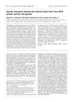
CD spectrum of rMrcp-20k in a 10 mm sodium phos-
phate buffer (pH 6.8) was also identical to that of
nMrcp-20k; both showed the presence of a mixture of
b-turn and random coil structures [9,10]. These spectra
were remarkably different from that observed after a
reducing treatment, probably due to denaturation of
the protein (Fig. 3).
Adsorption of rMrcp-20k to underwater material
surfaces
The adsorption of rMrcp-20k to several underwater
material surfaces was investigated, and the findings
are summarized in Fig. 4. The protein was adsorbed
to calcite in artificial seawater (ASW), whereas it was
not adsorbed to glass, gold, polystyrene, or benzo-
guanamine-formaldehyde resin, which is a positively
charged synthetic polymer. The protein was also
adsorbed to a limited extent to metal oxides such as
zinc oxide and magnetite. The amount adsorbed to
calcite in pure water was almost the same as that in
ASW.
A
B
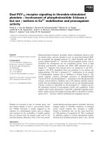
Fig. 2. Purification of rMrcp-20k. (A) Samples were separated by
using the 16.5% T Tris ⁄ Tricine buffer system of SDS ⁄ PAGE [30].
Lane 2: crude extract of bacterial cells. Lane 3: rMrcp-20k fused
with a tag in the vector construct. Lane 4: rMrcp-20k. Lane 1, low
molecular mass markers (Bio-Rad; aldolase, 45.0 kDa; carbonic
anhydrase, 31.0 kDa; soybean trypsin inhibitor, 21.5 kDa; lysozyme,
14.4 kDa). (B) SDS ⁄ PAGE of rMrcp-20k with (left) and without
(right) pretreatment with the reducing agent 2-mercaptoethanol.
A
B
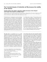
Fig. 1. Characterization of Mr cp-20k in the primary cement. (A)
Western blotting of fractions rendered soluble from the primary
cement by using the antibody to Mrcp-20k. Lane 1: GSF1 with
reduction pretreatment in SDS ⁄ PAGE. Lane 2: GSF2 with reduction
pretreatment. Lane 3: GSF1 without reduction pretreatment. Num-
bers on the left-hand side indicate molecular masses (kDa). (B)
Detection of Mrcp-20k in the peripheral shell of the barnacle by
using the antibody to Mrcp-20k. Two grams each (dry weight) of
the peripheral shell and calcareous base were decalcified and
subjected to dot-blotting. Lane 1: 2% acetic acid solution–soluble
fraction of the peripheral shell. Lane 2: GSF1 and GSF2 of the
peripheral shell. Lane 3: 2% acetic acid solution–soluble fraction of
the base. Lane 4: GSF1 and GSF2 of the base. Lane 5: rMrcp-20k
as positive control (1 lg). Lane 6: trypsin inhibitor from soybean as
negative control (1 lg; Wako Pure Chemical Industries).
Calcite-coupling protein in underwater adhesive Y. Mori et al.
6438 FEBS Journal 274 (2007) 6436–6446 ª 2007 The Authors Journal compilation ª 2007 FEBS
The relationship between the concentration of the
protein at the calcite surface and its solution concen-
tration is described by the adsorption isotherm. The
linearized forms of the isotherm for the adsorption to
calcite were C
eq
⁄ Q ¼ 0.3168 · 10
)3
+ 4.199C
eq
[corre-
lation coefficient (r
2
) of 0.97] in ASW and
C
eq
⁄ Q ¼ 1.7168 · 10
)3
+ 3.782C
eq
(r
2
of 0.98) in the
dilute buffer [C
eq
, equilibrium protein concentration;
Q, amount of absorbed protein (lmol) per m
2
of the
surface] (Fig. 5). The slope and intercept of the result-
ing lines enabled us to estimate the adsorption affinity
(K) and the maximum number of adsorption sites (N)
to be K ¼ 1.33 · 10
7
m
)1
and N ¼ 2.38 · 10
)7
molÆm
)2
in ASW, and K ¼ 2.20 · 10
6
m
)1
and N ¼
2.64 · 10
)7
molÆm
)2
in the dilute buffer solution. The
isotherms for adsorption to zinc oxide and magnetite
were not linear (r
2
of 0.75 and 0.58, respectively), so
that the adsorption to these surfaces seemed not to be
of the typical Langmuir type (supplementary Fig. S2).
The adsorption of rMrcp-20k to the barnacle shell
was visualized using the antibody to rMrcp-20k with
the secondary antibody conjugated by fluorochrome
(Fig. 6 and supplementary Fig. S3). A 10 min incuba-
tion with rMrcp-20k in ASW gave rise to fluorescence
emission at the barnacle shell, demonstrating the
Wavelength (nm)
[θ] (deg cm
-2
dmol
-1
)
200
-30
-20
-10
0
10
[θ] (deg cm
-2
dmol
-1
)
-30
-20
-10
0
10
[θ] (deg cm
-2
dmol
-1
)
-30
-20
-10
0
10
A
B
C
250 300 320
Wavelength (nm)
200 250 300 320
Wavelength (nm)
200
250 300 320
Fig. 3. Comparison of the CD spectra of rMrcp-20k and nMrcp-
20k. The spectra are shown of (A) rMrcp-20k, (B) nMrcp-20k and
(C) rMrcp-20k with the reducing pretreatment.
A
amount of adsorbed protein (ng/cm
2
)
0
50
100
150
200
250
300
BCDEFGH
Fig. 4. Adsorption of rMrcp-20k to various solid surfaces. The
adsorption of rMrcp-20k to the particles of several materials in
10 min at 25 °C was evaluated by measuring the decrease in pro-
tein amount remaining in the solution. Adsorption to (A) calcite in
ASW, (B) glass in ASW, (C) benzoguanamine–formaldehyde resin
in ASW, (D) zinc oxide in ASW, (E) magnetite in ASW, (F) gold in
ASW, (G) polystyrene in ASW, and (H) calcite in pure water. Error
bars indicate the standard deviation.
C
eq
(µmol/mL)
C
eq
/Q (m
2
/mL)
-5.2E-18
0
0.01
0.02
0.03
0.04
0.05
C
eq
/Q (m
2
/mL)
0
0.01
0.02
0.03
0.04
0.05
B
A
0.002 0.004 0.006 0.008 0.01
C
eq
(µmol/mL)
-2.08E-1 0.002 0.004 0.006 0.008 0.01
Fig. 5. Linearized adsorption isotherm for adsorption of rMrcp-20k
to calcite. (A) Isotherm in ASW. (B) Isotherm in 2.14 m
M sodium
carbonate (pH 8.2).
Y. Mori et al. Calcite-coupling protein in underwater adhesive
FEBS Journal 274 (2007) 6436–6446 ª 2007 The Authors Journal compilation ª 2007 FEBS 6439
successful adsorption of the protein to the calcareous
shell of the barnacle.
The distribution of the molecular size of
rMrcp-20k
The distribution of the molecular size of the recombi-
nant protein was evaluated by analytical ultracentrifu-
gation (Table 1).
Sedimentation velocity analyses indicated that the
protein exists as a single component in 100 mm to
500 mm NaCl solution. The sedimentation coefficient
of the component was estimated to be s $ 2.5.
The sedimentation equilibrium analyses gave nearly
20 kDa as the molecular mass in 100 mm to 1 m NaCl
solution, which is consistent with monomeric molecu-
lar mass of the protein. Therefore, the s $ 2.5 species
found by sedimentation velocity corresponds to the
monomeric form of the protein.
The possible change of intramolecular disulfide
bonds to intermolecular ones after a longer period of
incubation in ASW was evaluated by SDS ⁄ PAGE
analysis (Fig. 7). The molecular masses were mono-
meric for proteins in both the suspension and the lay-
ers adsorbed to calcite, thus confirming that there had
been no change of intramolecular disulfide bonds to
the intermolecular type in the protein.
Isolation of the homologous gene from Balanus
albicostatus
A PCR investigation of a homologous gene in three
barnacle species was attempted with several degener-
ated oligonucleotide primers based on the primary
structure of Mrcp-20k. All PCR trials with primers
designed from the primary structure of Mrcp-20k failed
to amplify homologous DNA, except for 3¢-RACE with
cDNA of Balanus albicostatus. The sequence of homo-
logous cDNA in B. albicostatus determined in this
study was 700 bp, and the coding region was deter-
mined to encode 125 amino acids (supplementary
Fig. S4). The first 20 amino acids are considered to
Fig. 6. Demonstration of the adsorption of Mrcp-20k to the barna-
cle peripheral shell. The protein adsorbed to the shell was treated
with the antibody, and visualized with the secondary antibody
linked to fluorochrome Cy3 (GE Healthcare Bio-Science). Images
under visible light (left) and those under reflected fluorescence
(right) are shown. The image pair was captured from the same
angle of the object. In the images under visible light, yellow areas
correspond to the shell, and white areas are transparent without
any object. Shell was incubated with rMrcp-20k, washed, and trea-
ted with the antibody to Mrcp-20k. No fluorescence was observed
in the control experiment (supplementary Fig. S3).
Table 1. The distribution of the molecular size of rMrcp-20k evalu-
ated by analytical ultracentrifugation. The sedimentation coefficients
and molecular masses of rMrcp-20k in several solvents were evalu-
ated by sedimentation velocity and sedimentation equilibrium,
respectively. Sedimentation coefficients were evaluated by sedi-
mentation velocity analyses and standardized with the
SEDNTERP pro-
gram [29]. Molecular masses were determined by sedimentation
equilibrium analyses.
NaCl concentration (
M)
s
20, W
(S)
Molecular
mass (kDa)
0.1 2.6 19.6
0.3 2.5 18.9
0.4 2.5 –
0.5 2.4 –
1.0 – 21.1
Fig. 7. Rearrangement of disulfide bonds in rMrcp-20k during long-
term incubation. The molecular masses of rMrcp-20k after several
treatments for 1 week at 25 °C were estimated by western blotting
with the antibody to Mrcp-20k antibody. rMrcp-20k was incubated
in ASW adjusted to pH 8.0 without calcite particles (lane 1), in a
dilute buffer adjusted to pH 8.0 without calcite particles (lane 2), or
in ASW with calcite particles (lane 3).
Calcite-coupling protein in underwater adhesive Y. Mori et al.
6440 FEBS Journal 274 (2007) 6436–6446 ª 2007 The Authors Journal compilation ª 2007 FEBS
correspond to the signal peptide, because of its high
hydrophobicity and the existence of a predicted signal
peptidase cleavage position [11]. The molecular mass
and isoelectric point of the mature polypeptide were
predicted to be 12 297.0 Da and 8.3, respectively,
assuming that all Cys residues were in the disulfide
form for prediction of the molecular mass. The amino
acid composition deduced from the cDNA indicated
that charged amino acids such as His (20%), Lys
(10%) and Cys (17%) are the dominant residues; the
contents of these residues appear to be significantly
higher than in the standard amino acid composition
[12]. The charged amino acids Asp, Glu, His, Lys and
Arg are estimated to comprise 42% of the total resi-
dues. Alignment of the Cys residues indicated that the
primary structure of the homologous protein in B. albi-
costatus consists of four repeated sequences (Fig. 8).
The difference between the B. albicostatus protein and
Mrcp-20k in their amino acid lengths depended on the
difference in the number of repeats. The similar Cys
spacing, the existence of Pro preceding the second
Cys, the presence of two amino acids after the second
Cys, and the sporadic insertion of clusters of charged
amino acids such as HKHHDHGK, HHHDD,
RHGKKH and HRKFH, are common characteristics
found in both proteins [4]. A BLAST search [13] of the
nonredundant database and a sequence profile-based
fold-recognition method for three-dimensional struc-
tural prediction [14] failed to provide any homologous
sequences and meaningful structure from currently
available databases.
Discussion
Although Mrcp-20k was found in the secondary cement
in the previous study, neither the presence of this
protein in the barnacle natural adhesive layer or pri-
mary cement, nor its specific function in underwater
attachment, has been characterized so far. The present
study was thus conducted to address these questions.
The conditions required for extracting the protein from
the insoluble primary cement, and its behavior in the
SDS ⁄ PAGE analysis, were similar to those of the pro-
tein from the secondary cement. The protein exhibited
a monomeric molecular mass on SDS ⁄ PAGE even
without a reducing pretreatment, a characteristic also
found for the protein from the secondary cement. The
amino acid composition of Mrcp-20k is characterized
by the unusually high contents of Cys (17%) and
charged amino acid residues [4], which suggests a possi-
ble role of polymerization via intermolecular disulfide
bonds for this protein in the process of underwater
adhesion [5]. The present study, however, excluded this
possibility. This was further supported by the fact that
long-term incubation of the bacterial recombinant pro-
tein in ASW did not give rise to any polymerized
molecular species by the conversion of disulfide bonds
to the intermolecular form. The abundance of Cys and
charged amino acid residues is reminiscent of proteins
involved in biomineralization. As the cement has
always been collected from the surface of the barnacle
calcareous base, some contamination of the proteins
used for calcification may have occurred. However, the
fact that Mrcp-20k could not be detected in the periph-
eral calcareous shell indicates that the protein is specific
to underwater attachment of the base, and does not
contribute to the calcification process. The protein con-
tains few hydrophobic residues, which would result in a
poor hydrophobic core in the structure; this may be a
reason for the introduction of abundant intramolecular
disulfide bonds to stabilize the structure in molecular
evolution. This was confirmed by the marked change in
the CD spectrum with the reducing treatment. The
limited number of hydrophobic residues may, in
turn, suggest the significance of the charged amino
acid residues in the function of the protein.
Mrcp-20k is a simple protein bearing no post-transla-
tional modifications [4]. This allowed us to express this
protein in bacteria under physiological conditions, and
to compare the characteristics of the recombinant pro-
tein with those of the native protein extracted with pure
water. Both proteins showed the same elution profiles
in column chromatography, the same behaviors as
analyzed by SDS ⁄ PAGE, MALDI-TOF MS and CD
spectra, and similar resistance to alkylation treatment
without any reducing treatment, indicating that both
proteins possessed similar molecular structures. We
therefore characterized the functional properties of the
recombinant protein. This is an unusual case in biotic
underwater adhesive studies, as all mussel foot proteins
(fps), which represent another model system, are
Fig. 8. Alignment of the repetitive sequences in Mr cp-20k and the
homologous protein in B. albicostatus. All Cys residues are shown
in black, and conserved Pro residues are shown in gray.
Y. Mori et al. Calcite-coupling protein in underwater adhesive
FEBS Journal 274 (2007) 6436–6446 ª 2007 The Authors Journal compilation ª 2007 FEBS 6441
subjected to heavy post-translational modifications [15],
so that the native activity of the simple recombinant
protein cannot be obtained. The present study repre-
sents the first report based on a recombinant protein
retaining almost the same structure as that of the native
protein in the study of biotic underwater adhesive.
The protein was adsorbed to calcite, a crystalline
form of calcium carbonate, but not to glass and syn-
thetic polymers. The isotherm for adsorption of the
recombinant protein to calcite followed the Langmuir
model, which has been extensively applied to the quan-
titative evaluation of the interaction between macro-
molecules and mineral interfaces [16]. Although the
protein was also adsorbed to some metal oxides to a
limited extent, this adsorption isotherm did not fit the
Langmuir model. These results suggest that the adsorp-
tion to calcite is a specific function of Mrcp-20k. This
may not be surprising if we consider that half of the
material to be attached is the organism’s own calcare-
ous base. The barnacle also prefers to attach itself to
the peripheral calcite shell of another barnacle, because
of the gregarious behavior of this species. It therefore
seems that the barnacle arranges a specific protein in
the cement to be adsorbed to the most typical target,
calcite, although it is not clear whether the target of the
protein is specific to the organism’s own base or the
foreign calcified shell, or both.
The adsorption isotherm for the attachment of
rMrcp-20k to calcite determined in the present study
indicated that the protein has an affinity for calcite that
is one magnitude of order higher than that of the ame-
logenin–hydroxyapatite interaction, whose adsorption
affinity was 1.97 · 10
6
m
)1
[17]. The calculated pI value
for rMrcp-20k is 4.7. The points for zero charge of
calcite and glass are 9.50 ± 0.50 [18] and 1.80, respec-
tively [19], so they are expected to possess positive and
negative net charges in seawater (pH 7.8–8.0). This may
suggest a simple electrostatic interaction between the
protein and calcite. However, the protein was not
adsorbed to a positively charged synthetic polymer in
seawater. Thus, the adsorption of rMrcp-20k to calcite
cannot be explained simply by the electrostatic inter-
action, and probably depends on the particular arrange-
ment of surface amino acids in the protein structure.
Comparison between the sequences of the gene from
M. rosa and a homologous gene from B. albicostatus
suggests that the abundance of charged amino acids
and Cys residues, and the repetitive primary structure,
are common features of this protein, whereas the num-
ber of repeated sequences was different between differ-
ent species. This may indicate that the characteristics
of the protein found in this study can also be applied
to the cp-20k protein in other barnacle cements.
The holdfast system of the barnacle showed no simi-
larity to that of the mussel, which is relatively well
characterized. There were no sequence similarities
among the protein components between the two
systems. The mussel holdfast system [15] depends
on several protein modifications, typically including
3,4-dihydroxyphenylalanine; however, no involvement
of 3,4-dihydroxyphenylalanine in the barnacle cement
was found [2]. The mussel attaches to an underwater
foreign substratum using a byssal thread as its hold-
fast. The tip of the byssus, called the disk, directly
attaches to the substratum. At least two proteins, fp-3
and fp-5, have been identified as surface-coupling
proteins of this disk [20]. Phosphorylation of the Ser
residues in fp-5 has prompted the suggestion that cal-
careous material-specific coupling is its functional role
[21]. There is a huge quantity of calcareous material in
the marine environment. Both the barnacle and mus-
sel, at least, seem to provide a specific coupling protein
for this frequently encountered material. They have
acquired distinct molecular features in the course of
evolution: the dependence on common amino acids
with a rigid three-dimensional structure in the barna-
cle, and the dependence on the function of the amino
acid side chains with post-translational modifications
in the mussel [15,22]. Moreover, Mrcp-20k may not be
covalently linked to other bulk proteins in the barnacle
cement; this is also different from the case in the mus-
sel, whose surface proteins seem to be covalently
linked to other bulk proteins in the disk [23].
Experimental procedures
Chemicals
The chemicals used were of the highest grade available and
purchased from Wako Pure Chemical Industries (Osaka,
Japan). ASW was prepared by dissolving Marine Art SF
(Senju Seiyaku Co., Osaka, Japan) in ultrapure water that
had been ultrafiltered through an MW3000-cutoff mem-
brane (YM3; Amicon-Millipore, Billerica, MA, USA).
Preparation of the cement samples
Specimens of M. rosa attached to a polyethylene substra-
tum were collected from Ryou-ishi Bay (Iwate, Japan). The
secondary cement was collected as previously reported [3].
The primary cement was prepared from animals that had
been carefully dislodged from the substratum by applying
vibration, only those specimens without any apparent dam-
age being used. The inner soft bodies were physically
removed and cleaned. The calcareous base and peripheral
shell were separately recovered, and each of them was
Calcite-coupling protein in underwater adhesive Y. Mori et al.
6442 FEBS Journal 274 (2007) 6436–6446 ª 2007 The Authors Journal compilation ª 2007 FEBS
weighed and decalcified by dialyzing against 2% (v ⁄ v) ace-
tic acid at 4 °C. The supernatant was recovered as the ace-
tic acid-soluble fraction, and the precipitate was rendered
soluble as previously reported [3]. Briefly, the cement was
suspended in a solution of 7 m guanidine hydrochloride
and 10 mm Hepes at pH 7 and 60 °C for 1 h; the superna-
tant of this corresponded to GSF1. The precipitate was ren-
dered soluble by reduction in a solution of 0.5 m
dithiothreitol, 7 m guanidine hydrochloride, 20 mm EDTA
and 1.5 m Tris at pH 8.5 and 60 °C for 1 h in a nitrogen
atmosphere; the supernatant was recovered as GSF2. Both
fractions were dialyzed against 5% (v ⁄ v) acetic acid at 4 °C
and then stored at ) 20 °C until needed. The protein in the
secondary cement was partially extracted even in water.
Therefore, nMrcp-20k was prepared by suspending the
cement in ultrapure water and agitating overnight at 4 °C.
The extract was recovered by centrifugation (21 600 g,
4 °C, 15 min), applied to a Mono-Q 5 ⁄ 50GL column (GE
Healthcare Bio-Sciences Corp., Piscataway, NJ, USA) that
had been equilibrated with 50 mm Tris ⁄ HCl at pH 7.4, and
eluted with 50 mm Tris ⁄ HCl at pH 7.4 with a 30 min linear
gradient of 1 m NaCl from 30% to 50%.
Preparation of rMrcp-20k
The Mrcp-20k recombinant system was constructed in bacte-
rial cells. cDNA encoding mature Mrcp-20k was first ampli-
fied by PCR with M. rosa cDNA [3] and Ex Taq (Takara
Bio, Shiga, Japan) as the template and enzyme, respectively.
The following oligo-DNA primers were designed from both
the N-terminal and C-terminal regions of mature Mrcp-20k
to create the NcoI and BamHI restriction sites, respectively:
5¢-AGTTG
CCATGGCGCACGAGGAGGA-3¢ and 5¢-TT
CTGTTC
GGATCCCAAGGCTTA-3¢. The amplified DNA
fragment was digested with both NcoI and BamHI, before
being inserted into pET32a (Novagen, Darmstadt, Germany)
with the same restriction sites. The sequence of the insert was
confirmed by using a Prism Dye Deoxy sequencing kit and
3700-DNA analyzer (Applied Biosystems, Foster City, CA,
USA). The resulting plasmid was transformed into E. coli
OrigamiB (DE3) (Novagen). The transformant was culti-
vated in a modified M9 medium [24] with 50 lgÆmL
)1
carben-
icillin and 0.75% (w ⁄ v) glucose at 37 °C for 16 h to reach
the mid-log phase with an attenuance of 0.6–0.9 at 600 nm.
Isopropyl thio-b-d-galactoside (0.4 mm) and 0.75% glucose
were added to the medium, and the cells were cultivated
at 30 °C for 6 h. A crude protein extract was prepared by
sonication in 100 mm Tris ⁄ HCl at pH 9.0 on ice, and
the supernatant was purified in an Ni-immobilized column
(Novagen) with the standard protocol. The protein was
eluted with 2 m imidazole, 500 mm NaCl and 50 mm
Tris ⁄ HCl at pH 7.9. The rMrcp-20k was dialyzed against a
buffer for enterokinase digestion, concentrated with Centri-
prep (Amicon-Millipore), and treated with recombinant
enterokinase [Novagen; enzyme ⁄ substrate ratio of 1 : 10
(molar ratio)] at 20 °C for 3 days. Final purification was car-
ried out in the Mono-Q 5 ⁄ 50GL column as already
described. The protein concentration was measured with a
bicinchonic acid protein assay kit (Pierce, Rockford, IL,
USA), with BSA used as a reference [25].
Immunochemical detection of Mrcp-20k
The recombinant C-terminal 79 amino acid region was pre-
pared as an antigen with a method similar to that used for
the whole length protein, except that the vector used was
pET30a (Novagen), and a 3.9 mm diameter · 150 mm
l-Bondasphere RP-HPLC column (C8, 300 A
˚
; Waters,
Milford, MA, USA) was used for the purification. For
PCR amplification of the C-terminal 79 amino acid region,
the following oligo-DNA primers were used: 5¢-AATGTA
CCATGGAAGCGCCGT-3¢ and 5¢-GCCTTCTGTTCGG
ATCCCAAGGCT-3¢. The polyclonal antibody was raised
in rabbits by serial subcutaneous injection (Takara Bio).
Immunochemical detection was carried out by dot-blotting
or electrotransfer to a nitrocellulose membrane (0.45 lm;
Bio-Rad, Hercules, CA, USA). Poly(vinylidene difluoride)
was not suitable for holding Mrcp-20k in our several trials,
probably due to the abnormal characteristics of this pro-
tein. A goat anti-rabbit IgG (H + L) horseradish peroxi-
dase (HRP) conjugate (Bio-Rad) was used as the secondary
antibody, and HRP-100 immunostaining (Konica-Minolta,
Tokyo, Japan) was used to develop the signal.
Characterization of rMrcp-20k
The N-terminal sequence of the recombinant protein was
confirmed with a protein sequencer (Procise 494 cLC;
Applied Biosystems), and the molecular mass was con-
firmed with MALDI-TOF MS. The sample was mixed with
synapic acid saturated in 30% (v ⁄ v) acetonitrile and then
analyzed with a Voyager-DE STR instrument (Applied
Biosystems, Foster City, CA, USA), using Calibration
Mixture 3 (Applied Biosystems) as the reference. The Lae-
mmli buffer system [26] was used for SDS ⁄ PAGE analysis.
The alkylation treatment of the protein was carried out as
described in a previous study [4]. A 5 lm amount of
rMrcp-20k was suspended in a solution of 7 m guanidine
hydrochloride, 20 mm EDTA and 1.5 m Tris ⁄ HCl at
pH 8.0. Monoiodo acetic acid (Wako Pure Chemical Indus-
tries) was then added to an amount 500 times the number
of cysteine residues in rMrcp-20k, and the mixture was
incubated in a nitrogen atmosphere in the dark at room
temperature for 2 h. The reaction mixture was purified by
RP-HPLC and then subjected to MALDI-TOF MS analy-
sis. The CD spectra of the protein (32 lgÆmL
)1
, dissolved
in 10 mm sodium phosphate at pH 6.8) were measured with
a J-725 spectropolarimeter (Jasco, Tokyo, Japan). The spec-
tra were scanned at 20 ° C from 200 nm to 320 nm, and
then integrated 128 times. Prior to the analysis, a reduction
Y. Mori et al. Calcite-coupling protein in underwater adhesive
FEBS Journal 274 (2007) 6436–6446 ª 2007 The Authors Journal compilation ª 2007 FEBS 6443
treatment was carried out with 100 mm dithiothrei-
tol ⁄ 10 mm sodium phosphate at pH 6.8 and 25 °C for 1 h,
with subsequent dialysis against 100 mm NaCl and 10 mm
sodium phosphate at pH 6.8.
Measurement of adsorption to underwater
material surfaces
The protein adsorption to underwater materials was mea-
sured by quantifying the protein amount in soluble fractions
after incubating with defined particles. Neither the adsorp-
tion of rMrcp-20k to a polypropylene tube nor any precipi-
tate formation was apparent. Thus, a polypropylene tube
was used to handle the protein solution. The particles used in
this study were as follows: calcite (2500 cm
2
surface areaÆg
)1
,
8 lm in diameter; Sankyo Seihun Co., Okayama, Japan),
glass (50 lm in diameter; Toshinriko Co., Tokyo, Japan),
benzoguanamine–formaldehyde resin (3000 cm
2
surface
areaÆg
)1
, 12.75 lm in diameter; Nippon Shokubai Co.,
Tokyo, Japan), zinc oxide (20 000 cm
2
surface areaÆg
)1
;
0.70 lm in diameter; Mitsui Mining and Smelting Co.,
Tokyo, Japan), magnetite (20 000 cm
2
surface areaÆg
)1
; Toda
Kogyo Co., Hiroshima, Japan), gold-coated polystyrene
(5.0 lm in diameter; Sekisui Chemical Co., Osaka, Japan),
and polystyrene (5.0 lm in diameter; Duke Scientific Corpo-
ration, Fremont, CA, USA). Each type of particle was
suspended in 20 lL of two-fold concentrated ASW or in
ultrapure water in a polypropylene tube and then incubated
at 25 °C for 10 min. The same volume of protein
(0.30 mgÆmL
)1
, dissolved in ultrapure water) was preincu-
bated at 25 °C, mixed with each type of particle, and incu-
bated at 25 °C for 10 min to allow adsorption. A 10 lL
aliquot of the supernatant was recovered by centrifugation,
and the protein concentration was measured using a bicinch-
oninic acid protein assay kit (Pierce) with an ‘enhanced pro-
tocol’ according to the manufacturer’s specifications. The
incubation time for adsorption was confirmed to be sufficient
for maximum adsorption in a preliminary experiment.
The adsorption affinity was determined by incubating
various concentrations of the protein with each type of par-
ticles (total surface area, 12.5 cm
2
each) in ASW, and then
evaluating the amount of free protein as described above
(N ¼ 3). Calibration curves were constructed as reported
elsewhere [17]. The amount of adsorbed protein (lmol) per
m
2
of the surface was calculated by the difference between
the initial (C
I
) and equilibrium (C
eq
) protein concentration
(lmolÆmL
)1
) according to the following equation:
Q ¼½ðC
I
À C
eq
ÞV=ðWSÞð1Þ
where V is the volume of the solution (0.04 mL), W is the
mass of the adsorbent, and S is the specific surface area of
the adsorbent. The amount of adsorbed protein reached a
plateau under the experimental conditions used. This type
of the isotherm can be described by the Langmuir model
with the following equation:
C
eq
=Q ¼ 1=NK þ C
eq
=N ð2Þ
where N is the maximum number of adsorption sites per
unit of surface area (molÆm
)2
) of the adsorbent, and K is
the affinity of the adsorbent molecules (LÆmol
)1
) for the
adsorption sites.
The protein adsorption to the barnacle peripheral shell
was visualized after removing the soft inner body of the
animal from the peripheral shell and physically cleaning it.
A10lL amount of rMrcp-20k (0.1 mgÆmL
)1
) in ASW was
dropped on to the outer surface of the peripheral shell.
After incubation at room temperature for 10 min, the shell
was immersed in ASW three times for 10 min each and
subjected to immunochemical detection with Cy3-labeled
anti-rabbit IgG (GE Healthcare Bio-Science Corp.) and
fluorescence microscopy.
Analyses to evaluate the distribution of the
molecular size
An Optima XL-I (Beckman Coulter Inc., Fullerton, CA,
USA) analytical ultracentrifuge with an AN60-Ti rotor was
used in all investigations. Sedimentation velocity experi-
ments at 20 °C were conducted at 42 000 r.p.m. The sample
cells were double sector charcoal-filled centerpieces equipped
with quartz windows. Concentration distributions were
acquired by scanning at 215 nm. Protein samples were dia-
lyzed against 20 mm NaCl solution, mixed with concentrated
NaCl solution in the cell, to form appropriate solutions.
The dcdt program in Beckman XLI data analysis soft-
ware was used to analyze groups of boundaries to derive
sedimentation coefficients. This method is based on the
time-derivative method developed by Stafford [27], which
fits Gaussian functions to the so-called g(s*) distribution
from the time derivative of the concentration distributions
(dc ⁄ dt), and the sedimentation coefficient was calculated on
the basis of the positions of Gaussian fits to the g(s*) ver-
sus s data. Results were confirmed by the method of Van
Holde & Weischet [28].
The sedimentation coefficient was corrected to standard
solvent conditions (the viscosity, and the density of water
at 20 °C) using the same program.
The sedimentation equilibrium runs were performed for
15 h before equilibrium absorbance measurements were
taken at 215 nm. Protein solutions at three concentrations
ranging from 12 to 22 lgÆmL
)1
in NaCl solution were cen-
trifuged at 21 000 r.p.m. at 20 °C. Molecular weights were
obtained using Beckman XLI data analysis software, in
which radial position versus absorbance data were fitted to
the following equilibrium equation using nonlinear least-
squares techniques:
AðrÞ¼A
0
ðr
0
Þ exp½HM
app
ðr
2
À r
2
0
Þ þ B ð3Þ
where H ¼ (1 ) mq)x
2
⁄ 2RT, m is partial specific volume of
sample, q is density of solvent, R is gas constant, T is
Calcite-coupling protein in underwater adhesive Y. Mori et al.
6444 FEBS Journal 274 (2007) 6436–6446 ª 2007 The Authors Journal compilation ª 2007 FEBS
temperature, x is angular velocity, A
0
is absorbance at a ref-
erence point r
0
, A(r) is absorbance at a position r cm from
the rotor center, and B is baseline correction. In this study,
the m of rMrcp-20k (0.6804 mLÆg
)1
) and q of the solvents
were calculated from the amino acid composition and solvent
composition, respectively, using the program sednterp [29].
In order to confirm whether or not intramolecular disul-
fide bonds were rearranged to intermolecular ones, rMrcp-
20k (0.1 mgÆmL
)1
)in10mm Tris ⁄ HCl (pH 8.0) or two-fold
concentrated ASW with 10 mm Tris ⁄ HCl (pH 8.0) were
incubated at 25 °C for 7 days, dialyzed against 10 mm
Tris ⁄ HCl at pH 6.8, separated on SDS ⁄ PAGE (15% T)
without any reduction treatment, and visualized by western
blotting with the rMrcp-20k-C antibody. Confirmation in
the adsorbent was carried out in a similar manner. rMrcp-
20k (0.1 mgÆmL
)1
) was incubated in ASW with 60 mg of
calcite particles at 25 °C for 7 days. After centrifuging
(21 600 g,25°C, 15 min) and washing with ASW, the par-
ticles were dialyzed against 5% (v ⁄ v) acetic acid to decal-
cify them and to release the adsorbed protein into solution.
The protein was then analyzed as described above after
evaporation.
PCR investigation of the gene homologous to
that encoding Mrcp-20k
B. albicostatus and Balanus amphitrite were collected from
Shimizu Bay (Shizuoka, Japan), and Balanus rostratus was
collected from Asamushi Bay (Aomori, Japan). RNA and
DNA manipulations were performed as previously
described [4]. 3¢-RACE was carried out with a degenerated
primer designed from the consensus sequence of the repeti-
tive sequences in Mrcp-20k by using a 3¢-RACE core kit
(Takara Bio). The degenerated primer used was 5¢-
CTG
ATCTAGAGGTACCGGATCCTGYAACGANGAKCAY
CCTG-3¢, where the underlining corresponds to the three-
site adaptor region of the kit. A 336 bp DNA fragment
was amplified only from B. albicostatus cDNA. Subsequent
5¢-RACE was carried out using a 5¢-RACE core kit (Taka-
ra Bio). The 5¢-RACE primers used were as follows:
5¢-(pG-TG CCA GCA CCG GTG G)-3¢ for reverse tran-
scription; 5¢-(AAA CAG TAA GGC CAG CGT AT)-3¢
and 5¢-(GCA TCA TGA TCA CGG AAA GA)-3¢ for the
first PCR amplification; and 5¢-(TGA TGG CAA TGT
GAT GTT GA)-3¢ and 5¢-(TGC TAC CAC TGC CAC
ACC GA)-3¢ for the second PCR amplification. The coding
region was finally confirmed by PCR amplification with the
primers 5 ¢-(CAA CAC TTC TGT GCT C)-3¢ and 5¢-(GGC
GTT CTC TCA GCC G)-3¢.
Acknowledgements
We thank Professor T. Watanabe of Niigata Univer-
sity and Dr T. Shimoyama for their advice on the
kinetic analysis and assistance with fluorescence
microscopy observations. We also thank Dr S. Kanai
and Ms N. Inoue of PharmaDesign, Inc., Japan for
bio-informatic analyses. Special thanks are given to
Professor J R. Shen of Okayama University for his
critical reading of this manuscript. Calcite, benzoguan-
amine–formaldehyde resin, zinc oxide, magnetite and
gold-coated particles were kindly provided by Sankyo
Seihun Co. Ltd, Nippon Shokubai Co. Ltd, Mitsui
Mining and Smelting Co. Ltd, Toda Kogyo Co. Ltd,
and Sekisui Chemical Co. Ltd, respectively. This work
was performed as part of an industrial science and
technology project entitled Technological Development
for Biomaterials Design Based on Self-organizing Pro-
teins, supported by the New Energy and Industrial
Technology Development Organization (NEDO).
References
1 Saroyan JR, Linder E, Dooley CA & Bleile HR (1970)
Repair and reattachment in the Balanidae as related to
their cementing mechanism. Ind Eng Chem Prod Res
Dev 9, 122–133.
2 Kamino K (2006) Barnacle underwater attachment. In
Biological Adhesives (Smith AM & Callow JA, eds),
pp. 145–166. Springer-Verlag, Berlin.
3 Kamino K, Inoue K, Maruyama T, Takamatsu N,
Harayama S & Shizuri Y (2000) Barnacle cement
proteins. J Biol Chem 275, 27360–27365.
4 Kamino K (2001) Novel barnacle underwater adhesive
protein is a charged amino acid-rich protein constituted
by a Cys-rich repetitive sequence. Biochem J 356, 503–507.
5 Weigemann M & Watermann B (2003) Peculiarities
of barnacle adhesive cured on non-stick surfaces.
J Adhesion Sci Technol 17, 1957–1977.
6 Waite JH (1987) Nature’s underwater adhesive special-
ist. Int J Adhes 7, 9–14.
7 Naldrett MJ (1993) The importance of sulphur cross-
links and hydrophobic interactions in the polymerization
of barnacle cement. J Mar Bio Assoc UK 73, 689–702.
8 Dougherty WJ (1990) Barnacle adhesion: reattachment
of the adult barnacle Chthamalus fragilis Darwin to
polystyrene surfaces followed by centrifugational shear-
ing,. J Crustacean Biol 10, 469–478.
9 Greenfield N & Fasman GD (1969) Computed circular
dichroism spectra for the evaluation of protein confor-
mation. Biochemistry 8, 4108–4116.
10 Brahms S & Brahms J (1980) Determination of protein
secondary structure in solution by vacuum ultraviolet
circular dichroism. J Mol Biol 138, 149–178.
11 von Heijne G (1986) A new method for predicting
signal sequence cleavage sites. Nucleic Acids Res 14,
4683–4690.
Y. Mori et al. Calcite-coupling protein in underwater adhesive
FEBS Journal 274 (2007) 6436–6446 ª 2007 The Authors Journal compilation ª 2007 FEBS 6445
12 Jones DT, Taylor WR & Thornton JM (1992) The
rapid generation of mutation data matrices from protein
sequences. J CABIOS 8, 275–282.
13 Altschul SF, Gish W, Miller W, Myers EW & Lipman
DJ (1990) Basic local alignment search tool. J Mol Biol
215, 403–410.
14 McGuffin LJ & Jones DT (2003) Improvement of the
GenTHREADER method for genomic fold recognition.
Bioinformatics 19, 874–881.
15 Sagert J, Sun C & Waite JH (2006) Chemical subtleties
of mussel and polychaete holdfasts. In Biological Adhe-
sives (Smith AM & Callow JA, eds), pp. 125–140.
Springer-Verlag, Berlin.
16 Wallwork M, Kirkham J, Zhang J, Brookes S, Shore R,
Wood S, Ryu O, Robinson C & Smith DA (2001) Bind-
ing of matrix proteins to developing enamel crystals: an
atomic force microscopy study,. Langmuir 17, 2508–
2513.
17 Bouropoulos N & Moradian-Oldak J (2003) Analysis of
hydroxyapatite surface coverage by amelogenin nano-
spheres following the Langmuir model for protein
adsorption. Calcif Tissue Int 72, 599–603.
18 Huang CP (1975) Adsorption of tryptophan onto cal-
cium carbonate surface. Environ Lett 9, 7–17.
19 Lokar WJ & Ducker WA (2004) Proximal adsorption at
glass surfaces: ionic strength, pH, chain length effects,.
Langmuir 20, 378–388.
20 Zhao H, Robertson NB, Jewhurst SA & Waite JH
(2006) Probing the adhesive footproteins of Mytilus cali-
fornianus byssus. J Biol Chem 281, 11090–11096.
21 Waite JH & Qin XX (2001) Polyphosphoprotein from
the adhesive pads of Mytilus edulis. Biochemistry 40,
2887–2893.
22 Lin Q, Gourdon D, Sun C, Holten-Anderson T, Waite
JH & Israelachvili JN (2007) Adhesion mechanisms of
the mussel foot proteins mfp-1 and mfp-3. Proc Natl
Acad Sci USA 104, 3782–3786.
23 Zhao H & Waite JH (2006) Linking adhesive and struc-
tural proteins in the attachment plaque of Mytilus cali-
fornianus. J Biol Chem 281, 26150–26158.
24 Cai M, Huang Y, Sakaguchi K, Clore GM, Gronen-
born AM & Craigie R (1998) An efficient and cost-
effective isotope labeling protocol for protein expressed
in Escherichia coli. J Biomol NMR 11, 97–102.
25 Smith PK, Krohn RI, Hermanson GT, Mallia AK,
Gartner FH, Provenzano MD, Fujimoto EK, Goeke
NM, Olson BJ & Klenk DC (1985) Measurement of pro-
tein using bicinchroninic acid. Anal Biochem 150 , 76–85.
26 Laemmli UK (1970) Cleavage of structural proteins
during the assembly of the head of bacteriophage T4.
Nature 227, 680–685.
27 Stafford WF (1992) Boundary analysis in sedimentation
transport experiments: a procedure for obtaining sedi-
mentation coefficient distributions using the time deriva-
tive of the concentration profile. Anal Biochem 203,
478–501.
28 Van Holde KE & Weischet WO (1978) Boundary analy-
sis of sedimentation velocity experiments with monodis-
perse and paucidisperse solutes. Biopolymers 72, 1287–
1403.
29 Laue TM, Shah BD, Ridgeway TM & Pelletier SL
(1992) In Analytical Ultracentrifugation in Biochemistry
and Polymer Science (Harding AJR & Horton JC, eds),
pp. 90–125. Royal Society of Chemistry, Cambridge.
30 Scha
¨
ger H & Jagow G (1987) Tricine–sodium dodecyl
sulfate-polyacrylamide gel electrophorcsis for the sepa-
ration of proteins in the range from 1 to 100 kDa. Anal
Biochem 166, 368–379.
Supplementary material
The following supplementary material is available
online:
Fig. S1. Comparison of rMrcp-20k and nMrcp-20k by
liquid chromatography.
Fig. S2. Adsorption isotherm for adsorption of rMrcp-
20k to a metal oxide surface.
Fig. S3. Control for Fig. 5.
Fig. S4. cDNA sequence of the homologous protein in
B. albicostatus.
This material is available as part of the online article
from
Please note: Blackwell Publishing is not responsible
for the content or functionality of any supplementary
materials supplied by the authors. Any queries (other
than missing material) should be directed to the corre-
sponding author for the article.
Calcite-coupling protein in underwater adhesive Y. Mori et al.
6446 FEBS Journal 274 (2007) 6436–6446 ª 2007 The Authors Journal compilation ª 2007 FEBS