Báo cáo khoa học: The F13 residue is critical for interaction among the coat protein subunits of papaya mosaic virus doc
Bạn đang xem bản rút gọn của tài liệu. Xem và tải ngay bản đầy đủ của tài liệu tại đây (958.32 KB, 11 trang )
The F13 residue is critical for interaction among the coat
protein subunits of papaya mosaic virus
M. E. Laliberte
´
Gagne
´
1
, K. Lecours
2
, S. Gagne
´
2
and D. Leclerc
1
1 Infectious Disease Research Centre, Laval University, Que
´
bec, Canada
2 Department of Biochemistry, Laval University, Que
´
bec, Canada
Papaya mosaic virus (PapMV) is a member of the
potexvirus family. Its virion is a flexuous rod that is
500 nm long and 13 nm in diameter. A PapMV parti-
cle is composed of 1400 subunits of the coat protein
(CP) [1] assembled around a 6656 nucleotide plus
strand of genomic RNA [2]. The CP is composed of
215 amino acids and has an estimated molecular mass
of 23 kDa. Until now, most of the information
obtained regarding assembly of potexvirus family
members has been obtained from studying partially
denatured CPs extracted from purified plant virus by
the acetic acid method [3]. Even though in vitro assem-
bly using this method has been studied extensively [3–
8], the nature of the interaction among CP subunits
and genomic RNA remains unknown.
Recently, we have shown that CP expression in
Escherichia coli leads to formation of nucleocapsid-like
particles (NLPs) that are very similar to wild-type
virus purified from plants [9]. Therefore, this system is
ideal for investigating virus assembly as well as for
mapping domains of CPs involved in this process. The
recombinant NLPs, with an average length of 50 nm,
represent 20–30% of the total purified proteins. The
remaining protein is essentially found as a 450 kDa
multimer that forms a 20 subunit disk. Recombinant
disks self-assemble in vitro in the presence of RNA [9].
We also showed that the affinity of disks for RNA
was important for protein self-assembly into NLPs.
Mutated K97A disks, which cannot bind RNA, are
incapable of self-assembly. Conversely, the E128A
mutant, which shows improved affinity for RNA,
makes longer NLPs than the wild-type protein [9]. In
another study, we have shown that deletion of 26
amino acids at the N-terminus of the CP leads to a
Keywords
Nucleocapsid-like particles (NLPs); PapMV;
papaya mosaic virus; potexvirus; virus
self-assembly
Correspondence
D. Leclerc, Infectious Disease Research
Centre, Laval University, QC, Canada
Fax: +1 418 654 2715
Tel: +1 418 654 2705
E-mail:
(Received 31 August 2007, revised 6
December 2007, accepted 21 January 2008)
doi:10.1111/j.1742-4658.2008.06306.x
Papaya mosaic virus (PapMV) coat protein (CP) in Escherichia coli was
previously showed to self-assemble in nucleocapsid-like particles (NLPs)
that were similar in shape and appearance to the native virus. We have also
shown that a truncated CP missing the N-terminal 26 amino acids is mono-
meric and loses its ability to bind RNA. It is likely that the N-terminus of
the CP is important for the interaction between the subunits in self-assem-
bly into NLPs. In this work, through deletion and mutation analysis, we
have shown that the deletion of 13 amino acids is sufficient to generate the
monomeric form of the CP. Furthermore, we have shown that residue F13
is critical for self-assembly of the CP subunits into NLPs. The replacement
of F13 with hydrophobic residues (L or Y) generated mutated forms of the
CP that were able to self-assemble into NLPs. However, the replacement
of F13 by A, G, R, E or S was detrimental to the self-assembly of the pro-
tein into NLPs. We concluded that a hydrophobic interaction at the N-ter-
minus is important to ensure self-assembly of the protein into NLPs. We
also discuss the importance of F13 for assembly of other members of the
potexvirus family.
Abbreviations
CP, coat protein; EMSA, electrophoretic mobility shift assay; NLP, nucleocapsid-like particle; PapMV, papaya mosaic virus; PVBV, pepper
vain banding virus; PVX, potato virus X.
1474 FEBS Journal 275 (2008) 1474–1484 ª 2008 The Authors Journal compilation ª 2008 FEBS
monomeric form of the protein [10]. This protein failed
to assemble, form disks or interact with RNA in vitro
[10]. On the basis of this result, we hypothesized that
the N-terminus of the CP is involved in contact among
NLP subunits.
In this study, we have established precisely which of
the N-terminal 26 amino acids are important in Pap-
MV CP multimerization. We found that deletion of
only 13 amino acids was sufficient to inhibit interac-
tion among CP subunits, thus leading to a monomeric
form. We provide evidence that the F13 residue plays
a crucial role in CP subunit interaction and assembly.
Results
Expression and purification of truncated and
mutated forms of PapMV CP
Our reference recombinant proteins are CP6–215 [9]
and CP27–215 [10], which will be compared with all
mutated forms described in this article. The expression
and purification of CP6–215 and CP27–215 have been
described elsewhere [9,10]. However, here we employed
a French press instead of sonication for bacterial lysis.
We generated two truncated versions of CP13–215 and
CP14–215 (Fig. 1A), and then expressed and purified
the recombinant proteins as reported previously using
a His6 tag [9]. As expected, we observed differences in
molecular mass among CP6–215, CP13–215, CP14–215
and CP27–215 as a consequence of deletion of a few
amino acids (Fig. 1B). In addition, we introduced
single amino acid changes at F13, made substitutions
with amino acids of increasing hydrophobicity, and
generated CP6–215 F13G, F13A, F13L, and F13Y
mutants (Fig. 1A), with charged residues and gener-
ated the F13R and F13E mutants (Fig. 1C), and
finally with a polar residue and generated the F13S
mutant (Fig. 1C).
Some of the mutations and deletions appear to have
an impact on the stability of the resulting recombinant
proteins. Indeed, only 24 h after purification, recombi-
nant CP13–215, CP14–215, F13A, F13G, F13R, F13E
and F13S showed signs of degradation, and bands of
lower molecular mass proteins appeared in western
blots (Fig. 1B,C).
Characterization of recombinant NLPs
As shown before, purified CP6–215 can self-assemble
in E. coli [9], and CP27–215 was found as a mono-
meric form [10]. To monitor the capacity of the differ-
ent mutated and truncated forms to produce NLPs, we
examined purified proteins by electron microscopy
(Fig. 2A–D). Three mutated forms, CP13–215, F13L
mutant, and F13Y mutant, could form NLPs. CP13–
215 NLPs were similar in shape and length to CP6–21
(Fig. 2B). Interestingly, the F13L and F13Y mutants
A
B
C
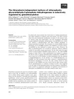
Fig. 1. PapMV CP mutants. (A) Schematic representation of Pap-
MV CP mutant constructs expressed in E. coli. All constructs pos-
sess a His6 tag. The dark rectangle in the schemata and the
underlined amino acids represent a small helix of six amino acids
that is predicted to occur between Q18 and S23 [10]. Amino acids
that are mutated in some constructs are in italics. (B, C) Expression
and purification of recombinant coat proteins on an SDS/PAGE gel.
The left panels represent Coomassie staining profiles and the right
panels represent western blots of purified proteins revealed with
IgG directed against PapMV CP.
M. E. Laliberte
´
Gagne
´
et al. F13 critical for interaction among the CP subunits
FEBS Journal 275 (2008) 1474–1484 ª 2008 The Authors Journal compilation ª 2008 FEBS 1475
formed NLPs that appeared to be longer than CP6–
215 and CP13–215 (Fig. 2C,D).
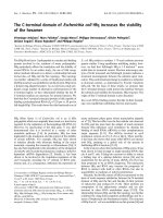
We determined the length of 250 NLPs for each
recombinant protein, and the average lengths are given
in Fig. 2E. As expected, CP6–215 and CP13–215 NLPs
were similar in length, measuring 50 nm. However,
NLPs comprising the F13L and F13Y mutants were
longer than CP6–215 NLPs. Indeed, F13L NLPs
appeared to be 2.5 times longer than CP6–215 NLPs,
whereas F13Y NLPs were four times longer.
Gel filtration analysis of recombinant proteins
Previously, we showed that when expressed in E. coli,
the CP6–215 protein occurred 80% of the time as a
450 kDa multimer (disks), and the remaining 20% was
in NLPs [9]. To measure the ability of our recombi-
nant CPs to form NLPs, we subjected purified proteins
to gel filtration (Figs 3 and 4). The Superdex 200 and
Superdex 75 FPLC profiles of recombinant CP6–215
and CP27–215 were compared with those of other
recombinant CPs. As shown before [9], the FPLC
Superdex 200 profile of CP6–215 first presents a peak
eluting at 42.7 mL, which corresponds to molecules
(larger than 670 kDa) that are excluded by the column
(Fig. 3A) where NLPs are found. A second peak elutes
at 50.5 mL; this corresponds to a multimer of
450 kDa, which corresponds to CP6–215 disks.
Finally, a third peak eluting at 78.8 mL corresponds
to low molecular mass molecules composed of
degraded forms of the CP that remain monomeric [9].
The respective percentages of the total proteins
0.2 µm
0.2 µm
0.2 µm
0.2 µm
0.2 µm
0.2 µm
0.2 µm
0.2 µm
A
B
C
E
D
350
300
250
150
Length of the NLPs (nm)
50
0
200
100
CP6–215 CP13–215 F13L F13Y
Fig. 2. Characterization of recombinant
NLPs self-assembled in E. coli. Electron
microscopy of (A) CP6–215, (B) CP13–215,
(C) F13L mutant and (D) F13Y high-speed
pellet. Bars are 200 nm. (E) Average length
of recombinant NLPs: CP6–215, CP13–215,
F13L, and F13Y (n = 250).
F13 critical for interaction among the CP subunits M. E. Laliberte
´
Gagne
´
et al.
1476 FEBS Journal 275 (2008) 1474–1484 ª 2008 The Authors Journal compilation ª 2008 FEBS
represented by the three forms were as follows: NLP,
33%; disks, 36%; and monomers, 31%. This current
profile differs slightly from the first one that we pub-
lished [9]. This is probably because the methods used
for bacterial lysis were different. Here, use of a French
press permitted recovery of more proteins that were
not previously detected when sonication was employed
to lyse the cells. It is likely that the heat generated by
sonication affected the protein and influenced the
recovery. CP27–215 was applied to a Superdex 75 26/
60 column (Fig. 3B), and eluted as a single peak at
164.61 mL, as previously reported [10]. The elution
AB
CD
E
F
0.2 µm
0.2 µm
0.2 µm
0.2 µm
Fig. 3. Gel filtration analysis of the truncated recombinant proteins and mutants F13A, F13G, F13L and F13Y mutants. (A) Black line,
CP
13–215
; gray line, CP6–215; 2 mg of the purified proteins was loaded onto an FPLC Superdex 200 16/60 column. (B) Black line, CP14–215;
gray line, CP
27–215
(21.2 kDa); 2 mg of the purified proteins was loaded onto an FPLC Superdex 75 26/60 column. (C) Gray line, F13L
mutant; dark line, F13Y mutant; dotted line, CP6–215; 2 mg of the recombinant proteins was loaded onto an FPLC Superdex 200 16/60 col-
umn. (D) Gray line, F13A mutant; black line, F13G mutant; dotted line, CP6–215; 2 mg of the recombinant proteins was loaded onto an FPLC
Superdex 200 16/60 column. Molecular markers are shown in the right (A, C, D) or left (B) corners. HMWF, high molecular weight forms
(> 670 000); disks, 20 subunits of the CP (450 000); LMWF: low molecular weight forms (< 230 000). Electron microscopy of the HMWF
fractions of (E) the F13A mutant and (F) the F13G mutant.
M. E. Laliberte
´
Gagne
´
et al. F13 critical for interaction among the CP subunits
FEBS Journal 275 (2008) 1474–1484 ª 2008 The Authors Journal compilation ª 2008 FEBS 1477
profile of CP13–215 was very similar to that of CP6–
215 (Fig. 3A), but showed a lower ratio of NLPs
(16%), a similar amount of disks (33%), and an
increase in the monomeric form of the protein (51%).
This might indicate lower stability of the protein,
which consequently impacts on the quantity of NLPs
produced. This result suggests that deletion of 12
amino acids at the N-terminus of PapMV CP does not
abolish its capacity to self-assemble and form NLPs.
Deletion of 13 amino acids in recombinant CP14–
215 led to a monomeric form, as shown by a single
peak at 158.31 mL obtained using the Superdex 75 26/
60 column. As expected, the recombinant CP14–215
eluted before the truncated CP27–215, as it is 13
amino acids longer. Both proteins were detected with
100% frequency as monomers.
Superdex 200 profiles of F13L and F13Y were also
compared with that of CP6–215 (Fig. 3C). The two
mutated forms were eluted in only two peaks, in con-
trast with three peaks for CP6–215. In both cases,
most of the protein was eluted in the first peak, which
occurred at 42.6 mL for the F13L mutant and at
41.7 mL for the F13Y mutant (Fig. 3C). These peaks
correspond to 80% and 90% of the total purified pro-
tein respectively. These fractions contain NLPs. Inter-
estingly, disks that normally elute at 50.5 mL were not
detected with these two mutants (Fig. 3C). Finally, a
peak eluted at 86.1 mL for the F13L mutant and
82.6 mL for the F13Y mutant. This peak is associated
with monomeric forms that probably represent a
degraded protein. These results suggest that the two
mutants are highly efficient at forming NLPs.
The F13A and F13G mutants were also subjected to
Superdex 200 (Fig. 3D) elution. The F13A and F13G
mutants eluted in two peaks (Fig. 3D). The first one
appeared at 42.1 mL for the F13A mutant and at
40.3 mL for the F13G mutant. The top of each peak
was collected and examined by electron microscopy.
Few NLPs were observed with the F13A mutant, as
most of the protein appeared as nonspecific aggregates
(Fig. 3E). For the F13G mutant, NLPs were not found
on the electron microscopy grids. Only nonspecific
aggregates were visible (Fig. 3F). In both cases, disks
were not found in the sample. A peak that eluted at
81.4 mL for the F13A mutant and at 81.5 mL for the
F13G mutant corresponds to a monomeric form
(Fig. 3D). In fact, most of the purified F13A (65%)
mutant was found to be monomeric. In contrast, only
20% of the F13G mutant eluted as a monomer. It
seems that the F13A mutation affects the capacity of
the recombinant CP to form NLPs, because a large
proportion of the recombinant purified protein is
found in low molecular mass forms. Also, even if 35%
of the protein eluted as a large molecular mass multi-
mer, the electron microscopy observation revealed that
the proteins form nonspecific aggregates that are ineffi-
cient in making NLPs. For the F13G mutant, the
mutation probably greatly affects its capacity to multi-
merize into disks and NLPs.
The F13R, F13E and F13S mutants were also sub-
jected to Superdex 200 (Fig. 4A) gel filtration. In this
experiment, we loaded smaller amount (150 lg) of
CP6–215 protein to separate the NLPs from the disks
into two distinct peaks. The F13R and F13S mutants
A B
0.2 µm
Fig. 4. Gel filtration of the F13R, F13E and F13S mutants. (A) Gel filtration analysis of recombinant proteins. Black dotted line, CP6–215;
bright gray line, F13E mutant; dark gray line, F13R mutant; black line, F13S mutant; 500 lg of the purified F13E, F13R and F13S mutant pro-
teins and 150 lg of the purified CP6–215 protein were loaded onto an FPLC Superdex 200 10/300 column. Molecular markers are shown in
the left corner. HMWF, high molecular weight forms (> 670 000); disks, 20 subunits of the CP (450 000); LMWF, low molecular weight
forms (< 230 000). (B) Electron microscopy of the HMWF fraction of the F13E mutant.
F13 critical for interaction among the CP subunits M. E. Laliberte
´
Gagne
´
et al.
1478 FEBS Journal 275 (2008) 1474–1484 ª 2008 The Authors Journal compilation ª 2008 FEBS
were found entirely in the low molecular mass frac-
tions and were unable to self-assemble into NLPs (data
not shown). Most of the protein of the F13E mutant
was found as low molecular mass forms, but a small
fraction was found in the exclusion fraction with CP6–
215 NLPs (Fig. 4A). However, NLPs were absent, and
only nonspecific aggregates could be observed by elec-
tron microscopy in this fraction (Fig. 4B). Therefore,
we concluded that the F13E mutant was unable to
self-assemble into an NLP.
1
H-
15
N HSQC spectrum analysis
To confirm that the CP14–215 monomer can be used
for NMR analysis, we uniformly labeled the protein
with
15
N and acquired preliminary NMR data that we
superimposed on similar spectra obtained previously
with the monomeric form of CP27–215 [10]. Conditions
determined previously to be optimal for NMR were
used [10]. In order to improve solubility and stability
for NMR sample analysis, a pH of 6.2 was selected. A
2D
1
H-
15
N HSQC spectrum of CP14–215 was acquired
at 600 MHz at 25 °C (Fig. 5). Good spectral dispersion
(3.5 p.p.m.) of backbone amide
1
H resonances indicates
that PapMV CP is well folded under the conditions
used. Furthermore, the peak line width and signal
intensity under the conditions used suggest that the
mutant CP14–215 is monomeric in solution, as expected
from the chromatography results. Superimposition of
spectra revealed that all peaks corresponding to struc-
tured regions of CP27–215 are present in the CP14–215
spectrum. This suggests that the structure of both trun-
cated forms is very similar. Moreover, the presence of
several peaks in the middle of the spectrum (corre-
sponding to unstructured regions) suggests that amino
acids 14–26 are not structured.
Gel shift assays
To evaluate whether the ability to form NLPs was
related to affinity for RNA, as we have shown previ-
ously with the E128A and K97A mutants [9], we mea-
sured the affinity of the mutant by electrophoretic
mobility shift assay (EMSA) (Fig. 6). The high-speed
supernatant (disks) of the purified proteins was incu-
bated with 165 fmol of an RNA probe labeled with
c-
32
P made from an 80 nucleotide RNA transcript
from the 5¢-end of PapMV. The disks of CP
6-215
and
CP13–215 interacted with the probe in a cooperative
manner and induced a shift when as little as 100 ng of
proteins was added (Fig. 6A,B). This result suggests
that differences between the ability of the two proteins
to form NLPs, as shown in Fig. 3A, are not related to
their affinity for RNA.
A similar experiment was performed with CP14–215
and CP27–215, two proteins known to form mono-
mers. As expected, both CP14–215 and CP27–215
failed to interact with the first 80 nucleotides of viral
RNA in vitro (Fig. 6C,D). We performed an EMSA
with the high-speed supernatant of F13A, and showed
that it failed to induce formation of a protein–RNA
complex (Fig. 6E). This is consistent with our electron
microscopy observations, which highlighted the inabil-
ity of this protein to self-assemble into NLPs.
As the F13L and F13Y mutants form only NLPs in
E. coli, we needed to disrupt NLPs using the widely
employed acetic acid treatment to isolate the disks as
previously described [3], to test their ability to bind
RNA. The same treatment was done with CP6–215
NLPs as a control. Previously, we proposed that puri-
fied protein NLP length was related directly to its
RNA-binding capacity [9]. Surprisingly, isolated disks
of these two proteins showed a lower affinity for
RNA than CP6–215 disks (Fig. 7A–C), even though
extracted disks looked normal at the electron micros-
copy level (supplementary Fig. S1). We did not test the
F13G, F13E, F13R and F13S mutants, because they
were unable to form NLPs and therefore did not bind
RNA.
Measurement of RNA content by spectroscopy
In addition to EMSA, we evaluated the difference
observed between the F13L and F13Y mutants and
Fig. 5. Superimposition of the
1
H-
15
N HSQC spectra of CP14–215
and CP
27–215
; 0.1 mM each protein was diluted in 10 mM dithiothrei-
tol, 10% D
2
O, 1· complete protease inhibitor cocktail, 0.1 mM
NaN
3
and 60 lM DSS at pH 6.2.
M. E. Laliberte
´
Gagne
´
et al. F13 critical for interaction among the CP subunits
FEBS Journal 275 (2008) 1474–1484 ª 2008 The Authors Journal compilation ª 2008 FEBS 1479
CP6–215 by spectroscopy using the A
280/260 nm
ratio of
different recombinant proteins. Measurement of the
A
280/260 nm
ratio, which was performed three times,
was very consistent, and the average is presented in
Table 1. Surprisingly, A
280/260 nm
ratios obtained for
the two recombinant proteins were closer to the one
obtained for PapMV than for CP6–215 NLPs. These
results suggest that F13L and F13Y NLPs are compe-
tent at binding RNA in spite of the lower affinity mea-
sured by EMSA.
The A
280/260 nm
ratio was also calculated for disks.
Results for PapMV disks and CP6–215 disks differed
from those for F13L and F13Y disks, and suggest that
there is still some RNA associated with recombinant
F13L and F13Y disks. This could partially explain the
decreased affinity of F13L and F13Y disks in EMSA.
Discussion
Previous studies on the PapMV CP indicated that an
essential domain for CP multimerization is located on
26 amino acids of the N-terminus [9,10]. In this work,
we investigated this region in detail, introducing dele-
tions and point mutations. All mutations incorporated
in the PapMV CP gene did not affect the secondary
structure prediction of the CPs (supplementary
Fig. S2). We have shown clearly that the N-terminal
12 amino acids are not important for self-assembly of
the PapMV CP. This result is consistent with the find-
ings of Zhang et al. [1], who showed that cleavage of
the N-terminus with trypsin did not affect virus parti-
cles. This region probably plays a role in protein sta-
bilization, rather than in NLP formation, as we found
more degraded monomers with CP13–215 than with
CP6–215 in the FPLC profiles (Fig. 3A).
Deletion of 13 amino acids, mutation of residue F13
for the less hydrophobic residues A or G, or replace-
ment with the charged residues R or E, or the polar
residue S, had a major detrimental impact on NLP
formation. This suggests that F13 is involved in a
hydrophobic interaction that is crucial for interplay
among the protein subunits and formation of the disks
AB
C
E
D
Fig. 6. EMSA with high-speed supernatant
of recombinant CPs. (A) CP6–215;
(B) CP
13–215
; (C) CP
27–215
; (D) CP14–215;
(E) F13A mutant. Increasing protein
amounts were incubated at 22 ° C for 1 h
with 165 fmol of an RNA probe labeled with
c-
32
P. The probe was made from an
80 nucleotide RNA transcript from the
5¢-end of the PapMV noncoding region. The
free probe and the RNA–protein complex
are indicated by arrows.
F13 critical for interaction among the CP subunits M. E. Laliberte
´
Gagne
´
et al.
1480 FEBS Journal 275 (2008) 1474–1484 ª 2008 The Authors Journal compilation ª 2008 FEBS
that are the building blocks with the RNA of the
NLPs. Interestingly, F13L and F13Y substitutions
increased NLP formation, probably through improve-
ment of the RNA-binding capacity of the proteins, as
shown by the A
280/260 nm
ratio (Table 1). EMSA analy-
sis of F13L and F13Y extracted disks did not show
improved affinity for RNA as compared with CP6–
215, probably because they were still bound tightly to
RNA, which interfered with RNA probe binding.
It appears that F13 plays an important role in the
aggregation state of the protein, as mutation of this
residue led to formation of either NLPs (F13Y and
F13L) or monomeric forms of the protein (F13G,
F13A, F13R, F13E, F13S), which were always
detrimental to accumulation of disks in bacteria. It is
possible that this regulation is important in PapMV-
infected plants to ensure that only viral RNA, and not
plant cellular RNA, gets encapsulated by the viral CP.
It is tempting to draw a parallel with tobacco mosaic
virus CP, even if this protein is not related to the Pap-
MV CP, where a hydrophobic interaction between the
CP subunits was shown to be important for self-assem-
bly of the virus into a rigid rod structure [11].
Comparison of 2D
1
H-
15
N HSQC spectra from two
monomeric forms, CP14–215 and CP27–215, indicates
that amino acids 14–26 are unstructured. This result
suggests that the small helix that was predicted by bio-
informatics to occur between residues 18 and 24 [10] is
probably unstable. We propose that the entire N-ter-
minus from residues 1 to 36 forms an unstructured coil
region.
A recent report showed that the CP of potato vir-
us X (PVX) can be truncated by 22 amino acids at its
N-terminus without affecting either virus infectivity or
formation of virus particles in plants [12]. The authors
took advantage of this mutant by fusing foreign pep-
tides to the surface of the virus. Alignment of the
N-terminus of the PVX CP with the PapMV CP
revealed that the PVX CP harbors an extension of
20 amino acids in the N-terminus as compared with
PapMV (Fig. 8). At position 33 of the PVX CP, we
find an F residue that aligns perfectly with the PapMV
A
BC
Fig. 7. EMSA with high-speed supernatant
of recombinant disks obtained from the dis-
ruption of the NLPs by use of the acetic
acid method [3]. (A) CP6–215; (B) F13L
mutant; (C) F13Y mutant. Increasing
amounts of proteins were incubated at
22 °C for 1 h with 165 fmol of an RNA
probe labeled with c-
32
P. The probe was
made from an 80 nucleotide RNA transcript
from the 5¢-end of the PapMV noncoding
region. The free probe and the RNA–protein
complex are indicated by arrows.
Table 1. Protein A
280/260 nm
ratio. Spectrophotometer absorbance measurements were taken three times with different protein preparations.
Results were consistent among measurements. Recombinant CP6–215 NLPs were isolated from the high-speed pellet. The absorbance
measurement was taken directly from the purified PapMV and purified F13L and F13Y recombinant proteins. The four proteins were treated
by acetic acid methodology [3] to generate disks that were used to calculate the A
280/260 nm
ratio.
Virus and NLPs Extracted disks
PapMV CP6–215 F13L F13Y PapMV CP6–215 F13L F13Y
A
280/260 nm
ratio
0.75 1.1 0.8 0.75 1.5 1.55 0.95 0.9
M. E. Laliberte
´
Gagne
´
et al. F13 critical for interaction among the CP subunits
FEBS Journal 275 (2008) 1474–1484 ª 2008 The Authors Journal compilation ª 2008 FEBS 1481
CP F13. Therefore, on the basis of our results, it is
likely that a deletion of 32 amino acids will be toler-
ated by PVX without disturbing the assembly process.
Alignment of this F residue is also shared with several
other potexviral CP sequences, as seven out of the
18 N-terminal sequences of the potexviruses showed
consensus for an F in the position that corresponds to
F13 of PapMV CP (Fig. 8). Also, an F is present in
the same area in the CP of bamboo mosaic virus. The
CP of mint virus X presents an L in this position,
which corresponds to a hydrophobic residue that could
substitute for an F in the PapMV CP. Therefore, on
the basis of the alignment, we propose that a hydro-
phobic residue at the position that corresponds to Pap-
MV CP F13 is preferred in half of the potexvirus CP.
It is likely that this residue also plays an important
role in the interactions between the subunits in the
potexviruses family.
Finally, our results agree with the assembly model
recently proposed for a potyvirus member of the Poty-
viridea family: the pepper vain banding virus (PVBV)
[13]. These authors proposed that the N-terminal
extension of a CP subunit interacts with the C-terminal
extension of an adjacent CP subunit in a head-to-tail
manner, thereby permitting formation of both the
ring-like intermediate and the NLPs into helix-like
structures. We propose that this model is applicable
for PapMV and probably all potexviruses. However, a
major difference between PapMV and PVBV is that
PapMV CP subunit assembly into disk structures is
based on a hydrophobic interaction, whereas PVBV
CP assembly into ring-like structures (disks) was pro-
posed to be driven by electrostatic interactions [13].
Experimental procedures
Cloning and expression of recombinant proteins
The PapMV CP gene CP6–215 has been described previously
[9], as has the truncated version of PapMV CP, CP27–215
[10]. The other truncated versions of PapMV, CP13–215 and
CP14–215, were amplified by PCR from the clone CP6–215
inserted into a pET-3d vector. The forward primers used for
these PCR reactions were CP13–215 forward, 5¢-ACGT
CA
TATGTTCCCCGCCATCACCCAG-3¢, and CP14–215 for-
ward, 5¢-ACGT
CATATGCCCGCCATCACCCAGGAA-3¢.
A reverse primer, 3¢-GAAATTCTTCCTCTATAT
GTA
TACTGCA-5¢, was used for both constructs. The PCR prod-
ucts were digested with NdeI, to generate the two truncated
CPs inserted into a pET-3d vector.
The F13A, F13E, F13G, F13L, F13R, F13S and F13Y
mutations were introduced by PCR into the CP6–215 clone
using the following oligonucleotides: forward (F13A),
5¢-
GCGCCCGCCATCACCCAGGAACAA-3¢; forward
(F13E), 5¢-
GAACCCGCCATCACCCAGGAACAA-3¢; for-
ward (F13G), 5¢-
GGCCCCGCCATCACCCAGGAACAA-
3¢; forward (F13L), 5¢-
CTGCCCGCCATCACCCAGGA
ACAA-3¢; forward (F13R), 5¢-
CGCCCCGCCATCACCC
Fig. 8. Alignment of a consensus sequence derived from 18 known potexvirus coat proteins and the PapMV CP in the N-terminal
region 1–27 of PapMV CP. Conserved hydrophobic residues that aligned with amino acid 13 of the PapMV CP are highlighted in bold. Align-
ment was done using the CP sequences of: bamboo mosaic virus (BaMV); cactus virus X (CVX); clover yellow mosaic virus (ClYMV); cas-
sava common mosaic virus (CsCMV); Cymbidium mosaic virus (CymMV); foxtail mosaic virus (FoMV); Hosta virus X (HVX); lily virus X (LVX);
mint virus X (MVX); narcissus mosaic virus (NMV); PapMV; potato aucuba mosaic virus (PAMV); pepino mosaic virus (PepMV); plantago asi-
atica mosaic virus (PlAMV); PVX; scallion virus X (ScaVX); strawberry mild yellow edge virus (SMYEV); tulip virus X (TVX); white clover
mosaic virus (WClMV).
F13 critical for interaction among the CP subunits M. E. Laliberte
´
Gagne
´
et al.
1482 FEBS Journal 275 (2008) 1474–1484 ª 2008 The Authors Journal compilation ª 2008 FEBS
AGGAACAA-3¢; forward (F13S), 5¢-AGCCCCGCCAT
CACCCAGGAACAA-3¢; forward (F13Y), 5¢-
TATCCCG
CCATCACCCAGGAACAA-3¢; and reverse (F13), 3¢-CG
TAGGTGTGGGTTGTATCGG-5¢. PCR products with
blunt ends were circularized to form the fourth mutated CP
inserted into a pET-3d vector.
Expression and purification of recombinant
proteins from E. coli
Expression and induction of proteins was conducted as
described previously [9]. Bacteria were harvested by centri-
fugation for 30 min at 9000 g. The pellet was resuspended
in ice-cold lysis buffer (50 mm NaH
2
PO
4
, pH 8.0, 300 mm
NaCl, 10 mm imidazole, 40 lm phenylmethanesulfonyl fluo-
ride and 0.2 mgÆmL
)1
lysosyme), and bacteria were lysed
by one passage through a French press. The lysate was
incubated with agitation for 15 min with 9000 units of
DNase and 1.5 mm MgCl
2
, and this was followed by two
centrifugations for 30 min at 10 000 g to eliminate cellular
debris. The supernatant was incubated with 3 mL of Ni–ni-
trilotriacetic acid (Qiagen, Turnberry Lane, Valencia, CA,
USA) under gentle agitation overnight at 4 °C. Proteins
were purified as described elsewhere [9], except that they
were incubated for 4 h with 2 mL of the elution buffer
(10 mm Tris/HCl, pH 8.0, supplemented with 1 m imidaz-
ole) before elution. Imidazole was eliminated by dialysis for
24 h. Protein purity was determined by SDS/PAGE and
confirmed by western immunoblot analysis using rabbit
polyclonal antibodies generated against purified PapMV
virus.
Separation of disks and NLPs
To separate the disks from NLPs, 1 mL of purified proteins
was subjected to a high-speed centrifugation for 2 h at
100 000 g in a Beckman SW60Ti rotor. The pellet that
comprised the NLPs was resuspended in 300 lLof10mm
Tris/HCl at pH 8.0. The supernatant with the disks and the
low molecular mass forms was retained for gel shift assays.
SDS/PAGE and electroblotting
Proteins were mixed with one-third of the final volume of
loading buffer containing 5% SDS, 30% glycerol, and
0.01% bromophenol blue. SDS/PAGE was performed as
described elsewhere [14].
Electron microscopy
Nucleocapsid-like particles or viruses were diluted in
10 mm Tris/HCl (pH 8.0) to a concentration of 50 ngÆlL
)1
,
and were absorbed for 6 min on carbon-coated formvar
grids. Grids were washed twice with 8 lL of water. Finally,
grids were incubated in darkness for 6 min with 8 lLof
2% uranyl acetate.
Acetic acid degradation
Isolation of disks from CP6–215, F13L and F13Y NLPs
was performed by acetic acid degradation as described pre-
viously [3]. Two volumes of glacial acetic acid were added
to the NLPs and incubated at 4 °C for 1 h. Centrifugation
at 10 000 g for 15 min removed insoluble RNA. The super-
natant was removed and subjected to high-speed centrifuga-
tion at 100 000 g for 2 h in a Beckman 50.2Ti rotor to
remove any residual NLPs. Proteins were dialyzed exten-
sively against 10 mm Tris/HCl (pH 8.0).
Gel filtration
Proteins were purified by gel filtration. Columns were first
calibrated with molecular weight markers (GE Healthcare,
Baie d’Urfe
´
, Canada). Superdex 75 26/60 (GE Healthcare),
Superdex 200 16/60 (GE Healthcare) and Superdex 200 10/
300 (GE Healthcare), pre-equilibrated with gel filtration buf-
fer (10 mm Tris/HCl, pH 8.0, supplemented with 150 mm
NaCl), were used. The volume of protein loaded into the
sample loop was 1.5 mL for Superdex 75 26/60, 1 mL for
Superdex 200 16/60, and 0.1 mL for Superdex 200 10/300.
NMR spectroscopy
The 600 lL sample used for NMR spectroscopy was
0.1 mm CP14–215 or CP27–215 in 90% H
2
O/10% D
2
O,
10 mm dithiothreitol (pH 6.2), 1· complete protease inhibi-
tor cocktail (Roche), with 0.1 mm NaN
3
and 60 lm 2,2-
dimethyl-2-silapentane-5-sulfonic acid (DSS) as the NMR
chemical shift reference. The
1
H-
15
N HSQ spectra were
obtained at 25 °C on a Varian Unity 600 MHz spectrome-
ter equipped with a triple-resonance cryoprobe and Z-axis
pulsed-weld gradient. The acquired data consisted of 768
complex data points in the acquisition domain and 128
complex data points in the indirectly detected domain. The
spectral width was 10 000 Hz in the
1
H dimension and
1680 Hz in the
15
N dimension. NMR spectra were pro-
cessed using NMRPipe [15]. Processing involved doubling
of the
15
N time domain by linear prediction, zero-filling to
2048 and 512 complex points in
1
H and
15
N, respectively, a
45° shifted sine-bell apodization in the
1
H dimension, and a
72° shifted sine-bell apodization in the
15
N dimension.
RNA transcripts and EMSA
The probe was generated as described before [9]. Labeled
RNA probe was incubated with various amounts of recom-
binant proteins at room temperature for 60 min. We used
165 fmol of RNA for each reaction in the in vitro assembly
M. E. Laliberte
´
Gagne
´
et al. F13 critical for interaction among the CP subunits
FEBS Journal 275 (2008) 1474–1484 ª 2008 The Authors Journal compilation ª 2008 FEBS 1483
buffer (10 mm Tris/HCl, 4% glycerol, 1 mm MgCl
2
,
0.5 mm dithiothreitol, 0.5 mm EDTA, 20 mm NaCl), which
contained 7.5 U of RNase inhibitor (27-0816-01; GE
Healthcare). The final reaction volume was 10 lL. Two
microliters of loading dye was added to the sample before
loading onto a 5% native polyacrylamide gel. Electrophore-
sis was performed in 0.5· Tris/borate/EDTA buffer for
90 min at 10 mA. The gel was dried and subjected to auto-
radiography for 16 h on Kodak Bio-Max MS film
(V8326886; GE Healthcare) and developed.
Acknowledgements
We thank the Natural Sciences and Engineering
Research Council of Canada (NSERC) and the ‘Fond
de Recherche sur la Nature et les Technologies’
(FQRNT) for funding our research program on
papaya mosaic virus, and Dr Paul Khan for critical
reading of our manuscript.
References
1 Zhang H, Todderud E & Stubbs G (1993) Crystalliza-
tion and preliminary X-ray analysis of papaya mosaic
virus coat protein. J Mol Biol 234, 885–887.
2 Sit TL, Abouhaidar MG & Holy S (1989) Nucleotide
sequence of papaya mosaic virus RNA. J Gen Virol 70
(Pt 9), 2325–2331.
3 Erickson JW, Bancroft JB & Horne RW (1976) The
assembly of papaya mosaic virus protein. Virology 72,
514–517.
4 Abouhaidar M & Bancroft JB (1978) The initiation of
papaya mosaic virus assembly. Virology 90, 54–59.
5 Abouhaidar MG & Bancroft JB (1980) The polarity of
assembly of papaya mosaic-virus and tobacco mosaic-
virus RNAs with PMV-protein under conditions of
nonspecificity. Virology 107, 202–207.
6 Erickson JW & Bancroft JB (1978) The self-assembly of
papaya mosaic virus. Virology 90, 36–46.
7 Erickson JW, Bancroft JB & Stillman MJ (1981) Circu-
lar dichroism studies of papaya mosaic virus coat pro-
tein and its polymers. J Mol Biol 147, 337–349.
8 Erickson JW, Hallett FR & Bancroft JB (1983) Sub-
assembly aggregates of papaya mosaic-virus protein.
Virology 129, 207–211.
9 Tremblay MH, Majeau N, Gagne
´
ME, Lecours K,
Morin H, Duvignaud JB, Bolduc M, Chouinard N,
Pare
´
C, Gagne
´
S et al. (2006) Effect of mutations K97A
and E128A on RNA binding and self assembly of
papaya mosaic potexvirus coat protein. FEBS J 273,
14–25.
10 Lecours K, Tremblay MH, Gagne
´
ME, Gagne
´
SM &
Leclerc D (2006) Purification and biochemical
characterization of a monomeric form of papaya
mosaic potexvirus coat protein. Protein Expr Purif 47,
273–280.
11 Bendahmane M, Fitchen JH, Zhang G & Beachy RN
(1997) Studies of coat protein-mediated resistance to
tobacco mosaic tobamovirus: correlation between
assembly of mutant coat proteins and resistance. J Virol
71, 7942–7950.
12 Donini M, Lico C, Baschieri S, Conti S, Magliani W,
Polonelli L & Benvenuto E (2005) Production of an
engineered killer peptide in Nicotiana benthamiana by
using a potato virus X expression system. Appl Environ
Microbiol 71, 6360–6367.
13 Anindya R & Savithri HS (2003) Surface-exposed
amino- and carboxy-terminal residues are crucial for
the initiation of assembly in Pepper vein banding virus:
a flexuous rod-shaped virus. Virology 316, 325–336.
14 Schagger H & von Jagow G (1987) Tricine-sodium
dodecyl sulfate-polyacrylamide gel electrophoresis for
the separation of proteins in the range from 1 to
100 kDa. Anal Biochem 166, 368–379.
15 Delaglio F, Grzsiek S, Vuister VW, Zhu G, Pfeifer J &
Bax A (1995) NMRPipe: a multidimensional spectral
processing system based on UNIX pipes. J Biomol
NMR 6, 277–293.
Supplementary material
The following supplementary material is available
online:
Fig. S1. Electron microscopy of disks extracted by ace-
tic acid methodology [3] of: (A) CP6–215; (B) F13L
mutant; and (C) F13Y mutant.
Fig. S2. Predicted secondary structure of recombinant
PapMV CPs.
This material is available as part of the online article
from
Please note: Blackwell Publishing are not responsible
for the content or functionality of any supplementary
materials supplied by the authors. Any queries (other
than missing material) should be directed to the corre-
sponding author for the article.
F13 critical for interaction among the CP subunits M. E. Laliberte
´
Gagne
´
et al.
1484 FEBS Journal 275 (2008) 1474–1484 ª 2008 The Authors Journal compilation ª 2008 FEBS