Báo cáo khoa học: Interaction between Lim15/Dmc1 and the homologue of the large subunit of CAF-1 – a molecular link between recombination and chromatin assembly during meiosis pot
Bạn đang xem bản rút gọn của tài liệu. Xem và tải ngay bản đầy đủ của tài liệu tại đây (442.25 KB, 10 trang )
Interaction between Lim15/Dmc1 and the homologue of
the large subunit of CAF-1 – a molecular link between
recombination and chromatin assembly during meiosis
Satomi Ishii*
,
†, Akiyo Koshiyama*, Fumika N. Hamada, Takayuki Y. Nara, Kazuki Iwabata,
Kengo Sakaguchi and Satoshi H. Namekawa
Department of Applied Biological Science, Faculty of Science and Technology, Tokyo University of Science, Japan
Keywords
chromatin assembly; chromatin assembly
factor 1 (CAF-1); Lim15/Dmc1; meiotic
recombination; proliferating cell nuclear
antigen (PCNA)
Correspondence
K. Sakaguchi, Department of Applied
Biological Science, Faculty of Science and
Technology, Tokyo University of Science,
2641 Yamazaki, Noda-shi, Chiba-ken
278-8510, Japan
Fax: +81 4 7123 9767
Tel: +81 4 7124 1501 (ext. 3409)
E-mail:
Website: />riko_app_bio.html
S. H. Namekawa, Department of Molecular
Biology, Massachusetts General Hospital,
and Department of Genetics, Harvard
Medical School, Boston, MA 02114, USA
Fax: +1 617 726 6893
Tel: +1 617 726 5966
E-mail:
Present address
†Quantum Beam Science Directorate, Japan
Atomic Energy Agency, Gunma, Japan
*These authors contributed equally to this work
(Received 7 January 2008, revised 24
February 2008, accepted 25 February 2008)
doi:10.1111/j.1742-4658.2008.06357.x
In eukaryotes, meiosis leads to genetically variable gametes through recom-
bination between homologous chromosomes of maternal and paternal ori-
gin. Chromatin organization following meiotic recombination is critical to
ensure the correct segregation of homologous chromosomes into gametes.
However, the mechanism of chromatin organization after meiotic recombi-
nation is unknown. In this study we report that the meiosis-specific
recombinase Lim15/Dmc1 interacts with the homologue of the largest
subunit of chromatin assembly factor 1 (CAF-1) in the basidiomycete
Coprinopsis cinerea (Coprinus cinereus). Using C. cinerea LIM15/DMC1
(CcLIM15) as the bait in a yeast two-hybrid screen, we have isolated the
C. cinerea homologue of Cac1, the largest subunit of CAF-1 in Saccharo-
myces cerevisiae, and named it C. cinerea Cac1-like (CcCac1L). Two-hybrid
assays confirmed that CcCac1L binds CcLim15 in vivo. b-Galactosidase
assays revealed that the N-terminus of CcCac1L preferentially interacts
with CcLim15. Co-immunoprecipitation experiments showed that these
proteins also interact in the crude extract of meiotic cells. Furthermore, we
demonstrate that, during meiosis, CcCac1L interacts with proliferating cell
nuclear antigen (PCNA), a component of the DNA synthesis machinery
recently reported as an interacting partner of Lim15/Dmc1. Taken
together, these results suggest a novel role of the CAF-1–PCNA complex
in meiotic events. We propose that the CAF-1–PCNA complex modulates
chromatin assembly following meiotic recombination.
Abbreviations
ATCC, American Type Culture Collection; Cac1, chromatin assembly complex 1; CAF-1, chromatin assembly factor 1; CcCac1L,
Coprinopsis cinerea Cac1-like; CPRG, chlorophenol red-b-
D-galactopyranoside; DSB, double-strand break; IPTG, isopropyl thio-b-D-galactoside;
PCNA, proliferating cell nuclear antigen; RLM-RACE, RNA ligase-mediated-RACE; RU, resonance unit; SPR, surface plasmon resonance.
2032 FEBS Journal 275 (2008) 2032–2041 ª 2008 The Authors Journal compilation ª 2008 FEBS
In eukaryotes, sexual reproduction is achieved by the
conjugation of genetically variable gametes, which are
generated during meiosis in the parental germline. Mei-
osis consists of two rounds of chromosome segrega-
tion, resulting in gametes with half the number of
chromosomes in order to prepare for conjugation.
During prophase of the first meiotic division, recombi-
nation takes place between homologous chromosomes
of maternal and paternal origin. This is followed by
the segregation of maternal and paternal copies of
each chromosome. A physical connection at the site of
homologous recombination, called the chiasma, orients
homologous chromosome pairs towards opposite spin-
dle poles at meiosis I [1]. Therefore, chromatin organi-
zation following meiotic recombination is required to
establish the chiasma and to segregate homologous
chromosomes.
Meiotic recombination comprises several steps
beginning with meiosis-specific double-strand breaks
(DSBs). A single-strand overhang is formed by exonu-
clease activity and invades the homologous double-
stranded region of the other allele. These steps
of homology search and recombination are catalysed
by two bacterial RecA homologues, Rad51 and
Lim15/Dmc1. Rad51 catalyses both somatic and
meiotic recombination, whereas Lim15/Dmc1 is meio-
sis-specific [2–5]. Rad51 and Lim15/Dmc1 are compo-
nents of a multiprotein complex at the site of
recombination [6,7]. In order to understand the mecha-
nisms of meiotic recombination, much effort has been
made to identify additional components of the Rad51
and Lim15/Dmc1 complex, in particular Lim15/Dmc1
interacting partners.
Recent analysis has identified various interacting
partners of Lim15/Dmc1, which seem to be involved in
homology search and strand exchange. Tid1/Rdh54,
an SWI2/SNF2 family of chromatin-remodelling
factors, promotes the co-localization of Rad51 and
Lim15/Dmc1 [8]. The heterodimeric complex of Hop2
and Mnd1 stimulates strand exchange of Lim15/Dmc1
[9–11]. The meiosis-specific proteins Mei5 and Sae3
form a complex with Lim15/Dmc1 and are necessary
for the assembly of Lim15/Dmc1 [12,13]. Furthermore,
the DNA mismatch repair protein MSH4 (MutS
homologue 4) [14], the tumor suppressor protein p53
[15], DNA topoisomerase II [16], the sumoylation pro-
tein Ubc9 [17] and the DNA synthesis-related factor
proliferating cell nuclear antigen (PCNA) [18] have
been reported to interact with Lim15/Dmc1. These
proteins seem to participate in the modulation of
Lim15/Dmc1. However, how chromatin is organized
following meiotic recombination has not been
described.
In order to explore chromatin organization after
meiotic recombination, we designed experiments to
investigate the possible interactions between recombi-
nation proteins and chromatin assembly factors. In
this article, we report that the largest subunit homo-
logue of chromatin assembly factor 1 (CAF-1) is a
novel interacting partner of Lim15/Dmc1. CAF-1 con-
sists of three subunits that are highly conserved
amongst yeast, plant, fly and human [19–23]. CAF-1
deposits histones H3 and H4 onto newly synthesized
DNA after replication and repair [24–26]. In addition,
the largest subunit of CAF-1 interacts with PCNA
during replication [27], in nucleotide excision repair
[28] and in DSB repair [29,30]. Despite much accumu-
lating evidence regarding the role of CAF-1 in chroma-
tin assembly following various DNA synthesis events,
its involvement in chromatin assembly following mei-
otic recombination is unknown. In this study, we test
the involvement of CAF-1 in meiotic events. We pro-
pose a novel role of the CAF-1–PCNA complex in
chromatin assembly following meiotic recombination.
Results
Isolation of Coprinopsis cinerea Cac1-like
(CcCac1L) by two-hybrid screening using
CcLim15 as bait
To isolate proteins that interact with CcLim15, we per-
formed a yeast two-hybrid screen using CcLim15 as
bait. A clone was isolated which had moderate amino
acid similarity with the largest subunit of human
CAF-1 (p150) [19] and the largest subunit of Saccharo-
myces cerevisiae CAF-1 (Cac1, chromatin assembly
complex 1) [20]. The sequence similarities of this clone
with human and S. cerevisiae homologues were found
to be 26% and 23%, respectively. Hence, this clone
was identified as C. cinerea Cac1-like (CcCac1L).
CcCac1L encodes a predicted product of 812 amino
acid residues with a molecular mass of 120 kDa. The
highly charged KER (lysine/glutamate/arginine-rich;
242–360 amino acids) and ED (glutamate/aspartate-
rich; 522–578 amino acids) domains in CcCac1L are
conserved amongst human and S. cerevisiae homo-
logues (Fig. 1A). The KER and ED domains are
known to interact directly with newly synthesized H3/
H4 histones [19,21].
CcCac1L interacts with CcLim15
To confirm the specificity of interaction between
CcCac1L and CcLim15, we performed yeast two-
hybrid and b-galactosidase assays (Fig. 1C,D). Next,
S. Ishii et al. Link between Lim15/Dmc1 and the CAF-1–PCNA complex
FEBS Journal 275 (2008) 2032–2041 ª 2008 The Authors Journal compilation ª 2008 FEBS 2033
we sought to determine which region of CcCac1L
was responsible for binding to CcLim15. The N-ter-
minus (CcCac1L-N; amino acids 1–381) contained
the KER domain, whereas the C-terminus (CcCac1L-
C; amino acids 382–812) contained the ED domain
(Fig. 1B). Two-hybrid assays demonstrated that
CcLim15 interacts with either of the truncated
mutants of CcCac1L in the mild selection medium
[SD3: lacking histidine, leucine and tryptophan
(–His/–Leu/–Trp)], and that CcLim15 preferentially
interacts with CcCac1L-N in the stringent selection
medium [SD4: lacking adenine, histidine, leucine
and tryptophan (–Ade/–His/–Leu/–Trp)] (Fig. 1C).
The interaction between the truncated mutants of
CcCac1L and CcLim15 was confirmed by b-galactosi-
dase assays, which demonstrated a higher binding
affinity of CcCac1L-N than CcCac1L-C to CcLim15
(Fig. 1D).
Characterization of CcCac1L during meiosis
The data above strongly suggest a novel function of
CAF-1 as a binding partner of Lim15/Dmc1. How-
ever, currently there are no observations available
describing the meiotic role of CAF-1. Therefore, we
sought to examine the distribution of CcCac1L dur-
ing meiosis. First, in order to determine the gene
expression profile of CcCac1L during meiotic develop-
ment, we performed northern analyses at each stage
during meiotic development. Total RNA was
extracted from basidia in synchronous culture at 1 h
intervals after the induction of meiosis. CcCac1L was
expressed at the premeiotic S phase, at the time of
genomic DNA replication (Fig. 2A). Homologous
chromosomes start to align at the leptotene/zygotene
stage. Then, fully synapsed homologues are observed
at the pachytene stage. CcCac1L began to accumulate
at the leptotene and zygotene stage, and decreased
after the pachytene stage (Fig. 2A). This expression
profile suggests the specific induction of CcCac1L
transcription during the meiotic prophase. Interest-
ingly, CcLIM15 showed specific expression during the
meiotic prophase [16,31], suggesting that CcCac1L
and CcLIM15 are expressed robustly at the same
stage.
Next, we examined the distribution of CcCac1L and
CcLim15 in the meiotic nuclei by immunostaining. We
raised a specific antibody against CcCac1L using a
purified fragment of CcCac1L, and confirmed its speci-
ficity in crude extracts of meiotic cells by western anal-
ysis (Fig. 2B). CcCac1L protein localized to nuclei
from the premeiotic S phase until the pachytene stage,
and then disappeared at metaphase I (Fig. 2C).
Consistent with our previous observations [16,17],
CcLim15 localized within nuclei from the leptotene/
zygotene stage to the pachytene stage, and disappeared
at metaphase I (Fig. 2C). Importantly, significant
amounts of CcCac1L and CcLim15 were localized
within the nuclei from the leptotene/zygotene stage to
the pachytene stage.
To examine the interaction between CcCac1L and
CcLim15 during meiosis, we performed co-immuno-
precipitation analysis using cell extracts from the
meiotic prophase in C. cinerea. CcLim15 was
co-immunoprecipitated by anti-CcCac1L IgG, but not
by control rabbit IgG (Fig. 2D). The reciprocal
A
B
C D
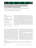
Fig. 1. Molecular cloning of CcCac1L and its interaction with CcLim15. (A) Schematic diagram of the CAF-1 large subunits in human, C. cine-
rea and S. cerevisiae. The KER and ED domains are represented by black and grey boxes, respectively. (B) Schematic diagram of the trunca-
tion mutants of CcCac1L. (C) Interaction between CcCac1L and CcLim15 in a yeast two-hybrid assay. The inserts in the activation domain
(AD) and DNA-binding domain (BK) are shown. +, binding; ), no binding. The mild selection medium (SD3: –His/–Leu/–Trp) and the stringent
selection medium (SD4: –Ade/–His/–Leu/–Trp) were tested. (D) Interaction between CcCac1L and CcLim15 in yeast using quantitative
b-galactosidase assays. b-Galactosidase assays with the other vector pairs in (C) showed little activity below the detection limit of
absorbance, and were not quantified.
Link between Lim15/Dmc1 and the CAF-1–PCNA complex S. Ishii et al.
2034 FEBS Journal 275 (2008) 2032–2041 ª 2008 The Authors Journal compilation ª 2008 FEBS
experiment confirmed the specific interaction of
CcCac1L and CcLim15 in the crude extracts of mei-
otic tissues (Fig. 2E). Taken together, these results
suggest that the interaction between CcLim15 and
CcCac1L is related to specific events during the mei-
otic prophase.
Interaction between CcCac1L and CcPCNA
during meiosis
CAF-1 forms a complex with PCNA to deposit
histones at the site of newly synthesized DNA during
replication and repair. The results above raised the
novel possibility that CAF-1 is involved in chromatin
assembly following recombination-associated DNA
synthesis during meiosis. If so, CAF-1 must form a
complex with PCNA in the meiotic prophase. PCNA
is expressed abundantly in meiotic prophase I [32].
Interestingly, recent analysis has revealed that PCNA
interacts with Lim15/Dmc1 at the time of meiotic
recombination [18]. To determine whether CcCac1L
interacts with CcPCNA during meiosis, we performed
co-immunoprecipitation analysis using cell extracts
from the meiotic prophase in C. cinerea. CcPCNA was
specifically co-immunoprecipitated by anti-CcCac1L
IgG, but not by control rabbit IgG (Fig. 3A). The
A
C
B
D
E
Fig. 2. Interaction between CcCac1L and CcLim15 during meiosis. (A) Northern analysis of CcCac1L expression at various stages during
meiosis. Each lane contained 20 lg of total RNA isolated from meiotic cells of C. cinerea at the premeiotic S phase and at every hour after
karyogamy (the initiation of meiosis) to 9 h after karyogamy. The blot was hybridized with either CcCac1L (top panel) or C. cinerea glyceral-
dehyde 3-phosphate dehydrogenase (CcG3PDH; bottom panel). (B) Western analysis of the rat anti-CcCac1L IgG. The cell extract at the mei-
otic prophase was examined. (C) Nuclear localization of CcLim15 and CcCac1L in the nuclei of C. cinerea meiotic cells. Meiotic nuclei were
stained with anti-CcCac1L IgG (red) and anti-CcLim15 IgG (green). The nuclei were then counterstained with 4¢,6-diamidino-2-phenylindole di-
hydrochloride n-hydrate (DAPI). The meiotic stages are indicated on the left. (D, E) Immunoprecipitation of CcCac1L and CcLim15 from the
cell extract at the meiotic prophase; 20 mg of cell extract was incubated with anti-CcLim15 IgG, anti-CcCac1L IgG or control rabbit serum-
conjugated beads. After washing the beads, the bound proteins were eluted and analysed by western analysis using anti-CcLim15 IgG (D) or
anti-CcCac1L IgG (E). Lane 1, 100 lg of crude extract was loaded.
S. Ishii et al. Link between Lim15/Dmc1 and the CAF-1–PCNA complex
FEBS Journal 275 (2008) 2032–2041 ª 2008 The Authors Journal compilation ª 2008 FEBS 2035
reciprocal experiment confirmed the specific interaction
of CcCac1L and CcPCNA in the crude extracts of
meiotic tissues (Fig. 3B).
Next, we sought to examine the binding affinity of
CcCac1L to CcPCNA by performing BIAcore analysis
with the truncated mutants of CcCac1L, as shown in
Fig. 1B. The BIAcore system enabled us to detect the
surface plasmon resonance (SPR), which measures the
interaction between a ligand on a detection surface
(sensor chip) and a ligand that is injected. First, we
conjugated CcPCNA to a sensor chip onto which
either CcCac1L-N or CcCac1L-C was injected. Consis-
tent with results from other organisms [27,33],
CcCac1L-N specifically bound to CcPCNA (Fig. 3C),
confirming the evolutionarily conserved CAF-1–PCNA
complex. From these results, we suggest a novel role
of the CAF-1–PCNA complex during the meiotic pro-
phase together with the meiosis-specific recombinase,
Lim15/Dmc1.
Discussion
In this study, we identified CcCac1L as a novel
interacting partner of CcLim15. Furthermore, it was
shown that CcCac1L interacts with CcPCNA during
the meiotic prophase. Several DNA synthesis events
take place during the meiotic prophase, even after
genome-wide replication at the premeiotic S phase
[32,34]. In the current model, DNA synthesis is
required in the molecular events of meiotic recombi-
nation [35,36]. Meiotic DSBs are processed to single-
strand overhangs, followed by single-strand invasion
to the other allele. Recombination results in either
crossover products (exchanging the flanking DNA
arms between homologues) or non-crossover products
(non-exchange of DNA arms). Both pathways
accompany DNA synthesis following recombination
[35,36]. Given the coordination of CAF-1 and PCNA
in various DNA synthesis events, a CAF-1–PCNA
complex may be involved in chromatin assembly fol-
lowing DNA synthesis events during the meiotic pro-
phase. Based on the current model, we propose the
role of the CAF-1–PCNA complex during meiosis
(Fig. 4). PCNA recruits DNA polymerase at the
end of single-strand regions that are coated by
Lim15/Dmc1 (Fig. 4A,B). Consistent with this model,
DNA polymerases and DNA ligases are active dur-
ing this stage [37–40]. After DNA synthesis, CAF-1
is recruited to the site of the Lim15/Dmc1–PCNA
complex and deposits histone H3 (or a histone vari-
ant) and H4 on the naked DNA to restore the
nucleosome structure (Fig. 4C). Because of the vari-
ous interactions of Lim15/Dmc1–CAF-1–PCNA, we
suggest that they act in multiple ways at the site of
meiotic recombination and contribute to the subse-
quent assembly of chromatin. Therefore, there may
be coordination between meiotic recombination and
CAF-1-dependent nucleosome assembly before the
resolution of Holliday junctions (Fig. 4C).
The CAF-1–PCNA complex senses DNA damage
and subsequently contributes to chromatin assembly
at the site of DNA repair [33], including nucleotide
excision repair [28] and DSB repair [29,30]. During
the process of chromatin assembly, CAF-1 deposits
new H3.1 histones on the site of repair-associated
DNA synthesis without the recycling of parental
histones; therefore, CAF-1-dependent chromatin
assembly results in a chromatin memory of damage
at a repair site [41]. Similarly, CAF-1 may establish
a chromatin memory at the site of DNA synthesis
A
B
C
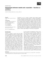
Fig. 3. Interaction between CcCac1L and CcPCNA during meiosis.
(A, B) Co-immunoprecipitation of CcCac1L and PCNA in the cell
extract at the meiotic prophase; 20 mg of cell extract was incu-
bated with anti-CcPCNA IgG, anti-CcCac1L IgG or control rabbit
serum-conjugated beads. After washing the beads, the bound
proteins were eluted and analysed by western analysis with anti-
PCNA IgG (A) or anti-CcCac1L IgG (B). Lane 1, 100 lg of crude
extract was loaded. (C) Detection of SPR using a Biacore assay.
Truncation mutants of CcCac1L were injected onto a CcPCNA
conjugated chip. The binding affinity is inversely related to the
dissociation constant (K
D
), which is a ratio of the dissociation (K
d
)
and association (K
a
) rates (K
D
= K
d
/K
a
). ND, not detected.
Link between Lim15/Dmc1 and the CAF-1–PCNA complex S. Ishii et al.
2036 FEBS Journal 275 (2008) 2032–2041 ª 2008 The Authors Journal compilation ª 2008 FEBS
following meiotic recombination. The site of cross-
over recombination becomes the chiasma, required
for the appropriate segregation of homologous chro-
mosomes. Chiasma formation involves the coordi-
nated local change of DNA and the surrounding
chromatin environment [42]. One tantalizing possibil-
ity is that CAF-1-dependent chromatin memory
directs chiasma formation to newly synthesized DNA
at the site of recombination. CAF-1-dependent his-
tone deposition is an established key early step for
chromatin organization in mitosis [19,24–26]. Multi-
ple steps are involved in organizing the chromatin
structure after histone deposition by CAF-1. There-
fore, the CAF-1–PCNA complex may be the central
player establishing the memory of recombination,
leading to unique nuclear organization during
meiosis.
Materials and methods
Culture of C. cinerea and collection of fruiting
bodies
The basidiomycete Coprinopsis cinerea (Coprinus cinereus)
(strain #56838) was purchased from the American Type
Culture Collection (ATCC), Manassas, VA, USA. The
culture methods and procedures for the photoinduction of
meiosis were performed as described previously [38,43].
Yeast two-hybrid screening
The C. cinerea cDNA library in meiotic tissues was con-
structed using a Time Saver cDNA Synthesis Kit (GE
Healthcare UK Ltd, Little Chalfont, UK). Yeast
two-hybrid screening was carried out using the MATCH-
MAKER GAL4 Two-Hybrid System 3 (Clontech, Moun-
tain View, CA, USA). The cDNA encoding full-length
CcLim15 was fused in-frame with the GAL4 DNA-binding
domain in the pBKDT7 vector as bait. The cDNA library
was subsequently cloned into the pGADT7 vector encoding
the GAL4 activation domain, and used as prey in the two-
hybrid experiments. Both the GAL4 fusion bait and the
prey plasmids were transformed into the yeast strain,
AH109 (Clontech), by standard lithium acetate transforma-
tion. Putative interacting clones were subsequently isolated
based on their ability to activate the expression of the
GAL4 selectable marker genes, thus producing growth on
SD minimal medium lacking adenine, histidine, leucine
and tryptophan (SD4: –Ade/–His/–Leu/–Trp). To confirm
galactosidase activity, colonies that grew under this selective
condition were plated onto SD4 medium with X-a-galacto-
sidase. Purified plasmids from yeast clones were electropo-
rated into Escherichia coli DH10B. After the plasmid DNA
had been prepared, the cDNA inserts were sequenced and
the corresponding gene was identified by blast analysis.
cDNA cloning of CcCac1L
One of the interacting factors identified in our screen was
found to encode the CcCac1L C-terminus, consisting of
the amino acid region 382–812 (CcCac1L-C) (Fig. 1B). To
obtain the full-length CcCac1L cDNA, 5¢RNA ligase-medi-
ated-RACE (5¢RLM-RACE) (Ambion, Austin, TX, USA)
and 3¢RLM-RACE (Invitrogen, Carlsbad, CA, USA)
experiments were performed, each according to the manu-
facturer’s protocol. The DDBJ/EMBL/GenBank accession
number of the nucleotide sequence for CcCac1L reported
in this study is AB074897.
A
B
C
Fig. 4. Model of chromatin assembly following meiotic recombina-
tion. (A) After DSB formation, Lim15/Dmc1 coats the single-strand
end during strand invasion. (B) PCNA recruits the DNA polymerase
to the site of Lim15/Dmc1. The broken line represents newly syn-
thesized DNA. (C) CAF-1 forms a complex with Lim15/Dmc1 and
PCNA. CAF-1 deposits histones H3 and H4 or other factors, such
as histone variants (indicated as ‘?’), on the newly synthesized
DNA.
S. Ishii et al. Link between Lim15/Dmc1 and the CAF-1–PCNA complex
FEBS Journal 275 (2008) 2032–2041 ª 2008 The Authors Journal compilation ª 2008 FEBS 2037
Two-hybrid assay
To confirm the direct interaction between proteins or pro-
tein fragments, the appropriate bait and prey constructs
were co-transformed into yeast cells, and two-hybrid assays
were performed using the MATCH-MAKER Kit (Clon-
tech), according to the manufacturer’s instructions. The
full-length CcLim15, CcCac1L, CcCac1L-N and CcCac1L-
C fragments were cloned into pGADT7 and pGBKT7. The
vector pairs indicated in Fig. 1C were co-transformed into
the yeast strain AH109. Controls for self-activating fusion
proteins were carried out in each of these assays by trans-
formation of specific expression constructs with a pGBKT7
or pGADT7 empty vector. Transformants were then plated
onto three types of selection medium: SD2, –Leu/–Trp;
SD3, –His/–Leu/–Trp; SD4, –Ade/–His/–Leu/–Trp.
b-Galactosidase assays were performed in chlorophenol
red-b-d-galactopyranoside (CPRG)-based liquid culture
using the individual colonies that grew in SD3 medium,
according to the Yeast Protocols Handbook (Clontech).
Northern blotting
Northern blotting was performed as described previously
[44]. The region of the CcCac1L cDNA corresponding to
1146–2346 bp was used as a probe.
Antibodies
A polyclonal antibody against the CcCac1L protein was
raised in rabbit and rat using the purified 382–812 amino
acid fragment expressed as a His-CcCac1L-C protein in
E. coli. The specificity of the antibodies was confirmed by
western analysis as described previously [44,45]. A poly-
clonal antibody against CcLim15 was also raised as
described previously [45]. Anti-CcPCNA IgGs and purified
recombinant His-tagged CcPCNA (His-CcPCNA) have
been described previously [44].
In vivo co-immunoprecipitation
Rabbit anti-CcCac1L polyclonal IgGs rabbit anti-
CcLim15 polyclonal IgG or control rabbit serum was
coupled with CNBr-activated sepharose beads, according
to the manufacturer’s instructions {20 mg aliquots of
crude extracts from meiotic tissues were prepared in buf-
fer D [buffer C, as described below, with 0.6 m NaCl and
protease inhibitors (1 mm phenylmethanesulfonyl fluoride,
1 lm leupeptin and 1 lm pepstatin A)]}. The extracts in
buffer D were then incubated with either 70 lL of pri-
mary antibody or with control rabbit serum-conjugated
beads for 1 h at 4 °C. The beads were then collected by
centrifugation at 800 g for 30 s. After resuspension of the
beads in buffer E (0.15 m NaCl in buffer D), the superna-
tant was removed by centrifugation at 9100 g for 30 s. The
bound material was eluted from the beads with 20 lL
of buffer F (50 mm glycine/HCl, pH 2.5, and 0.01%
Triton X-100). After neutralization of the pH by the addition
of 1 m Tris/HCl, pH 7.5, the bound material was analysed by
immunoblotting with either anti-CcCac1L or anti-CcLim15
IgG, both at a dilution of 1 : 1000. To test the interaction
between CcCac1L and CcPCNA in vivo, anti-CcCac1L and
anti-CcPCNA IgGs were used and in vivo immunoprecipita-
tion experiments were performed as described previously
[44]. The CcCac1L cDNA corresponding to 1146–2346 bp
was used as a probe.
Immunostaining of nuclei of C. cinerea meiotic
cells
Immunostaining of nuclei of C. cinerea meiotic cells was
performed as described previously [38]. A 1 : 100 dilution
was used of both rabbit anti-CcLim15 and rat anti-
CcCac1L primary IgGs. We also employed a 1 : 1000 dilu-
tion of both anti-rabbit IgG conjugated with Alexa Fluoro
488 (Invitrogen) for anti-CcLim15 and anti-rat IgG conju-
gated with Alexa Fluoro 568 (Invitrogen) for anti-CcCac1L
as secondary antibodies.
Proteins
A truncated cDNA corresponding to the N-terminus (resi-
dues 1–381, as shown in Fig. 1B) of CcCac1L (CcCac1L-N)
was cloned into the Bam HI and NotI sites of the expression
vector pET21a(+) (Novagen, Gibbstown, NJ, USA). The
C-terminal insert of CcCac1L (CcCac1L-C, residues 382–
812) was cloned into the NcoI and XhoI sites of the
pET21d(+) expression vector (Novagen). The following
primer pairs were used for subsequent PCR amplification
of these cDNAs. CcCac1L-N: 1F, 5¢-CGGGATCCA
TGTCGGGAGCAGATTCA; 381R, 5¢-TGCTACTTCTC
TCAGCGGCCGCATTCTTAT. CcCac1L-C: 382F, 5¢-CA
TGCCATGGTGTCAGGGGATGTAGAAATG; 812R,
5¢-GAGATTTCAGTTTCGTCACTCGAGCGG. To over-
express N-terminal hexahistidine-tagged CcCac1L-N
(His-CcCac1L-N) and CcCac1L-C (His-CcCac1L-C), E. coli
BL21 cells (DE3) (Novagen) carrying the expression plasmid
for each gene were grown in 2 · YT medium (16 gÆL
)1
poly-
peptone, 10 gÆL
)1
yeast extract, 5 gÆL
)1
NaCl) containing
1 lgÆmL
)1
ampicillin at 37 °C. After reaching an absorbance
at 600 nm of 0.6, isopropyl thio-b-d-galactoside (IPTG) was
added to these cultures at a final concentration of 1 mm,
and the cells were incubated for an additional 5 h at 25 °C.
The bacterial cells were then harvested by centrifugation at
4500 g for 15 min, and the resulting cell pellet was resus-
pended in 15 mL of ice-cold buffer A [20 mm Tris/HCl,
pH 7.9, 10% glycerol, 0.5 m NaCl, 5 mm imidazole con-
taining protease inhibitors (1 mm phenylmethanesulfonyl
Link between Lim15/Dmc1 and the CAF-1–PCNA complex S. Ishii et al.
2038 FEBS Journal 275 (2008) 2032–2041 ª 2008 The Authors Journal compilation ª 2008 FEBS
fluoride, 1 lm leupeptin and 1 lm pepstatin A)]. The cells
were then lysed by the addition of 1 mgÆmL
)1
lysozyme, stir-
red on ice for 30 min and sonicated. Insoluble material was
removed by centrifugation at 26 000 g for 15 min. Proteins
were loaded onto a 5 mL Hi-trap chelating column (GE
Healthcare UK Ltd.), and bound proteins were eluted with
a 20 mL linear gradient of 0.05–1 m imidazole in buffer B
(buffer A with 0.1% Nonidet P40). The eluted protein frac-
tion was then dialysed against buffer C (50 mm Tris/HCl,
pH 7.5, 0.05 m NaCl, 1 mm EDTA, 5 mm 2-mercaptoetha-
nol, 10% glycerol, 0.1% Nonidet P40), and the dialysate
was loaded onto a heparin column (GE Healthcare UK
Ltd.) equilibrated with 0.05 m NaCl in buffer B. After
washing, fractions were collected with a 20 mL linear gradi-
ent of 0–0.5 m NaCl in buffer B. The eluted protein was
then dialysed against 0.05 m NaCl in buffer B, and loaded
onto a MonoQ HR5/5 column (GE Healthcare UK Ltd).
After washing, the fractions were again collected with
20 mL of a linear gradient of 0–0.5 m NaCl in buffer B.
Fractions containing the recombinant proteins were verified
by SDS-PAGE, pooled and then dialysed against storage
buffer (NaCl/P
i
, pH 7.4, 50% glycerol). Recombinant His-
tagged CcLim15 (His-CcLim15) was expressed in E. coli
and purified as described previously [31].
Surface plasmon resonance
Analysis of both His-CcCac1L-N and His-CcCac1L-C
binding to His-CcPCNA was performed using a BIAcore
Biosensor instrument (GE Healthcare Bio-Sciences,
Uppsala, Sweden), according to the manufacturer’s proto-
col. A sensor chip (CM 5 research grade) was activated by
the N-hydroxysuccinimide/N-ethyl-N¢-(dimethylaminopro-
pyl)carbodiimide coupling reaction, and 55 lL of coupling
buffer (10 mm sodium acetate, pH 4.0) containing the
His-CcPCNA protein (625 nm) was injected over the chip
at a rate of 20 lLÆmin
)1
. His-CcPCNA was covalently
bound to the sensor chip surface via carboxyl moieties on
the dextran. Unreacted N-hydroxysuccinimide ester groups
were inactivated using 1 m ethanolamine/HCl (pH 8.0).
HBS-EP buffer (10 mm Hepes, pH 7.4, 150 mm NaCl,
3mm EDTA, 0.005% Tween 20) was passed continuously
over the sensor chip. The binding levels were measured in
resonance units (RU); 1000 RU of protein corresponds to
a surface concentration alteration of approximately
1ngÆmm
)2
[46]. In this experiment, approximately 6600
RU of His-CcPCNA was immobilized onto the chip
surface. The binding of His-CcPCNA to either
His-CcCac1L-N or His-CcCac1L-C was performed in a
reaction containing 20 lL of HBS-EP buffer with three
different concentrations of His-CcCac1L-N or His-
CcCac1L-C (250 nm, 500 nm or 1 lm). The running buffer
(HBS-EP buffer) flow rate was 5 lLÆmin
)1
at 37 °C. All
data were monitored and analysed using the manufac-
turer’s software (GE Healthcare Bio-Sciences).
Acknowledgements
We thank Montserrat Anguera, Jennifer Erwin and
Janice Ahn for critical reading of the manuscript, and
all members of Sakaguchi Laboratory for help and dis-
cussions. S. H. N. is a research fellow of the Japan
Society for Promotion of Science.
References
1 Kleckner N (2006) Chiasma formation: chromatin/axis
interplay and the role(s) of the synaptonemal complex.
Chromosoma 115, 175–194.
2 Masson JY & West SC (2001) The Rad51 and Dmc1
recombinases: a non-identical twin relationship. Trends
Biochem Sci 26, 131–136.
3 Namekawa SH, Iwabata K, Sugawara H, Hamada FN,
Koshiyama A, Chiku H, Kamada T & Sakaguchi K
(2005) Knockdown of LIM15/DMC1 in the mushroom
Coprinus cinereus by double-stranded RNA-mediated
gene silencing. Microbiology 151, 3669–3678.
4 Neale MJ & Keeney S (2006) Clarifying the mechanics
of DNA strand exchange in meiotic recombination.
Nature 442, 153–158.
5 Villeneuve AM & Hillers KJ (2001) Whence meiosis?
Cell 106, 647–650.
6 Bishop DK (1994) RecA homologs Dmc1 and Rad51
interact to form multiple nuclear complexes prior to
meiotic chromosome synapsis. Cell 79, 1081–1092.
7 Tarsounas M, Morita T, Pearlman RE & Moens PB
(1999) RAD51 and DMC1 form mixed complexes asso-
ciated with mouse meiotic chromosome cores and syn-
aptonemal complexes. J Cell Biol 147, 207–220.
8 Shinohara M, Gasior SL, Bishop DK & Shinohara A
(2000) Tid1/Rdh54 promotes colocalization of rad51
and dmc1 during meiotic recombination. Proc Natl
Acad Sci USA 97, 10814–10819.
9 Chen YK, Leng CH, Olivares H, Lee MH, Chang YC,
Kung WM, Ti SC, Lo YH, Wang AH, Chang CS et al.
(2004) Heterodimeric complexes of Hop2 and Mnd1
function with Dmc1 to promote meiotic homolog juxta-
position and strand assimilation. Proc Natl Acad Sci
USA 101, 10572–10577.
10 Enomoto R, Kinebuchi T, Sato M, Yagi H, Kurumi-
zaka H & Yokoyama S (2006) Stimulation of DNA
strand exchange by the human TBPIP/Hop2-Mnd1
complex. J Biol Chem 281, 5575–5581.
11 Petukhova GV, Pezza RJ, Vanevski F, Ploquin M,
Masson JY & Camerini-Otero RD (2005) The Hop2
and Mnd1 proteins act in concert with Rad51 and
Dmc1 in meiotic recombination. Nat Struct Mol Biol
12, 449–453.
12 Hayase A, Takagi M, Miyazaki T, Oshiumi H,
Shinohara M & Shinohara A (2004) A protein complex
containing Mei5 and Sae3 promotes the assembly of the
S. Ishii et al. Link between Lim15/Dmc1 and the CAF-1–PCNA complex
FEBS Journal 275 (2008) 2032–2041 ª 2008 The Authors Journal compilation ª 2008 FEBS 2039
meiosis-specific RecA homolog Dmc1. Cell 119, 927–
940.
13 Tsubouchi H & Roeder GS (2004) The budding yeast
mei5 and sae3 proteins act together with dmc1 during
meiotic recombination. Genetics 168, 1219–1230.
14 Neyton S, Lespinasse F, Moens PB, Paul R, Gaudray
P, Paquis-Flucklinger V & Santucci-Darmanin S (2004)
Association between MSH4 (MutS homologue 4) and
the DNA strand-exchange RAD51 and DMC1 proteins
during mammalian meiosis. Mol Hum Reprod 10, 917–
924.
15 Habu T, Wakabayashi N, Yoshida K, Yomogida K,
Nishimune Y & Morita T (2004) p53 Protein interacts
specifically with the meiosis-specific mammalian RecA-
like protein DMC1 in meiosis. Carcinogenesis 25, 889–
893.
16 Iwabata K, Koshiyama A, Yamaguchi T, Sugawara H,
Hamada FN, Namekawa SH, Ishii S, Ishizaki T, Chiku
H, Nara T et al. (2005) DNA topoisomerase II interacts
with Lim15/Dmc1 in meiosis. Nucleic Acids Res 33,
5809–5818.
17 Koshiyama A, Hamada FN, Namekawa SH, Iwabata
K, Sugawara H, Sakamoto A, Ishizaki T & Sakaguchi
K (2006) Sumoylation of a meiosis-specific RecA homo-
log, Lim15/Dmc1, via interaction with the small ubiqu-
itin-related modifier (SUMO)-conjugating enzyme
Ubc9. Febs J 273, 4003–4012.
18 Hamada FN, Koshiyama A, Namekawa SH, Ishii S,
Iwabata K, Sugawara H, Nara TY, Sakaguchi K &
Sawado T (2007) Proliferating cell nuclear antigen
(PCNA) interacts with a meiosis-specific RecA homo-
logue, Lim15/Dmc1, but does not stimulate its strand
transfer activity. Biochem Biophys Res Commun 352,
836–842.
19 Kaufman PD, Kobayashi R, Kessler N & Stillman B
(1995) The p150 and p60 subunits of chromatin
assembly factor I: a molecular link between newly
synthesized histones and DNA replication. Cell 81,
1105–1114.
20 Kaufman PD, Kobayashi R & Stillman B (1997) Ultra-
violet radiation sensitivity and reduction of telomeric
silencing in Saccharomyces cerevisiae cells lacking chro-
matin assembly factor-I. Genes Dev 11, 345–357.
21 Tyler JK, Collins KA, Prasad-Sinha J, Amiott E, Bulger
M, Harte PJ, Kobayashi R & Kadonaga JT (2001) Inter-
action between the Drosophila CAF-1 and ASF1 chro-
matin assembly factors. Mol Cell Biol 21, 6574–6584.
22 Verreault A, Kaufman PD, Kobayashi R & Stillman B
(1996) Nucleosome assembly by a complex of CAF-1
and acetylated histones H3/H4. Cell 87, 95–104.
23 Kaya H, Shibahara KI, Taoka KI, Iwabuchi M, Still-
man B & Araki T (2001) FASCIATA genes for chro-
matin assembly factor-1 in Arabidopsis maintain the
cellular organization of apical meristems. Cell 104, 131–
142.
24 Gaillard PH, Martini EM, Kaufman PD, Stillman B,
Moustacchi E & Almouzni G (1996) Chromatin assem-
bly coupled to DNA repair: a new role for chromatin
assembly factor I. Cell 86, 887–896.
25 Smith S & Stillman B (1989) Purification and character-
ization of CAF-I, a human cell factor required for chro-
matin assembly during DNA replication in vitro. Cell
58, 15–25.
26 Stillman B (1986) Chromatin assembly during SV40
DNA replication in vitro. Cell 45, 555–565.
27 Shibahara K & Stillman B (1999) Replication-depen-
dent marking of DNA by PCNA facilitates CAF-1-cou-
pled inheritance of chromatin. Cell 96, 575–585.
28 Green CM & Almouzni G (2003) Local action of the
chromatin assembly factor CAF-1 at sites of nucleotide
excision repair in vivo. EMBO J 22
, 5163–5174.
29 Linger J & Tyler JK (2005) The yeast histone chaperone
chromatin assembly factor 1 protects against double-
strand DNA-damaging agents. Genetics 171, 1513–1522.
30 Nabatiyan A, Szuts D & Krude T (2006) Induction of
CAF-1 expression in response to DNA strand breaks in
quiescent human cells. Mol Cell Biol 26, 1839–1849.
31 Nara T, Yamamoto T & Sakaguchi K (2000) Charac-
terization of interaction of C- and N-terminal domains
in LIM15/DMC1 and RAD51 from a basidiomycete,
Coprinus cinereus. Biochem Biophys Res Commun 275,
97–102.
32 Hotta Y, Ito M & Stern H (1966) Synthesis of DNA
during meiosis. Proc Natl Acad Sci USA 56, 1184–1191.
33 Moggs JG, Grandi P, Quivy JP, Jonsson ZO, Hubscher
U, Becker PB & Almouzni G (2000) A CAF-1–PCNA-
mediated chromatin assembly pathway triggered by
sensing DNA damage. Mol Cell Biol 20, 1206–1218.
34 Lu BC & Jeng DY (1975) Meiosis in Coprinus VII. The
prekaryogamy S-phase and the postkaryogamy DNA
replication in C. lagopus. J Cell Sci 17, 461–470.
35 Allers T & Lichten M (2001) Differential timing and
control of noncrossover and crossover recombination
during meiosis. Cell 106, 47–57.
36 Hunter N & Kleckner N (2001) The single-end invasion:
an asymmetric intermediate at the double-strand break
to double-Holliday junction transition of meiotic recom-
bination. Cell 106, 59–70.
37 Namekawa S, Hamada F, Ishii S, Ichijima Y, Yamagu-
chi T, Nara T, Kimura S, Ishizaki T, Iwabata K, Ko-
shiyama A et al. (2003) Coprinus cinereus DNA ligase I
during meiotic development. Biochim Biophys Acta
1627, 47–55.
38 Namekawa S, Hamada F, Sawado T, Ishii S, Nara T,
Ishizaki T, Ohuchi T, Arai T & Sakaguchi K (2003)
Dissociation of DNA polymerase alpha-primase com-
plex during meiosis in Coprinus cinereus. Eur J Biochem
270, 2137–2146.
39 Sakaguchi K & Lu BC (1982) Meiosis in Coprinus:
characterization and activities of two forms of DNA
Link between Lim15/Dmc1 and the CAF-1–PCNA complex S. Ishii et al.
2040 FEBS Journal 275 (2008) 2032–2041 ª 2008 The Authors Journal compilation ª 2008 FEBS
polymerase during meiotic stages. Mol Cell Biol 2, 752–
757.
40 Sawado T & Sakaguchi K (1997) A DNA polymerase
alpha catalytic subunit is purified independently from
the tissues at meiotic prometaphase I of a basidiomy-
cete, Coprinus cinereus. Biochem Biophys Res Commun
232, 454–460.
41 Polo SE, Roche D & Almouzni G (2006) New histone
incorporation marks sites of UV repair in human cells.
Cell 127, 481–493.
42 Blat Y, Protacio RU, Hunter N & Kleckner N (2002)
Physical and functional interactions among basic chro-
mosome organizational features govern early steps of
meiotic chiasma formation. Cell 111, 791–802.
43 Nara T, Saka T, Sawado T, Takase H, Ito Y, Hotta Y
& Sakaguchi K (1999) Isolation of a LIM15/DMC1
homolog from the basidiomycete Coprinus cinereus and
its expression in relation to meiotic chromosome pair-
ing. Mol Gen Genet 262, 781–789.
44 Hamada F, Namekawa S, Kasai N, Nara T, Kimura S,
Sugawara F & Sakaguchi K (2002) Proliferating cell
nuclear antigen from a basidiomycete, Coprinus cinere-
us. Alternative truncation and expression in meiosis.
Eur J Biochem 269, 164–174.
45 Nara T, Hamada F, Namekawa S & Sakaguchi K
(2001) Strand exchange reaction in vitro and DNA-
dependent ATPase activity of recombinant LIM15/
DMC1 and RAD51 proteins from Coprinus cinereus.
Biochem Biophys Res Commun 285, 92–97.
46 Wong CW, Komm B & Cheskis BJ (2001) Structure–
function evaluation of ER alpha and beta interplay
with SRC family coactivators. ER selective ligands.
Biochemistry 40, 6756–6765.
S. Ishii et al. Link between Lim15/Dmc1 and the CAF-1–PCNA complex
FEBS Journal 275 (2008) 2032–2041 ª 2008 The Authors Journal compilation ª 2008 FEBS 2041