Báo cáo khoa học: Molecular basis of perinatal hypophosphatasia with tissue-nonspecific alkaline phosphatase bearing a conservative replacement of valine by alanine at position 406 Structural importance of the crown domain potx
Bạn đang xem bản rút gọn của tài liệu. Xem và tải ngay bản đầy đủ của tài liệu tại đây (535.31 KB, 11 trang )
Molecular basis of perinatal hypophosphatasia with
tissue-nonspecific alkaline phosphatase bearing
a conservative replacement of valine by alanine at
position 406
Structural importance of the crown domain
Natsuko Numa
1
, Yoko Ishida
2
, Makiko Nasu
3
, Miwa Sohda
2
, Yoshio Misumi
4
, Tadashi Noda
1
and Kimimitsu Oda
2,5
1 Division of Pediatric Dentistry, Niigata University Graduate School of Medical and Dental Sciences, Japan
2 Division of Oral Biochemistry, Niigata University Graduate School of Medical and Dental Sciences, Japan
3 Division of Oral Health in Aging and Fixed Prosthodontics, Niigata University Graduate School of Medical and Dental Sciences, Japan
4 Department of Cell Biology, Fukuoka University School of Medicine, Japan
5 Center for Transdisciplinary Research, Niigata University, Japan
Keywords
crown domain; glycosylphosphatidylinositol;
hypophosphatasia; raft; tissue-nonspecific
alkaline phosphatase
Correspondence
K. Oda, Division of Oral Biochemistry,
Niigata University Graduate School of
Medical and Dental Sciences, 2-5274,
Gakkocho-dori, Niigata 951-8514, Japan
Fax: +81 25 227 0805
Tel: +81 25 227 2827
E-mail:
(Received 30 November 2007, revised 18
January 2008, accepted 19 March 2008)
doi:10.1111/j.1742-4658.2008.06414.x
Hypophosphatasia, a congenital metabolic disease related to the tissue-non-
specific alkaline phosphatase gene (TNSALP), is characterized by reduced
serum alkaline phosphatase levels and defective mineralization of hard tis-
sues. A replacement of valine with alanine at position 406, located in the
crown domain of TNSALP, was reported in a perinatal form of hypophos-
phatasia. To understand the molecular defect of the TNSALP (V406A)
molecule, we examined this missense mutant protein in transiently trans-
fected COS-1 cells and in stable CHO-K1 Tet-On cells. Compared with the
wild-type enzyme, the mutant protein showed a markedly reduced alkaline
phosphatase activity. This was not the result of defective transport and
resultant degradation of TNSALP (V406A) in the endoplasmic reticulum,
as the majority of newly synthesized TNSALP (V406A) was conveyed to
the Golgi apparatus and incorporated into a cold detergent insoluble frac-
tion (raft) at a rate similar to that of the wild-type TNSALP. TNSALP
(V406A) consisted of a dimer, as judged by sucrose gradient centrifugation,
suggestive of its proper folding and correct assembly, although this mutant
showed increased susceptibility to digestion by trypsin or proteinase K.
When purified as a glycosylphosphatidylinositol-anchorless soluble form,
the mutant protein exhibited a remarkably lower K
cat
⁄ K
m
value compared
with that of the wild-type TNSALP. Interestingly, leucine and isoleucine,
but not phenylalanine, were able to substitute for valine, pointing to the
indispensable role of residues with a longer aliphatic side chain at position
406 of TNSALP. Taken together, this particular mutation highlights the
structural importance of the crown domain with respect to the catalytic
function of TNSALP.
Abbreviations
Endo H, endo-b-N-acetylglucosaminidase H; GPI, glycosylphosphatidylinositol; TNSALP (V406A), TNSALP with a valine to alanine substitution
at position 406; TNSALP, tissue-nonspecific alkaline phosphatase.
FEBS Journal 275 (2008) 2727–2737 ª 2008 The Authors Journal compilation ª 2008 FEBS 2727
Hypophosphatasia is caused by various mutations of
the tissue-nonspecific alkaline phosphatase (TNSALP)
gene (EC 3.1.3.1) [1–6]. To date a total of 191 distinct
mutations have been reported worldwide, and about
80% of these mutations are missense (http://www.
sesep.uvsq.fr./Database.html). Hypophosphatasia is
characterized by reduced levels of serum alkaline phos-
phatase activity and defective mineralization in bone
and tooth, and clinical severity is inversely correlated
to serum alkaline phosphatase levels [1,2,7]. Patients
suffering from severe hypophosphatasia, such as the
perinatal or infantile forms, develop severe defects in
skeletal bone mineralization, unequivocally demon-
strating that TNSALP is physiologically involved in
the mineralization process of bone. Consistent with
this concept, TNSALP-deficient mice are reported to
develop rickets and osteomalacia [8–10].
During the course of our study on several TNSALP
mutant proteins associated with the severe form of
hypophosphatasia, we found that the cell-surface
expression of the TNSALP mutants is remarkably
reduced. The mutant proteins often fail to undergo
proper folding and correct assembly, resulting in accu-
mulation in the early stage of the secretory pathway
and eventual degradation in the endoplasmic reticulum
[11–15]. However, the extent to which each TNSALP
mutant protein reaches the cell surface varies from one
mutation to another, depending on the position of the
mutation in the gene and the amino acid residue that
is replaced. Fleisch et al. proposed that TNSALP regu-
lates mineralization by hydrolyzing inorganic pyro-
phosphate, a poison of hydroxyapatite crystal [16,17],
at the site of biomineralization. According to this pro-
posal, it is likely that defective bone formation occur-
ring in severe hypophosphatasia is closely related to
the number of cell-surface TNSALP mutant molecules
and their residual pyrophosphate-cleaving activity
[4,18,19].
Replacement of valine at position of 406 with
alanine was reported in a patient diagnosed with peri-
natal hypophosphatasia who was a compound hetero-
zygote for this mutation and A99T [20]. The valine
residue at position 406 is located in a unique domain
called the crown domain [21]. This domain shows the
lowest degree of homology among alkaline phospha-
tase isoenzymes, and isoenzyme-specific properties,
such as uncompetitive inhibition, heat stability and
allosteric behavior, are attributed to residues located
in this domain of each isoenzyme [4,22]. Besides, this
crown domain is responsible for interacting with
extracellular matrix proteins, including collagen [4,21–
24]. Here we demonstrate that, in contrast to other
missense mutations associated with severe hypophos-
phatasia, the majority of TNSALP (V406A) molecules
are capable of reaching the cell surface at a rate simi-
lar to that of the wild-type enzyme, thus excluding
the possibility that transport incompetence is a major
molecular defect of TNSALP (V406A). Rather, it is
likely that this particular mutation affects the active
site of TNSALP through imposing a subtle change
on the crown domain, rendering TNSALP (V406A)
less efficient for its catalytic function.
Results
Transient expression of TNSALP (V406A)
When expressed transiently, TNSALP (V406A) pro-
duced only a weak cytochemical reaction product
compared with the wild-type enzyme (Fig. 1A). In
agreement with these staining patterns, the specific
alkaline phosphatase activity of the cell homogenate
expressing the mutant protein was less than one-quar-
ter of that of the cell homogenate expressing the
wild-type enzyme (Fig. 1B). Immunoblotting con-
firmed that both the wild-type protein and the
TNSALP (V406A) mutant consisted of a 66-kDa and
an 80-kDa molecular species (Fig. 1C), which repre-
sent an immature form bearing high mannose-type
N-linked oligosaccharides and a mature form bearing
complex-type oligosaccharides, respectively [11].
Besides, the amount of TNSALP (V406A) mutant
was similar to that of the wild-type protein in trans-
fected cells, indicating that the lower expression level
does not account for the low specific enzyme activity
of the former. Upon incubation with phosphatidylino-
sitol-specific phospholipase C, both the wild-type pro-
tein and the TNSALP (V406A) mutant were released
into the medium (Fig. 1D, lanes 4 and 8), confirming
that they are anchored to the cell surface via glyco-
sylphosphatidylinositol (GPI). By contrast, the 66-kDa
form was the only molecular species observed within
the cells expressing TNSALP (D289V) (Fig. 1C),
which is arrested and forms the disulfide-bonded
aggregate in the endoplasmic reticulum [15], and is
not able to gain access to the cell surface (Fig. 1D,
lane 12).
Expression of TNSALP (V406A) in a stable
cell line
In the transient expression system, even the wild-type
enzyme formed a disulfide-bonded aggregate, probably
as a result of the synthesis of an excess amount of
TNSALP (Fig. 1C, lanes 4–6, see the top of gel). In
the present experiment, TNSALPs were expressed
Molecular basis of perinatal form of hypophosphatasia N. Numa et al.
2728 FEBS Journal 275 (2008) 2727–2737 ª 2008 The Authors Journal compilation ª 2008 FEBS
under control of the CMV I.E. Enhancer ⁄ Promoter.
We observed the same aggregate also in a previous
experiment using a different expression vector contain-
ing the SV40 early promoter [11]. Shortage of a pre-
cursor of GPI in the endoplasmic reticulum may be
one of the reasons why a small but significant fraction
of the wild-type TNSALP forms the aggregate in
transfected COS-1 cells [25]. To circumvent this draw-
back of transient expression, we established CHO-K1
Tet-On cells harboring a plasmid encoding TNSALP
(V406A). When incubated with doxycycline (an ana-
logue of tetracycline) TNSALP (V406A) appeared on
the cell surface (Fig. 2A, panel a) and exhibited weak
enzyme activity (Fig. 2A, panel c). The protein was
induced only with doxycycline, and no disulfide-
bonded aggregate was found on the top of the gel
(Fig. 2B). Note that most of the cellular TNSALP
(V406A) was present as the 80-kDa mature form. This
is in marked contrast to the transiently transfected
cells, where the 66-kDa form was a predominant
molecular species (Fig. 1C). This immunoblotting pat-
tern of the CHO-K1 Tet-On cells resembles that of
Saos-2 cells [14] – osteosarcoma producing a large
amount of TNSALP. Figure 3A shows pulse–chase
labeling experiments in combination with endo-b-N-
glucosaminidase H (Endo H) digestion. The wild-type
enzyme was synthesized as the 66-kDa Endo H-sensi-
tive form, which quickly became the 80-kDa Endo H-
resistant form. The processing of the newly synthesized
wild-type enzyme was complete by the end of the 2-h
chase period. This was also the case for TNSALP
(V406A) with only a small fraction being sensitive,
even at the end of the 2-h chase. Compatible with this
result, both the wild-type protein and the mutant pro-
tein were partitioned into a cold Triton X-100-insolu-
ble fraction (the raft) at a similar rate (Fig. 3B),
further supporting that the folding and assembly pro-
cess and subsequent intracellular trafficking of
TNSALP (V406A) are largely normal in the stable cell
line.
Kinetics of the soluble form of TNSALP (V406A)
Consistent with the biosynthetic studies in Fig. 3, the
expression level in the CHO-K1 Tet-On cell of
TNSALP (V406A) was similar to that of the wild-type
protein, as shown in Fig. 4A,B. However, the
CHO-K1 Tet-On cells expressing TNSALP (V406A)
Fig. 1. Transient expression of TNSALP mutant proteins in COS-1 cells. (A) COS-1 cells expressing the wild-type TNSALP or the TNSALP
(V406A) mutant were stained for alkaline phosphatase activity. Each dish was incubated in a reaction mixture for 10 min at room tempera-
ture. In panels B and C, cell homogenates prepared from the transfected COS-1 cells were assayed for alkaline phosphatase (abscissa
enzyme activity expressed in unitsÆmg
)1
of protein) or analyzed by SDS-PAGE under reducing (Red) or non-reducing (Nonred) conditions, fol-
lowed by immunoblotting using anti-TNSALP serum. The arrowhead indicates the top of the resolving gel. The values are the means of two
independent experiments. (D) COS-1 cells expressing the wild-type TNSALP, or the TNSALP (V406A) or TNSALP (D289V) mutants were
labeled with [
35
S]methionine and incubated further in the absence (lanes 1, 2, 5, 6, 9 and 10) or presence (lanes 3, 4, 7, 8, 11 and 12) of
phosphatidylinositol-specific phospholipase C. Both cell lysates (C) and media (M) were subjected to immunoprecipitation, followed by SDS-
PAGE (under reducing condition) ⁄ fluorography. Left lane:
14
C-methylated protein markers of 200, 97.5, 66, 46 and 30 kDa, from the top of
the gel.
N. Numa et al. Molecular basis of perinatal form of hypophosphatasia
FEBS Journal 275 (2008) 2727–2737 ª 2008 The Authors Journal compilation ª 2008 FEBS 2729
Fig. 3. Biosynthesis of TNSALP (V406A) in CHO-K1 Tet-On cells. Established CHO-K1 Tet-On cells harboring a plasmid encoding the wild-
type TNSALP or the TNSALP (V406A) mutant, which had been cultured in the presence of 0.5 lgÆmL
)1
of doxycycline for 14 h, were pulse-
labeled with [
35
S]methionine for 30 min and then the cells were collected at the indicated chase periods. The cells were lysed in the lysis
buffer and subjected to immunoprecipitation in (A). The immunoprecipitates on beads were incubated with or without Endo H prior to analy-
sis by SDS-PAGE (reducing condition) ⁄ fluorography. Some degradation products were observed in the samples during incubation with the
glycosidase. Left lane:
14
C-methylated protein markers of 200, 97.5, 66, 46 and 30 kDa, from the top of the gel. In panel B, the metabolically
labeled cells were lysed in cold 1% Triton X-100 and centrifuged at 15 000 g for 10 min. Triton X-100 soluble (S) and insoluble (I) fractions
were separated. The latter was further lysed in the lysis buffer and incubated at 37 °C for 20 min to extract TNSALP from the raft. Both sol-
uble and insoluble fractions were subjected to immunoprecipitation, followed by SDS-PAGE (reducing condition) ⁄ fluorography. Left lane:
14
C-methylated protein markers of 200, 97.5, 66, 46 and 30 kDa, from the top of the gel.
80 kD
a
DOX + – + –
Red
BA
ab
cd
Nonred
Fig. 2. Expression of TNSALP (V406A) in a CHO-K1 Tet-On cell line. (A) Established CHO-K1 Tet-On cells harboring a plasmid encoding
TNSALP (V406A) were cultured for 24 h in the absence (b, d) or presence (a, c) of doxycycline (1 lgÆmL
)1
) and stained for immunofluores-
cence using anti-TNSALP serum (a, b) or stained for alkaline phosphatase activity (c, d). (B) CHO-K1 Tet-On cells were cultured for 24 h in
the absence or presence of doxycycline (DOX) and analyzed by SDS-PAGE in the absence (Nonred) or presence (Red) of 2-mercaptoethanol,
followed by immunoblotting. An arrowhead indicates the top of the gel.
Molecular basis of perinatal form of hypophosphatasia N. Numa et al.
2730 FEBS Journal 275 (2008) 2727–2737 ª 2008 The Authors Journal compilation ª 2008 FEBS
showed remarkably lower enzyme activity than those
expressing the wild-type enzyme, indicating that
TNSALP (V406A) has a compromised enzyme activity.
Next, in an attempt to compare enzymatic properties
of the wild-type and TNSALP (V406A) proteins in
detail, we purified both the enzymes as GPI-anchorless
soluble forms. We engineered the codons in TNSALP
cDNA to obtain a consecutive region of six histidine
residues and a premature stop codon upstream of a
putative C-terminal GPI-anchor signal sequence, as
described previously [26]. Both wild-type and mutant
proteins were secreted into the medium from transfect-
ed COS-1 cells, and the proteins were applied to a
Ni-chelate column. Eluted protein bands were appar-
ently homogeneous with a molecular mass of 70
(Fig. 4C) and no disulfide-bonded aggregate was found
in this secreted form (data not shown).K
m
and V
max
values were determined graphically using the direct lin-
ear plot of Eisenthal & Cornish-Bowden. TNSALP
(V406A) showed a reduced K
m
value in association
with a marked reduction in the K
cat
value, resulting in
a K
cat
⁄ K
m
value that was < 10% of that of the wild-
type enzyme (Table 1), indicating that the conversion
of valine to alanine at position 406 in the crown
domain compromises the catalytic function of
TNSALP.
Protease sensitivity of TNSALP (V406A)
As shown in Fig. 5A, both the wild-type protein and
the mutant protein migrated at exactly the same posi-
tion, as judged by sucrose-density-gradient centrifuga-
tion, demonstrating that both the mutant protein and
the wild-type protein form a homodimer. However,
TNSALP (V406A) was found to be much more suscep-
tible to trypsin or proteinase K than the wild-type pro-
tein (Fig. 5B), suggesting that the conformation of the
crown domain of TNSALP (V406A) may be altered so
that each protease degrades the mutant protein more
easily, although its overall structure is not markedly
different from the wild-type protein.
Mutation analysis of the residue at position 406
Valine and alanine are usually classified into the same
amino acid group with a hydrophobic side chain.
Therefore, we hypothesized that not only hydrophobic-
ity, but also the length of the alkyl side chain of the
amino acid at position 406, is crucial to the catalytic
efficiency of TNSALP. This was the case. Leucine and
isoleucine, but not phenylalanine, successfully substi-
tuted for the valine residue (Fig. 6A). Replacement
with phenylalanine resulted in a low enzyme activity,
even though TNSALP (V406F) was processed to the
80-kDa mature form similarly to TNSALP (V406A)
and appeared on the cell surface like the wild-type
protein (data not shown).
Fig. 4. Enzyme activity of wild-type TNSALP and the TNSALP
(V406A) mutant. Established CHO-K1 Tet-On cells harboring a plas-
mid encoding the wild-type TNSALP or the TNSALP (V406A) mutant
were cultured in the presence of 1 lgÆmL
)1
of doxycycline. After
24 h, the cells were homogenized and subjected to the alkaline phos-
phatase assay (abscissa: enzyme activity expressed in unitsÆmg
)1
of
protein; the values are the means of two independent experiments)
(A) or analyzed by SDS-PAGE (under reducing conditions), followed
by immunoblotting (5 lg each loaded) (B). (C) COS-1 cells were
transfected with the plasmid encoding the soluble form of the wild-
type TNSALP or the TNSALP (V406A) mutant. After 48 h, the media
were collected and applied to the Ni-nitrilotriacetic acid column.
TNSALP was eluted with 250 m
M imidazole. Each eluate was ana-
lyzed by SDS-PAGE (reducing condition), followed by silver staining
(100 or 200 ng of protein loaded). Left lane: molecular mass markers
(200, 6, 46 and 30 kDa, from the top of the gel).
Table 1. Kinetic parameters of soluble forms of the wild-type
TNSALP and the TNSALP (V406A) mutant. The assay was carried
out using p-nitrophenylphosphate as the substrate in 0.1
M 2-
amino-2-methyl-1,3-propanediol ⁄ HCl buffer (pH 10.5) containing
5m
M MgCl
2
and 0.1% Triton X-100.
K
m
K
cat
(s
)1
)
K
cat
⁄ K
m
· 10
3
(M
)1
s
)1
)
Wild-type TNSALP 0.21 971 4624
TNSALP (V406A)
mutant
0.09 34 377
N. Numa et al. Molecular basis of perinatal form of hypophosphatasia
FEBS Journal 275 (2008) 2727–2737 ª 2008 The Authors Journal compilation ª 2008 FEBS 2731
Discussion
Hypophosphatasia is an inborn error of metabolism
related to bone and tooth. The disease is caused by
various mutations on the TNSALP gene [1–6], which
is located on chromosome 1 (p34-p36.1). Reduction in
serum alkaline phosphatase levels is a biochemical hall-
mark of mutations in TNSALP, and patients develop
a variable degree of defective bone and tooth minerali-
zation. The disease is categorized into five groups: (a)
perinatal hypophosphatasia, (b) infantile hypophos-
phatasia, (c) childhood hypophosphatasia, (d) adult
hypophosphatasia and (e) odonto hypophosphatasia
[1–4]. Perinatal and infantile forms of hypophosphata-
sia are severe and are usually transmitted as a recessive
trait, whereas the other three forms of hypophosphata-
sia are mild and are transmitted recessively or domi-
nantly. So far, we have found that several missense
mutations, which were reported in patients with severe
hypophosphatasia, affect the folding and assembly
process of the TNSALP molecule. As a result, these
TNSALP mutant proteins fail to acquire transport
competence and accumulate in the early stages of the
secretory pathway, followed by degradation in an
ubiquitin ⁄ proteasomal pathway [13–15]. This leads to
decreased levels of expression of TNSALP mutants on
the cell surface, although the degree by which
TNSALP mutants reach the cell surface differs from
one mutation to another: TNSALP (R54C), TNSALP
(N153D), TNSALP (E218G), TNSALP (D289V) and
TNSALP (G317A) were totally absent from the cell
surface [12–15], whereas TNSALP (A162T) and
TNSALP (D277A) were present at the cell surface)
[11,12]. Residual activities of the latter mutant enzymes
may contribute to a highly variable clinical expressivity
of hypophosphatasia [4]. Improper folding and resul-
tant delayed trafficking are also molecular phenotypes
of TNSALP having missense mutations such as E174K,
G438S, I473F, G232V, I201T and F310L [27,28].
Recently we have characterized a unique mutation
associated with infantile hypophosphatasia that appar-
ently does not impair the trafficking of TNSALP [29].
Fig. 5. Molecular properties of the TNSALP
(V406A) mutant. Established CHO-K1 Tet-
On cells harboring a plasmid encoding the
wild-type TNSALP or the TNSALP (V406A)
mutant were cultured in the presence of
1 lgÆmL
)1
of doxycycline for 24 h. (A) Cells
were lysed and directly applied to a sucrose
density gradient. After centrifugation, 13
fractions were collected and assayed for
alkaline phosphatase activity. The figure
combines the results from two gradients:
black bar, wild-type TNSALP; white bar,
TNSALP (V406A) mutant. Abscissa: units of
enzyme activityÆmL
)1
of each fraction. b, a
and c denote bovine albumin (68 kDa), alco-
hol dehydrogenase (141 kDa) and catalase
(250 kDa), respectively, which were centri-
fuged separately. (B) The cells were col-
lected and homogenized in 10 m
M Tris-HCl
(pH 8.0) using a sonicator. The homogen-
ates were incubated with increasing concen-
trations (lgÆmL
)1
) of trypsin or proteinase K
in an ice ⁄ water bath for 30 min. Each sam-
ple was analyzed by SDS-PAGE (under
reducing conditions), followed by immuno-
blotting using anti-TNSALP serum.
Molecular basis of perinatal form of hypophosphatasia N. Numa et al.
2732 FEBS Journal 275 (2008) 2727–2737 ª 2008 The Authors Journal compilation ª 2008 FEBS
TNSALP (R433C) forms a disulfide-bridged dimer
instead of a non-covalently assembled dimer like the
wild-type enzyme. Although this mutant appears on
the cell surface at similar kinetics to the wild-type
enzyme, this novel covalent cross-linkage, but not the
replacement of the amino acid residue per se, is the
cause of the decreased enzyme activity of TNSALP
(R433C).
TNSALP (V406A) was reported in a patient diag-
nosed with perinatal hypophosphatasia, who is a com-
pound heterozygote carrying V406A and A99T [20].
A99T was also found in milder forms of the disease,
such as adult hypophosphatasia and odonto hypophos-
phatasia, and is known to be transmitted dominantly
[30]. In this report we focused on the V406A missense
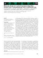
mutation. Figure 7 is a proposed 3D structural model
of human TNSALP, based on the crystallographic
analysis of human placental alkaline phosphatase, in
which both Val406 and Arg433 residues are high-
lighted. Valine at position 406 is located in the crown
domain consisting of 65 residues [4,21]. We have dem-
onstrated that TNSALP (V406A) is another allele with
severe effects, but does not show defective trafficking
like TNSALP (R433C). The rate of the intracellular
transport of the mutant protein was similar to that of
the wild-type protein, as assessed by the acquisition of
Endo H resistance. Besides, the mutant protein was
found to be incorporated into the raft at a kinetic rate
similar to that of the wild-type enzyme. GPI-anchored
protein is well known to be incorporated into the raft
in the Golgi apparatus [31], thus being ferried to the
apical surface of differentiated epithelial cells [32].
These findings strongly suggest that the cause of this
severe hypophosphatasia is not a defect in transport,
but the decreased catalytic activity of TNSALP
(V406A) itself. This was indeed confirmed by the
kinetic analysis of a purified GPI-anchorless soluble
version of the mutant protein. The K
cat
⁄ K
m
value of
the mutant TNSALP was less than one-tenth of the
K
cat
⁄ K
m
value of the soluble form of the wild-type
enzyme, indicating that the replacement of valine with
alanine at position 406 in the crown domain somehow
strongly affects the catalytic efficiency of TNSALP.
Considering that TNSALP (V406A) migrates at the
same position as the wild-type enzyme, as judged by
sucrose-density-gradient centrifugation, it is likely that
the overall structure of the mutant protein is not
grossly changed. Nevertheless, enhanced susceptibility
to trypsin, or especially to proteinase K, suggests a
subtle distortion of the crown domain of the mutant
protein. Currently we do not have a definite answer as
to how this missense mutation at position 406 in the
crown domain results in a remarkable decrease in the
catalytic efficiency of TNSALP. In this respect, it is
worth pointing out that residues in the crown domain
are closely related to the enzymatic properties of
TNSALP [22]. Interestingly, when valine at posi-
tion 406 is replaced with leucine or isoleucine, both of
which have a longer aliphatic chain than alanine, these
TNSALP mutant proteins were processed to the 80-
kDa form and exhibited enzyme activity similar to that
of the wild-type protein. This leads us to speculate that
Crown domain
V406
V406
R433R433
Active site
Active site
Fig. 7. 3D model of human TNSALP. Valine at position 406 and
arginine at position 433 in the crown domain are highlighted.
Fig. 6. Expression of TNSALP (V406L), TNSALP (V406I) and
TNSALP (V406F) in COS-1 cells. COS-1 cells were transfected with
the plasmid encoding the wild-type TNSALP or the TNSALP
(V406A), TNSALP (V406L), TNSALP (V406I) or TNSALP (V406F)
mutants, for 24 h. Cell homogenates were assayed for alkaline
phosphatase activity (abscissa: unit activityÆmg of protein
)1
) (upper
panel). The same samples were analyzed by SDS-PAGE (under
non-reducing conditions), followed by immunoblotting using anti-
TNSALP serum (lower panel).
N. Numa et al. Molecular basis of perinatal form of hypophosphatasia
FEBS Journal 275 (2008) 2727–2737 ª 2008 The Authors Journal compilation ª 2008 FEBS 2733
the valine residue at position 406 on one subunit may
interact with the counterpart on the other subunit
through their long aliphatic hydrocarbon chains, thus
contributing to assume a proper conformation of the
crown domain, which is presumably indispensable for
the efficient catalytic function of TNSALP. In support
of our hypothesis, the cysteine residue at position 433
in one subunit of TNSALP (R433C) becomes cross-
linked to the counterpart of the other subunit [29],
implying that the two cysteine residues are sufficiently
close to stretch out to form a covalent linkage in the
crown domain. However, our hypothesis is not com-
patible with the current TNSALP structural model
shown in Fig. 7. It seems that the side chains of two
valine residues at position 406 are too far apart to
interact with each other in the crown domain. Never-
theless, it is worth noting that the overall homology of
the crown domain between TNSALP and the placental
isoenzyme is around 72% at the amino acid level,
while that of the loop comprising residues 405–435 is
only 50% [21]. Another possibility is that valine at
position 406 is involved in a substrate–enzyme interac-
tion instead of interacting with the counterpart of the
other subunit. Obviously, the definite conclusion
awaits the crystallographic analysis of human
TNSALP, although our findings imply a close func-
tional relationship between the active site and the
crown domain of the TNSALP molecule, which may
be physiologically relevant to biomineralization.
Materials and methods
Materials
Express
35
S
35
S protein labeling mix (> 1000 CiÆmmol
)1
)
was obtained from Dupont-New England Nuclear (Boston,
MA, USA); and
14
C-methylated proteins and the enhanced
chemiluminescence (ECL
Ò
) western blotting detection
reagent, peroxidase-conjugated donkey anti-(rabbit IgG)
and protein A–Sepharose CL-4B were obtained from Amer-
sham Pharmacia Biotech (Arlington Heights, IL, USA);
pALTER
Ò
-MAX, Altered sites
Ò
II mammalian mutagenesis
system was obtained from Promega (Madison, WI, USA);
QuikChange II Site-Directed Mutagenesis kit was obtained
from Stratagene (La Jolla, CA, USA); G418 and pansorbin
were obtained from Calbiochem (La Jolla CA, USA); Lipo-
fectamine Plus Reagent was obtained from Invitrogen
(Carlsbad, CA, USA); phosphatidylinositol-specific phos-
pholipase C was obtained from BIOMOL International,
L.P. (Plymouth Meeting, PA, USA); aprotinin, doxycycline
and saponin (Quillaja Bark) and l-1-tosylamide-2-phenyl-
ethyl-chloromethylketone-treated bovine pancreas trypsin
were obtained from Sigma Chemical Co. (St Louis, MO,
USA); proteinase K was obtained from Roche Diagnostics
(London, UK); Ni-nitrilotriacetic acid resin and the plas-
mid Midi-kit were obtained from Qiagen (Hilden, Ger-
many); antipain, chymostatin, elastatinal, leupeptin and
pepstatin A were obtained from Protein Research Founda-
tion (Osaka, Japan); and hygromycin B and (p-amidinophe-
nyl) methanesulfonylfluoride were obtained from Wako
Pure Chemicals (Tokyo, Japan). Antiserum against recom-
binant human TNSALP was raised in rabbits as described
previously [26]. The pTRE2 and BD
Ò
CHO-K1 Tet-On cell
line and Tet systems approved fetal bovine serum from BD
Biosciences Clontech (Palo Alto, CA, USA).
Plasmids and transfection
The pALTER-MAX
Ò
encoding the wild-type TNSALP was
constructed as described previously [14]. Mutations were
introduced at specific sites using the Altered sites
Ò
II mam-
malian mutagenesis system, as described previously [14,15].
The oligonucleotides used were: TNSALP (V406A), 5¢-TTC
ACC GCC CAC TGC CTT GTA GCC AGG-3¢ and a sol-
uble form of TNSALP (V406A), 5¢-GCA GCA AGG CTG
CCT GCC TAG TGA TGG TGA TGG TGA TGG CTG
GCA GGA GCA CA-3¢. TNSALP (V406L), TNSALP
(V406I) and TNSALP (V406F) were created using the
QuikChange II Site-Directed Mutagenesis kit with the
following primers: V406L, 5¢-CCT GGC TAC AAG CTG
GTG GGC GGT G-3¢ and 5¢-CAC CGC CCA CCA GCT
TGT AGCCAG G-3¢; V406I, 5¢-CCT GGC TAC AAG
ATA GTG GGC GGT GAA-3¢ and 5¢-TTC ACC GCC
CAC TAT CTT GTA GCC AGG-3¢; and V406F, 5¢-CCT
GGC TAC AAG TTC GTG GGC GGT G-3¢ and 5¢-CAC
CGC CCA CGA ACT TGT AGC CAG G-3¢, respectively.
The DNA sequence of the mutation sites was verified by
DNA sequence analyses. The cDNA encoding TNSALP
(V406A) was further subcloned into pTRE2 to establish
stable cell lines. Transfection and screening of stable cell
lines were performed essentially according to the manufac-
turer’s protocol. CHO-K1 Tet-On cells, which successfully
produced the mutant TNSALP in the presence of doxy-
cycline, but not in its absence, were identified using
immunofluorescence. Establishment and characterization of
CHO-K1 Tet-On cells expressing the wild-type TNSALP
will be published elsewhere. Established CHO-K1 Tet-On
cells were cultured and passaged in the absence of doxycy-
cline until they were used for experiments. For immuno-
blotting or immunofluorescence studies, the cells were
cultured in the presence of 1 lgÆmL
)1
of doxycycline for
24 h before use. Alternatively, cells were cultured in
0.2–0.5 lgÆmL
)1
of doxycycline for 14 h before biosynthetic
experiments. For transient expression, COS-1 cells
(1.0–1.3 · 10
5
cells per 35-mm dish) were transfected with
0.5–0.8 lg of each plasmid using Lipofectamine Plus,
according to the manufacturer’s protocol, as described
Molecular basis of perinatal form of hypophosphatasia N. Numa et al.
2734 FEBS Journal 275 (2008) 2727–2737 ª 2008 The Authors Journal compilation ª 2008 FEBS
previously [14,15], and the transfected cells were incubated
for 24 h in a 5% CO
2
⁄ 95% air incubator before use. For
purification of the soluble forms of enzymes, five 100-mm
dishes were transfected and cultured for 48 h. COS-1 cells
were cultured in DMEM supplemented with 10% fetal
bovine serum [9].
Metabolic labeling and immunoprecipitation
For pulse-chase experiments, cells were pre-incubated for
0.5–1 h in methionine ⁄ cysteine-free DMEM and labeled
with 50–100 lCi of [
35
S]methionine ⁄ cysteine for 0.5 h in the
fresh methionine ⁄ cysteine-free MEM. After a pulse period,
cells were washed and chased in the DMEM as described
previously [15,29]. After metabolic labeling, the medium
was removed and the cells were lysed in 0.5 mL of lysis
buffer [1% (w ⁄ v) Triton X-100 ⁄ 0.5% (w ⁄ v) sodium deoxy-
cholate ⁄ 0.05% (w ⁄ v) SDS in NaCl ⁄ P
i
]. For separation of
Triton X-100-soluble and Triton X-100-insoluble fractions,
labeled cells were lysed in 1% Triton X-100 in NaCl ⁄ P
i
instead of in the lysis buffer and were centrifuged at
15 000 g for 10 min before lysing the insoluble fraction fur-
ther in the lysis buffer for immunoprecipitation. A prote-
ase-inhibitor cocktail (antipain, aprotinin, chymostatin,
elastatinal, leupeptin, pepstatin A) was added to cell lysates
and media (10 lg of each protease inhibitor per mL). The
lysates were incubated for 20 min at 37 °C to extract
TNSALP. The lysates and media were subjected to immu-
noisolation, as described previously [11,12]. The immune
complexes ⁄ Protein A beads were boiled in the absence or
presence of 1% (v ⁄ v) 2-mercaptoethanol and were then
analyzed by SDS ⁄ PAGE [9% (w ⁄ v) gels], followed by fluo-
rography [11].
Enzyme digestion
Endo H digestion and protease digestion using trypsin or
proteinase K were carried out as described previously
[11,12,29].
3D structure of TNSALP
A 3D model based on the crystal structure of human
placental alkaline phosphatase (./
Database.html) was downloaded and the program pymol
(http://pymol sourceforge.net/) was used to generate the
figure.
Miscellaneous procedures
Immunofluorescence for alkaline phosphatase was per-
formed as described previously [12,15]. Sucrose-density-gra-
dient centrifugation was performed as described previously
[14,25,29]. Transfer of proteins and subsequent procedures
were as described previously [25,29]. Proteins on mem-
branes were detected using ECL
Ò
western blotting detection
reagents. Purification of the soluble forms of the wild-type
TNSALP and TNSALP (V406A) was carried out essentially
as described previously [26]. Protein and alkaline phospha-
tase assays were performed as described previously [11,12].
One unit of alkaline phosphatase activity is defined as nmol
of p-nitrophenylphosphate hydrolyzed per min at 37 °C.
Acknowledgements
We are grateful to Dr Etienne Mornet for advice on the
graphical presentation of 3D structure of TNSALP. We
thank Miyako Okamura for her technical assistance.
This work was supported in part by a Grant-in-Aid for
Scientific Research from the Ministry of Education,
Culture, Sports and Technology of Japan (to KO).
References
1 Harris H (1989) The human alkaline phosphatases:
what we know and what we don’t know. Clin Chim
Acta 186, 133–150.
2 Whyte MP (2001) Hypophosphatasia. In The Metabolic
and Molecular Basis of Inherited Disease (Scriver CR,
Beaudet AL, Sly WS, Valle D, Childs B, Kinzler KW &
Vogelstein B, eds), 8th edn., vol. 4. pp. 5313–5329.
McGraw-Hill, New York, NY.
3 Mornet E (2000) Hypophosphatasia: the mutations in
the tissue-nonspecific alkaline phosphatase gene. Hum
Mutat 15, 309–315.
4 Millan JL (2006) Mammalian Alkaline Phosphatases:
From Biology to Applications in Medicine and Biotech-
nology. WILEY-VCH Verlag GmbH & Co. KGaA,
Weinheim.
5 Weiss MJ, Cole DE, Ray K, Whyte MP, Lafferty MA,
Mulivor RA & Harris H (1988) A missense mutation in
the human liver ⁄ bone ⁄ kidney alkaline phosphatase
gene causing a lethal form of hypophosphatasia. Proc
Natl Acad Sci USA 85, 7666–7669.
6 Mumm S, Jones J, Finnegan P & Whyte MP (2001)
Hypophosphatasia: molecular diagnosis of Rathbun’s
original case. J Bone Miner Res 16, 1724–1727.
7 Zurutuza L, Muller F, Gibrat JF, Taillandier A,
Simon-Bouy B, Serre JL & Mornet E (1999) Correla-
tions of genotype and phenotype in hypophosphatasia.
Hum Mol Genet 8, 1039–1046.
8 Waymire KG, Mahuren JD, Jaje JM, Guilarte TR,
Coburn SP & MacGregor GR (1995) Mice lacking
tissue non-specific alkaline phosphatase die from
seizures due to defective metabolism of vitamin B-6.
Nat Genet 11, 45–51.
9 Narisawa S, Frohlander N & Millan JL (1997) Inactiva-
tion of two mouse alkaline phosphatase genes and
N. Numa et al. Molecular basis of perinatal form of hypophosphatasia
FEBS Journal 275 (2008) 2727–2737 ª 2008 The Authors Journal compilation ª 2008 FEBS 2735
establishment of a model of infantile hypophosphatasia.
Dev Dyn 208, 432–446.
10 Fedde KN, Blair L, Silverstein J, Coburn SP, Ryan
LM, Weinstein RS, Waymire K, Narisawa S, Millan
JL, MacGregor GR et al. (1999) Alkaline phosphatase
knock-out mice recapitulate the metabolic and skeletal
defects of infantile hypophosphatasia. J Bone Mineral
Res 14, 2015–2026.
11 Shibata H, Fukushi M, Igarashi A, Misumi Y, Ikehara
Y, Ohashi Y & Oda K (1998) Defective intracellular
transport of tissue-nonspecific alkaline phosphatase
with an Ala
162
fi Thr mutation associated with
lethal hypophosphatasia. J Biochem (Tokyo) 123,
968–977.
12 Fukushi-Irie M, Ito M, Amaya Y, Amizuka N, Ozawa
H, Omura S, Ikehara Y & Oda K (2000) Possible inter-
ference between tissue-non-specific alkaline phosphatase
with an Arg
54
fi Cys substitution and a counterpart
with an Asp
277
fi Ala substitution found in a com-
pound heterozygote associated with severe hypophos-
phatasia. Biochem J 348, 633–642.
13 Fukushi M, Amizuka N, Hoshi K, Ozawa H, Kumagai
H, Omura S, Misumi Y, Ikehara Y & Oda K (1998)
Intracellular retention and degradation of tissue-nonspe-
cific alkaline phosphatase with a Gly
317
fi Asp substitu-
tion associated with lethal hypophosphatasia. Biochem
Biophys Res Commun 246, 613–618.
14 Ito M, Amizuka N, Ozawa H & Oda K (2002) Reten-
tion at the cis-Golgi and delayed degradation of
tissue-non-specific alkaline phosphatase with an
Asn
153
fi Asp substitution, a cause of perinatal hypo-
phosphatasia. Biochem J 361, 473–480.
15 Ishida Y, Komaru K, Ito M, Amaya Y, Kohno S &
Oda K (2003) Tissue-nonspecific alkaline phosphatase
with an Asp
289
fi Val mutation fails to reach the cell
surface and undergoes proteasome-mediated degrada-
tion. J Biochem (Tokyo) 134, 63–70.
16 Fleisch H, Straumann F, Schenk R, Bizaz S & Allgower
M (1966) Effect of condensed phosphates on calcifica-
tion of chick embryo femurs in tissue culture. Am J
Physiol 211, 821–825.
17 Russell RG, Bisaz S, Donath A, Morgan DB & Fleisch
H (1971) Inorganic pyrophosphate in plasma in normal
persons and in patients with hypophosphatasia, osteo-
genesis imperfect, and other disorders of bone. J Clin
Invest 50, 961–969.
18 Hessle L, Johnson KA, Anderson HC, Narisawa S, Sali
A, Goding JW, Terkeltaub R & Millan JL (2002)
Tissue-nonspecific alkaline phosphatase and plasma cell
membrane glycoprotein-1 are central antagonistic regu-
lators of bone mineralization. Proc Natl Acad Sci USA
99, 9445–9449.
19 Murshed M, Harmey D, Millan JL, MaKee MD &
Karsenty G (2005) Unique coexpression in osteoblasts
of broadly expressed genes accounts for the spatial
restriction of ECM mineralization to bone. Genes Dev
19, 1093–1104.
20 Taillandier A, Lia-Baldini AS, Mouchard M, Robin B,
Muller F, Simon-Bouy B, Serre JL, Bera-Louville A,
Bonduelle M, Eckhardt J et al. (2001) Twelve novel
mutations in the tissue-nonspecific alkaline phosphatase
gene (ALPL) in patients with various forms of hypo-
phosphatasia. Hum Mutat 18, 83–84.
21 Mornet E, Stura E, Lia-Baldin AS, Stigbrand T, Menez
A & Le Du MH (2001) Structural evidence for a func-
tional role of human tissue nonspecific alkaline phos-
phatase in bone mineralization. J Biol Chem 276,
31171–31178.
22 Bossi M, Hoylaerts MF & Millan JL (1993) Modifica-
tions in a flexible surface modulate the isozyme-specific
properties of mammalian alkaline phosphatase. J Biol
Chem 268, 25409–25416.
23 Vittur FN, Stagni L, Moro L & der Bernard B (1984)
Alkaline phosphatase binds to collagen; a hypothesis on
the mechanism of extravascular mineralization in epihy-
seal cartilage. Experientia 40, 836–837.
24 Wu LNY, Genge BR, Lloyd GC & Wuthier RE (1991)
Collagen-binding proteins in collagenase-released matrix
vesicles from cartilage Interaction between matrix vesi-
cle proteins and different types of collagen. J Biol Chem
266, 1195–1203.
25 Komaru K, Ishida Y, Amaya Y, Goseki-Sone M,
Orimo H & Oda K (2005) Novel aggregate formation
of a frame-shift mutant protein of tissue- nonspecific
alkaline phosphatase is ascribed to three cysteine
residues in the C-terminal extension. Retarded secre-
tion and proteasomal degradation. FEBS J 272,
1704–1717.
26 Oda K, Amaya Y, Fukushi-Irie M, Kinameri Y,
Ohsuye K, Kubota I, Fujimura S & Kobayashi J (1999)
A general method for rapid purification of soluble ver-
sions of glycosylphosphatidylinositol-anchored proteins
expressed in insect cells: an application for human
tissue-nonspecific alkaline phosphatase. J Biochem
(Tokyo) 126, 694–699.
27 Brun-Heath I, Lia-Baldini AS, Maillard S, Taillandier
A, Utsch B, Nunes ME, Serre JL & Mornet E (2007)
Delayed transport of tissue-nonspecific alkaline
phosphatase with missense mutations causing
hypophosphatasia. Eur J Med Genet 50, 367–378.
28 Cai G, Michigami T, Yamamoto T, Yasui N, Stomura
K, Yamagata M, Shima M, Nakajima S, Mushiake S,
Okada S et al. (1998) Analysis of localization of
mutated tissue-nonspecific alkaline phosphatase proteins
associated with neonatal hypophosphatasia using green
fluorescent protein chimeras. J Clin Endocrinol Metab
83, 3936–3942.
29 Nasu M, Ito M, Ishida Y, Numa N, Komaru K,
Nomura S & Oda K (2006) Aberrant interchain
disulfide bridge of tissue-nonspecific alkaline
Molecular basis of perinatal form of hypophosphatasia N. Numa et al.
2736 FEBS Journal 275 (2008) 2727–2737 ª 2008 The Authors Journal compilation ª 2008 FEBS
phosphatase with an Arg433-Cys substitution associ-
ated with severe hypophosphatasia. FEBS J 273,
5612–5624.
30 Lia-Baldini AS, Muller F, Taillandier A, Gibrat JF,
Mouchard M, Robin B, Simon-Bouy B, Serre JL, Ayls-
worth AS, Bieth E et al. (2001) A molecular approach
to dominance in hypophosphatasia. Hum Genet 109,
99–108.
31 Brown DA & Rose JK (1992) Sorting of GPI-anchored
proteins to glycolipid-enriched membrane subdomains
during transport to the apical cell surface. Cell 68,
533–542.
32 Paladino S, Pocad T, Cafino MA & Zurzolo C (2006)
GPI-anchored proteins are directly targeted to the
apical surface in fully polarized MDCK cells. J Cell
Biol 172, 1023–1034.
N. Numa et al. Molecular basis of perinatal form of hypophosphatasia
FEBS Journal 275 (2008) 2727–2737 ª 2008 The Authors Journal compilation ª 2008 FEBS 2737