Báo cáo khoa học: Nuclear localization of human spermine oxidase isoforms – possible implications in drug response and disease etiology pptx
Bạn đang xem bản rút gọn của tài liệu. Xem và tải ngay bản đầy đủ của tài liệu tại đây (342.46 KB, 12 trang )
Nuclear localization of human spermine oxidase
isoforms – possible implications in drug response and
disease etiology
Tracy Murray-Stewart
1
, Yanlin Wang
1
, Andrew Goodwin
1
, Amy Hacker
1
, Alan Meeker
2
and Robert
A. Casero Jr
1
1 Department of Oncology, Johns Hopkins University School of Medicine and the Sidney Kimmel Comprehensive Cancer Center at Johns
Hopkins, Baltimore, MD, USA
2 Department of Pathology, Johns Hopkins University School of Medicine and the Sidney Kimmel Comprehensive Cancer Center at Johns
Hopkins, Baltimore, MD, USA
The naturally occurring polyamines, spermine, spermi-
dine and putrescine, are polycations that are abundant
and essential in both prokaryotic and eukaryotic cells.
These molecules have been implicated in multiple cellu-
lar functions and processes, including proliferation,
differentiation, apoptosis, gene expression and cell
Keywords
carcinogenesis; H
2
O
2
; oxidation; polyamine;
SMO
Correspondence
R. A. Casero Jr, Sidney Kimmel
Comprehensive Cancer Center at Johns
Hopkins, Bunting Blaustein Building, Room
551, 1650 Orleans Street, Baltimore, MD
21231, USA
Fax: +1 410 614 9884
Tel: +1 410 955 8580
E-mail:
(Received 7 December 2007, revised 14
March 2008, accepted 25 March 2008)
doi:10.1111/j.1742-4658.2008.06419.x
The recent discovery of the direct oxidation of spermine via spermine oxi-
dase (SMO) as a mechanism through which specific antitumor polyamine
analogues exert their cytotoxic effects has fueled interest in the study of the
polyamine catabolic pathway. A major byproduct of spermine oxidation is
H
2
O
2
, a source of toxic reactive oxygen species. Recent targeted small
interfering RNA studies have confirmed that SMO-produced reactive oxy-
gen species are directly responsible for oxidative stress capable of inducing
apoptosis and potentially mutagenic DNA damage. In the present study,
we describe a second catalytically active splice variant protein of the
human spermine oxidase gene, designated SMO5, which exhibits substrate
specificities and affinities comparable to those of the originally identified
human spermine oxidase-1, SMO ⁄ PAOh1, and, as such, is an additional
source of H
2
O
2
. Importantly, overexpression of either of these SMO iso-
forms in NCI-H157 human non-small cell lung carcinoma cells resulted in
significant localization of SMO protein in the nucleus, as determined by
confocal microscopy. Furthermore, cell lines overexpressing either SMO ⁄
PAOh1 or SMO5 demonstrated increased spermine oxidation in the
nucleus, with accompanying alterations in individual nuclear polyamine
concentrations. This increased oxidation of spermine in the nucleus there-
fore increases the production of highly reactive H
2
O
2
in close proximity to
DNA, as well as decreases nuclear spermine levels, thus altering the protec-
tive roles of spermine in free radical scavenging and DNA shielding, and
resulting in an overall increased potential for oxidative DNA damage in
these cells. The results of these studies therefore have considerable signifi-
cance both with respect to targeting polyamine oxidation as an antineo-
plastic strategy, and in regard to the potential role of spermine oxidase in
inflammation-induced carcinogenesis.
Abbreviations
BENSpm, bis(ethyl)norspermine; CPENSpm, N
1
-ethyl-N
11
-(cyclopropyl)methyl-4,8,diazaundecane; DAPI, 4¢,6¢-diamidino-2-phenylindole; LSD1,
lysine-specific demethylase 1; ROS, reactive oxygen species; SMO ⁄ PAOh1, human spermine oxidase-1; SMO5, spermine oxidase-5.
FEBS Journal 275 (2008) 2795–2806 ª 2008 The Authors Journal compilation ª 2008 FEBS 2795
signaling [1–6]. The strict maintenance of polyamine
homeostasis is critical for proper cell function, and is
regulated at the levels of biosynthesis, uptake, efflux
and catabolism. In cancer cells, the regulatory compo-
nents of polyamine homeostasis are often disrupted,
leading to increased levels of intracellular polyamines,
thus promoting increased growth and proliferation,
and, potentially, tumor progression [7–11]. This tumor-
associated dysregulation of polyamine metabolism has
led to the development of several classes of polyamine
analogues targeted towards inducing the catabolic
enzymes, which in turn deplete intracellular polyam-
ines, and arrest tumor cell growth [7,12–16].
Until recently, it was presumed that polyamine
catabolism was solely regulated by the rate-limiting
enzyme spermidine ⁄ spermine N
1
-acetyltransferase [17].
The resulting acetylated polyamines, N
1
-acetylspermi-
dine and N
1
-acetylspermine, are substrates for either
cellular export or further back-conversion via the con-
stitutively-expressed N
1
-acetylpolyamine oxidase [18].
Recent cloning and characterization of N
1
-acetylpoly-
amine oxidase has confirmed its preference for the
acetylated polyamines as substrates, and no significant
activity is observed when spermine is used as substrate
[19,20]. Our cloning of a second polyamine oxidase,
human spermine oxidase-1 (SMO ⁄ PAOh1), and its
confirmation as a spermine oxidase, identified a second
mechanism through which the polyamines are catabo-
lized, and has led to increased interest in the exploita-
tion of polyamine catabolism for antitumor therapy
[21–23].
SMO ⁄ PAOh1 is an FAD-containing enzyme (EC
1.5.3.–) that directly catabolizes spermine as its pre-
ferred substrate, resulting in spermidine, 3-aminoprop-
anal and, importantly, H
2
O
2
(Fig. 1). H
2
O
2
is a readily
diffusible source of cellular reactive oxygen species
(ROS), and has been linked to the cytotoxicity
observed in specific tumor cell types following treat-
ment with antitumor, polyamine catabolism-inducing
polyamine analogues [2,12,24–26]. The finding that
spermine oxidase activity, and thus H
2
O
2
production,
can be induced in a cell and tumor type-specific man-
ner by certain polyamine analogues [22,25,27] adds
considerably to the importance of investigating this
new pathway member because its regulation may con-
tribute to the facilitation of tumor cell apoptosis [5].
Additionally, studies overexpressing the mouse sper-
mine oxidases have provided evidence that SMO
directly induces DNA damage via H
2
O
2
-related oxida-
tive stress, and that this damage renders the cell more
sensitive to subsequent radiation exposure and apopto-
sis [28]. Furthermore, induction of spermine oxidase in
gastric and lung epithelial cells by Helicobacter pylori
and tumor necrosis factor-a, respectively, has been
linked to increased ROS production and DNA damage
[29,30], thus implicating SMO as a potential molecular
link between chronic inflammation and epithelial carci-
nogenesis. In addition, we have recently demonstrated
elevated SMO expression in prostatic epithelial tissue
from prostate cancer patients compared to prostate
disease-free control patients [31].
Subsequent to the initial characterization of SMO ⁄
PAOh1, three additional splice variants have been
identified [32]; however, the purified recombinant pro-
teins of these variants have failed to exhibit significant
oxidase activity on the natural polyamines. Based on
the exon structures of previously identified human
spermine oxidases, we suspected the existence of an
additional isoform that possesses a combination of the
gene segments present in the two longest variants: the
active SMO ⁄ PAOh1 and the inactive PAOh4 (Fig. 2A)
[32]. However, this hypothetical isoform was not iden-
tified when using the standard reverse transcription
PCR protocol that produced spermine oxidases 1–4. A
mouse spermine oxidase isoform, mSMOl, has been
identified that possesses an exon structure identical to
that of our hypothetical spermine oxidase-5, or SMO5.
Importantly, this mouse isoform has high spermine
oxidase activity, and exhibits a significantly greater
degree of localization in the nucleus than the other
mouse polyamine oxidases thus far described [33]. We
therefore sought to determine whether the correspond-
ing SMO5 isoform exists in human cells.
In the present study, we confirmed the existence of
SMO5 in human normal and tumor cell lines. SMO5
cDNA was subcloned and sequenced, and active pro-
tein was produced, purified and assayed for kinetic
properties and substrate specificities. NCI-H157 human
non-small cell lung carcinoma cells were stably trans-
fected, and individual clones selected that overexpress
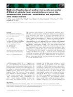
Fig. 1. Catalysis of spermine oxidation by human spermine oxi-
dase. SMO catalyzes the cleavage of spermine, resulting in back-
conversion to spermidine, and the generation of 3-aminopropanal
and H
2
O
2
.
Cellular localization of human spermine oxidase T. Murray-Stewart et al.
2796 FEBS Journal 275 (2008) 2795–2806 ª 2008 The Authors Journal compilation ª 2008 FEBS
each of the isoforms of interest to confirm cellular
localization. In contrast to results observed with the
homologous mouse SMO isoforms, each of the three
human isoforms examined were present in similar
amounts in the nucleus, with SMO ⁄ PAOh1 and SMO5
both demonstrating functional oxidase activity in both
nuclear and cytoplasmic compartments, as determined
by measurement of H
2
O
2
production, as well as HPLC
analysis of localized polyamine pools.
Previous reports have described transient overexpres-
sion of SMO ⁄ PAOh1 in the transformed human kid-
ney cell line HEK-293 [22], and Amendola et al. [28]
have described the establishment of stable populations
overexpressing several of the mouse SMO isoforms in
a mouse neuroblastoma line. The cell lines created in
the present study, however, represent the first stable
overexpression of any of the human spermine oxidase
isoforms in human tumor cells. Furthermore, we
report the first direct visualization of the localization
of human spermine oxidase in a human tumor cell line.
We also identify and classify SMO5 as a third human
polyamine catabolic enzyme that possesses the ability
to produce reactive oxygen species as a toxic byprod-
uct, while altering intracellular polyamine concentra-
tions. Most importantly, the surprising abundance of
both active isoforms, SMO ⁄ PAOh1 and SMO5, in the
nucleus, emphasizes the potential of these ROS-pro-
ducing enzymes to either modulate cancer cell response
to the antitumor polyamine analogues, or, in cells
stimulated by infection and ⁄ or inflammation, act as
the source of ROS with the potential to produce muta-
genic oxidative DNA damage, thus providing a link
between inflammation and carcinogenesis.
Results
SMO5 is expressed in human lung cell lines
To determine whether the SMO5 splice variant of the
spermine oxidase gene is present in human tissue, we
performed RT-PCR using a primer pair specific to
SMO5. Specifically, primers were located in the inter-
nal region of exon V and in exon VIa, both of which
are only found together in the SMO5 splice variant
(Fig. 2A). The resulting 653 bp amplification product
was detected in all cell lines tested, including the non-
small cell lung carcinoma lines, NCI-H157 and NCI-
A549, as well as the non-tumorigenic lung epithelial
cell line, Beas2B (Fig. 2B).
Real-time PCR using the same primers as above for
SMO5, as well as primers specific for SMO ⁄ PAOh1
(Fig. 2A), suggested that SMO5 mRNA is expressed
to a much lower extent than SMO ⁄ PAOh1 in both
H157 and A549 cell lines (data not shown), possibly
accounting for the fact that SMO5 was not identified
during our initial cloning of the spermine oxidase iso-
forms from NCI-H157 cells. We confirmed the relative
abundance of the two proteins using western blot anal-
ysis of A549 cells, which have previously been shown
to express relatively high levels of SMO ⁄ PAOh1 [27].
In both untreated cells and in those treated with
bis(ethyl)norspermine (BENSpm), a polyamine ana-
logue known to induce spermine oxidase, a 65 kDa
band corresponding to SMO5 was apparent at a much
lower intensity than the 61.9 kDa SMO ⁄ PAOh1
protein band (Fig. 2C).
SMO5 protein purification
To further study and characterize the SMO5 protein,
SMO5 cDNA was subcloned into the pET15b bacterial
expression vector. The 1984 bp cDNA (GenBank
A
BC
Fig. 2. Expression of human spermine oxidase splice variants in
lung epithelial cells. (A) Exon structures of spermine oxidase iso-
forms. SMO5 includes both the internal region of exon V that is
absent from the inactive PAOh4 isoform, as well as exon VIa,
which is absent from the active isoform, SMO ⁄ PAOh1. SMO5 and
the active, mouse isoform, mSMOl, have identical exon structures.
Arrows indicate isoform-specific primers utilized for RT- and real-
time PCR. (B) Expression of SMO5 mRNA in human lung cells.
Total RNA was extracted from: (1) A549; (2) A549 + 10 l
M BENS-
pm; (3) H157; or (4) Beas2B cells. One lg of each RNA sample
was used for RT-PCR with primers specific for SMO isoform 5,
resulting in 653 bp fragments that were separated and visualized
on a 1% agarose gel stained with ethidium bromide. (C) Western
blot of endogenous SMO ⁄ PAOh1 and SMO5 protein. To visualize
relative levels of the two active SMO isoforms, A549 cells were
treated with 10 l
M BENSpm for 24 h, total protein was extracted,
and immunoblotting was performed using a human SMO antibody
that recognizes both isoforms.
T. Murray-Stewart et al. Cellular localization of human spermine oxidase
FEBS Journal 275 (2008) 2795–2806 ª 2008 The Authors Journal compilation ª 2008 FEBS 2797
accession no. EF032141) includes both the internal
region of exon V that exists in SMO ⁄ PAOh1, as well
as exon VIa that is present in PAOh4 (Fig. 2A). Both
of these regions are also present in the nuclear-local-
ized mouse spermine oxidase isoform, mSMOl, which
shares 89% nucleotide identity with the human SMO5
cDNA. Isopropyl thio-b-d-galactoside induction of
transformed BL
21
(DE
3
) Escherichia coli resulted in the
production of SMO5 protein, the largest of the SMO
isoforms, consisting of 586 amino acids, with a pre-
dicted molecular mass of approximately 65 kDa, as
observed by SDS ⁄ PAGE analysis (Fig. 3A). As previ-
ously reported for the PAOh1 ⁄ SMO protein [23], the
majority of SMO5 protein produced in this bacterial
expression system localizes to inclusion bodies, and
therefore was denatured and refolded prior to analysis.
All SMO5 protein production and purification steps
were performed in parallel with SMO ⁄ PAOh1 and
PAOh4 proteins for comparison.
Polyamine oxidase activity and substrate
specificity of SMO5
Polyamine oxidase activity assays of the purified recom-
binant SMO5 and SMO ⁄ PAOh1 proteins revealed
nearly identical substrate specificities for the two iso-
forms. Both enzymes clearly exhibit a strong preference
for spermine as the primary substrate over all other nat-
urally occurring polyamines, with SMO ⁄ PAOh1 having
a specific activity approximately 2.5-fold greater than
that of SMO5 (Fig. 3B). N
1
-acetylspermine was the only
other polyamine to be oxidized by the proteins, but this
activity was less than 10% of that observed when using
spermine with the same enzyme. Similar to SMO ⁄
PAOh1, SMO5 exhibited virtually no oxidase activity
when using N
1
,N
12
-diacetylspermine as substrate, and
there was a complete absence of oxidation when
N
1
-acetylspermidine, N
8
-acetylspermidine, spermidine,
or the polyamine analogues, BENSpm or N
1
-ethyl-
N
11
-(cyclopropyl)methyl-4,8,diazaundecane (CPENSpm),
were presented as potential substrates. MDL72,527, an
inhibitor of the polyamine oxidases, was also effective
as an inhibitor of SMO5 activity, as demonstrated by a
greater than 99% reduction in the oxidation of
spermine.
Purified, recombinant PAOh4 was inactive as an
oxidase with all substrates tested (data not shown).
The only difference between PAOh4 and SMO5 is the
absence of a 53 amino acid central region of exon V,
including residues 283–335. According to structure
analyses and molecular modeling of homologous pro-
teins, many residues in the 3¢ half of this region are
0
1
2
3
4
5
6
7
8
9
Spm N1AcSpm DASpm
250 µ
M
Substrate
Spermine oxidase activity
(µmol H
2
O
2
·mg protein
–1
·min
–1
)
SMO/PAOh1
SMO/PAOh1 + MDL72,527
SMO5
SMO5 + MDL72,527
75 kDa
50 kDa
1 A
C
D
B
2 3 4
Lane Sample MW
1
2
3
SMO5
SMO/PAOh1
PAOh4
65.0 kDa
61.9 kDa
59.2 kDa 4
marker
R
2
= 0.9953
R
2
= 0.9978
R
2
= 0.9791
R
2
= 0.9852
–10
–5
0
5
10
15
20
25
–1 –0.5 0 0.5 1 1.5
1/N
1
AcSpm (µM)
1/v
SMO/PAOh1
SMO5
–1
–0.5
0
0.5
1
1.5
2
–10 –5 0 5 10 15
1/Spm (µM)
1/v
SMO/PAOh1
SMO5
Fig. 3. Characteristics of recombinant
SMO5 protein. (A) Recombinant SMO ⁄
PAOh1, PAOh4 or SMO5 protein was
expressed and purified from transformed
E. coli cells. Dialyzed, refolded proteins
were analyzed by SDS ⁄ PAGE, visualized by
staining with Coomassie blue, and photo-
graphed. (B) Substrate specificity and
specific activities of purified SMO5 versus
SMO ⁄ PAOh1. Oxidase activity was
assessed for each purified protein using
250 l
M of various potential substrates ± an
equimolar concentration of the polyamine
oxidase inhibitor, MDL72,527. Substrates
used were spermine (Spm), N
1
-acetylsper-
mine (N1AcSpm), or N
1
,N
12
-diacetylsper-
mine (DASpm). Data represent the
mean ± SE of three separate experiments,
each performed in triplicate. (C) Representa-
tive Lineweaver–Burk plots of purified
SMO ⁄ PAOh1 or SMO5 using spermine or
N
1
-acetylspermine as substrates. Increasing
concentrations of each substrate were
added to each of the purified enzymes and
spermine oxidase activity was determined
as a function of lmol H
2
O
2
Æmg
protein
)1
Æmin
)1
.
Cellular localization of human spermine oxidase T. Murray-Stewart et al.
2798 FEBS Journal 275 (2008) 2795–2806 ª 2008 The Authors Journal compilation ª 2008 FEBS
highly conserved components of the FAD-binding
domain, and are therefore essential for catalysis
[28,33–35].
Kinetic properties of SMO5
Polyamine oxidase activity of SMO5 and SMO ⁄
PAOh1 was measured using increasing concentrations
of either spermine or N
1
-acetylspermine as substrate
(Fig. 3C). SMO5 and SMO ⁄ PAOh1 displayed very
similar affinities for spermine, as determined by
Lineweaver–Burk transformation of the Michaelis–
Menton equation, with K
m
values of 0.5 and 0.6 lm,
respectively. Both enzymes possessed a calculated K
m
of approximately 3.0 lm when N
1
-acetylspermine was
used as substrate. SMO ⁄ PAOh1 consistently demon-
strated a k
cat
value approximately 2.4-fold that of
SMO5 with either of the substrates examined. Specifi-
cally, SMO ⁄ PAOh1 exhibited k
cat
values for spermine
and N
1
-acetylspermine of 7.55 s
)1
and 0.28 s
)1
, respec-
tively, whereas those for SMO5 measured 3.11 s
)1
and
0.12 s
)1
. The most dramatic kinetic differences, how-
ever, were seen in the velocities at which each enzyme
was capable of oxidizing spermine as opposed to
N
1
-acetylspermine. With both enzymes, the k
cat
value
of spermine is approximately 27-fold that of N
1
-acetyl-
spermine.
Overexpression of SMO in NCI-H157 cells
Expression vectors containing coding sequences of
SMO ⁄ PAOh1, PAOh4 and SMO5 were constructed to
determine localization of each protein following stable
transfection into H157 human non-small cell lung car-
cinoma cells. Human SMO antibody [30,31] was used
to screen individual stable clones for overexpression of
the three SMO isoforms via western blotting. This pro-
cess was facilitated by the fact that basal expression of
SMO in H157 cells is nearly undetectable by western
blot analysis. Clones with the highest amounts of each
exogenous isoform were selected for further experi-
ments, and designated SMO1, SMO4 and SMO5
(Fig. 4A). Real-time PCR using isoform-specific primer
pairs verified specific expression of individual splice
variants (data not shown).
SMO activity assays of total cellular protein were
used to verify that these cell lines were overexpressing
functional spermine oxidase proteins. Not surprisingly,
the SMO ⁄ PAOh1 clone, SMO1, displayed the highest
activity (Fig. 4B). Cells overexpressing SMO5 also dis-
played significant spermine oxidase activity. As
expected, cells overexpressing the inactive splice vari-
ant, PAOh4 (clone SMO4), by western blot and real-
0
2
4
6
8
10
v SMO4 SMO1 SMO5
Intracellular polyamines
(nmol·mg protein
–1
)
Put
Spd
Spm
*
*
*
*
v
SMO1
SMO4
SMO5
pmolH
2
O
2
·mg protein
–1
min
–1
9.7 + 5.4
13006.9 + 3511.6
25.8 + 5.9
515.0 + 66.8
50 kDa
75 kDa
5
Actin
SMO
v
4 1
SMO isoform A
B
C
Fig. 4. Overexpression of SMO isoforms in NCI-H157 cells. (A)
Western blot of total cellular protein from empty vector-transfect-
ed control H157 cells (v) and H157 cells stably overexpressing
SMO ⁄ PAOh1 (SMO1), PAOh4 (SMO4) or SMO5 (SMO5). Total
protein (30 lg per lane) was separated on a 10% Novex gel, and
immunoblotting was performed using SMO and actin antibodies.
Dye-conjugated secondary antibodies were used to detect bound
proteins using the Odyssey infrared detection system. The illus-
trated blot is representative of three separate experiments.
(B) For SMO activity assays, 50 lL of cell lysate from each
overexpressing cell line was used per reaction, in triplicate,
and SMO activity was calculated relative to milligrams of total
cellular protein, as determined by the Bradford assay. Graph
represents means with standard errors of three separate experi-
ments. (C) Intracellular levels of the polyamines putrescine
(Put), spermidine (Spd) and spermine (Spm) were determined by
HPLC analysis of dansyl chloride-labeled cell lysates. Data
represents the means ± SE of four separate determinations.
*Statistically significant differences in individual polyamine levels
of SMO overexpressing cells relative to vector control cells
(P < 0.02).
T. Murray-Stewart et al. Cellular localization of human spermine oxidase
FEBS Journal 275 (2008) 2795–2806 ª 2008 The Authors Journal compilation ª 2008 FEBS 2799
time PCR, demonstrated spermine oxidase activity no
greater than that of the empty vector-transfected
control cells.
Polyamine pool analysis of the overexpressing
SMO cells further confirmed the increased SMO
activity, with decreases in intracellular spermine pools
and increased spermidine levels in SMO1 and SMO5
cells. These changes in spermine and spermidine levels
in both SMO1 and SMO5 overexpressing cell lines
were statistically significant compared to those levels
in the vector control cell line (P < 0.02). As
expected, cells overexpressing PAOh4 exhibited a
polyamine pool profile similar to control cells con-
taining the empty expression vector (Fig. 4C). Impor-
tantly, in spite of the much larger amount of SMO
activity observed in the SMO ⁄ PAOh1 clone relative
to the SMO5 clone when assayed in vitro with satu-
rating substrate conditions, there are only slight
differences in the intracellular polyamine pools of the
two, indicating that the activity of SMO5 is sufficient
to catabolize the amount of available spermine in
the cell.
Localization of SMO in H157 cells
Western blot analysis and quantification of nuclear
and cytoplasmic protein extracts indicated that all
three isoforms, SMO ⁄ PAOh1, SMO5 and PAOh4,
exhibit similar localization patterns when transfected
into H157 cells, with significant amounts of spermine
oxidase protein present in the nucleus, as well as the
cytoplasm (Fig. 5A). This is in contrast to the data
regarding the mouse SMO isoforms, of which only
the SMO5 homologue has been shown to exist in the
nucleus. Because all other mouse spermine oxidases,
including the predominant splice variant homologue,
show localization exclusively in the cytoplasm [33],
our finding that SMO ⁄ PAOh1 is present in the
nucleus was novel and unexpected. SMO activity
assays of SMO ⁄ PAOh1 and SMO5 overexpressing
nuclear and cytoplasmic extracts corroborate these
data, showing spermine oxidase activity and H
2
O
2
generation in both nuclear and cytoplasmic extracts
(Fig. 5B). Polyamine pool analyses also verified func-
tional spermine oxidase activity in both fractions,
with cells overexpressing SMO ⁄ PAOh1 and SMO5
displaying increased nuclear and cytoplasmic spermi-
dine, with significantly diminished spermine levels,
compared to vector control or PAOh4-overexpressing
cells (Fig. 5C). Furthermore, confocal microscopy of
immunofluorescent staining of SMO in cells overex-
pressing each of the isoforms provided direct visuali-
zation and confirmation of protein localization
without the possibility of contamination of one
fraction with another during protein preparation
(Fig. 5D).
Discussion
Several studies have confirmed that the oxidation of
polyamines by SMO plays an important role in the
antitumor effects of multiple antitumor polyamine ana-
logues that act as inducers of SMO expression
[2,12,21,22,25,27]. More recently, a series of studies
have implicated SMO activity as a source for poten-
tially mutagenic ROS production and as a direct link
between infection, inflammation and carcinogenesis
[29–31]. Consequently, a better understanding of the
physical cellular localization of SMO has gained in
importance. A highly active mouse spermine oxidase
isoform, mSMOl, was recently reported by Cervelli
et al. [33] to be localized in both the nucleus and cyto-
plasm, and its nuclear localization was considered to
be unique among the mouse SMO isoforms [28,33].
Although we had previously cloned a number of
human spermine oxidase splice variants, as well as
truncated proteins, no homologue of the mouse
mSMOl, which is essentially a combination of the
human isoforms 1 and 4, had been identified. The dis-
covery of its existence in the mouse suggested the like-
lihood of a human homologue, and led us to pursue
the identification of human SMO5. Furthermore, the
implications of spermine oxidase activity in the nucleus
prompted us to further investigate the localization of
the human SMO isoforms.
SMO5 mRNA was detected by RT-PCR in all
human lung cell lines examined. The addition of
exon VIa to SMO ⁄ PAOh1 to make SMO5 had little
effect on the substrate affinities of the purified pro-
teins. However, the k
cat
values of SMO5 were some-
what lower for both spermine and N
1
-acetylspermine
than were those of SMO ⁄ PAOh1, thus accounting for
the observed difference in specific activities. Because
the only difference in the gene structures of the two
enzymes is the presence of exon VIa, it appears that
its presence may actually hamper the efficiency with
which spermine is oxidized by human SMO. Molecu-
lar modeling of homologous mouse isoforms [28]
place this specifically mammalian, highly conserved,
31 amino acid region on the surface loop of the
enzyme in close proximity to the FAD-binding
domain, where it may potentially interfere with the
reaction. In spite of this difference, by possessing a
specific activity of approximately 3 lmol H
2
O
2
Æmg
protein
)1
Æmin
)1
, purified SMO5 protein still very effi-
ciently oxidizes spermine. Furthermore, it should be
Cellular localization of human spermine oxidase T. Murray-Stewart et al.
2800 FEBS Journal 275 (2008) 2795–2806 ª 2008 The Authors Journal compilation ª 2008 FEBS
noted that these proteins have been denatured and
refolded; thus, the possibility of altered activities can-
not be excluded.
A second region that varies among the three
enzymes studied, located within exon V, is present in
the active SMO ⁄ PAOh1 and SMO5 enzymes, but
absent from the inactive PAOh4 protein. This entire 53
amino acid region is highly conserved in mammals,
and can be subdivided into a 5¢-end that is suggested
to be involved in nuclear localization of the mouse
spermine oxidase [28], and a 3¢ region, which structural
modeling of homologous proteins has demonstrated to
play an essential role in the oxidase activity of the
enzyme through interacting with the FAD cofactor
[34,35]. A mouse SMOl mutant protein was purified
that lacks the 31 residues of the 5¢ end of this region,
while retaining all of its catalytic activity [28]. Intrigu-
ingly, the human version of this entire 53 amino acid
region is flanked by splice sites, resulting in the pro-
duction of several catalytically inactive SMO proteins
[32], including the PAOh4 protein utilized in the pres-
ent study. Although many isoforms have been identi-
fied for the mouse SMO protein, none display this
same pattern of splicing. Analysis of nucleotide
sequences surrounding this region reveal a base change
(G to A) at the 3¢ end of the removed human
1.0 + 0.0
10.1 + 0.4
0.5 + 0.0
2.7 + 0.2
v
SMO1
SMO4
SMO5
1.1 + 0.1
1157.6 + 31.7
1.5 + 0.1
62.9 + 1.1
CytoplasmicNuclear
NNNNCCCC
SMO
Actin
v SMO1 SMO4 SMO5M
A
C
B
D
Nuclear polyamines
0
2
4
6
8
Put
Spd
Spm
*
*
Cytoplasmic polyamines
0
2
4
6
8
V SMO4 SMO1 SMO5
V SMO4 SMO1 SMO5
nmol·mg protein
–1
nmol·mg protein
–1
Put
Spd
Spm
*
V
SMO1
SMO5
SMO DAPI Merge
Fig. 5. Functional SMO ⁄ PAOh1 and SMO5 are found in both the cytoplasm and nucleus of overexpressing NCI-H157 cells. (A) Western blot
analysis of nuclear and cytoplasmic SMO protein from H157 cells overexpressing SMO ⁄ PAOh1 (SMO1), PAOh4 (SMO4), SMO5 (SMO5) or
empty vector (v). Nuclear (N) or cytoplasmic (C) proteins (30 lg per lane) were separated by SDS ⁄ PAGE, transferred to poly(vinylidene difluo-
ride), and incubated with an antibody specific for human SMO. Blot is representative of three independent experiments. M, molecular
weight marker. (B) Fold-change in SMO activity of nuclear and cytoplasmic proteins. Nuclear or cytoplasmic protein (25 lL per reaction) was
assayed for oxidase activity on spermine, in triplicate. Values were calculated relative to protein amount, as determined by BioRad DC quan-
tification, and represent means ± SD calculated within a single, representative experiment that was repeated three times. (C) Localized poly-
amine pool analysis. Aliquots of nuclear and cytoplasmic extracts used above for SMO activity assays were dansylated and analyzed for
localized polyamine content via HPLC. Concentrations of putrescine (Put), spermidine (Spd), and spermine (Spm) represent duplicate injec-
tions in three separate experiments, with standard errors. Cells lines SMO1 and SMO5 both demonstrate active spermine oxidase in both
fractions, as depicted by the observed decreases in spermine with accumulation of spermidine. Again, asterisks indicate statistically signifi-
cant differences in spermine levels of SMO overexpressing cells relative to vector control cells (P < 0.05). Although spermidine levels do
appear to be elevated in the cell lines overexpressing active SMO isoforms, these changes were not found to be statistically significant. (D)
Both cytoplasm and nuclei stain for SMO ⁄ PAOh1 and SMO5 in overexpressing H157 cells. Stably transfected H157 cells were fixed on
chamber slides, permeabilized, and incubated with the SMO antibody (1 : 500), followed incubation with Alexa Fluor488 anti-rabbit secondary
serum, and DAPI staining. Confocal microscopy was performed with a ·60 objective. Column 1, SMO; column 2, DAPI staining of nuclei,
column 3, columns 1 and 2 merged.
T. Murray-Stewart et al. Cellular localization of human spermine oxidase
FEBS Journal 275 (2008) 2795–2806 ª 2008 The Authors Journal compilation ª 2008 FEBS 2801
sequence, resulting in the creation of a 3¢ splice
consensus sequence. This base change is present only
in the human SMO sequence, as even the chimpanzee
(Pan troglodytes) sequence (XM 001163724.1), which
is approximately 99% homologous to human SMO,
retains the guanine nucleotide that has also been
reported for Canis familiaris (XM 542910.2), Bos
taurus (XM 577020.2), Rattus norvegicus (XM
001079707.1) and Mus musculus (AF495853.1). The
essential bases of the 5¢ splice consensus sequence are
present in human, chimp, rat and mouse sequences,
whereas those of the dog and cow show variations in
one of the two critical bases.
Despite the findings that, in the cell line studied,
endogenous SMO5 appears to be less abundant than
SMO ⁄ PAOh1, and the maximum velocity of the puri-
fied protein is somewhat lower than that of SMO ⁄
PAOh1, it is imperative to recognize that SMO5 is an
active, efficient oxidase in the polyamine catabolic
pathway. This is especially demonstrated by the altered
polyamine pool concentrations detected in cells overex-
pressing SMO5 compared to those transfected with
SMO ⁄ PAOh1. In spite of the lower expression level of
exogenous spermine oxidase displayed by the SMO5
cells as detected by western blot, as well as the lower
specific activity of SMO5 and potential for splicing to
the inactive form, these cells still exhibit a decrease in
spermine and increase in spermidine pools that is only
slightly less than that observed in cells overexpressing
SMO ⁄ PAOh1. This difference is even smaller when
considering the 25-fold difference in activity between
the two overexpressing cell lines when lysates are
assayed in vitro with saturating spermine concentra-
tions. It appears that the oxidase activity of SMO5 is
sufficient to efficiently oxidize almost all spermine
available in the cell, whereas the extremely high activ-
ity of SMO ⁄ PAOh1 is limited by intracellular substrate
amounts.
In the present study, we demonstrate that both active
human isoforms, SMO5 and the predominant splice
variant, SMO ⁄ PAOh1, exist in the nucleus. By con-
trast, Cervelli et al. [33] previously reported the exis-
tence of only mSMOl, the mouse SMO5 homologue,
in the nucleus, whereas the other mouse SMO splice
variants localized exclusively in the cytoplasm. The dif-
ferences in localization observed may be a result of the
indirect detection of the mouse SMO isoforms through
interaction with a V5 tag antibody [28,33], as opposed
to our detection using an antibody to human spermine
oxidase [30,31]. The immunofluorescent detection of
SMO in the vector control H157 cells presented here
provides direct evidence of nuclear localization of
endogenous spermine oxidase in these cells. As noted
above, two regions of the human SMO gene are present
or absent in different combinations in the three iso-
forms studied here. Bianchi et al. [28] have suggested
that, in the mouse SMO protein, which does not con-
tain evidence of a nuclear localization sequence [33],
both the 5¢ end of the internal region of exon V, and
exon VIa must be present for nuclear localization of
their tagged construct. Although highly conserved, with
an amino acid identity of 70% to the mouse sequence,
exon VIa is apparently not essential for localization of
the human protein because SMO ⁄ PAOh1 shows signifi-
cant translocation to, and activity in, the nucleus
without it. Furthermore, no apparent differences in
localization are observed between SMO ⁄ PAOh1 and
SMO5, the latter of which contains the extra exon.
The present studies also demonstrated the nuclear
presence of the inactive isoform, PAOh4, when overex-
pressed in H157 cells. It is possible that SMO proteins
that are metabolically inactive as oxidases are serving
other functions in the nucleus and in the cell in general.
A homologous protein, lysine-specific demethylase 1
(LSD1) [36], was recently described which also func-
tions in the nucleus. Although it possesses little oxidase
activity on the polyamines, it does play a very impor-
tant role as a histone lysine demethylase component of
transcriptional repression complexes [36]. LSD1 is an
FAD-dependent enzyme that acts on mono- and
dimethylated lysine 4 of histone H3 through an oxidase
reaction almost identical to the activity of SMO [36,37].
Furthermore, the active site of LSD1 is highly homolo-
gous to that of SMO ⁄ PAOh1 and SMO5 [36]. There-
fore, the possibility exists that the spermine oxidase
splice variants that do not oxidize spermine may oxi-
dize other, as yet undefined, substrates.
Most importantly, our experiments demonstrating
that spermine oxidase activity exists in the nucleus, in
the forms of both SMO ⁄ PAOh1 and SMO5, empha-
sizes the functional significance of these SMO enzymes
in relation to the production of H
2
O
2
and subsequent
ROS. Generally, spermine oxidase activity is very low
in cells. However, in cancer cells, where polyamine lev-
els are frequently increased, spermine oxidase activity
can be induced by antitumor polyamine analogues, in
a tumor-specific manner, leading to ROS generation,
lethal DNA damage and apoptotic cell death, as previ-
ously reported [2,25,27].
At least equally important to analogue-induced sper-
mine oxidation as a means for chemotherapeutic inter-
vention are our recent discoveries that ROS produced
by SMO causes potentially mutagenic DNA damage in
cells stimulated by inflammatory cytokines or the
infectious agent, H. pylori, the causative agent of pep-
tic ulcers and gastric cancer [29,30,38]. These studies
Cellular localization of human spermine oxidase T. Murray-Stewart et al.
2802 FEBS Journal 275 (2008) 2795–2806 ª 2008 The Authors Journal compilation ª 2008 FEBS
have significant implications in that they strongly
implicate spermine oxidase as one direct link between
inflammation and carcinogenesis. As such, the proxi-
mal nature of SMO-generated H
2
O
2
to DNA may
prove to be critical [39].
Either pharmacologically via polyamine analogue
treatment, or as a result of inflammation and ⁄ or infec-
tion, the induction of SMO-produced H
2
O
2
in the
nucleus has the obvious potential to increase apoptotic
effects, due to the proximity of the ROS source to the
target DNA, where the possibility of detoxification of
the ROS prior to damage is reduced. Additionally, the
oxidation of spermine to spermidine diminishes nuclear
spermine pools. Because spermine has essential roles in
the the protection of DNA, including free radical scav-
enging and DNA shielding, this reduction would fur-
ther contribute to the likelihood of detrimental DNA
damage [1]. Our discovery that two active spermine
oxidase isoforms, including the predominant splice var-
iant, produce highly reactive H
2
O
2
proximal to DNA
may be essential to understanding the mechanism of
action of the antitumor polyamine analogues, as well
as may further contribute to our understanding of the
role of spermine oxidase as a link between inflamma-
tion and carcinogenesis.
Experimental procedures
Cell lines and culture conditions
Cell lines used were NCI-H157 human non-small cell lung
carcinoma, A549 human lung adenocarcinoma and Beas2B
transformed, non-tumorigenic, human lung epithelium
(ATCC, Manassas, VA, USA). H157 and A549 cells were
maintained in RPMI 1640 media (Mediatech, Inc., Hern-
don, VA, USA) containing 9% iron-supplemented fetal
bovine serum (Hyclone, Logan, UT, USA) and 1% penicil-
lin and streptomycin. Beas2B cells were maintained in
LHC-9 serum-free medium (Invitrogen, Carlsbad, CA,
USA) with 1% penicillin and streptomycin. All cells were
kept at 37 °C, 5% CO
2
. A549 cells were also treated for
24 h with 10 lm BENSpm, an antitumor polyamine ana-
logue that induces expression of SMO ⁄ PAOh1 mRNA in
these cells.
SMO splice variant-specific mRNA expression in
human lung cell lines
The existence of SMO5 mRNA in various human lung cell
lines was verified using RT-PCR with primers specific for
the exon structure of SMO5 to amplify a 653 bp fragment
(Fig. 1). Primer sequences used were: 5¢-GATCCCGGCGG
ACCATGTGATTGTG-3¢ and 5¢-TTTACGGCGCCCCTG
TTAGCATCC-3¢. Total cellular RNA was isolated from
cells using Trizol reagent according the manufacturer’s
instructions, and reverse transcriptase PCR was performed
using SuperScriptII One-Step RT-PCR with Platinum Taq
(Invitrogen).
Expression levels were further quantified using SYBR
green-mediated real-time PCR with primer pairs specific for
SMO5, SMO ⁄ PAOh1, PAOh4 or GAPDH. Trizol-extracted
RNA was treated with DNAse I, and cDNA was produced
using M-MLV reverse transcriptase with an oligo d(T) pri-
mer (Invitrogen). QuantiTect SYBR green Taq polymerase
was purchased from Qiagen (Valencia, CA, USA), and
real-time PCR was performed on a BioRad iCycler My IQ
single color real-time PCR detection system (Hercules, CA,
USA). Primer sequences used for SMO5 were the same as
those described above for RT-PCR. SMO ⁄ PAOh1 real-
time primers were: 5¢-GATCCCGGCGGACCATGTGATT
GTG-3¢ and 5¢-CCTGCATGGGCGCTGTCTTTG-3¢.
PAOh4 primers were: 5¢-GCCCCGGGGTGTGCTAAA
GAG-3¢ and 5¢-TTTACGGCGCCCCTGTTAGCATCC-3¢.
GAPDH primers were: 5¢-GAAGGTGAAGGTCGGA
GTC-3¢ and 5¢-GAAGATGGTGATGGGATTTC-3¢. All
primers were manufactured by Invitrogen.
Human SMO antibody production and western
blot analysis
Rabbit, polyclonal, anti-human SMO serum was produced
by standard methods using the peptide sequence
Ac-CIHWDQASARPRGPEIEPR-amide, and antisera were
collected and affinity purified [30]. The peptide corresponds
to amino acids 263–281 of SMO ⁄ PAOh1, located in the
5¢ region of exon 5, thus recognizing splice variants 1, 4
and 5.
For detection of endogenous SMO5 and PAOh1 ⁄ SMO
proteins, A549 cells were treated with BENSpm as above
and total cellular protein extracted. Expression of SMO
was detected by western blotting using 30 lg of total pro-
tein per lane on 10% Bis-Tris Novex gels (Invitrogen) as
previously reported [30]. Briefly, gels were run at 200 V in
Mops buffer, transferred onto activated Immunblot
poly(vinylidene difluoride) membrane (BioRad) for 1 h at
30 V, and blocked for 1 h in Odyssey blocking buffer
(LI-COR Biosciences, Lincoln, NE, USA). Rabbit SMO
and mouse actin (Sigma, St Louis, MO, USA) primary
antibodies were then added together at dilutions of 1 : 1000
and 1 : 1500, respectively, with 0.1% Tween 20 in blocking
buffer for 1 h at room temperature. Following washes with
NaCl ⁄ P
i
-Tween, blots were incubated with rabbit IgG4 IR-
Dye800 (Rockland Immunochemicals, Gilbertsville, PA,
USA) and mouse IgG4 Alexa Fluor680 (Molecular Probes,
Carlsbad, CA, USA) dye-conjugated secondary antibodies
(1 : 4000 each, 0.1% Tween 20, in blocking buffer, pro-
tected from light, for 45 min), which allowed detection of
T. Murray-Stewart et al. Cellular localization of human spermine oxidase
FEBS Journal 275 (2008) 2795–2806 ª 2008 The Authors Journal compilation ª 2008 FEBS 2803
each protein using an Odyssey infrared detection system
and software (LI-COR Biosciences).
Construction and purification of recombinant
SMO5 ⁄ pET15b
PAOh4 cDNA was cloned into the pET15b bacterial expres-
sion vector (Novagen, Madison, WI, USA) using the
method previously described for SMO ⁄ PAOh1 [23]. The
resulting pET15b constructs, containing either SMO ⁄
PAOh1 or PAOh4, were then combined to produce SMO5 ⁄
pET15b. Specifically, the XbaItoKpnI fragment of
SMO ⁄ PAOh1 in pET15b was isolated and ligated to the
purified KpnItoXbaI fragment of PAOh4 in pET15b. The
resultant SMO5 ⁄ pET15b cDNA therefore contains exons of
SMO ⁄ PAOh1 that are 5¢ of the KpnI site, as well as all
exons of PAOh4 that are 3¢ of the KpnI site (Fig. 1A). The
SMO5 ⁄ pET15b cDNA sequence was verified using an ABI
Prism automated sequencer (Applied Biosystems, Foster
City, CA, USA). Restriction and modification enzymes were
purchased from New England Biolabs (Beverly, MA, USA).
BL(21)DE3 chemically competent E. coli cells were trans-
formed with the SMO ⁄ PAOh1, PAOh4 or SMO5 expression
construct DNA, and transformants were selected on LB agar
plates in the presence of 50 lgÆmL
)1
ampicillin. Liquid LB
cultures were grown and recombinant protein production
was induced by the addition of 1 mm isopropyl thio-b-d-
galactoside for 3 h at 37 °C. Each protein was denatured,
purified by affinity chromatography with Ni-NTA resin
(Qiagen), and refolded in the presence of 0.2 lm FAD using
a Slide-a-lyzer dialysis cassette (Pierce, Rockford, IL, USA)
as previously described [23]. Approximate protein size was
verified on a precast 10% Bis-Tris polyacrylamide Novex gel
(Invitrogen) stained with Coomassie brilliant blue and
photographed using a Kodak EDAS 290 scientific imaging
system (New Haven, CT, USA).
Spermine oxidase activity and substrate
specificity
Purified recombinant SMO5, along with SMO ⁄ PAOh1, was
assayed for oxidase activity using a chemiluminescent detec-
tion method as previously described [23]. Various polyam-
ines, as well as synthetic polyamine analogues, were assayed
as potential substrates at a concentration of 250 lm. Sper-
mine, spermidine, N
1
,N
12
-diacetylspermine, N
8
-acetylspe-
rmidine and N
1
-acetylspermidine were purchased from
Sigma, and N
1
-acetylspermine was purchased from Fluka
(Buchs, Switzerland). The polyamine analogues, CPENSpm
and BENSpm, were synthesized as previously described
[40]. The polyamine oxidase inhibitor, MDL72,527 [41],
was also utilized in these experiments, at equimolar concen-
tration as substrate. Protein concentration was determined
using the method of Bradford with reagents from BioRad.
Kinetic analysis of polyamine oxidase activity
Oxidase activity was assessed using fixed concentrations of
purified enzymes with concentrations of spermine or
N
1
-acetylspermine in the range 0.1–250 lm. Values were
plotted and kinetic parameters were determined using the
Lineweaver–Burk transformation of the Michaelis–Menton
equation.
Construction of spermine oxidase mammalian
expression plasmids.
For expression studies in mammalian cell lines, SMO5,
SMO ⁄ PAOh1, or PAOh4 cDNAs were each cloned into the
phCMV3 vector (Gene Therapy Systems, San Diego, CA,
USA) using PCR with the primer pair 5¢-CAATC
CTC
GAGTATGCAAAGTTGTGAATCCAG-3¢ and 5¢-TAA
T
AAGCTTTGGTCCCCTGCTGGAAGAGGTC-3¢ and
each cDNA in pET15b as the template. PCR products were
restriction digested with XhoI and HindIII (underlined
primer sequences), and ligated into the same restriction sites
of phCMV3. Sequences were verified by sequencing.
Overexpression of human spermine oxidase
isoforms in NCI-H157 cells.
NCI-H157 human non-small cell lung carcinoma cells were
seeded in six-well plates and transfected for 4.5 h with each
of the SMO expression constructs from above using 1.3 lg
of DNA with 3.7 lL Lipofectin per well (Invitrogen).
Stable colonies were isolated following selection with
0.4 mgÆmL
)1
G418. Colonies were screened for increased
expression of SMO by western blotting using 30 lg of total
protein per lane as described above. Overexpressing clones
for each isoform were further analyzed for spermine oxi-
dase isoform-specific mRNA expression using real-time
PCR as described above, as well as for spermine oxidase
activity [23] and polyamine pool levels [42], as previously
described.
Cellular localization of SMO isoforms
The western blotting method and antibodies described
above were also used to detect the presence of the SMO
protein in nuclear versus cytoplasmic protein fractions,
which were isolated using Pierce NE-PER nuclear and
cytoplasmic extraction reagents, and quantified using Bio-
Rad DC reagents. Thirty micrograms of lysate was
loaded per lane and immunoblotting was performed as
described above. These same lysates were further analyzed
for spermine oxidase activity, using 25 lL of nuclear or
cytoplasmic lysate per reaction in the luminol-based assay
previously described [23]. Additionally, 50 lL of nuclear
Cellular localization of human spermine oxidase T. Murray-Stewart et al.
2804 FEBS Journal 275 (2008) 2795–2806 ª 2008 The Authors Journal compilation ª 2008 FEBS
or cytoplasmic lysate were dansylated and analyzed by
HPLC for localized quantitation of polyamine levels [42].
Statistical significance of changes in individual polyamine
pools was analyzed using Student’s t-test.
To further validate localization of the chosen isoforms,
we used immunofluorescent staining of the transfected
H157 cells. Cells were plated on eight-well chamber slides,
allowed to attach overnight at 37 °C, 5% CO
2
and fixed in
10% formalin for 4 h. Slides were placed in 1% Tween 20
for 1 min, then steamed for 40 min in high temperature
target retrieval solution (Dako, Carpinteria, CA, USA).
SMO antibody was added at 1 : 500 in antibody dilution
buffer (Dako) and incubated for 45 min at room tempera-
ture. Following NaCl ⁄ P
i
-Tween washes, anti-rabbit Alexa
Fluor488 dye-conjugated secondary serum (Molecular
Probes) was added at a dilution of 1 : 100 in Dulbecco’s
NaCl ⁄ P
i
and incubated for 30 min. After additional
NaCl ⁄ P
i
-Tween washes, nuclei were counterstained for
1 min with 4¢,6¢-diamidino-2-phenylindole (DAPI) (Sigma),
and cells were observed and analyzed using a Nikon C1si
confocal microscope system (Melville, NY, USA) with ·60
oil immersion magnification.
Acknowledgements
This work was funded by NIH Grants CA51085 and
CA98454, the Samuel Waxman Cancer Research
Foundation, and the Patrick C. Walsh Prostate Cancer
Research Fund, for which R. A. C. is the Schwartz
Scholar.
References
1 Ha HC, Sirisoma NS, Kuppusamy P, Zweier JL,
Woster PM & Casero RA Jr (1998) The natural
polyamine spermine functions directly as a free radical
scavenger. Proc Natl Acad Sci U S A 95, 11140–11145.
2 Ha HC, Woster PM, Yager JD & Casero RA Jr (1997)
The role of polyamine catabolism in polyamine ana-
logue-induced programmed cell death. Proc Natl Acad
Sci U S A 94, 11557–11562.
3 Pegg AE & McCann PP (1982) Polyamine metabolism
and function. Am J Physiol 243, C212–C221.
4 Seiler N & Raul F (2005) Polyamines and apoptosis.
J Cell Mol Med 9, 623–642.
5 Thomas T & Thomas TJ (2003) Polyamine metabolism
and cancer. J Cell Mol Med 7, 113–126.
6 Wallace HM, Fraser AV & Hughes A (2003) A perspec-
tive of polyamine metabolism. Biochem J 376, 1–14.
7 Davidson NE, Mank AR, Prestigiacomo LJ, Bergeron
RJ & Casero RA Jr (1993) Growth inhibition of hor-
mone-responsive and -resistant human breast cancer
cells in culture by N1, N12-bis(ethyl)spermine. Cancer
Res 53, 2071–2075.
8 Casero RA Jr & Marton LJ (2007) Targeting polyamine
metabolism and function in cancer and other hyperpro-
liferative diseases. Nat Rev Drug Discov 6, 373–390.
9 Huang Y, Pledgie A, Casero RA Jr & Davidson NE
(2005) Molecular mechanisms of polyamine analogs in
cancer cells. Anticancer Drugs 16, 229–241.
10 Kingsnorth AN, Wallace HM, Bundred NJ & Dixon
JM (1984) Polyamines in breast cancer. Br J Surg 71,
352–356.
11 Wallace HM, Duthie J, Evans DM, Lamond S, Nicoll
KM & Heys SD (2000) Alterations in polyamine
catabolic enzymes in human breast cancer tissue. Clin
Cancer Res 6, 3657–3661.
12 Casero RA Jr, Wang Y, Stewart TM, Devereux W,
Hacker A, Wang Y, Smith R & Woster PM (2003) The
role of polyamine catabolism in anti-tumour drug
response. Biochem Soc Trans 31, 361–365.
13 Casero RA Jr & Woster PM (2001) Terminally alkyl-
ated polyamine analogues as chemotherapeutic agents.
J Med Chem 44, 1–26.
14 Casero RA Jr, Celano P, Ervin SJ, Porter CW, Berger-
on RJ & Libby PR (1989) Differential induction of
spermidine ⁄ spermine N1-acetyltransferase in human
lung cancer cells by the bis(ethyl)polyamine analogues.
Cancer Res 49, 3829–3833.
15 Porter CW, Ganis B, Libby PR & Bergeron RJ (1991)
Correlations between polyamine analogue-induced
increases in spermidine ⁄ spermine N1-acetyltransferase
activity, polyamine pool depletion, and growth inhibi-
tion in human melanoma cell lines. Cancer Res 51,
3715–3720.
16 Chang BK, Liang Y, Miller DW, Bergeron RJ, Porter
CW & Wang G (1993) Effects of diethyl spermine ana-
logues in human bladder cancer cell lines in culture.
J Urol 150, 1293–1297.
17 Casero RA Jr & Pegg AE (1993) Spermidine ⁄ spermine
N1-acetyltransferase – the turning point in polyamine
metabolism. FASEB J 7, 653–661.
18 Seiler N (1995) Polyamine oxidase, properties and func-
tions. Prog Brain Res 106
, 333–344.
19 Vujcic S, Liang P, Diegelman P, Kramer DL & Porter
CW (2003) Genomic identification and biochemical
characterization of the mammalian polyamine oxidase
involved in polyamine back-conversion. Biochem J 370,
19–28.
20 Wu T, Yankovskaya V & McIntire WS (2003) Cloning,
sequencing, and heterologous expression of the murine
peroxisomal flavoprotein, N1-acetylated polyamine oxi-
dase. J Biol Chem 278, 20514–20525.
21 Wang Y, Devereux W, Woster PM, Stewart TM,
Hacker A & Casero RA Jr (2001) Cloning and charac-
terization of a human polyamine oxidase that is induc-
ible by polyamine analogue exposure. Cancer Res 61,
5370–5373.
T. Murray-Stewart et al. Cellular localization of human spermine oxidase
FEBS Journal 275 (2008) 2795–2806 ª 2008 The Authors Journal compilation ª 2008 FEBS 2805
22 Vujcic S, Diegelman P, Bacchi CJ, Kramer DL &
Porter CW (2002) Identification and characterization of
a novel flavin-containing spermine oxidase of mamma-
lian cell origin. Biochem J 367, 665–675.
23 Wang Y, Murray-Stewart T, Devereux W, Hacker A,
Frydman B, Woster PM & Casero RA Jr (2003) Prop-
erties of purified recombinant human polyamine oxi-
dase, PAOh1 ⁄ SMO. Biochem Biophys Res Commun 304,
605–611.
24 McCloskey DE, Yang J, Woster PM, Davidson NE &
Casero RA Jr (1996) Polyamine analogue induction of
programmed cell death in human lung tumor cells. Clin
Cancer Res 2, 441–446.
25 Pledgie A, Huang Y, Hacker A, Zhang Z, Woster PM,
Davidson NE & Casero RA Jr (2005) Spermine oxidase
SMO(PAOh1), Not N1-acetylpolyamine oxidase PAO,
is the primary source of cytotoxic H2O2 in polyamine
analogue-treated human breast cancer cell lines. J Biol
Chem 280, 39843–39851.
26 Chen Y, Kramer DL, Diegelman P, Vujcic S & Porter
CW (2001) Apoptotic signaling in polyamine analogue-
treated SK-MEL-28 human melanoma cells. Cancer Res
61, 6437–6444.
27 Devereux W, Wang Y, Stewart TM, Hacker A, Smith
R, Frydman B, Valasinas AL, Reddy VK, Marton LJ,
Ward TD et al. (2003) Induction of the PAOh1 ⁄ SMO
polyamine oxidase by polyamine analogues in human
lung carcinoma cells. Cancer Chemother Pharmacol 52,
383–390.
28 Bianchi M, Amendola R, Federico R, Polticelli F &
Mariottini P (2005) Two short protein domains are
responsible for the nuclear localization of the mouse
spermine oxidase mu isoform. Febs J 272, 3052–3059.
29 Xu H, Chaturvedi R, Cheng Y, Bussiere FI, Asim M,
Yao MD, Potosky D, Meltzer SJ, Rhee JG, Kim SS
et al. (2004) Spermine oxidation induced by Helicobact-
er pylori results in apoptosis and DNA damage:
implications for gastric carcinogenesis. Cancer Res 64,
8521–8525.
30 Babbar N & Casero RA Jr (2006) Tumor necrosis fac-
tor-alpha increases reactive oxygen species by inducing
spermine oxidase in human lung epithelial cells: a
potential mechanism for inflammation-induced carcino-
genesis. Cancer Res 66, 11125–11130.
31 Goodwin AC, Jadallah S, Toubaji A, Lecksell K, Hicks
JL, Kowalski J, Bova GS, deMarzo AM, Netto GJ &
Casero RA (2008) Increased spermine oxidase expression
in human prostate cancer and prostatic intraepithelial
neoplasia tissues. Prostate, 68, doi: 10.1002/pros.20735.
32 Murray-Stewart T, Wang Y, Devereux W & Casero RA
Jr (2002) Cloning and characterization of multiple
human polyamine oxidase splice variants that code for
isoenzymes with different biochemical characteristics.
Biochem J 368, 673–677.
33 Cervelli M, Bellini A, Bianchi M, Marcocci L, Nocera
S, Polticelli F, Federico R, Amendola R & Mariottini P
(2004) Mouse spermine oxidase gene splice variants.
Nuclear subcellular localization of a novel active
isoform. Eur J Biochem 271, 760–770.
34 Binda C, Coda A, Angelini R, Federico R, Ascenzi P &
Mattevi A (1999) A 30-angstrom-long U-shaped cata-
lytic tunnel in the crystal structure of polyamine
oxidase. Structure 7, 265–276.
35 Binda C, Newton-Vinson P, Hubalek F, Edmondson
DE & Mattevi A (2002) Structure of human mono-
amine oxidase B, a drug target for the treatment of
neurological disorders. Nat Struct Biol 9, 22–26.
36 Shi Y & Whetstine JR (2007) Dynamic regulation of
histone lysine methylation by demethylases. Mol Cell
25, 1–14.
37 Forneris F, Binda C, Vanoni MA, Mattevi A &
Battaglioli E (2005) Histone demethylation catalysed by
LSD1 is a flavin-dependent oxidative process. FEBS
Lett
579, 2203–2207.
38 Chaturvedi R, Cheng Y, Asim M, Bussiere FI, Xu H,
Gobert AP, Hacker A, Casero RA Jr & Wilson KT
(2004) Induction of polyamine oxidase 1 by Helico-
bacter pylori causes macrophage apoptosis by hydrogen
peroxide release and mitochondrial membrane depolari-
zation. J Biol Chem 279, 40161–40173.
39 Babbar N, Murray-Stewart T & Casero RA Jr (2007)
Inflammation and polyamine catabolism: the good, the
bad and the ugly. Biochem Soc Trans 35, 300–304.
40 Bergeron RJ, Neims AH, McManis JS, Hawthorne TR,
Vinson JR, Bortell R & Ingeno MJ (1988) Synthetic
polyamine analogues as antineoplastics. J Med Chem
31, 1183–1190.
41 Bey P, Bolkenius FN, Seiler N & Casara P (1985)
N-2,3-Butadienyl-1,4-butanediamine derivatives: potent
irreversible inactivators of mammalian polyamine
oxidase. J Med Chem 28, 1–2.
42 Kabra PM, Lee HK, Lubich WP & Marton LJ (1986)
Solid-phase extraction and determination of dansyl
derivatives of unconjugated and acetylated polyamines
by reversed-phase liquid chromatography: improved
separation systems for polyamines in cerebrospinal
fluid, urine and tissue. J Chromatogr 380, 19–32.
Cellular localization of human spermine oxidase T. Murray-Stewart et al.
2806 FEBS Journal 275 (2008) 2795–2806 ª 2008 The Authors Journal compilation ª 2008 FEBS