Báo cáo khoa học: Spatio-temporal distribution of fatty acid-binding protein 6 (fabp6) gene transcripts in the developing and adult zebrafish (Danio rerio) pptx
Bạn đang xem bản rút gọn của tài liệu. Xem và tải ngay bản đầy đủ của tài liệu tại đây (1.68 MB, 10 trang )
Spatio-temporal distribution of fatty acid-binding protein 6
(fabp6) gene transcripts in the developing and adult
zebrafish (Danio rerio)
Fernanda A. Alves-Costa
1,
*, Eileen M. Denovan-Wright
2
, Christine Thisse
3
, Bernard Thisse
3
and Jonathan M. Wright
1
1 Department of Biology, Dalhousie University, Halifax, Canada
2 Department of Pharmacology, Halifax, Canada
3 Department of Cell Biology, University of Virginia Health Sciences Center, Charlottesville, VA, USA
Intracellular lipid-binding proteins (iLBPs) are encoded
by a highly conserved multigene family, and include
fatty acid-binding proteins (FABP ⁄ Fabps), cellular ret-
inol-binding proteins (CRBPs) and cellular retinoic
acid-binding proteins (CRABPs) [1,2]. Currently, 16
paralogous iLBP genes have been identified in animals,
but no member of this multigene family has thus far
been identified in plants and fungi. Schaap et al. [2]
Keywords
adrenal gland; conserved gene synteny;
ileum; in situ hybridization; linkage group
assignment
Correspondence
J. M. Wright, Department of Biology,
Dalhousie University, Halifax, NS, Canada
B3H 4J1
Fax: +1 902 494 3736
Tel: +1 902 494 6468
E-mail:
Website: />*Present address
Departamento de Gene
´
tica, Instituto de Bio-
cie
ˆ
ncias, UNESP – Universidade Estadual
Paulista, CEP 18618, Botucatu, Sa˜o Paulo,
Brazil
Database
Sequences for the six fabp6 clones have
been submitted to the
DDBJ ⁄ EMBL ⁄ GenBank databases under the
accession numbers EU665309, EU665310,
EU665311, EU665312, EU665313 and
EU665314
(Received 20 February 2008, revised 21
March 2008, accepted 24 April 2008)
doi:10.1111/j.1742-4658.2008.06480.x
We have determined the structure of the fatty acid-binding protein 6
(fabp6) gene and the tissue-specific distribution of its transcripts in
embryos, larvae and adult zebrafish (Danio rerio). Like most members of
the vertebrate FABP multigene family, the zebrafish fabp6 gene contains
four exons separated by three introns. The coding region of the gene and
expressed sequence tags code for a polypeptide of 131 amino acids
(14 kDa, pI 6.59). The putative zebrafish Fabp6 protein shared greatest
sequence identity with human FABP6 (55.3%) compared to other orthol-
ogous mammalian FABPs and paralogous zebrafish Fabps. Phylogenetic
analysis showed that the zebrafish Fabp6 formed a distinct clade with the
mammalian FABP6s. The zebrafish fabp6 gene was assigned to linkage
group (chromosome) 21 by radiation hybrid mapping. Conserved gene
synteny was evident between the zebrafish fabp6 gene on chromosome 21
and the FABP6 ⁄ Fabp6 genes on human chromosome 5, rat chromo-
some 10 and mouse chromosome 11. Zebrafish fabp6 transcripts were first
detected in the distal region of the intestine of embryos at 72 h postfertil-
ization. This spatial distribution remained constant to 7-day-old larvae,
the last stage assayed during larval development. In adult zebrafish, fabp6
transcripts were detected by RT-PCR in RNA extracted from liver, heart,
intestine, ovary and kidney (most likely adrenal tissue), but not in RNA
from skin, brain, gill, eye or muscle. In situ hybridization of a fabp6
riboprobe to adult zebrafish sections revealed intense hybridization signals
in the adrenal homolog of the kidney and the distal region of the intes-
tine, and to a lesser extent in ovary and liver, a transcript distribution
that is similar, but not identical, to that seen for the mammalian
FABP6 ⁄ Fabp6 gene.
Abbreviations
EST, expressed sequence tag; FABP (mammals) ⁄ Fabp6 (zebrafish), fatty acid-binding protein 6; hpf, hours postfertilization; iLBP, intracellular
lipid-binding protein; SNP, single nucleotide polymorphism.
FEBS Journal 275 (2008) 3325–3334 ª 2008 The Authors Journal compilation ª 2008 FEBS 3325
have therefore suggested that the first iLBP gene
emerged after the divergence of animals from plants
and fungi approximately 930 million years ago. This
ancestral iLBP gene presumably then underwent a ser-
ies of duplication events followed by sequence diver-
gence, giving rise to the extant iLBP multigene family.
To date, 11 isoforms of FABP ⁄ Fabps or their genes,
or both, have been identified in vertebrate species [3].
Originally, these proteins were named according to the
tissue from which they were initially isolated, e.g. liver-
type fatty acid-binding protein (L-FABP), brain-type
fatty acid-binding protein (B-FABP), intestinal-type
fatty acid-binding protein (I-FABP), etc. However, this
nomenclature has become confusing because different
types of FABPs have been isolated from the same
tissue, and some orthologous FABPs from different
species exhibit distinctly different tissue-specific patterns
of distribution [4,5]. Furthermore, two so-called liver-
type fabp genes, fabp1a and fabp1b (based on phyloge-
netic analysis and conserved gene synteny), are not
expressed in the liver of teleost fishes [6]. In this paper,
we have used the alternative nomenclature proposed by
Hertzel and Bernlohr [4], which uses numerals to distin-
guish the FABP proteins and genes corresponding to
the chronological order of their discovery, e.g., FABP1
(liver-type FABP), FABP2 (intestinal-type FABP),
FABP3 (heart-type FABP), etc. We have also followed
the gene and protein designations for mammalian and
teleost fish genes and proteins according to the recom-
mendations of the Zebrafish Model Organism Database
(http://zfin.org), in which zebrafish genes and proteins
are represented as fabp6 and Fabp6, respectively,
human genes and proteins are given in upper-case let-
ters, e.g. FABP6 and FABP6, respectively, and the
mouse gene is designated Fabp6 and its protein FABP6.
Phylogenetic studies have identified three main
groups for the FABPs: group 1 includes FABP1,
FABP6 and Fabp10 (Fabp10 has only been found in
non-mammalian vertebrates), group 2 consists of a sin-
gle protein, FABP2, and group 3 consists of FABP4,
FABP5, FABP8, FABP9 and Fabp11 (Fabp11 may be
unique to teleost fishes [3]). Schaap et al. [2] estimate
that the FABPs from group 1 diverged from the last
common ancestral FABP gene approximately 679
million years ago.
Although the first FABP, FABP1, was described
almost four decades ago [7], and extensive studies have
focused on the tissue distribution and binding activities
of FABPs and regulation of FABP genes, including
FABP gene knock-out experiments [8], our under-
standing of the physiological function(s) of these pro-
teins remains limited or, in many cases, unknown.
However, sufficient evidence exists to strongly suggest
the following roles for FABPs: (a) uptake of fatty
acids across the plasma membrane and transport to
various subcellular organelles, (b) modulation of the
activity of enzymes involved in fatty acid metabolism,
(c) protection of enzymes and membranes from the
detergent effects of excess fatty acids by sequestering
them, and (d) modulation of cell growth and differenti-
ation by transport of fatty acids to the nucleus where
they activate specific gene transcription [4,8,9].
Here we report studies on the fabp6 gene from zebra-
fish, the first fabp6 gene described for non-mammalian
vertebrates. Previous work has reported the cloning
and sequencing of mammalian FABP6 ⁄ Fabp6 genes
and cDNAs, and their expression in mammalian species
including human [10], rat [11–13], mouse [14,15] and
pig [16]. Over the years, FABP6 has been given a vari-
ety of names, such as ileal lipid-binding protein, intesti-
nal 15 kDa protein, ileal bile acid-binding protein and
gastrotropin, reflecting the speculations of authors on
its intracellular function(s). In vitro binding assays
revealed a surprisingly low affinity of recombinant-
derived human FABP6 and rat Fabp6 for long-chain
fatty acids, such as palmitate and oleate, despite these
proteins having a common three-dimensional structural
motif with other FABP ⁄ Fabps known to bind long-
chain fatty acids [10,17]. Work by Gong et al. [12]
suggests that the ligands of FABP6 are bile salts and
that FABP6 is involved in their uptake from the ileal
epithelium. However, other studies have detected mam-
malian FABP6⁄ Fabp6 gene transcripts and encoded
proteins in the ovary and steroid endocrine cells of the
adrenal gland, leading to speculation that FABP6 may
also function in steroid metabolism [11]. Comparative
studies of FABP6 ⁄ Fabp6 ⁄ fabp6 gene expression in
mammals and teleost fishes may provide additional evi-
dence for the role of this protein in cellular physiology.
In this paper, we describe the structure of the zebrafish
fabp6 gene, its linkage group (chromosome) assign-
ment, the conserved gene synteny with mammalian or-
thologs, and the tissue-specific distribution of fabp6
gene transcripts in embryos, larvae and adults.
Results and Discussion
Identification of zebrafish cDNA and genomic
fabp6 sequences
Following BLAST searches of GenBank at the
National Center for Biotechnology Information
(NCBI), we identified an expressed sequence tag (EST)
(GenBank accession number NM_001002076) [3] for
which the deduced amino acid sequence showed high-
est percentage sequence identity to the amino acid
The zebrafish fabp6 gene F. A. Alves-Costa et al.
3326 FEBS Journal 275 (2008) 3325–3334 ª 2008 The Authors Journal compilation ª 2008 FEBS
sequences of mammalian FABP6 ⁄ Fabp6s (see below).
Using this EST sequence as a query, we retrieved
numerous other ESTs coding for zebrafish Fabp6 from
NCBI and the sequence for the zebrafish fabp6 gene
from the genomic DNA assembly Zv7, scaffold 296.3
(ENSDARG00000044566), at the Wellcome Trust San-
ger Institute ( />index.html). In order to generate a hybridization probe
for further study of the tissue-specific distribution of
fabp6 transcripts in zebrafish embryos, we amplified
the fabp6 transcript by RT-PCR of total RNA
extracted from a whole adult zebrafish. The resulting
DNA of the expected size was cloned, and six indepen-
dent clones were sequenced and found to be identical
(GenBank accession numbers EU665309–EU665314).
Five single nucleotide polymorphisms (SNP) were seen
between the sequence of the fabp6 transcripts cloned
by us and the coding sequence of the fabp6 gene
(Fig. 1). Only one SNP in the coding sequence, located
at nucleotide (nt) position +1633 in Fig. 1, changed
the deduced amino acid sequence of Fabp6, a change
of valine (GTC) to isoleucine (ATC). We attribute the
five SNPs to differences between established strains of
zebrafish. The coding sequence derived from the fabp6
gene at the Wellcome Trust Sanger Institute was
derived from the Tu
¨
bingen strain of zebrafish, while
the sequences for the fabp6 cDNA generated in this
study were derived from the AB strain of zebrafish
(see http://zfin.org for strain details).
The coding sequence of the fabp6 gene contained an
open reading frame of 393 bp (not including the stop
codon), with 5¢ and 3¢ untranslated regions of 50 bp
and 74 bp, respectively (Fig. 1). The open reading
frame codes for a polypeptide of 131 amino acids, with
a molecular mass of 14 406 Da and an isoelectric point
of 6.59. With the exception of some Fabp10s, which
have an isoelectric point of 8.8–9.0, all other FABPs
have isoelectric points of approximately 6 [18].
The zebrafish fabp6 gene consists of four exons of
112, 174, 89 and 141 bp, coding for 22, 59, 30 and 20
amino acids, respectively. The intron ⁄ exon structure of
the fabp6 gene from zebrafish is consistent with that of
all other fabp genes studied to date [1], with the excep-
tion of the muscle-type FABP (M-FABP) gene from
desert locust [19], which lacks intron II, and the fabp1b
gene from zebrafish, which contains an additional
intron in the 5¢-untranslated region [6]. Each of the
intron ⁄ exon splice junctions in the zebrafish fabp6 gene
conform to the GT ⁄ AG rule proposed by Breathnach
and Chambon [20].
Alignment of the zebrafish Fabp6 sequence with
mammalian FABP6 sequences (human, rat, mouse and
pig) and to paralogs of other zebrafish Fabps and
human FABPs showed that zebrafish Fabp6 shared
greatest sequence identity with human (55.3%), mouse
(50%), rat (50%) and pig (49.2%) FABP6 sequences
(Fig. 2). Sequence identity of the zebrafish Fabp6 with
paralogous zebrafish Fabps and human FABPs varied
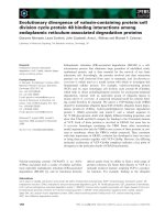
Fig. 1. The nucleotide sequence of the
zebrafish fabp6 gene. The coding sequence
is shown in upper-case letters, with the
deduced amino acid sequence below. The
stop codon is indicated by an asterisk. The
size of each intron is shown, with the exo-
n ⁄ intron splice junctions (gt ⁄ ag) shown in
bold and underlined. The 5¢ upstream
sequence of the fabp6 gene is shown in
lower-case letters, with a putative TATA box
in upper-case letters, underlined and in bold.
SNPs based on differences between the
fabp6 gene derived from the Tu
¨
bingen strain
and cDNA sequences from the AB strain of
zebrafish are shown in bold above the geno-
mic sequence. The polyadenylation signal
sequence AATAAA is underlined and in
bold.
F. A. Alves-Costa et al. The zebrafish fabp6 gene
FEBS Journal 275 (2008) 3325–3334 ª 2008 The Authors Journal compilation ª 2008 FEBS 3327
from 42.1% to 21.8%. Phylogenetic analysis revealed
the inclusion of zebrafish Fabp6 with human, rat,
mouse and pig FABP6s in a distinct clade with a robust
bootstrap value of 99 ⁄ 100 (Fig. 3). The phylogenetic
tree indicates a closer evolutionary relationship between
FABP6s and FABP1s, and a more distant relationship
between FABP6s and FABP7s, a finding consistent
with early phylogenetic studies of rat and human
FABP6 and FABP1 [10,12]. The sequence alignment
(Fig. 2) and phylogenetic analysis (Fig. 3) strongly sug-
gests that the ESTs and genomic sequence retrieved
from DNA assembly Zv7, scaffold 296.3 (Wellcome
Trust Sanger Institute zebrafish genome sequence)
described above, code for Fabp6 in zebrafish.
Linkage group assignment of the zebrafish fabp6
gene by radiation hybrid mapping and its
conserved gene synteny with mammalian
FABP6/Fabp6 genes
To provide additional evidence that the gene located
on the DNA assembly Zv.7, scaffold 296.3, indeed
codes for zebrafish Fabp6, we determined the linkage
group (chromosome) assignment of the zebrafish
fabp6 gene and examined its conserved gene synteny
with the human, rat and mouse FABP6 ⁄ Fabp6 genes.
Using the LN54 panel of radiation hybrids [21] and
specific primers to exon 2 and intron 2, repectively
(see Fig. 1 and Experimental procedures), the zebra-
fish fabp6 gene was mapped to linkage group (chro-
mosome) 21 at a distance of 26.79 cR from the
marker fc08c06, with an LOD (logarithm of the odds
[to the base 10]) of 10.8 (mapping data available at
This result is
consistent with the chromosomal location of fabp6 on
Zv6 in the Wellcome Trust Sanger Institute database,
but not with the latest version, Zv7, which places the
zebrafish fabp6 gene on chromosome 3. We have pre-
viously observed incompatibilities between radiation
hybrid mapping data for other zebrafish fabp genes
and their chromosomal assignment in the Wellcome
Trust Sanger Institute genome sequence database for
zebrafish. Later, versions of the zebrafish genome
sequence have been corrected in agreement with the
chromosomal assignment of fabp genes by radiation
hybrid mapping.
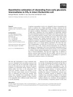
Fig. 2. Sequence alignment and amino acid sequence identity of zebrafish Fabp6 and FABP6s from various species, and paralogs of other
zebrafish Fabps and human FABPs. The deduced amino acid sequence of the zebrafish Fabp6 (Ensembl peptide ID ENSDARP00000065447)
was compared to sequences of FABP6s from human (HU FABP6; GenBank accession number U19869), mouse (MO FABP6; CAI24826),
rat (RA FABP6; NP_058794), pig (PI FABP6; P10289), and to zebrafish Fabp paralogs FABP1A (ZF FABP1A; DQ062095), FABP1B (ZF FABP1B;
DQ062096), FABP2 (ZF FABP2; AAH75970), FABP3 (ZF FABP3; NP_694493), FABP7A (ZF FABP7A; NP_571680), FABP7B (ZF
FABP7B; AAQ92970), and human FABPs FABP1 (Hu FABP1; M10617), FABP2 (HU FABP2; M18079), FABP3 (HU FABP3; X56549), FABP4 (HU
FABP4; NP_00133) and FABP7 (HU FABP7; CAI15449). Dots indicate amino acid identity. Gaps (dashes) have been introduced to maximize align-
ment. The percentage amino acid sequence identities between the zebrafish Fabp6 and other FABPs are shown at the end of each sequence.
The zebrafish fabp6 gene F. A. Alves-Costa et al.
3328 FEBS Journal 275 (2008) 3325–3334 ª 2008 The Authors Journal compilation ª 2008 FEBS
The conserved gene synteny between the zebrafish
fabp6 gene on chromosome 21 and human FABP6
gene on chromosome 5 is extensive (Table 1). Con-
served gene synteny was also evident between the
zebrafish fabp6 gene and the Fabp6 genes on rat chro-
mosome 10 and mouse chromosome 11. Not all the
genes that show conserved gene synteny between
zebrafish chromosome 21 and human chromosome 5
are located on rat chromosome 10 and mouse chromo-
some 11. Other genes are located on rat chromosomes
2, 17, 18 and 20, and mouse chromosomes 13, 15 and
18, suggesting chromosomal rearrangements or trans-
locations in these regions after divergence of the
human and rodent lineages. Despite these chromo-
somal rearrangements, the conserved gene synteny
shown in Table 1 strongly indicates that a common
linkage group containing the FABP6 ⁄ Fabp6 ⁄ fabp6
gene was inherited from a common ancestor of fishes
and mammals. The conserved gene synteny (Table 1),
sequence identity (Fig. 2) and phylogenetic analysis
(Fig. 3) provide compelling evidence that the putative
zebrafish fabp6 gene described here and the mamma-
lian FABP6 ⁄ Fabp6 genes are orthologs.
Distribution of fabp6 gene transcripts in zebrafish
embryos and larvae
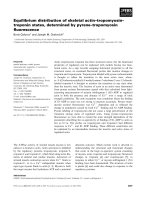
To determine the spatio-temporal distribution of
fabp6 transcripts during zebrafish embryonic and lar-
val development, we performed whole-mount in situ
hybridization to zebrafish embryos and larvae at var-
ious developmental stages (Fig. 4). fabp6 transcripts
were not detected in embryos at 48 h postfertilization
(hpf), but a very strong hybridization signal was
detected in the distal region of the zebrafish intestine
at 72 hpf (Fig. 4A), indicating that initiation of
fapb6 gene transcription occurred between 48 and
72 hfp. The distribution of fabp6 transcripts
remained constant in the distal region of the intes-
tine of zebrafish larvae from 3 to 7 days postfertil-
ization (Fig. 4A–C). A transverse section of a 4-day-
old larva showed the presence of fabp6 transcripts
located predominately in epithelial cells of the intes-
tine (Fig. 4B).
To our knowledge, only two studies have investi-
gated the tissue-specific distribution of FABP6 ⁄
Fabp6 transcripts during embryogenesis [14,15]. In
the mouse, Sacchettini et al. [14] showed by dot-blot
hybridization that no Fabp6 transcripts were detected
in any tissues during fetal life, or throughout the
suckling period of 1–12 postnatal days. Mouse Fabp6
transcripts were first detected and restricted to the
ileum at the beginning of the suckling ⁄ weaning tran-
sition at postnatal days 12–14. In contrast, Crossman
et al. [15] did detect Fabp6 transcripts in mouse
embryos. They used Northern blot analysis and
quantified mRNA steady-state levels by scanning au-
toradiograms of RNA extracted from total intestine
and sections along the entire length of the intestine
(i.e. from the gastroduodenal junction to the rec-
tum). Fabp6 transcripts were first detected in RNA
from total intestine at E18, which is the stage at
which the ‘proximal-to-distal wave of cytodifferentia-
tion of the pseudo-stratified gut epithelium to a
monolayer had reached the ileum’ [14]. During post-
natal development, Fabp6 transcripts were restricted
to the distal third of the small intestine and cecum.
No Fabp6 transcripts were detected in the duode-
num, jejunum or 12 other extraintestinal tissues
(the latter tissues were not specified). The transcrip-
tional initiation of the zebrafish fabp6 gene in the
distal region of the intestine at around 72 hpf, prior
to hatching (Fig. 5), occurs at approximately the
same developmental stage as the transcriptional initi-
ation of the mouse Fabp6 gene in the ileum at E18
[14].
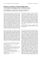
Fig. 3. A neighbor-joining tree showing the phylogenetic relation-
ship of zebrafish Fabp6 with selected paralogous and orthologous
Fabp ⁄ FABPs from zebrafish and mammals. The bootstrap values,
as percentage (based on 100 replicates), are indicated at the nodes.
The sequences used were zebrafish FABP6 (Ensembl peptide ID
ENSDARP00000065447), mammalian sequences for FABP6 from
human (HU FABP6, GenBank accession number U19869), mouse
(MO FABP6, CAI24826), rat (RA FABP6, NP_058794) and pig (PI
FABP6, P10289), and sequences for zebrafish FABP1A (ZF Fabp1A,
DQ062095), FABP1B (ZF Fabp1B, DQ062096), FABP2 (ZF Fabp2,
AAH75970), FABP3 (ZF Fabp3, NP_694493), FABP7A (ZF Fabp7A,
NP_571680) and FABP7B (ZF Fabp7B, AAQ92970) and human
FABP1 (Hu FABP1, M10617), FABP2 (HU FABP2, M18079), FABP3
(HU FABP3, X56549), FABP4 (HU FABP4, NP_00133) and FABP7
(HU FABP7, CAI15449). The distinct clade of FABP6 ⁄ Fabp6s is
shaded in gray. Scale bar = 0.2 substitutions per site.
F. A. Alves-Costa et al. The zebrafish fabp6 gene
FEBS Journal 275 (2008) 3325–3334 ª 2008 The Authors Journal compilation ª 2008 FEBS 3329
Tissue-specific distribution of the fabp6 gene
transcript in adult zebrafish
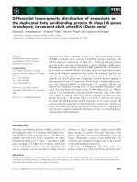
We explored the tissue-specific distribution of fabp6
transcripts in adult zebrafish by RT-PCR amplification
from total RNA extracted from various tissues and by
in situ hybridization of a fabp6-specific antisense oligo-
nucleotide probe to sections of adult zebrafish. A
fabp6-specific RT-PCR product of expected size was
amplified from total RNA extracted from liver, heart,
intestine, ovary and kidney (Fig. 5, top panel). No
fabp6-specific RT-PCR product was amplified from
total RNA extracted from the skin, brain, gill, eye or
muscle. As a positive control to determine the integrity
of the RNA samples used in these assays, transcripts
for the constitutively expressed elongation factor 1a
(ef1a) gene were amplified by RT-PCR. A product of
the expected size was generated from RNA extracted
from all tissues assayed (Fig. 5, bottom panel).
Table 1. Conserved gene synteny between zebrafish linkage group (chromosome) 21 and human chromosome 5, rat chromosome 10 and
mouse chromosome 11.
Chromosomal position
Genes
Zebrafish Human Rat Mouse
Linkage
group 5 2 10 17 ⁄ 20 18 11 13 15 18
c6 21 5p13 2q16 3.0 cM
c7 21 5p13 2q16 3.0 cM
rpl37 21 5p13 2q16 A1
fgf10 21 5p13-p12 2q15-q16 75.0 cM
taf9 21 5q11.2-q13.1 2q12 D1
f2rl1 21 5q13 2q12 75.0 cM
thbs4 21 5q13 2q12 46.99 cM
bhmt 21 5q13.1-q15 2q12 D1
glrx 21 5q14 2q11 44.0 cM
ell2 21 5q15 2q11 C1
pcsk1 21 5q15-q21 2q11-q12 44.0 cM
rnf14 21 5q23.3-q31 18p11 17.0 cM
cdc23 21 5q31 18p12 17.0 cM
pou4f3 21 5q31 18p11 24.0 cM
sept8 21 5q31 10q22 28.5 cM
skp1a 21 5q31 10q22 31.0 cM
vdac1 21 5q31 10q22 29.0 cM
cnot8 21 5q31-q33 10q22 B1.3
ddx46 21 5q31.1 17p14 B2
pdlim4 21 5q31.1 – – – – 28.5 cM
rapgef6 21 5q31.1 10q22 B1.3
sara2 21 5q31.1 10q22 B1.3
tcf7 21 5q31.1 10q22 28.0 cM
zcchc10 21 5q31.1 10q22 28.5 cM
spry4 21 5q31.3 18p11 18.0 cM
zmat2 21 5q31.3 18p11 B2
rbm22 21 5q33.1 18q12.1 D2
larp1 21 5q33.2 10q22 B2
sap301 21 5q33.2 10q22 B2
rnf145 21 5q33.3 – – – – B1.1
fabp6 21 5q33.3-q34 10q21 24.0 cM
sgcd 21 5q33.3-q34 10q21 B1.2
mat2b 21 5q34-q35 10q12 A5
drd1 21 5q35.1 20p12 32.0 cM
fgfr4 21 5q35.1 17p14 33.0 cM
rars 21 5q35.1 10q12 A4
ubtd2 21 5q35.1 – – – – A4
cnot6 21 5q35.3 10q22 B1.2
nola2 21 5q35.3 10q22 28.5 cM
The zebrafish fabp6 gene F. A. Alves-Costa et al.
3330 FEBS Journal 275 (2008) 3325–3334 ª 2008 The Authors Journal compilation ª 2008 FEBS
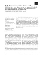
In situ hybridization of an antisense fabp6 oligonu-
cleotide probe to adult zebrafish sections revealed an
intense hybridization signal in the distal region of the
intestine (Fig. 6, 1A) and in the adrenal homolog of
the kidney (Fig. 6, 1D). Less-intense hybridization sig-
nals were observed in the liver (Fig. 6, 1B) and the
ovary (Fig. 6, 1C). Despite the difference in sensitivity
of the two methods employed, the tissue distribution
of fabp6 transcripts in adult zebrafish assayed by
RT-PCR and by in situ hybridization was identical.
In adult mammals, the reported tissue distributions
of FABP6 ⁄ Fabp6 gene transcripts and its protein have
varied, probably due to the assay techniques used. For
example, Fujita et al. [10] used Northern blot analysis
to detect a single-sized FABP6 transcript in RNA
extracted from the terminal region of the human
ileum, whereas RT-PCR generated an abundant
FABP6-specific product from total RNA extracted
from the ileum, and to a much lesser extent from
RNA extracted from the human ovary and placenta.
Unfortunately, the authors do not state whether other
tissues were assayed by RT-PCR in which FABP6
transcripts were not detected. Rat Fabp6 transcripts
were detected by Northern blot analysis of RNA
extracted from the ileum and ovary, but not in RNA
extracted from the stomach, jejunum, colon, adrenal,
brain, heart or liver [12]. Iseki et al. [11] used immuno-
cytochemistry to localize the rat Fabp6 protein and
in situ hybridization to localize Fabp6 transcripts to
the enterocytes of the ileum, luteal cells of the ovary
and a subpopulation of steroid endocrine cells of the
adrenal gland. Sato et al. [13] also detected rat FABP6
in the adrenal gland and ovary. In adult mouse, Fabp6
transcripts were only detected by blot hybridization in
the intestine, and not in the liver, stomach, pancreas,
kidney, spleen, testis, skeletal muscle, heart or lung [14].
With the exception of one report [12], these studies
consistently show that the FABP6 ⁄ Fabp6 gene tran-
scripts are expressed at high levels in the ileum and to a
lesser extent in the ovary and adrenal gland of adult
mammals. In zebrafish, we showed by RT-PCR and
AB
C
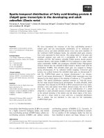
Fig. 4. The spatio-temporal distribution of fabp6 transcripts during
zebrafish embryonic and larval development was determined by
whole-mount in situ hybridization. fabp6 transcripts were first
detected at 72 h postfertilization in the distal region of the intestine
(A) and remained confined to this region of the intestine up to
7 days postfertilization (C), the last time point assayed. (B) Distribu-
tion of fabp6 transcripts throughout the enterocytes of the intestine
in a transverse section at 4 days postfertilization.
Fig. 5. RT-PCR detection of fabp6 transcripts in RNA extracted
from tissues of adult zebrafish. RT-PCR generated a fabp6 mRNA-
specific product from RNA extracted from adult zebrafish liver (L),
heart (H), intestine (I), ovary (O) and kidney (K). No fabp6 mRNA-
specific product was generated by RT-PCR of RNA extracted from
adult zebrafish skin (S), brain (B), gills (G), eyes (E), muscle (M), or
the negative control ()) lacking RNA template. As a positive control
for the integrity of each RNA template, an ef1a mRNA-specific
product was generated from all the adult zebrafish tissues ana-
lyzed.
2
A
D
C
B
1
C
B
A
C
D
Fig. 6. Tissue-specific detection of fabp6 gene transcripts by in situ
hybridization of an antisense riboprobe to sections of adult zebra-
fish. Panel 1 shows the distribution of fapb6 transcripts in the distal
region of the intestine (A), liver (B), ovary (C) and the adrenal homo-
log of the fish kidney (D). Panel 2 shows the locations of the distal
region of the intestine (A), liver (B), ovary (C) and the adrenal homo-
log of the fish kidney (D) in an adjacent tissue section stained with
cresyl violet.
F. A. Alves-Costa et al. The zebrafish fabp6 gene
FEBS Journal 275 (2008) 3325–3334 ª 2008 The Authors Journal compilation ª 2008 FEBS 3331
in situ hybridization that fabp6 transcripts were detected
at high levels in the distal region of the intestine, the tis-
sue homologous to the mammalian ileum. The presence
of fabp6 transcripts suggests that Fabp6 may well play a
role in the uptake of lipids from the distal region of the
zebrafish intestine, which is similar to the suggested role
for FABP6 in the uptake of bile salts from the mam-
malian ileum [11,12]. Zebrafish fabp6 transcripts were
shown by RT-PCR assay (Fig. 5) and in situ hybridiza-
tion (Fig. 6) to be abundant in the ovary and kidney of
adult zebrafish, similar to the distribution of mamma-
lian FABP6 ⁄ Fabp6 transcripts, which are also generally
found in the ovary and adrenal gland. In fishes, the
adrenal homolog is not as compact as the adrenal gland
found in mammals. In fishes, adrenal tissue exists as
aminergic chromaffin and inter-regnal cells, mostly
inside the head kidney, with the two tissues being either
mixed, adjacent, or completely separated [22]. The dis-
tribution of the hybridization signal for zebrafish fabp6
transcripts in the adrenal homolog of the kidney (Fig. 6,
1D) is consistent with the structure of the adrenal homo-
log in teleost fishes. With the exception of the zebrafish
liver and heart, the overall pattern of adult tissue distri-
bution of zebrafish fapb6 and mammalian FABP6 ⁄ -
Fabp6 gene transcripts, and the transcriptional initiation
of these genes at similar embryonic stages of develop-
ment, is surprisingly concordant, in contrast to some
other members of the multigene family of iLBP genes
(e.g., fabp1a ⁄ b, fabp10, fabp11, rbp2) [3,6,16,23,24]. As
FABP6 has been implicated in human colorectal cancer
[25] and type 2 diabetes [26], zebrafish may serve as a
useful model experimental system to investigate the role
of FABP6 in these disease states.
Experimental procedures
Husbandry of zebrafish
The AB strain of zebrafish was used throughout this work
and maintained according to established procedures [27].
Experimental protocols were reviewed by the Animal Care
Committee of Dalhousie University in accordance with
guidelines set down by the Canadian Committee on Animal
Care.
Nucleotide sequence of the zebrafish fabp6 cDNA
and gene
We retrieved a previously uncharacterized Ensembl gene
(ENSDAR00000044566) by a BLASTn search of the
zebrafish genome sequence database at the Wellcome Trust
Sanger Institute (version Zv7, scaffold 296.3, http://www.
ensembl.org/Danio_rerio/index.html), using NM_001002076
(GenBank accession number) as the query sequence. This
sequence was also used in BLASTn searches for other ESTs
coding for zebrafish Fabp6. Based on the NM_001002076
sequence, primers were designed for RT-PCR amplification
of this transcript from total RNA extracted from a whole
adult zebrafish of strain AB (forward primer, 5¢-CTC
TTCTTCTCCGCTCAA-3¢; reverse primer, 5¢-ATCAGTT
TAGCTCGTACA-3¢). The resulting product of expected size
as estimated by agarose gel electrophoresis was cloned into
the pGEM-T vector (Promega, Madison, WI, USA) and six
clones were sequenced. To identify SNPs, the cDNA
sequences obtained by us were compared to the coding
sequence of the zebrafish fabp6 gene retrieved from the
Zebrafish Genome Sequence Database at the Wellcome Trust
Sanger Institute by alignment using clustalw [28]. The
molecular mass and isoelectric point of the Fabp6 polypep-
tide encoded by clone NM_001002076 was determined using
the program at />Phylogenetic analysis
Sequence alignment and determination of percentage amino
acid sequence identity of FABP ⁄ Fabp sequences from
zebrafish and other vertebrates was performed using bio-
edit (version 7.0.9) [29]. Phylogenetic analysis was per-
formed using clustalw [28] to generate a neighbor-joining
tree. Bootstrap values were based on 100 replicates.
Linkage group (chromosome) assignment
by radiation hybrid mapping of the zebrafish
fabp6 gene
Radiation hybrids of the LN54 panel were used to assign the
fabp6 gene to a specific zebrafish linkage group according to
the protocol described by Hukriede et al. [21]. Two primers
were designed (forward primer, 5¢-TAGGCAAAGAGAG
CCACATGCAGA-3¢; reverse primer, 5¢-TGCTCAAATCC
TGACACCATGGAC-3¢) to PCR-amplify a portion of the
zebrafish fabp6 gene from genomic DNA samples isolated
from the LN54 hybrid panel using Platinum PCR Super Mix
(Invitrogen, Burlington, Canada).
Whole-mount in situ hybridization to zebrafish
embryos and larvae
Whole-mount in situ hybridization using a cloned fabp6
cDNA to generate an antisense riboprobe was performed
according to the methods described previously [30].
Detection of fabp6 transcripts in adult zebrafish
tissues by RT-PCR
RT-PCR was used to determine the tissue distribution of
fabp6 transcripts in RNA extracted from tissues of adult
The zebrafish fabp6 gene F. A. Alves-Costa et al.
3332 FEBS Journal 275 (2008) 3325–3334 ª 2008 The Authors Journal compilation ª 2008 FEBS
zebrafish. RNA was extracted from tissue using Trizol
reagent (Invitrogen). Following synthesis of cDNA using the
Omniscript RT kit (Qiagen, Mississauga, Canada), the zebra-
fish fabp6 transcripts were amplified by PCR from total
RNA extracted from various tissues using the forward pri-
mer, 5¢-TAGGCAAAGAGAGCCACATGGAGA-3¢, and
the reverse primer, 5¢-GCGGTTAAACCTTCTTGCTTGT
GC-3¢, according to the protocol described by Liu et al. [23].
The constitutively expressed gene for elongation factor
1a (ef1a) was used as a positive control to assay the inte-
grity of RNA extracted from each tissue. The primers
and RT-PCR conditions employed have been described
previously [31].
Detection of fabp6 transcript in adult sections of
zebrafish by in situ hybridization
A synthetic antisense probe, 5¢-GTACTGGGTCCATGT
GAAGTCATCTCCGTTC-3¢, was used for in situ hybrid-
ization to detect fabp6 transcripts in sections of adult zebra-
fish according to the method described by Denovan-Wright
et al. [32].
Acknowledgements
The authors wish to thank Santhosh Karanth (Depart-
ment of Biology, Dalhousie University, Halifax,
Canada) and Paul Wright (University of Virginia
Health Sciences Center, Charlottesville, VA, USA) for
technical assistance during the course of these studies.
This work was supported by funds from the Natural
Sciences and Engineering Research Council of Canada
(to J. M. W.), the Canadian Institutes of Health
Research (to E. D-W.), and the National Institutes of
Health ⁄ European Commission as part of the ZF-Mod-
els integrated project in the 6th Framework Program
(to B. T. and C. T.). F. A-C. was the recipient of a
scholarship from FAPESP (Fundac¸ a
˜
o de Amparo a
`
Pesiquisa do Estado de Sa
˜
o Paulo), Brazil.
References
1 Bernlohr DA, Simpson MA, Hertzel AV & Banaszak
LJ (1997) Intracellular lipid-binding proteins and their
genes. Annu Rev Nutr 17, 277–303.
2 Schaap FG, Van der Vusse GJ & Glatz JFC (2002)
Evolution of the family of intracellular lipid binding
proteins in vertebrates. Mol Cell Biochem 239,
69–77.
3 Agulleiro MJ, Andre
´
M, Morais S, Cedra
`
J & Babin PJ
(2007) High transcript level of fatty acid-binding pro-
tein 11 but not of very low-density lipoprotein receptor
is correlated to ovarian follicle atresia in a teleost fish
(Solea senegalensis). Biol Reprod 77, 504–516.
4 Hertzel AV & Bernlohr DA (2000) The mammalian
fatty acid-binding protein multigene family: molecular
and genetic insights into function. Trends Endocrine
Metab 11, 175–180.
5 Zimmerman AW & Veerkamp JH (2002) New insights
into the structure and function of fatty acid-binding
proteins. Cell Mol Life Sci 11, 1096–1116.
6 Sharma MK, Liu R–Z, Thisse C, Thisse B, Denovan-
Wright EM & Wright JM (2006) Hierarchical subfunc-
tionalization of fabp1a, fabp1b, and fabp10 tissue-
specific expression may account for retention of these
duplicated genes in the zebrafish (Danio rerio) genome.
FEBS J 273, 3216–3229.
7 Ockner RK, Manning JA, Poppenhausen RB & Ho
WK (1972) A binding protein for fatty acids in cytosol
of intestinal mucosa, liver, myocardium, and other tis-
sues. Science 177, 56–58.
8 Haunerland NH & Spener F (2004) Fatty acid-binding
proteins – insights from genetic manipulations. Prog
Lipid Res 43, 328–349.
9 Storch J & Thumser AEA (2000) The fatty acid trans-
port function of fatty acid-binding proteins. Biochim
Biophys Acta 1486, 28–44.
10 Fujita M, Fujii H, Kanda T, Sato E, Hatakeyama K &
Ono T (1995) Molecular cloning, expression and char-
acterization of a human intestinal 15-kDa protein. Eur
J Biochem 233, 406–413.
11 Iseki S, Amano O, Kanda T, Fujii H & Ono T (1993)
Expression and localization of intestinal 15 kDa protein
in the rat. Mol Cell Biochem 123, 113–120.
12 Gong Y-Z, Everett ET, Schwartz DA, Norris JS &
Wilson FA (1994) Molecular cloning, tissue distribu-
tion, and expression of a 14_kDa bile acid-binding pro-
tein from rat ileal cytosol. Proc Natl Acad Sci USA 91,
4741–4745.
13 Sato E, Fujii H, Fujita M, Kanda T, Iseki S, Hatakey-
ama K, Tanaka T & Ono T (1995) Tissue-specific
regulation of the expression of rat intestinal bile acid-
binding protein. FEBS Lett 374, 184–186.
14 Sacchettini JC, Hauft SM, Van Camp SL, Cistola DP
& Gordon JI (1990) Developmental and structural
studies of an intracellular lipid binding protein
expressed in the ileal epithelium. J Biol Chem 265,
19199–19207.
15 Crossman MW, Hauft SM & Gordon JI (1994) The
mouse ileal lipid-binding protein gene: a model for
studying axial patterning during gut morphogenesis.
J Cell Biol 126, 1547–1564.
16 Walz DA, Wider MD, Snow JW, Dass C & Desiderio
DM (1988) The complete amino acid sequence of por-
cine gastrotropin, an ileal protein which stimulates gas-
tric acid and pepsinogen secretion. J Biol Chem 28,
14189–14195.
17 Kanda T, Odani S, Tomoi M, Matsubara Y & Ono
T (1991) Primary structure of the 15-kDa protein
F. A. Alves-Costa et al. The zebrafish fabp6 gene
FEBS Journal 275 (2008) 3325–3334 ª 2008 The Authors Journal compilation ª 2008 FEBS 3333
from rat intestinal epithelium. Eur J Biochem 197,
759–768.
18 Denovan-Wright EM, Pierce M, Sharma MK & Wright
JM (2000) cDNA sequence and tissue-specific expres-
sion of a basic liver-type fatty acid binding protein in
adult zebrafish (Danio rerio). Biochim Biophys Acta
1492, 227–232.
19 Wu Q, Andolfatto P & Haunerland NH (2001) Cloning
and sequence of the gene encoding the muscle fatty acid
binding protein from desert locus, Schistocerca gregaria.
Insect Biochem Mol Biol 31, 553–563.
20 Breathnach R & Chambon P (1981) Organization and
expression of eukaryotic split genes coding for proteins.
Annu Rev Biochem 31, 349–383.
21 Hukriede NA, Joly L, Tsang M, Miles J, Tellis P, Epstein
JA, Barbazuk WB, Li FN, Paw B, Postlethwait JH et al.
(1999) Radiation hybrid mapping of the zebrafish gen-
ome. Proc Natl Acad Sci USA 96, 9745–9750.
22 Gallo VP & Civinini A (2003) Survey of the adrenal
homolog in teleosts. Int Rev Cytol 230, 89–187.
23 Liu R-Z, Denovan-Wright EM & Wright JM (2003)
Structure, linkage mapping and expression of the heart-
type fatty acid-binding protein gene (fabp3) from zebra-
fish (Danio rerio). Eur J Biochem 270, 3223–3234.
24 Liu R-Z, Denovan-Wright EM, Degrave A, Thisse C,
Thisse B & Wright JM (2004) Spatio-temporal distribu-
tion of cellular retinol-binding protein gene transcripts
(CRBPI and CRBPII) in the developing and adult
zebrafish (Danio rerio). Eur J Biochem 271, 339–348.
25 Ohmachi T, Inoue H, Mimori K, Tanaka F, Sasaki A,
Kanda T, Fujii H, Yanaga K & Mori M (2006) Fatty
acid binding protein 6 is overexpressed in colorectal
cancer. Clin Cancer Res 12, 5090–5095.
26 Fisher E, Nitz I, Lindner I, Rubin D, Boeing H, Mo
¨
hlig
M, Hampe J, Schreiber S, Schrezenmeir J & Do
¨
ring F
(2007) Candidate gene association study of type 2 dia-
betes in a nested case-control study of the EPIC-Pots-
dam cohort – role of fat assimilation. Mol Nutr Food
Res 51, 185–191.
27 Westerfield M (1995) The Zebrafish Book: A Guide for
the Laboratory Use of Zebrafish (Danio rerio), 3rd edn.
University of Oregon Press, Eugene, OR.
28 Thompson JD, Gibson TJ, Plewniak F, Jeanmougin F
& Higgins HG (1997) The CLUSTALW windows inter-
face: flexible strategies for multiple sequence alignment
aided by quality analysis tools. Nucleic Acids Res 24,
4876–4882.
29 Hall T (1999) BioEdit: a user-friendly biological
sequence alignment editor and analysis program for
Windows 95 ⁄ 98 ⁄ NT. Nucleic Acids Symp Ser 41, 95–98.
30 Thisse C & Thisse B (2008) High-resolution in situ
hybridization to whole-mount zebrafish embryos. Nat
Protoc 3, 59–69.
31 Pattyn F, Robbrecht P, Speleman F, De Paepe A &
Vandesompele J (2006) RTPrimerDB: the real-time
PCR primer and probe database, major update 2006.
Nucleic Acids Res 34, D684–D688.
32 Denovan-Wright EM, Newton RA, Armstrong JM,
Babity JM & Robertson HA (1998) Acute administra-
tion of cocaine, but not amphetamine, increases the
level of synaptotgmin IV mRNA in the dorsal striatum
of rat. Mol Brain Res 55, 350–354.
The zebrafish fabp6 gene F. A. Alves-Costa et al.
3334 FEBS Journal 275 (2008) 3325–3334 ª 2008 The Authors Journal compilation ª 2008 FEBS