Báo cáo khoa học: Global shape and pH stability of ovorubin, an oligomeric protein from the eggs of Pomacea canaliculata pot
Bạn đang xem bản rút gọn của tài liệu. Xem và tải ngay bản đầy đủ của tài liệu tại đây (443.98 KB, 9 trang )
Global shape and pH stability of ovorubin, an oligomeric
protein from the eggs of Pomacea canaliculata
Marcos S. Dreon
1
, Santiago Ituarte
1
, Marcelo Ceolı
´n
2
and Horacio Heras
1
1 Instituto de Investigaciones Bioquı
´
micas de La Plata (INIBIOLP), CONICET-UNLP, Argentina
2 Instituto de Investigaciones Fı
´
sico-Quı
´
micas, Teo
´
ricas y Aplicadas (INIFTA), CONICET-UNLP, La Plata, Argentina and Universidad Nacional
del Noroeste de Buenos Aires, Pergamino, Argentina
Pomacea canaliculata (Architaenioglossa: Ampullarii-
dae) is a freshwater snail native to the Amazon and
Plata basins, where its seasonal reproduction is mostly
affected by changes in environmental temperatures and
the availability of water [1–3]. During the 1980s, it was
introduced into Asia, where it has both become a pest
for rice crops and a vector for human eosinophilic
meningoencephalitis, a parasitic disease that is rapidly
expanding worldwide [4].
Most gastropod eggs have perivitellin fluid sur-
rounding the fertilized oocyte that represents the major
supply of nutrients during embryogenesis [5]. Ovorubin
is the major protein in the perivitellin fluid of the eggs
of P. canaliculata, previously described by Comfort [6]
and Cheesman [7] as a carotenoprotein. It is a lipo-
glyco-carotenoprotein with a molecular mass of
300 kDa, composed of three subunits of 28, 32 and
35 kDa [8], and it represents 60% of the total perivitel-
lin fluid protein content [9]. The carotenoid content of
ovorubin is mainly composed of astaxanthin (ASX), a
potent membrane antioxidant [10] in its free and esteri-
fied forms. Ovorubin, besides its function as an energy
and structural precursor donor, acts by transporting
and stabilizing these labile antioxidants in the perivi-
Keywords
carotenoprotein; mollusk; protease inhibitor;
protein stability; protein structure
Correspondence
H. Heras, INIBIOLP – Fac. Cs. Me
´
dicas, 60 y
120, La Plata (1900), Argentina
Fax: +54 221 4258988
Tel: +54 221 4824894
E-mail:
(Received 16 May 2008, revised 3 July
2008, accepted 11 July 2008)
doi:10.1111/j.1742-4658.2008.06595.x
Ovorubin, a 300-kDa thermostable oligomer, is the major egg protein from
the perivitellin fluid that surrounds the embryos of the apple snail Poma-
cea canaliculata. It plays essential roles in embryo development, including
transport and protection of carotenoids, protease inhibition, photoprotec-
tion, storage, and nourishment. Here, we report ovorubin dimensions and
global shape, and test the role of electrostatic interactions in conforma-
tional stability by analyzing the effects of pH, using small-angle X-ray scat-
tering (SAXS), transmission electron microscopy, CD, and fluorescence
and absorption spectroscopy. Analysis of SAXS data shows that ovorubin
is an anisometric particle with a major axis of 130 A
˚
and a minor one
varying between 63 and 76 A
˚
. The particle shape was not significantly
affected by the absence of the cofactor astaxanthin. The 3D model pre-
sented here is the first for an invertebrate egg carotenoprotein. The quater-
nary structure is stable over a wide pH range (4.5–12.0). At a pH between
2.0 and 4.0, a reduction in the gyration radius and a loss of tertiary struc-
ture are observed, although astaxanthin binding is not affected and only
minor alterations in secondary structure are observed. In vitro pepsin diges-
tion indicates that ovorubin is resistant to this protease action. The high
stability over a considerable pH range and against pepsin, together with
the capacity to bear temperatures > 95 °C, reinforces the idea that
ovorubin is tailored to withstand a wide variety of conditions in order to
play its key role in embryo protection during development.
Abbreviations
ASX, astaxanthin; R
g,
gyration radius; SAXS, small-angle X-ray scattering; TEM, transmission electron microscopy.
4522 FEBS Journal 275 (2008) 4522–4530 ª 2008 The Authors Journal compilation ª 2008 FEBS
tellin fluid [11]. In addition, Norden [12] has described
this carotenoprotein as having trypsin, chymotrypsin
and other protease inhibitor activity, another unusual
function for a perivitellin.
In contrast to most invertebrate carotenoproteins,
ovorubin does not suffer destabilization when its carot-
enoid is removed [11]. Moreover, the stabilities of apo-
ovorubin and holo-ovorubin are virtually the same as
regards structure stability against temperature; they
remain stable over 95 °C and are affected only by
molar concentrations of urea and guanidinium hydro-
chloride [13].
Except for the detailed studies on crustacyanin, the
lobster carapace carotenoprotein, there is little infor-
mation on the structure and stability of this interesting
group of proteins, and there is no information in
mollusks [14,15].
In this work, we report the first 3D low-resolution
model of ovorubin obtained by small-angle X-ray scat-
tering (SAXS). Ovorubin stability with regard to pH
was also studied using SAXS, CD, intrinsic tryptophan
fluorescence and absorption spectroscopy, in an
attempt to further test its structural features.
Results
Global shape of ovorubin
Figure 1A shows the SAXS curves obtained for holo-
ovorubin and apo-ovorubin normalized for protein
concentration. Clearly, the two curves virtually over-
lap, indicating that both ovorubin forms have nearly
the same shape and size. From the Guinier plot for
holo-ovorubin and apo-ovorubin (Fig. 1A), it was pos-
sible to fit gyration radii of 43.0 ± 0.7 A
˚
and
44.0 ± 0.1 A
˚
, respectively. The Kratky plots (Fig. 1B)
are bell-shaped, as expected for globular proteins. The
gyration radii obtained are quite compatible with a
compact oligomer of about 300 kDa, which is a mole-
cular mass determined previously for ovorubin.
Figure 1C shows the pair distribution curves obtained
by means of the regularization technique implemented
in gnom4.5 [16]. Holo-ovorubin showed a maximum
at 52 A
˚
with a well-defined D
max
of 122 A
˚
, which is
compatible with an anisometric particle. Apo-ovorubin
showed a slightly displaced maximum and a higher
contribution at longer distances, probably due to some
degree of aggregation induced by the lack of the cofac-
tor. A low-resolution model, obtained by averaging 16
calculated models using the algorithm implemented in
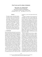
dammin [17], is depicted in Fig. 2A–C. This ab initio
theoretical model fits satisfactorily with the experimen-
tal scattering intensity data (Fig. 2D). The particle
shows an anisometric shape, with a major axis of
130 A
˚
and a minor one varying between 63 and 76 A
˚
.
Image analysis of transmission electron microscopy
(TEM) data provided a size distribution curve of these
particles showing a bimodal shape with two maxima,
which account for more than 75% of the total
(Fig. 3B). The diameter obtained from the first maxi-
mum, 112 A
˚
, is in general agreement with the maxi-
mum pair distance obtained from SAXS results. The
second maximum of the size distribution, 162 A
˚
,is
most likely an artefact resulting from sample process-
ing. The absence of supramolecular aggregates
observed by TEM is consistent with the SAXS results.
Structural stability of ovorubin with regard to pH
The gyration radius, R
g
, of holo-ovorubin at different
pH values is shown in Fig. 4A, where a constant value
of 45 ± 2 A
˚
can be observed between pH 12.0 and
pH 4.5. The isoelectric point determined for holo-
ovorubin was 4.9, and below this pH, a sudden
A
B
Q(Å
–1
)
C
R (Å)
Ln (I(Q)/C)
Q(Å
–2
)
Log (Q
2
.l (Q)/C)
Log (l(Q)/C)P (R)
Fig. 1. Study of holo-ovorubin and apo-ovorubin solution structure
by SAXS. (A) Raw SAXS data [I(Q)]. Inset: Guinier region in linear-
ized variables. (B) Kratky plot [I(Q)Q
2
] of data depicted in A. (C)
Pair–distance distribution obtained from data in (A) using the pro-
gram
GNOM v4.5. Solid line: holo-ovorubin. Dotted line: apo-ovorubin.
M. S. Dreon et al. Structure and pH stability of snail egg ovorubin
FEBS Journal 275 (2008) 4522–4530 ª 2008 The Authors Journal compilation ª 2008 FEBS 4523
increase in R
g
was observed before the onset of oligo-
mer disassembly, observed from pH 4.0 to pH 2.0 as a
constant decrease in the R
g
value. The Kratky plots
also showed a progressive loss of globularity at low
pH values (Fig. 4B).
The absorption spectra of the protein at different
pH values are displayed in Fig. 5. Only slight changes
in the fine structure of the spectrum were observed at
pH 2.0. Interestingly, neither red nor blue shifts were
observed at all pH values assayed. It is known that the
UV spectrum of ASX undergoes a large bathochromic
shift, due to ASX binding to ovorubin, attributed to
strong structural deformations of the carotenoid struc-
ture [18,19]. Lack of hypsochromism indicates that
ASX remains bound to its binding site even under very
acidic conditions.
The tryptophan fluorescence spectra between pH 2.0
and pH 12.0 (Fig. 6) show a red shift of its emission
maxima (from 330 to 338 nm) and an intensity
decrease at pH 2.0, indicative of the exposure of
some of the tryptophan residues to the aqueous envir-
onment.
A
10
Z
X
Y
X
1
0.1
0.1 0.2
Q (Å
–1
)
log I (Q)
B
D
C
Y
X
Fig. 2. Three-dimensional model of ovorubin from the eggs of P. canaliculata, obtained by analyzing the scattering data using the DAMMIN
program in three different views. Referred to (A) view, (B) rotated 90° around x-axis, and (C) rotated 90° around z-axis. (D) Scattering inten-
sity of experimental data for ovorubin (solid line) and theoretical ab initio dummy atom model (dotted line).
Count
Particle diameter (Å)
100 nm
A B
Fig. 3. Electron microscopy analysis of
ovorubin from the eggs of the apple snail.
(A) Electron micrograph of negatively
stained ovorubin sample. Final magnification
· 50 000. (B) Size distribution curve of
ovorubin molecules. See Experimental pro-
cedures for details. Bar: 100 nm.
Structure and pH stability of snail egg ovorubin M. S. Dreon et al.
4524 FEBS Journal 275 (2008) 4522–4530 ª 2008 The Authors Journal compilation ª 2008 FEBS
On the basis of the above results, the CD spectra in
the near-UV and far-UV region were only recorded at
pH 2.0 and pH 6.0 (Fig. 7). In the far-UV region
(200–260 nm), both spectra were nearly coincident,
indicating that the secondary structure of holo-ovoru-
bin remains intact even at a low pH (Fig. 7A). Regard-
ing the near-UV region (260–320 nm), a general loss of
structure can be appreciated in the spectrum obtained
at pH 2.0 in comparison with the one obtained at
pH 6.0. No preferential loss of signal in the region of
any of the aromatic residues was observed, suggesting
a global loss of the tertiary structure of ovorubin.
Enzymatic digestion with pepsin was performed at
acidic pH and at different preincubation times. It was
observed that the oligomer was resistant to hydrolysis
after a 150 min incubation, but degraded when prein-
cubated for 48 h at pH 2.5 (Fig. 8).
Discussion
Size and solution structure of ovorubin
Ovorubin and crustacyanin are, so far, the only inver-
tebrate carotenoproteins for which a 3D structure has
been resolved, and a comprehensive body of infor-
mation on the protein is available [11,13,19–24]. It is
evident from these studies that the molluskan ovorubin
complex differs in properties and molecular features
from the crustacean carotenoprotein. Regarding the
3D structure, analysis of the SAXS scattering spectral
data reveals that lobster crustacyanin has a cylindrical
shape [21], whereas ovorubin is an anisometric protein.
Another difference is the role that the carotenoid
pigment ASX plays in the structural stability of these
A
pH
B
I(q *q
2
) Rg (Å)
q (Å
–1
)
Fig. 4. Effect of pH on native ovorubin size and shape. (A) R
g
of
the particle as determined by SAXS. (B) Kratky plots for ovorubin at
different pH values. Solid line: pH 6.0. Dotted line: pH 4.5. Dashed
line: pH 2.0.
Absorbance (au)
λ
λ
(nm)
Fig. 5. Absorption spectra of ovorubin from P. canaliculata at differ-
ent pH values. Solid line: pH 6.0. Dashed line: pH 2.0. Dotted line:
pH 12.0.
Fluorescense yield (au)
λ
λ
(nm)
Fig. 6. Tryptophan fluorescence spectra of ovorubin at different pH
values. Dashed line: pH 2.0. Solid line: pH 6.0. Dotted line:
pH 12.0.
M. S. Dreon et al. Structure and pH stability of snail egg ovorubin
FEBS Journal 275 (2008) 4522–4530 ª 2008 The Authors Journal compilation ª 2008 FEBS 4525
proteins: ASX is essential for crustacyanin integrity
[21], which contrasts with the situation for ovorubin,
where it plays virtually no role in the stability of the
oligomer [11,13], thus indicating a very different inter-
action between subunits in the two carotenoproteins.
Using the ab initio program dammin, we have modeled
the shape of ovorubin as a compact complex of
130 · 76 A
˚
. Negatively stained purified ovorubin
appeared in electron micrographs also as anisometric
particles with a maximum size of 112 A
˚
(assuming that
the larger particles are experimental artefacts). This is
convergent with the SAXS data regarding global shape
and dimensions, and differs from data on other inver-
tebrate carotenoproteins such as the lobster crusta-
cyanin (a cylinder of 238 · 95 A
˚
) [21] and the starfish
linckiacyanin (a spring-like structure with a diameter
of 200–260 A
˚
) [25], which have functions quite differ-
ent from the role of ovorubin in the eggs of apple
snails (Table 1).
Physiological and biophysical implications of
stability with regard to pH
Overall, carotenoproteins belong to a group of pro-
teins that are stable over a relatively wide pH range
[26]. Although this fact has not been previously studied
in the phylum Mollusca, there are several examples in
crustaceans and echinoderms (Table 1).
Ovorubin, the first molluskan carotenoprotein so far
studied shows structural stability over a wider pH
range than that of the crustaceans or echinoderm
proteins. Remarkably, ovorubin is the only caroteno-
protein stable at pH 12. At this pH, the lysyl and argi-
nyl residues are neutralized, usually affecting the
quaternary structure. The high stability of ovorubin
oligomers might be due to a shift of the pK of the
amino acid residues beyond 12, owing to their involve-
ment in salt bridges. At acidic pH, the stability of
ovorubin was similar to that of all other caroteno-
proteins (Table 1).
As mentioned above, electrostatic forces are crucial
for stabilization of the ovorubin quaternary structure,
as suggested by the strong decrease in the R
g
at pH
values below 4.0.
The sharp increase in R
g
obsrved at pH 4.5 is
probably due to partial unfolding of the subunits,
leading to their dissociation. In addition, the
isoelectric point determined at pH 4.9 suggests that
alterations in the charge of the molecule are taking
part in the R
g
change. All these results indicate that,
around this pH, the native structure of ovorubin
becomes unstable, leading to the disassembly
observed at a lower pH.
Ellipticity (mdeg) Ellipticity (mdeg)
A
B
λ
λ
(nm)
Fig. 7. CD spectra of ovorubin at different pH values. Spectra in
the (A) near-UV region (260–320 nm) and (B) the far-UV region
(200–260 nm). Solid line: pH 6.0. Dashed line: pH 2.0.
Fig. 8. Pepsin resistance of ovorubin analyzed on 4–20%
SDS ⁄ PAGE. Lane 1: negative control ovorubin incubated for
150 min at pH 2.5 (6 lg). Lane 2: pepsin-digested ovorubin (6 lg).
Lane 3: ovorubin (6 lg) preincubated for 48 h at pH 2.5 and then
digested with pepsin. Lane 4: molecular mass markers.
Structure and pH stability of snail egg ovorubin M. S. Dreon et al.
4526 FEBS Journal 275 (2008) 4522–4530 ª 2008 The Authors Journal compilation ª 2008 FEBS
The lack of differences in the absorption spectra of
ovorubin in the pH range assayed clearly indicate that
residues in the ASX-binding site were not charged,
suggesting that the residues involved in ASX binding,
responsible for the bathochromic effect, are not ioniz-
able polar residues. This is in agreement with previous
studies of tryptophan resonance energy transfer to
ASX, which indicate that the carotenoid-binding site is
a nonpolar environment [13].
In other words, at pH 2.0 there is a decrease in R
g
,
indicative of disassembly of the particle, but there are
no changes in the absorption spectrum of ovorubin,
indicating that ASX is not located in the subunit inter-
face involved in the stabilization of the oligomer. This
is in agreement with previous reports on the stability
of apo-ovorubin and holo-ovorubin against tempera-
ture and chaotropes [13]. Other serine protease inhibi-
tors have a similarly high stability, ranging from pH 2
to pH 12 [27]. It must be remarked that the major loss
of tertiary and quaternary structure was not enough to
promote the detachment of the ASX molecule from
ovorubin, indicating that the structure of the caroten-
oid-binding site is mainly dominated by secondary
structure elements. Moreover, an indirect indication
that ovorubin is susceptible to hydrolysis at acidic pH
came from the pepsin digestion experiment. When
ovorubin was preincubated for 48 h at pH 2.5, it lost
its resistance towards the enzyme that was observed at
short incubation times.
Eggs of P. canaliculata have a conspicuous warning
coloration that signals to potential predators the pres-
ence of unpalatable or toxic compounds [28]. Snail
eggs were therefore thought to be unpalatable [29],
and in fact have a small number of predators. The pH
stability of ovorubin is within the pH range of verte-
brate digestive tract fluids [30,31], and the present
results indicate that the protein can withstand the com-
bined effect of low pH values and enzymatic attack for
more than 2 h. Thus, if the eggs are ingested by a
predator, ovorubin could reach the intestine in a fully
active form and exert its potent trypsin inhibitor
action, formerly thought to be only antimicrobial [12].
It could therefore be speculated that ovorubin is
actively involved in the chemical defense of the
embryos by limiting the predator’s ability to digest and
use essential nutrients from the eggs, thus rendering
the ingestion of P. canaliculata eggs antinutritive.
The ovorubin complex, despite its large size and
oligomeric nature, now appears to be a protein tai-
lored to withstand a variety of extreme conditions,
reinforcing the idea it plays a key role in embryo
development.
Ongoing research is looking further into the anti-
trypsin properties of the molecule.
Experimental procedures
Egg collection
Adults of P. canaliculata were collected in streams or ponds
near La Plata, a province of Buenos Aires, Argentina. Eggs
were collected from females either raised in our laboratory
or taken from the wild between November and April
(reproductive season). Embryo development was checked in
each egg mass microscopically [8], and only egg masses
having embryos developed to no more than the morula
stage were used.
Ovorubin isolation and purification
Fertilized eggs were repeatedly rinsed with ice-cold 20 mm
Tris ⁄ HCl (pH 6.8), containing 0.8 lm aprotinin (Trasylol,
Mobay Chemical Co., New York, Ny, USA) and homo-
genized in a Potter-type homogenizer (Thomas Sci.,
Swedesvoro, NJ, USA) in the dark and under an N
2
atmo-
sphere. The buffer ⁄ sample ratio was kept at 5 : 1 v ⁄ w [32].
The crude homogenates were then sonicated for 15 s and
centrifuged at 10 000 g for 30 min, and then at 100 000 g
for 60 min. The pellet was discarded, and the supernatant
was stored at )70 °C until analysis. Protein content was
determined by the method of Bradford et al. [33], using
BSA as standard.
The soluble protein fraction obtained using the above
procedure was purified in a Merck-Hitachi high-perfor-
mance liquid chromatograph (Hitachi Ltd, Tokyo, Japan)
Table 1. Stability with regard to pH of aquatic invertebrate carotenoproteins.
Taxa Species Carotenoprotein ⁄ location pH range Ref.
Arthropoda: Crustacea Procambarus clarkii Blue ⁄ carapace 5.5–8.0 [26]
Arthropoda: Crustacea Upogebia pusilla Blue ⁄ carapace 5.5–9.0 [41]
Arthropoda: Crustacea Homarus americanus Crustacyanin ⁄ carapace 5.0–8.5 [42]
Echinodermata: Asteroidea Marthasterias glacialis Blue ⁄ skin 4.0–8.5 [43]
Echinodermata: Asteroidea Marthasterias glacialis Purple ⁄ skin 3.5–8.5 [43]
Arthropoda: Crustacea Homarus americanus Ovoverdin ⁄ egg 4.0–9.0 [44]
Mollusca: Gastropoda Pomacea canaliculata Ovorubin ⁄ egg 4.0–12.0 Present paper
M. S. Dreon et al. Structure and pH stability of snail egg ovorubin
FEBS Journal 275 (2008) 4522–4530 ª 2008 The Authors Journal compilation ª 2008 FEBS 4527
with an L-6200 Intelligent Pump and an L-4200 UV detec-
tor set at 280 nm. A serial HPLC purification was
performed. First, the sample was analyzed in a Mono
QHR10⁄ 10 (Amersham-Pharmacia, Uppsala, Sweden),
using a gradient of 0–1 m NaCl in 20 mm Tris buffer. The
ovorubin peak was then further purified by size exclusion
chromatography (Superdex 200 HR 10 ⁄ 20; Amersham-
Pharmacia, Uppsala, Sweden), using an isocratic gradient
of 50 mm sodium phosphate buffer and 150 mm NaCl
(pH 7.6). The purity of the single peak obtained was
checked by native electrophoresis.
A solution of 2 mgÆmL
)1
apo-ovorubin was prepared as
previously described [13].
Gel electrophoresis
Nondissociating electrophoresis was performed on a 4–20%
polyacrylamide gradient [34,35]. The gels were stained with
Coomassie Brilliant Blue R-250 (Sigma Chemical Co, St
Louis, MO, USA).
SAXS
SAXS experiments were performed either at the D11A-
SAXS1 or the D02A-SAXS2 lines operating in the
Laboratorio Nacional de Luz Syncrotron, Campinas (SP,
Brazil). The scattering pattern was detected either using a
gas-filled one-dimensional position-sensitive detector with
an active window of 80 mm (SAXS1) or a MARCCD
bidimensional charge-coupled device assisted by fit 2d
software ( />(SAXS2). The experiments were performed using a wave-
length of 1.448 A
˚
for the incident X-ray beam to mini-
mize carbon absorption. The distance between the sample
and the detector was kept at 1044 mm, allowing a
Q-range between 0.012 and 0.25 A
˚
)1
(D
max
£ 260 A
˚
). The
temperature was controlled using a circulating water
bath, and kept at 15 °C. Each individual run was cor-
rected for sample absorption, photon flux, buffer scatter-
ing, and detector homogeneity. At least three independent
curves were averaged for each single experiment. SAXS
experiments in a protein range of 2.4–0.20 mgÆmL
)1
were
performed to rule out a concentration effect in the data.
The final experiments were performed at 0.24 mgÆmL
)1
.
The distance distribution function P(r) was calculated by
the Fourier inversion of the scattering intensity I(q) using
the gnom 4.5 program [16]. The low-resolution model of
ovorubin was obtained from the algorithm built in the
program dammin [36]. The program dammin uses
simulated annealing optimization to generate a bead
model giving the best fit to the scattering intensity. The
resulting dummy atom model represents the shape of
the scattering particle. To increase the reliability of the
results, the final model for the dummy atom modeling
was obtained by a spatial average of 16 independent
low-resolution models, calculated with the package
program damaver [37].
TEM
Samples for TEM of native ovorubin of 3 mgÆmL
)1
in
20 mm phosphate buffer (pH 7.4) were stained with 1%
(w ⁄ v) sodium phosphotungstate (pH 7.4), blotted and air-
dried. Images were recorded under low-dose conditions in a
JEM-1200 EX transmission electron microscope (Tokyo,
Japan). Statistical analysis of the particle size distribution
was carried out using the tools built into the program
imagej 1.36b ( />Ovorubin stability with regard to pH
In order to evaluate the influence of pH on ovorubin struc-
ture, solutions of 0.24 mgÆmL
)1
of the protein at different
pH values (from 2 to 12) were prepared using sodium phos-
phate salts and citric acid. All buffers employed were 0.1 m
sodium phosphate salts, except for the pH 4 buffer, which
was prepared by mixing 0.1 m sodium citrate and 0.2 m
Na
2
HPO
4
[38].
After 48 h of incubation, samples were analyzed by
SAXS, CD, and fluorescence and absorption spectroscopy.
Ovorubin isoelectric point determination by 2D
electrophoresis
Immobiline DryStrips (7 cm; pH 4–7, GE Healthcare, Upp-
sala, Sweden) were rehydrated overnight with rehydration
buffer (0.5% immobilized pH gradient buffer 4–7 in Milli-
Q water, and traces of bromophenol blue) containing
approximately 0.5 lg of purified ovorubin. Running was
performed in an Ettan IPGphor 3 IEF system from GE
Healthcare. Electrical conditions were as described by the
supplier. After the first-dimension run, the immobilized pH
gradient gel strips were incubated at room temperature in
3 mL of equilibration buffer (50 mm Tris, pH 6.8, and
traces of bromophenol blue) prior to separation in the sec-
ond dimension. The second-dimension PAGE electrophore-
sis was performed in a vertical system with uniform 10%
separating gel, at 25 °C. The ovorubin spot in the 2D gel
was visualized by Coomassie Brilliant Blue R-250 stain
(Sigma Chemicals).
Pepsin resistance
To analyze pepsin resistance, 20 lg of ovorubin was incu-
bated for 150 min in 0.02 mL of 150 mm NaCl (pH 2.5),
adjusted with 1 m HCl in the presence or absence of 1 lg
of pepsin (Sigma; product No. P6887) [39]. Assays were
performed with preincubation of ovorubin at pH 2.5 for
48 h before pepsin was added. The proteins were analyzed
by 4–20% SDS ⁄ PAGE.
Structure and pH stability of snail egg ovorubin M. S. Dreon et al.
4528 FEBS Journal 275 (2008) 4522–4530 ª 2008 The Authors Journal compilation ª 2008 FEBS
CD and visible absorption spectroscopy
measurements
CD spectra were made either in a Jasco Inc. J-720 spectro-
polarimeter or in a J-810 spectropolarimeter (USA), using
0.2 mm cells placed in a thermostated cell holder at 15 °C.
Samples were measured at a concentration of 0.06 mgÆmL
)1
in 0.1 m phosphate buffer at pH 6 and pH 2. Scanning was
performed with a 1 nm bandwidth, a 100-nmÆmin
)1
scan
speed, and a 4s average time. Each spectrum was obtained
by averaging at least five individual runs, and corrected for
buffer optical activity. Secondary structure content was
estimated by analysis of the molar ellipticities with the k2d
algorithm [40].
Fluorescence and absorption spectroscopy
measurements
Tryptophan fluorescence spectra of ovorubin at pH 2, pH 6
and pH 12 in 0.1 m phosphate buffer were recorded in
emission scanning mode (SLM Aminco, Urbana, IL, USA).
Tryptophan emission was excited at 290 nm (5 nm slit) and
recorded between 310 and 410 nm (5 nm slit). The measure-
ments were made in 5 mm optical path length quartz cells
placed in a thermostated cell holder kept at 20 °C. Each
spectrum was corrected for buffer fluorescence and aver-
aged from at least two independent runs. Similarly, absorp-
tion spectra (350–650 nm) for each pH value were taken.
Acknowledgements
This work was partially supported by CONICET PIP
No. 5888. M. S. Dreon is a member of Carrera del
Investigador CICBA, Argentina. H. Heras and
M. Ceolı
´
n are members of Carrera del Investigador
CONICET, Argentina. S. Ituarte is a doctoral
fellow of CONICET, Argentina. We also thank LNLS
– Brazilian Synchrotron Light Laboratory ⁄ MCT for
access to their facilities and partial financial support
(Projects D11A-SAXS1-5207 ⁄ 06 and 5267).
We thank Dr M. Erma
´
cora for kindly providing
access to the CD equipment.
References
1 Albrecht EA, Carren
˜
o NB & Castro-Vazquez A (1999)
A quantitative study of environmental factors influenc-
ing the seasonal onset of reproductive behaviour in the
south American apple-snail Pomacea canaliculata (Gas-
tropoda: Ampullariidae). J Molluscan Stud 65, 241–250.
2 Pizani NV, Estebenet AL & Martin PR (2005) Effects
of submersion and aerial exposure on clutches and
hatchlings of Pomacea canaliculata (Gastropoda: Amp-
ullariidae). Am Malacol Bull 20, 55–63.
3 Albrecht EA, Koch E, Carren
˜
o NB & Castro-Vazquez A
(2005) Control of the seasonal arrest of copulation and
spawning in the apple snail Pomacea canaliculata
(Prosobranchia: Ampullariidae): differential effects of
food availability, water temperature, and day length.
Veliger 47, 169–174.
4 Cowie RH (2002) Apple snails as agricultural pests:
their biology, impacts, and management. In Molluscs as
Crop Pests (Baker GM, ed), pp. 145–192. CABI, Wal-
lingford.
5 de Jong-Brink M, Boer HH & Joosse J (1983) Mollusca.
In Reproductive Biology of Invertebrates. Oogenesis Ovi-
position and Oosorption (Adiyodi KG & Adiyodi RG,
eds), pp. 297–355. Wiley, New York, NY.
6 Comfort A (1947) Lipochromes in the ova of Pila.
Nature 160, 333–334.
7 Cheesman DF (1958) Ovorubin, a chromoprotein from
the eggs of the gastropod mollusk Pomacea canaliculata.
Proc R Soc Lond [Biol] 149, 571–587.
8 Garı
´
n CF, Heras H & Pollero RJ (1996) Lipoprotein of
the egg perivitellin fluid of Pomacea canaliculata snails
(Mollusca: Gastropoda). J Exp Zool 276, 307–314.
9 Dreon MS, Heras H & Pollero RJ (2003) Metabolism
of ovorubin, the major egg lipoprotein from the apple
snail. Mol Cell Biochem 243, 9–14.
10 Palozza P & Krinsky NI (1992) Astaxanthin and canta-
xanthin are potent antioxidants in a membrane model.
Arch Biochem Biophys 297, 291–295.
11 Dreon MS, Schinella G, Heras H & Pollero RJ (2004)
Antioxidant defense system in the apple snail eggs, the
role of ovorubin. Arch Biochem Biophys 422, 1–8.
12 Norden DA (1972) The inhibition of trypsin and some
other proteases by ovorubin, a protein from the eggs
of Pomacea canaliculata. Comp Biochem Physiol 42,
569–576.
13 Dreon MS, Ceolı
´
n M & Heras H (2007) Astaxanthin
binding and structural stability of the apple snail
carotenoprotein ovorubin. Arch Biochem Biophys 460,
107–112.
14 Zagalsky PF, Wright CE & Parsons M (1995) Crystalli-
sation of alpha-crustacyanin, the lobster carapace
astaxanthin-protein: results from EURECA. Adv Space
Res 16, 91–94.
15 Zagalsky PF, Clark RJH & Fairclough DP (1983)
Resonance Raman and circular dichroism studies of the
copepod (Anomalocera patersoni) and siphonophore
(Porpita sp.) astaxanthin-proteins with an identical
absorption maximum at 650 nm. Comp Biochem Physiol
75, 169–170.
16 Svergun DI (1992) GNOM 4.5. J Appl Crystallogr 25,
495–503.
17 Svergun DI, Petoukhov MV & Koch MHJ (2001)
Determination of domain structure of proteins from
x-ray solution scattering. Biophys J 80, 2946–2953.
M. S. Dreon et al. Structure and pH stability of snail egg ovorubin
FEBS Journal 275 (2008) 4522–4530 ª 2008 The Authors Journal compilation ª 2008 FEBS 4529
18 Clark RJH, D’Urso NR & Zagalsky PF (1980) Exita-
tion profiles, absorption and resonance Raman spectra
of the carotenoprotein ovorubin, and a resonance
Raman study of some other astaxanthin proteins. JAm
Chem Soc 102, 6693–6698.
19 Zagalsky PF, Eliopoulos EE & Findlay JB (1990) The
architecture of invertebrate carotenoproteins. Comp Bio-
chem Physiol 97, 1–18.
20 Chayen NE, Cianci M, Olczak A, Raftery J, Rizkallah
PJ, Zagalsky PF & Helliwell JR (2000) Apocrustacya-
nin A1 from the lobster carotenoprotein alpha-crustacy-
anin: crystallization and initial X-ray analysis involving
softer X-rays. Acta Crystallogr 56, 1064–1066.
21 Dellisanti CD, Spinelli S, Cambillau C, Findlay JBC,
Zagalsky PF, Finet S & Receveur-Brechot V (2003)
Quaternary structure of alpha-crustacyanin from lobster
as seen by small-angle X-ray scattering. FEBS Lett 544,
189–193.
22 Dreon MS, Heras H & Pollero RJ (2004) Characteriza-
tion of the major egg glycolipoproteins from the perivi-
tellin fluid of the apple snail Pomacea canaliculata. Mol
Reprod Dev 68, 359–364.
23 Heras H, Dreon MS, Ituarte S & Pollero RJ (2007) Egg
carotenoproteins in neotropical Ampullariidae (Gastro-
poda: Arquitaenioglossa). Comp Biochem Physiol 146,
158–167.
24 Zagalsky PF, Mummery RS, Eliopoulos EE & Findlay
JBC (1990) The quaternary structure of the lobster car-
apace carotenoprotein, crustacyanin: studies using
cross-linking agents. Comp Biochem Physiol 97, 837–
848.
25 Zagalsky PF, Haxo F, Hertzberg S, Hertzberg S &
Liaaen-Jensen S (1989) Studies on a blue caroteno-
protein, linckiacyanin, isolated from the starfish Linckia
laevigata (Echinodermata: Asteroidea). Comp Biochem
Physiol 93, 339–353.
26 Garate AM, Milicua JCG, Gomez R, Macarulla JM &
Britton G (1986) Purification and characterization of
the blue carotenoprotein from the carapace of the cray-
fish Procambarus clarkii (Girard). Biochim Biophys Acta
881, 446–455.
27 Terada S, Fujimura S, Kino S & Kimoto E (1994) Puri-
fication and characterization of three proteinase inhibi-
tors from Canavalia lineata seeds. Biosci Biotechnol
Biochem 58, 371–375.
28 Gittleman J & Harvey PH (1980) Why are distasteful
prey not cryptic? Nature 286, 149–150.
29 Snyder NFR & Snyder HA (1971) Defenses of the
Florida apple snail Pomacea paludosa. Behaviour 40,
175–215.
30 Randall D, Burggren W & French K (1997) Energy
acquisition: feeding, digestion and metabolism. In Eck-
ert Animal Physiology. Mechanisms and Adaptations
(Randall D, Burggren W & French K, eds), pp.
683–724. Freeman, New York, NY.
31 Denbow DM (2000) Gastrointestinal anatomy and
physiology. In Sturkie’s Avian Physiology (Whittow
GC, ed), pp. 299–325. Academic Press, Boston, MA.
32 Heras H, Garı
´
n CF & Pollero RJ (1998) Biochemical
composition and energy sources during embryo devel-
opment and in early juveniles of the snail Pomacea
canaliculata (Mollusca: Gastropoda). J Exp Zool 280
,
375–383.
33 Bradford MM (1976) A rapid and sensitive method for
the quantitation of microgram quantities of protein uti-
lizing the principle of protein-dye binding. Anal Bio-
chem 72, 248–274.
34 Davis B (1964) Disc electrophoresis. II. Method and
application to human serum proteins. Ann N Y Acad
Sci 121, 404–428.
35 Margolis J & Wrigley CW (1975) Improvement of pore
gradient electrophoresis by increasing the degree of
cross-linking at high acrylamide concentration. J Chro-
matogr 105, 204–209.
36 Svergun DI (1999) Restoring low resolution structure of
biological macromolecules from solution scattering
using simulated annealing. Biophys J 76, 2879–2886.
37 Volkov VV & Svergun DI (2003) Uniqueness of ab
initio shape determination in small-angle scattering.
J Appl Crystallogr 36, 860–864.
38 Deutscher MP (1990) Section II. General methods for
handling proteins and enzymes. In Guide to Protein
Purification. Methods in Enzymology (Deutscher MP,
ed), pp. 19–92. Academic Press, New York, NY.
39 Moreno FJ, Maldonado BM, Wellner N & Mills EN
(2005) Thermostability and in vitro digestibility of a
purified major allergen 2S albumin (Ses i 1) from white
sesame seeds (Sesamum indicum L.). Biochim Biophys
Acta 1752, 142–153.
40 Andrade MA, Chaco
´
n P, Merelo JJ & Mora
´
n F (1993)
Evaluation of secondary structure of proteins from UV
circular dichroism using an unsupervised learning neural
network. Prot Eng 6, 383–390.
41 Villarroel A, Garate AM, Gomez R & Milicua JCG
(1985) A blue carotenoprotein from Upogebia pusilla.
Purification, characterization and properties. Comp
Biochem Physiol 81, 547–550.
42 Chayen NE, Cianci M, Grossmann JG, Habash J,
Helliwell JR, Nneji GA, Raftery J, Rizkallah PJ &
Zagalsky PF (2003) Unravelling the structural chemistry
of the colouration mechanism in lobster shell. Acta
Crystallogr 59, 2072–2082.
43 Armitt GM (1981) Studies on invertebrate carotenopro-
teins. PhD Thesis, University of Liverpool, Liverpool,
UK.
44 Salares VR, Young NM, Bernstein HJ & Carey PR
(1979) Mechanisms of spectral shifts in lobster carote-
noproteins. The resonance raman spectra of ovoverdin
and the crustacyanins. Biochim Biophys Acta 576, 176–
191.
Structure and pH stability of snail egg ovorubin M. S. Dreon et al.
4530 FEBS Journal 275 (2008) 4522–4530 ª 2008 The Authors Journal compilation ª 2008 FEBS