Báo cáo khoa học: A kinetic approach to the dependence of dissimilatory metal reduction by Shewanella oneidensis MR-1 on the outer membrane cytochromes c OmcA and OmcB potx
Bạn đang xem bản rút gọn của tài liệu. Xem và tải ngay bản đầy đủ của tài liệu tại đây (344.47 KB, 11 trang )
A kinetic approach to the dependence of dissimilatory
metal reduction by Shewanella oneidensis MR-1 on the
outer membrane cytochromes c OmcA and OmcB
Jimmy Borloo*, Bjorn Vergauwen*, Lina De Smet, Ann Brige
´
, Bart Motte, Bart Devreese
and Jozef Van Beeumen
Laboratory for Protein Biochemistry and Protein Engineering, Ghent University, Belgium
Shewanella oneidensis MR-1 is a Gram-negative c-pro-
teobacterium with an extremely versatile anaerobic res-
piratory metabolism. Under anaerobic conditions, this
organism reduces a variety of organic and inorganic
substrates, including fumarate, nitrate, trimethylamine
N-oxide, dimethylsulfoxide, sulfite and thiosulfate, as
well as various polyvalent metal ions and radio-
nuclides, including iron(III), manganese(IV), chro-
mium(VI), vanadium(V), selenium(VI), uranium(VI),
and tellurium(VI) [1–7]. Bacterial dissimilatory metal
Keywords
kinetic enzyme parameters; metal reduction;
outer membrane cytochromes c OmcA and
OmcB; Shewanella oneidensis MR-1;
terminal reductases
Correspondence
J. Borloo, Laboratory for Protein
Biochemistry and Protein Engineering,
Ghent University, K.L. Ledeganckstraat 35,
B-9000 Ghent, Belgium
Fax: +32 9 264 52 73
Tel: +32 9 264 51 26
E-mail:
Website: nt.
be/index.html
*These authors contributed equally to this
work
(Received 28 April 2007, revised 25 May
2007, accepted 30 May 2007)
doi:10.1111/j.1742-4658.2007.05907.x
The Gram-negative bacterium Shewanella oneidensis MR-1 shows a
remarkably versatile anaerobic respiratory metabolism. One of its hall-
marks is its ability to grow and survive through the reduction of metallic
compounds. Among other proteins, outer membrane decaheme cyto-
chromes c OmcA and OmcB have been identified as key players in metal
reduction. In fact, both of these cytochromes have been proposed to be ter-
minal Fe(III) and Mn(IV) reductases, although their role in the reduction
of other metals is less well understood. To obtain more insight into this,
we constructed and analyzed omcA, omcB and omcA ⁄ omcB insertion
mutants of S. oneidensis MR-1. Anaerobic growth on Fe(III), V(V), Se(VI)
and U(VI) revealed a requirement for both OmcA and OmcB in Fe(III)
reduction, a redundant function in V(V) reduction, and no apparent
involvement in Se(VI) and U(VI) reduction. Growth of the omcB
–
mutant
on Fe(III) was more affected than growth of the omcA
–
mutant, suggesting
OmcB to be the principal Fe(III) reductase. This result was corroborated
through the examination of whole cell kinetics of OmcA- and OmcB-
dependent Fe(III)-nitrilotriacetic acid reduction, showing that OmcB is
$ 11.5 and $ 6.3 times faster than OmcA at saturating and low nonsaturat-
ing concentrations of Fe(III)-nitrilotriacetic acid, respectively, whereas the
omcA
–
omcB
–
double mutant was devoid of Fe(III)-nitrilotriacetic acid
reduction activity. These experiments reveal, for the first time, that OmcA
and OmcB are the sole terminal Fe(III) reductases present in S. oneidensis
MR-1. Kinetic inhibition experiments further revealed vanadate (V
2
O
5
)to
be a competitive and mixed-type inhibitor of OmcA and OmcB, respect-
ively, showing similar affinities relative to Fe(III)-nitrilotriacetic acid. Nei-
ther sodium selenate nor uranyl acetate were found to inhibit OmcA- and
OmcB-dependent Fe(III)-nitrilotriacetic acid reduction. Taken together
with our growth experiments, this suggests that proteins other than OmcA
and OmcB play key roles in anaerobic Se(VI) and U(VI) respiration.
Abbreviation
FR, fumarate reductase.
3728 FEBS Journal 274 (2007) 3728–3738 ª 2007 The Authors Journal compilation ª 2007 FEBS
reduction is known to account for the majority of the
valence transitions of Fe(III) to Fe(II) in anoxic, non-
sulfidogenic and low-temperature environments. Fur-
thermore, microbial metal reduction represents a
potential strategy for the in situ immobilization and
containment of contaminant metals and radionuclides
in aqueous waste streams and subsurface environ-
ments, as some of these metals precipitate upon reduc-
tion [6,8].
Although the importance of bacterial dissimilatory
metal reduction in controlling the fate and transport
of metals and their potential for remediation purposes
are well recognized, the terminal reductases involved
are not yet identified, and nor are they sufficiently
characterized, as kinetic information on metal reduc-
tion is scarce. The electron transport chain involved in
the reduction of either Fe(III) or Mn(IV) in MR-1 is
thought to be composed of cytochromes and a qui-
none, located in both the cytoplasmic membrane
(CymA and menaquinone) and the outer membrane
(OmcB, and a partial role for OmcA) [4,9–11]. The
21 kDa tetraheme cytochrome c CymA (SO_4591) and
menaquinone are believed to be common central com-
ponents in the electron transport chain that branch to
several reductases downstream, as cymA
–
or menaqui-
none-deficient strains lose their ability to grow anaero-
bically on Fe(III), Mn(IV), V(V), nitrate, fumarate and
dimethylsulfoxide [9,10,12]. OmcA (SO_1779) and
OmcB (SO_1778) are outer membrane decaheme lipo-
protein cytochromes c [13,14] that are specifically
involved in metal reduction, although distinct func-
tions have been proposed. OmcB-negative MR-1
mutants are heavily affected in either Fe(III), Mn(IV)
or V(V) reduction, whereas the absence of OmcA
results in metal reduction rates that are 55% and 62%
of those of the MR-1 parent strain for Mn(IV) and
V(V), respectively [10]. Purified and dithionite-reduced
preparations of both outer membrane proteins were
recently shown to directly transfer electrons to chelated
Fe(III) at comparable rates (k
cat
values ranging
between 1.5 and 4.1 s
)1
), whereas only reduced OmcB
was shown to be oxidized by uranyl acetate
(k
cat
< 0.01 s
)1
) [15]. Taken together, OmcA and
OmcB function as metal reductases in MR-1, albeit
apparently behaving kinetically differently and display-
ing a rather undefined metal specificity.
To address these latter issues, we constructed omcA,
omcB and omcA ⁄ omcB insertion mutants of MR-1,
and analyzed them in terms of dissimilatory reduction
of a variety of metals, i.e. Fe(III), V(V), U(VI), and
Se(VI). A ‘whole cell’ kinetics approach was used to
determine the kinetic parameters for OmcA- and
OmcB-dependent chelated Fe(III) reduction, which are
shown to corroborate the results of inhibition and
liquid growth experiments. These results identify
OmcA and OmcB, for the first time to our knowledge,
as the sole terminal Fe(III) reductases, and additionally
provide novel insights into the dependence of dissimila-
tory metal reduction by MR-1 on OmcA and OmcB.
Results
Growth analyses of anaerobically metal-respiring
omcA
–
, omcB
–
and omcA
–
omcB
–
MR-1R mutants
relative to their MR-1R parent
To study the substrate specificities of the outer
membrane decaheme cytochromes OmcA and OmcB
in the process of dissimilatory metal reduction, omcA
–
,
omcB
–
and omcA
–
omcB
–
MR-1R mutants were con-
structed and evaluated in liquid broth growth experi-
ments with lactate as electron donor and either
Fe-nitrilotriacetic acid, Fe-citrate, V
2
O
5
,Na
2
SeO
4
or
UO
2
(CH
3
COO)
2
.2H
2
O as the terminal electron accep-
tor. Complete growth curves were recorded for each
experiment; those of MR-1R grown on the different
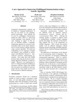
metals are shown in Fig. 1B, whereas the increases in
density at day 3 of MR-1R and of all mutants are
summarized in Fig. 1A. For chelated forms of Fe(III)
and for V
2
O
5
, culture turbidities gradually decreased
in the order MR-1R > omcA
–
>> omcB
–
> omcA
–
omcB
–
, with the greatest effect being caused by the
omcB disruption. OmcA and OmcB are collectively
essential for chelated Fe(III) dissimilatory reduction,
as the omcA
–
omcB
–
double mutant cannot grow on
either Fe(III)-nitrilotriacetic acid or Fe(III)-citrate,
whereas they appear to have an important, although
redundant, function as a terminal V(V) reductase, as
the omcA
–
omcB
–
double mutant still reaches $ 50%
of the MR-1R turbidity. Knocking out either omcA or
omcB turned out to have no significant growth pheno-
type with either U(VI) or Se(VI) as the terminal elec-
tron acceptor. These results therefore provide evidence
that there are differences between the electron transfer
pathways towards chelated Fe(III) on the one hand
and either U(VI) or Se(VI) on the other. Redundancy
between these pathways may explain the growth curves
observed for V(V) reduction.
Decaheme cytochrome c quantification of
anaerobically Fe(III)-respiring omcA
–
, omcB
–
and
omcA
–
omcB
–
MR-1R mutants relative to their
MR-1R parent
The major impact on Fe(III) respiration by OmcB relat-
ive to OmcA can be explained by one or a combination
J. Borloo et al. Shewanella oneidensis MR-1 OmcA and OmcB kinetics
FEBS Journal 274 (2007) 3728–3738 ª 2007 The Authors Journal compilation ª 2007 FEBS 3729
of the following possibilities: (a) the steady-state OmcB
concentration is greater than that of OmcA; (b) OmcB
is differentially produced (upregulated) by the omcA
insertional inactivation, but not vice versa; (c) OmcA
and OmcB show different behavior patterns in terms
of kinetics; and (d) OmcB is required to obtain
functional OmcA. These possibilities are discussed
below.
A heme-staining approach was used to reveal the
decaheme cytochrome c pools present in Fe(III)-respir-
ing MR-1 omcA
–
, omcB
–
and omcA
–
omcB
–
mutants
relative to their MR-1R parent. Figure 2B shows the
absence of mature OmcA (83 kDa) and OmcB
(78 kDa) in an omcA
–
and an omcB
–
background,
respectively, a complete lack of both proteins in the
omcA
–
omcB
–
double mutant, and approximately equal
amounts of either decaheme cytochrome c in an
MR-1R extract. Relative to MR-1R, Fig. 2B does not
suggest compensatory induction of either OmcB or
OmcA in an omcA
–
or omcB
–
background, respect-
ively.
To calculate the OmcA and OmcB content in
Fe(III)-respiring MR-1R and single mutants, differen-
tial absorption spectra for reduced-minus-oxidized
heme were recorded (Fig. 2D). As these spectra are
based on total heme content, it is imperative that all
the other heme-containing proteins in the cells are not
subjected to regulation in the respective mutants. Fig-
ure 2B,C shows that, apart from OmcA and OmcB,
the periplasmic fumarate reductase (FR), the cytoplas-
mic CymA and other, smaller (< 20 kDa), cyto-
chromes are highly abundant c-type cytochromes in
MR-1R, and thus contribute substantially to the
554 nm absorbance. Although not fully linear and sat-
urating with increasing cytochrome content, the heme
staining experiments are indicative of the fact that
these cytochromes are not subjected to upregulation or
downregulation in the analyzed mutants. We further-
more monitored and compared FR activities in wild-
type MR-1R and mutants. The enzyme assay yielded
activity values of (in lmolÆmin
)1
Æmg
)1
) 43.8 ± 0.90,
42.9 ± 0.58, 43.0 ± 0.24 and 44.3 ± 0.70 for
MR-1R, omcA
–
, omcB
–
and omcA
–
omcB
–
, respect-
ively, indicating no upregulation or downregulation of
FR (P ¼ 0.83). On the basis of the fact that FR is not
subjected to regulation under the applied conditions,
and deducing from Fig. 2C that all other c-type cyto-
chromes are also invariantly produced in the respective
mutants, we feel safe to extract OmcB and OmcA
concentrations from omcA
–
and omcB
–
mutant heme
values minus omcA
–
omcB
–
double mutant values,
respectively. The concentrations of OmcA and OmcB
were subsequently calculated on the basis on the
known stoichiometry of 10 heme groups per OmcA or
OmcB molecule [16]. This approach is valid, because
no alterations other than the expected disappearance
of either or both OmcA and OmcB in the respective
mutants are apparent from the heme-staining gels. The
omcA
–
background contains 4.00 pmol of OmcB per
10
9
cells, which, as to be expected from the heme stain
in Fig. 2B, is similar to the OmcA concentration cal-
culated for the omcB
–
background (3.43 pmol per
10
9
cells).
By subtracting the heme concentration of the
omcA
–
omcB
–
double mutant from that of MR-1R
cells, we calculated a decaheme cytochrome c content
(OmcA + OmcB) in MR-1R of about 6.68 pmol per
10
9
cells. This value matches the sum of both deca-
heme cytochrome c concentrations in the respective
single mutants, again showing that neither decaheme
cytochrome c is upregulated in the absence of the
Fig. 1. Anaerobic liquid growth experiments assess the role of
OmcA and OmcB in dissimilatory metal reduction. Anaerobic liquid
growth of MR-1R, omcA
–
, omcB
–
and omcA
–
omcB
–
mutant cul-
tures with either Fe(III)-nitrilotriacetic acid, Fe(III)-citrate, V(V),
U(VI) or Se(VI) as terminal electron acceptor is represented as
the increase in density reached after 3 days of growth (A).
Complete curves of MR-1R grown on the different metals are pro-
vided in (B).
Shewanella oneidensis MR-1 OmcA and OmcB kinetics J. Borloo et al.
3730 FEBS Journal 274 (2007) 3728–3738 ª 2007 The Authors Journal compilation ª 2007 FEBS
other. Statistical analysis (Student’s t-test) between the
MR-1R values and the sum of the values of the omcA
–
and the omcB
–
mutants revealed that there is no statis-
tically significant difference (P ¼ 0.43).
Whole cell kinetics of OmcA- and
OmcB-dependent chelated Fe(III) reduction
To establish whether differential kinetics and ⁄ or syner-
gism explain the dominance of OmcB over OmcA in
dissimilatory chelated Fe(III) reduction, we determined
the kinetic parameters for each decaheme cytochrome
c using intact actively Fe(III)-respiring cells (Table 1).
Maximal activities were converted to turnover numbers
on the basis of either the OmcA or OmcB concentra-
tions calculated in the above paragraph for the omcB
–
and omcA
–
single mutants, respectively. As explained
in Experimental procedures, Monod-based kinetic
models for whole cell kinetics simplify to Michaelis–
Menten models under the conditions applied in this
study.
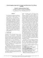
Figure 3A shows Fe(III)-nitrilotriacetic acid satura-
tion curves obtained using either omcA
–
[OmcB-
dependent Fe(III) reduction], omcB
–
[OmcA-dependent
Fe(III) reduction] or MR-1R [OmcA + OmcB-
dependent Fe(III) reduction] cells. In the absence of
Table 1. Enzymatic properties of OmcA- and OmcB-dependent-
Fe(III)-nitrilotriacetic acid reduction. Values represent the average of
triplicate experiments ± SD.
Enzymatic properties OmcA OmcB
Fe(III)-nitrilotriacetic acid
K
m
(lM) 15.3 ± 2.1 28.0 ± 0.9
k
cat
(s
)1
) 17.8 ± 0.4 205 ± 3.0
k
cat
⁄ K
m
(M
)1
Æs
)1
) 1.17 · 10
6
7.33 · 10
6
V
2
O
5
Inhibition type Competitive Mixed type
K
ic
22.5 ± 1.0 65.9 ± 0.1
K
iu
11.5 ± 0.6
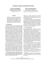
Fig. 2. Heme quantifications reveal unaltered protein production
profiles of both OmcA and OmcB in the respective single mutants
relative to the wild-type. (A) RT-PCR confirming the absence of
polar effects in mutants omcA
–
and omcB
–
. Specific oligonucleo-
tides were used to amplify omcA (lane 2), omcB (lane 3), mtrA
(lane 4) and mtrB (lane 5) in the omcA
–
mutant, and omcA (lane 7),
omcB (lane 8), mtrA (lane 9) and mtrB (lane 10) in the omcB
–
mutant. MR-1R was used as a positive control to display omcA
(lane 1) and omcB (lane 6). DNA standards are indicated at the left
and right of the agarose gels. (B) Visualization and separation of
high molecular mass cytochromes c through heme staining of a
Tris ⁄ glycine SDS ⁄ PAGE gel loaded with 4 · 10
7
whole cells from
anaerobically grown overnight cultures of MR-1R (lane 1), mutants
omcA
–
(lane 2), omcB
–
(lane 3), and omcA
–
omcB
–
(lane 4), and
complemented strains omcA
–
⁄ pBAD202 ⁄ D-TOPOomcA (lane 5)
and omcB
–
⁄ pBAD202 ⁄ D-TOPOomcB (lane 6). A molecular mass
standard is indicated at the right. (C) Visualization of low molecular
mass cytochromes c through heme staining of a Tricine ⁄
SDS ⁄ PAGE gel loaded with 4 · 10
7
whole cells from anaerobically
grown overnight cultures of MR-1R (lane 1), and mutants omcA
–
(lane 2), omcB
–
(lane 3), and omcA
–
omcB
–
(lane 4). A molecular
mass standard is indicated at the left. (D) Bar graph represen-
tation of the cytochrome content, normalized to 10
9
CFU, and
calculated from reduced-minus-oxidized heme absorption differ-
ences at 554 nm (a peak) using the absorption coefficient of
21 400
M
)1
Æcm
)1
. The differences in peak height reflect the
concentrations of OmcA and OmcB in omcB
–
and omcA
–
cells,
respectively.
A
B
C
D
J. Borloo et al. Shewanella oneidensis MR-1 OmcA and OmcB kinetics
FEBS Journal 274 (2007) 3728–3738 ª 2007 The Authors Journal compilation ª 2007 FEBS 3731
synergism, the OmcA- and OmcB-dependent substrate
saturation curves should add up to form the MR-1R
(OmcA + OmcB) curve; this is a valid assumption, as
we could not identify differential protein production
profiles as mentioned in the previous paragraph. At
full Fe(III)-nitrilotriacetic acid saturation, the modeled
summation function corresponds well with the MR-1R
curve, whereas it shows slightly lower than experiment-
ally determined activities at nonsaturating Fe(III)-
nitrilotriacetic acid concentrations. This suggests that
OmcA might synergistically enhance, albeit slightly,
the affinity of OmcB for its metal substrate. However,
the curves totally refute the reverse possibility, i.e. that
OmcB is needed to get functional OmcA.
On the other hand, the derived kinetic parameters
for OmcA- and OmcB-dependent chelated Fe(III)
reduction summarized in Table 1 do rationalize the
dominance of OmcB in dissimilatory Fe(III) reduction:
under physiologically relevant low micromolar concen-
trations of Fe(III), OmcA should outnumber OmcB
six-fold to catalyze electron transfer at a similar rate.
Complementation of the omcA
–
and omcB
–
mutants
restored Fe(III)-nitrilotriacetic acid reduction activity
to MR-1R levels (Fig. 3B).
Inhibition assays of OmcA- and OmcB-dependent
chelated Fe(III) reduction as a measure of enzyme
specificity
To determine whether the lack of phenotype of
omcA
–
omcB
–
strains observed during anaerobic
growth on either of the electron acceptors U(VI) and
Se(VI) is due to the decaheme cytochromes c not
recognizing either of these electron acceptors, we
probed the relative affinities via competition assays.
Figure 4 shows the IC
50
plots of the inhibition data of
whole cell OmcA- and OmcB-dependent Fe(III)-nitrilo-
triacetic acid reduction by either V(V), U(VI), or
Se(VI). Only V(V) appears to significantly inhibit
Fe(III) reduction, as characterized by IC
50
s of 10.7 lm
and 81.4 lm for inhibition of OmcA and OmcB,
respectively.
Modes of inhibition of either OmcA or OmcB
by V(V)
The modes of inhibition of either OmcA- or OmcB-
dependent Fe(III)-nitrilotriacetic acid reduction by
V(V) were investigated for the two following reasons:
(a) to derive the relevant inhibition constants; and (b)
to establish whether both decaheme cytochromes c
may differ mechanistically. Fe(III)-nitrilotriacetic acid
saturation curves in the absence and in the presence of
two different concentrations of V(V) were plotted and
modeled to obtain the apparent V
max
and K
m
values
(Fig. 5A,B). These parameters were subsequently used
to generate double-reciprocal Lineweaver–Burk plots
to easily determine inhibitor modality (Fig. 5C,D;
Table 1).
OmcA inhibition by V(V) is characterized by an
increase in apparent K
m
and no change in apparent
Fig. 3. Kinetic characterization of OmcA- and OmcB-dependent
Fe(III)-nitrilotriacetic acid reduction rationalizes the dominance of
OmcB in anaerobic ferric iron respiration. (A) Monod-based kinetic
model curves [34] for Fe(III)-nitrilotriacetic acid reduction by MR-1R
cells (inverted triangles), omcA
–
cells (squares), and omcB
–
cells
(triangles). As explained in Experimental procedures, the two latter
curves simplify to the Michaelis–Menten formulation under the con-
ditions applied. Adding up these curves generates the dotted-line
curve, which, as explained in Experimental procedures, should
resemble the MR-1R curve. Because this assumption is only valid
at saturating Fe(III)-nitrilotriacetic acid concentrations, slight synergy
may modulate activity when both OmcA and OmcB are present in
the outer membrane. (B) In trans complementation of omcA
–
and
omcB
–
cells restores Fe(III) reductase activity to MR-1R levels. See
Experimental procedures for details.
Shewanella oneidensis MR-1 OmcA and OmcB kinetics J. Borloo et al.
3732 FEBS Journal 274 (2007) 3728–3738 ª 2007 The Authors Journal compilation ª 2007 FEBS
V
max
, generating Lineweaver–Burk lines with intersect-
ing y-axis intercepts, which is the characteristic signa-
ture of competitive inhibition. We calculated a K
i
value of 22.5 lm, suggesting that the kinetics of V
2
O
5
binding to OmcA are similar to those for binding of
Fe(III)-nitrilotriacetic acid.
Fig. 4. Competition assays of OmcA- (left panel) and OmcB-dependent (right panel) Fe(III)-nitrilotriacetic acid reduction with other metals
show that only V(V) may represent an alternative substrate for both cytochromes. Fe(III)-nitrilotriacetic acid reductase activity in the absence
of a competing metal substrate is set to 100%. Relative activities are plotted as a function of increasing concentrations of either V(V) (as
vanadate; red), U(VI) (as uranyl acetate; green), or Se(VI) (sodium selenate; purple). Inhibition curves were fitted to the standard hyperbolic
inhibition equation (see Experimental procedures).
Fig. 5. Analysis of the modes of inhibition of OmcA- and OmcB-dependent Fe(III) reduction by V(V) reveals mechanistic differences between
the two cytochromes. (A, B) Direct plots of the steady-state velocities of OmcA-dependent (A) and OmcB-dependent (B) Fe(III)-nitrilotriacetic
acid reduction in the absence and the presence of two increasing V(V) concentrations. (C, D) Theoretical double reciprocal plots using the
kinetic parameters obtained by fitting the data from the direct plots.
J. Borloo et al. Shewanella oneidensis MR-1 OmcA and OmcB kinetics
FEBS Journal 274 (2007) 3728–3738 ª 2007 The Authors Journal compilation ª 2007 FEBS 3733
OmcB inhibition by V(V) is characterized by a
decrease in apparent K
m
and V
max
. By plugging the
values of the modeled apparent kinetic parameters into
the double-reciprocal Lineweaver–Burk equation and
plotting the resulting linear functions, we obtained the
graph in Fig. 5D. The lines intersect at negative values
of 1 ⁄ [S] and 1 ⁄ v, which is a characteristic signature
of noncompetitive inhibition. Thus, V(V) apparently
binds both the free OmcB enzyme and the binary
OmcB–Fe(III)-nitrilotriacetic acid complex, and the
binding is kinetically favored upon Fe(III)-nitrilotriace-
tic acid binding. We calculated K
ic
and K
iu
values of
65.9 lm and 11.5 lm, respectively, which again appears
to have physiologic significance. Hence, besides having
significantly different turnover rates, OmcA and OmcB
may also behave differently in terms of binding their
metallic substrates.
Discussion
In the present study, we could not detect an-
aerobic Fe(III)-nitrilotriacetic acid respiration for
omcA
–
omcB
–
double mutant cells. Virtually no biomass
was generated in minimal medium containing lactate
and Fe(III)-nitrilotriacetic acid as the electron donor
and acceptor, respectively (Fig. 1), and baseline reduc-
tion of Fe(III)-nitrilotriacetic acid was seen in the ferro-
zine-based whole cell kinetic approach (data not
shown). The collective action of both decaheme cyto-
chromes c, OmcA and OmcB, appears to be crucial for
anaerobic soluble Fe(III) respiration, and, because
of their outer membrane localization, one or both
cytochromes probably function as terminal Fe(III)
reductases. Both these outer membrane-localized
cytochromes are reduced through an as yet incompletely
identified electron transport chain, which at an early
point receives electrons from the NADH pool, in our
study obtained by lactate supplementation. In a recent
study, Marshall et al. [15] established almost equally
fast direct electron transfer from either dithionite-
reduced MR-1 OmcA or OmcB to chelated Fe(III), pro-
viding the first biochemical evidence that both decaheme
cytochromes c are in fact functional Fe(III) reductases.
As an OmcA ⁄ OmcB double mutant strain does not
show any Fe(III) reduction activity, our study not only
strengthens, but also exceeds, this evidence, in that
OmcA and OmcB are found to be the sole Fe(III) reduc-
tases present in MR-1. Furthermore, the outer mem-
brane localization and partial extracellular exposure of
both cytochromes c, combined with the fact that the
result of adding up the OmcA and OmcB Fe(III)-nitrilo-
triacetic acid reduction curves conforms to the MR-1R
curve, allow us to deduce that the electron transport
chain does not bifurcate any further, but ends at this
point before transferring electrons to the subject metal
species, indicating that OmcA and OmcB are the ter-
minal Fe(III) reductases in MR-1. Other MR-1 cyto-
chromes c, previously shown to be ferric iron reductases
in vitro, such as MtrA [17] and Ifc3 in S. frigidimarina
[18], appear to be not directly involved in the process of
anaerobic chelated Fe(III) respiration.
Notably, the apparent maximal rate reported for
Fe(III)-nitrilotriacetic acid-dependent OmcB oxidation
is approximately 50 times slower than the k
cat
for
OmcB-dependent Fe(III)-nitrilotriacetic acid reduction
(205 s
)1
), determined here using a whole cell kinetics
approach, which has the advantages of: (a) maintain-
ing the complete electron transport chain used during
metal respiration; and (b) keeping the terminal reduc-
tases in their native cellular compartment. For OmcA,
the in vitro K
obs
values determined by Marshall et al.
[15] and the in vivo k
cat
values determined in our study
also differ, although to a lesser extent (six-fold). This dis-
crepancy can most likely be accounted for by the fact
that the purified cytochromes used in the in vitro
approach lack some factor(s), such as one or more pro-
tein partners or lipids that generate maximal activity.
Reduced activity due to detergent-based solubilization
of the outer membrane cytochromes is an alternative
explanation.
Growth experiments as well as the whole cell Fe(III)
reduction kinetics presented here agree with previous
findings that OmcB is more important than OmcA in
anaerobic Fe(III) respiration [19]. Using a heme-quanti-
fication approach, we have presented evidence showing
that this relative difference is not based on differential
protein production profiles of either the omcA or omcB
gene in the presence or absence of the other. Shi et al.
[19] provided evidence for synergistic complex forma-
tion between both decaheme cytochromes, which may
explain the dominance of OmcB over OmcA in dissimi-
latory Fe(III) reduction. Our whole cell-based kinetic
analysis, however, refutes the possibility that OmcB is
necessary to reconstitute fully functional OmcA, as the
Fe(III)-reducing activities of omcA
–
and omcB
–
cells add
up to the counterpart activities of MR-1R cells. A per-
fect fit, however, only becomes possible after slightly
increasing the affinity of OmcB for its chelated Fe(III)
substrate (Fig. 3A). Complex formation may thus cause
some synergism only at low micromolar and therefore
physiologically relevant substrate concentrations.
The kinetics for OmcA- and OmcB-dependent
Fe(III)-nitrilotriacetic acid reduction (Table 1) do
rationalize the different roles of these proteins in Fe(III)
respiration. Both cytochromes have similar low micro-
molar affinities for their Fe(III) substrate; however,
Shewanella oneidensis MR-1 OmcA and OmcB kinetics J. Borloo et al.
3734 FEBS Journal 274 (2007) 3728–3738 ª 2007 The Authors Journal compilation ª 2007 FEBS
completion of the electron transfer pathway takes
$ 11.5 times longer for OmcA than for OmcB. Taking
into account the specificity constants, OmcA should out-
number OmcB about six-fold if it is to substitute for the
latter in anaerobic Fe(III) respiration at physiologic fer-
ric iron concentrations, a hypothesis that will be pursued
further in our laboratory. Note that the division of labor
established here for OmcA and OmcB cytochromes
should not necessarily apply to homologs from different
backgrounds; the OmcA homolog from S. frigidimarina,
for example, has been found to be as fast (206 s
)1
)as
the S. oneidensis MR-1 OmcB reductase [20].
It has previously been recognized that both cyto-
chromes, OmcA and OmcB, appear to have some sub-
strate specificity, as purified reduced batches lack
activity towards nitrite, nitrate and, in the case of
OmcA, uranyl acetate [15]. OmcB was shown to have
some activity towards U(VI); however, the turnover
number (K
obs1
¼ 0.039 s
)1
) is more than 100 times
lower than that for Fe(III)-nitrilotriacetic acid
(K
obs1
¼ 4.1 s
)1
) [15]. Our anaerobic growth experi-
ments show that neither decaheme cytochrome c is
necessary for dissimilatory uranyl acetate reduction
(Fig. 1). OmcA, as expected, but also OmcB does not
bind U(VI) in the competition assay shown in Fig. 4.
The 100-fold lower K
obs1
for U(VI) reduction com-
pared to Fe(III)-nitrilotriacetic acid reduction reported
by Marshall et al. [15] thus appears to result not from
disturbed catalysis, but rather from hampered sub-
strate binding. Of the other metals tested in this study
[V(V) and Se(VI)], only vanadate was shown to be a
substrate for either OmcA or OmcB. Inhibition experi-
ments suggest that Fe(III) and V(V) bind both cyto-
chromes with similar efficiencies (Table 1). However,
whereas omcA
–
omcB
–
double mutant cells did not
grow on chelated Fe(III), they do grow on V(V) to
about 50% of the MR-1R stationary-phase density
(Fig. 1). In the case of V(V), the electron transport
chain may thus bifurcate to one or several other, as
yet unrecognized, terminal reductases. Redundancy in
terminal metal reductases has been clearly shown here,
as MR-1 does not suffer from the omcA
–
omcB
–
dou-
ble mutants in anaerobic growth on the terminal elec-
tron acceptors Se(VI) and U(VI), and as none of these
metals inhibits OmcA- and OmcB-dependent whole
cell Fe(III)-nitrilotriacetic acid reduction. In summary,
metal reduction appears to be a selective process in
which the reduction potential and the topology and
accessibility of the presented metal play crucial roles in
terms of binding efficiencies and subsequent reduction
by the appropriate enzyme. The identification and
characterization of alternative terminal metal reductas-
es will be the subject of future research.
Experimental procedures
Bacterial strains
S. oneidensis MR-1 was originally isolated from Oneida
Lake sediments (Oneida Lake, NY, USA) [21], and was
obtained from the LMG culture collection (LMG 19005;
Ghent, Belgium). S. oneidensis MR-1R is a spontaneous rif-
ampicin-resistant mutant of strain MR-1 that was isolated
in-house. Escherichia coli strain TAM1pir
+
and E. coli S17-
1kpir cells were used for cloning purposes and conjugation
experiments, respectively.
Growth conditions
MR-1R, omcA
–
, omcB
–
and omcA
–
omcB
–
S. oneidensis cul-
tures were routinely grown overnight at 28 °C in LB broth
and subsequently inoculated in M1 defined medium [22] sup-
plemented with l-serine (1 lg ÆmL
)1
), l-arginine (1 lgÆmL
)1
),
l-glutamate (1 lgÆmL
)1
), lactate (15 mm), and fumarate
(20 mm). For growth experiments, fumarate was replaced
by either Fe(III)-citrate (2 mm), Fe(III)-nitrilotriacetic acid
(0.5 mm), Na
2
SeO
4
(1 mm), or UO
2
(CH
3
COO)
2
.2H
2
O
(0.5 mm) (all products: Sigma-Aldrich, Bornem, Belgium).
Growth on V(V) was studied using VM medium [23]. Anae-
robicity was achieved using a Coy anaerobic chamber (Coy
Laboratories, Grass Lake, MI) containing 90% N
2
,8%CO
2
,
and 2% H
2
. The presence of H
2
in the anaerobic chamber
did not affect metal reduction (data not shown). Growth
curves were recorded by measuring the attenuance (D
655
)of
the cultures at regular time intervals for 3 days. The average
rise in density after 3 days ± SEM for triplicate readings are
summarized in Fig. 1A, whereas the growth curves for MR-
1R grown on the different metals are shown in Fig. 1B.
Construction of the omcA
–
and omcB
–
single
mutants and of the omcA
–
omcB
–
double mutant
strains of MR-1
Single omcA
–
and omcB
–
mutants and a double omcA
–
omcB
–
mutant strain of MR-1 were generated by insertional inacti-
vation using the pKNOCK-based system [24]. The primers
used in this study are summarized in Table 2. Briefly, internal
PCR-amplified fragments of the omcA and omcB genes were
5¢-phosphorylated and cloned into EcoRV-digested and
calf intestinal phosphatase-treated pKNOCK-Km and
pKNOCK-Cm, respectively, using T4 DNA Ligase (all
enzymes: New England Biolabs, Ipswich, MA), yielding
pKNOCK-Km-omcA and pKNOCK-Cm-omcB. These con-
structs were transformed into E. coli S17-1kpir cells. Equal
amounts of overnight-grown transformed E. coli S17-1kpir
cells and rifampicin-resistant S. oneidensis cells were mixed
and spotted on LB ⁄ Rif plates (10 lgÆmL
)1
). After a 6 h incu-
bation period (necessary for the conjugation to take place),
the cells were resuspended in 500 lL of LB broth [25] and
J. Borloo et al. Shewanella oneidensis MR-1 OmcA and OmcB kinetics
FEBS Journal 274 (2007) 3728–3738 ª 2007 The Authors Journal compilation ª 2007 FEBS 3735
plated on LB ⁄ Rif plates containing either kanamycin
(25 lgÆmL
)1
) or chloramphenicol (25 lgÆmL
)1
) (Duchefa,
Haarlem, The Netherlands). After overnight incubation
at 28 °C, colonies were analyzed via PCR using the oligo-
nucleotides OMCA-F ⁄ OMCA-R and OMCB-F ⁄ OMCB-R
(Table 2), designed to amplify the entire omcA gene and
omcB gene, respectively. Homology-based insertional integ-
ration of the pKNOCK constructs enlarged the omcA
(2207 bp) and omcB (2015 bp) gene amplicons by 2700 and
2500 bp, respectively (data not shown). The omcA
–
omcB
–
double mutant was constructed by applying a similar proce-
dure to that described above, using the omcA
–
mutant as the
recipient strain in conjugation. As omcA and omcB are part
of the gene cluster mtrDEF–omcA–mtrCAB (omcB is also
known as mtrC), and the genes mtrCAB form a single
operon, we expected polar effects to occur when disrupting
omcB. RT-PCR experiments proved the absence of such
polar effects (Fig. 2A) and confirmed that we had obtained
the omcA
–
and the omcB
–
mutants.
Complementation of the MR-1 omcA
–
and omcB
–
mutant strains
Oligonucleotides OMCA-PBAD-F ⁄ OMCA-PBAD-R and
OMCB-PBAD-F ⁄ OMCB-PBAD-R (Table 2) were used to
amplify the omcA and omcB genes from MR-1 genomic
DNA, respectively. These genes were subsequently cloned
into vector pBAD202 ⁄ D-TOPO (Invitrogen, Carlsbad,
CA), and the constructs were transformed into the appro-
priate omcA
–
or omcB
–
mutants of MR-1 by electropora-
tion, generating the in trans complemented strains. As
pBAD202 ⁄ D-TOPO carries a kanamycin resistance region,
the ability to complement the omcA
–
mutant was shown
using a pKNOCK-Cm-based omcA
–
mutant, instead of
the pKNOCK-Km-based mutant that was applied in all
other experiments. Full complementation of either the omcA
or omcB insertional mutation by the wild-type genes,
controlled by an arabinose promoter [26], was achieved as
visualized by heme staining of SDS ⁄ PAGE gels (Fig. 2B),
as well as at the level of activity (see further).
Visualization of c-type cytochromes using heme
staining
High and low molecular mass c-type cytochromes were
resolved by SDS ⁄ PAGE according to Laemmli [27] and
Schaegger & von Jagow [28] (tricine gels), respectively. In
either case, 4 · 10
7
whole cells of anaerobically grown over-
night cultures were applied to the gels, which were then
heme stained according to Thomas et al. [29]. The outer
membrane cytochromes c OmcA and OmcB, the periplas-
mic FR, and the cytoplasmic tetraheme cytochrome c
CymA were unambiguously identified via MS from heme-
stained Tris ⁄ glycine gels and tricine gels, respectively.
Spectral quantification of the outer membrane
decaheme cytochromes c OmcA and OmcB
The heme content of whole cells was determined using the
difference absorption coefficient of 21 400 m
)1
Æcm
)1
[16] at
554 nm for the pyridine ferrohemochrome minus pyridine
ferrihemochrome spectrum. In that study, the difference
absorption coefficient was determined at pH 8.0, whereas
all our experiments were carried out at pH 7.5. We observed
no differences between spectra measured at pH 8.0 and 7.5
(data not shown). Sodium dithionite was used as the redu-
cing chemical. Overnight anaerobically grown cells (with
20 mm fumarate as the electron acceptor) were washed with
and suspended in an equal volume of air-saturated NaCl ⁄ P
i
(pH 7.5), and incubated at room temperature for 1 h to
ensure oxidation of the outer membrane cytochromes.
Absorption spectra of 1 mL fractions were recorded at
554 nm using a double-beam spectrophotometer (Uvikon,
Kontron, Herts, UK) in the absence and the presence of a
few crystals of sodium dithionite (Sigma-Aldrich). The
decaheme cytochrome c concentration was calculated as
explained in Results, taking into account 10 heme groups
per molecule of either OmcA or OmcB and our experiment-
ally derived correlation between D
655
and cell concentration
(a 1 mL MR-1 culture with a D
655
of 1.0 contains
1.44 · 10
9
cells). The values presented are means of tripli-
cate experiments ± SEM. To quantify FR, lysed MR-1R
omcA
–
, omcB
–
and omcA
–
omcB
–
cells were assayed for this
specific enzyme activity according to Maklashina et al. [30].
Whole cell kinetics of ferric iron reduction
The Fe(III) reductase activity of whole cells was measured
using the ferrozine-based method [31]. The chromophore
formed by ferrous iron and ferrozine was measured at
562 nm [32]. Whole cells for the Fe(III) reductase assays were
Table 2. Synthetic oligonucleotides used in this study.
Oligonucleotide name Sequence (5¢-to3¢)
OMCA-KO-F CACACTGCAACCTCTGGT
OMCA-KO-R ACTGTCAATAGTGAAGGT
OMCB-KO-F CCCCATGTCGCCTTTAGT
OMCB-KO-R TCGCTAGAACACATTGAC
OMCA-F ATGATGAAACGGTTCAAT
OMCA-R TTAGTTACCGTGTGCTTC
OMCB-F CTGCTGCTCGCAGCAAGT
OMCB-R GTGTGATCTGCAACTGTT
OMCA-PBAD-F CACCGAGGAATAATAAATGATG
AAACGGTTCAATTTC
OMCA-PBAD-R TTAGTTACCGTGTGCTTC
OMCB-PBAD-F CACCGAGGAATAATAAATGATG
AACGCACAAAAATCA
OMCB-PBAD-R TTACATTTTCACTTTAGT
Shewanella oneidensis MR-1 OmcA and OmcB kinetics J. Borloo et al.
3736 FEBS Journal 274 (2007) 3728–3738 ª 2007 The Authors Journal compilation ª 2007 FEBS
prepared as follows. Anaerobically grown cells (with fuma-
rate as the terminal electron acceptor) were collected by
centrifugation at 10 000 g (Beckman Coulter Avanti J-301
centrifuge, JA-30.50 rotor), washed twice with NaCl ⁄ P
i
sup-
plemented with 1 mm lactate (unless otherwise mentioned),
and placed on ice. These preparations retained full activity
for at least 4 h. Comparison of the reduced-minus-oxidized
spectra of anaerobically grown MR-1R cells washed with
NaCl ⁄ P
i
(pH 7.5) on the one hand, or water on the other,
revealed no differences in heme content, indicating that the
salt treatment did not lead to unwanted release of outer
membrane cytochromes. Assays were conducted in microtiter
plates at 25 °C in a final volume of 200 lL of NaCl ⁄ P
i
(pH 7.4), and were monitored using a Bio-Rad model 680
microplate reader (Bio-Rad, Hercules, CA). A standard reac-
tion mixture contained 1 mm 3-(2-pyridyl)-5,6-bis(4-phenyl-
sulfonic acid)-1,2,4-triazine monosodium salt (ferrozine;
Sigma-Aldrich), 1 mm lactate (unless otherwise mentioned),
a 1 : 100 dilution of the washed cell preparation, and Fe(III)-
nitrilotriacetic acid at concentrations ranging from 0.5 lm to
1.5 mm. Phosphate did not interfere with the reduction assay
(data not shown), which is in accordance with the results
reported by Ruebush [33]. For inhibition studies, the stand-
ard reaction mixture containing 100 lm Fe(III)-nitrilotriace-
tic acid (unless otherwise mentioned) was supplemented with
either V(V) (as V
2
O
5
), Se(VI) (as Na
2
SeO
4
) or U(VI) [as
UO
2
(CH
3
COO)
2
.2H
2
O], ranging in concentration from
0.5 lm to 1 m m. Inhibition curves were fitted using a least
squares algorithm (graphpad prism Version 4.00; GraphPad
Software, Inc., San Diego, CA) to the equation:
m
r
¼ 100 ÀðI
max
½Me=ðIC
50
þ½MeÞÞ
where v
r
is the relative activity, I
max
is the maximal
response amplitude, [Me] is the supplemented initial con-
centration of inhibiting metallic substrate, and IC
50
is the
half-maximal concentration of inhibiting metallic substrate.
To analyze kinetic data, we used Monod-based kinetic
models [34] that actually simplify to a Michaelis–Menten for-
mulation under the applied conditions. The kinetic rate is
determined solely by the electron acceptor, as the electron
donor used (lactate, 1 mm) is supplied in excess. The effect of
bacterial growth on Fe(III)-nitrilotriacetic acid reduction can
be neglected, as the initial cell concentration used was high,
and growth-supporting nutrients were excluded. We also
assumed that cell decay can be neglected, because the activity
proceeded linearly during our 1 h analyses. Therefore, the
Monod model takes a form similar to the Michaelis–Menten
expression v ¼ V
m
S ⁄ (K
s
+ S), where V
m
equals the maximal
activity for the initial bacterial concentration, S is the initial
Fe(III)-nitrilotriacetic acid concentration, and K
s
is the half-
velocity constant. As we have determined the OmcA and
OmcB concentrations present in omcB
–
and omcA
–
cells,
respectively, and because omcA
–
omcB
–
double mutant cells
completely lack Fe(III) reductase activity, we can, using the
single mutants, convert V
m
values to k
cat
values, and safely
assume K
s
to be K
m
, the familiar Michaelis–Menten constant
for enzyme-catalyzed reactions. Activity data were fitted to
the regular Michaelis–Menten equation using graphpad
prism Version 4.00. For MR-1R- and OmcA-dependent
kinetics, the Michaelis–Menten equation was adjusted for
substrate inhibition.
Acknowledgements
This work was supported by a personal grant to
J. Borloo from the Institute for the Promotion of
Innovation by Science and Technology in Flanders
(IWT-Vlaanderen). J. Van Beeumen and B. Devreese
are indebted to the Fund for Scientific Research (FWO-
Vlaanderen) for granting research project G.0190.04, as
well as to the Bijzonder Onderzoeksfonds of Ghent Uni-
versity for Concerted Research Action GOA 120154.
References
1 Krause B & Nealson KH (1997) Physiology and enzy-
mology involved in denitrification by Shewanella putre-
faciens. Appl Environ Microbiol 63, 2613–2618.
2 Moser DP & Nealson KH (1996) Growth of the faculta-
tive anaerobe Shewanella putrefaciens by elemental sul-
fur reduction. Appl Environ Microbiol 62, 2100–2105.
3 Myers CR, Carstens BP, Antholine WE & Myers JM
(2000) Chromium(VI) reductase activity is associated
with the cytoplasmic membrane of anaerobically grown
Shewanella putrefaciens MR-1. J Appl Microbiol 88,
98–106.
4 Myers JM & Myers CR (2001) Role for outer mem-
brane cytochromes OmcA and OmcB of Shewanella
putrefaciens MR-1 in reduction of manganese dioxide.
Appl Environ Microbiol 67, 260–269.
5 Myers JM & Myers CR (2003) Overlapping role of the
outer membrane cytochromes of Shewanella oneidensis
MR-1 in the reduction of manganese(IV) oxide. Lett
Appl Microbiol 37, 21–25.
6 Carpentier W, Sandra K, De Smet I, Brige A, De Smet
L & Van Beeumen J (2003) Microbial reduction and
precipitation of vanadium by Shewanella oneidensis.
Appl Environ Microbiol 69, 3636–3639.
7 Klonowska A, Heulin T & Vermeglio A (2005) Selenite
and tellurite reduction by Shewanella oneidensis. Appl
Environ Microbiol 71, 5607–5609.
8 Davis JA, Kent DV, Rea BA, Maest AS & Garabedial
SP (1993) Influence of redox environment and aqueous
speciation on metal transport in groundwater: prelimin-
ary results of tracer injection studies. In Metals in
ground water (Allen HE, Perdue EM & Brown DS, eds),
pp. 223–273. Lewis Publishers, Chelsea, MI.
9 Schwalb C, Chapman SK & Reid GA (2003) The
tetraheme cytochrome CymA is required for anaerobic
J. Borloo et al. Shewanella oneidensis MR-1 OmcA and OmcB kinetics
FEBS Journal 274 (2007) 3728–3738 ª 2007 The Authors Journal compilation ª 2007 FEBS 3737
respiration with dimethyl sulfoxide and nitrite in Shewa-
nella oneidensis. Biochemistry 42, 9491–9497.
10 Myers JM, Antholine WE & Myers CR (2004) Vana-
dium(V) reduction by Shewanella oneidensis MR-1
requires menaquinone and cytochromes from the cyto-
plasmic and outer membranes. Appl Environ Microbiol
70, 1405–1412.
11 Saffarini DA, Blumerman SL & Mansoorabadi KJ
(2002) Role of menaquinones in Fe(III) reduction by
membrane fractions of Shewanella putrefaciens. J Bacte-
riol 184, 846–848.
12 Myers CR & Myers JM (1997) Cloning and sequence of
cymA, a gene encoding a tetraheme cytochrome c
required for reduction of iron(III), fumarate, and nitrate
by Shewanella putrefaciens MR-1. J Bacteriol 179,
1143–1152.
13 Myers CR & Myers JM (2003) Cell surface exposure of
the outer membrane cytochromes of Shewanella one-
idensis MR-1. Lett Appl Microbiol 37, 254–258.
14 Myers CR & Myers JM (2004) The outer membrane
cytochromes of Shewanella oneidensis MR-1 are lipo-
proteins. Lett Appl Microbiol 39, 466–470.
15 Marshall MJ, Beliaev AS, Dohnalkova AC, Kennedy
DW, Shi L, Wang Z, Boyanov MI, Lai B, Kemner
KM, McLean JS et al. (2006) c-Type cytochrome-
dependent formation of U(IV) nanoparticles by Shewa-
nella oneidensis. PLoS Biol 4, 1324–1333.
16 Ozawa T, Tanaka M & Shimomura Y (1980) Crystal-
lization of the middle part of the mitochondrial electron
transfer chain: cytochrome bc1–cytochrome c complex.
Proc Natl Acad Sci USA 77, 5084–5086.
17 Pitts KE, Dobbin PS, Reyes-Ramirez F, Thomson AJ,
Richardson DJ & Seward HE (2003) Characterization
of the Shewanella oneidensis MR-1 decaheme cyto-
chrome MtrA: expression in Escherichia coli confers the
ability to reduce soluble Fe(III) chelates. J Biol Chem
278, 27758–27765.
18 Gordon EH, Pike AD, Hill AE, Cuthbertson PM,
Chapman SK & Reid GA (2000) Identification and
characterization of a novel cytochrome c(3) from
Shewanella frigidimarina that is involved in Fe(III) res-
piration. Biochem J 349, 153–158.
19 Shi L, Chen B, Wang Z, Elias DA, Mayer MU, Gorby
YA, Ni S, Lower BH, Kennedy DW, Wunschel DS
et al. (2006) Isolation of a high-affinity functional pro-
tein complex between OmcA and MtrC: two outer
membrane decaheme c-type cytochromes of Shewanella
oneidensis MR-1. J Bacteriol 188, 4705–4714.
20 Field SJ, Dobbin PS, Cheesman MR, Watmough NJ,
Thomson AJ & Richardson DJ (2000) Purification and
magneto-optical spectroscopic characterization of
cytoplasmic membrane and outer membrane multiheme
c-type cytochromes from Shewanella frigidimarina
NCIMB400. J Biol Chem 275, 8515–8522.
21 Myers CR & Nealson KH (1988) Bacterial manganese
reduction and growth with manganese oxide as the sole
electron-acceptor. Science 240, 1319–1321.
22 Myers CR & Nealson KH (1990) Respiration-linked
proton translocation coupled to anaerobic reduction of
manganese(IV) and iron(III) in Shewanella putrefaciens
MR-1. J Bacteriol 172, 6232–6238.
23 Carpentier W, De Smet L, Van Beeumen J & Brige A
(2005) Respiration and growth of Shewanella oneidensis
MR-1 using vanadate as the sole electron acceptor.
J Bacteriol 187, 3293–3301.
24 Alexeyev MF (1999) The pKNOCK series of broad-host-
range mobilizable suicide vectors for gene knockout and
targeted DNA insertion into the chromosome of gram-
negative bacteria. Biotechniques 26, 824–826, 828.
25 Sambrook J, Fritsch EF & Maniatis T (1989) Molecular
Cloning: a Laboratory Manual, 2nd edn. Cold Spring
Harbor Laboratory Press, Cold Spring Harbor, NY.
26 Shi L, Lin JT, Markillie LM, Squier TC & Hooker BS
(2005) Overexpression of multi-heme C-type cyto-
chromes. Biotechniques
38, 297–299.
27 Laemmli UK (1970) Cleavage of structural proteins
during the assembly of the head of bacteriophage T4.
Nature 227, 680–685.
28 Schaegger HA & von Jagow G (1987) Tricine-sodium
dodecyl sulfate-polyacrylamide gel electrophoresis for
the separation of proteins in the range from 1 to 100
kDa. Anal Biochem 166, 368–379.
29 Thomas PE, Ryan D & Levin W (1976) An improved
staining procedure for the detection of the peroxidase-
activity of cytochrome-P450 on sodium dodecyl sulfate
polyacrylamide gels. Anal Biochem 75, 168–176.
30 Maklashina E, Iverson TM, Sher Y, Kotlyar V,
Andrell J, Mirza O, Hudson JM, Armstrong FA,
Rothery RA, Weiner JH et al. (2006) Fumarate reduc-
tase and succinate oxidase activity of Escherichia coli
complex II homologs are perturbed differently by
mutation of the flavin binding domain. J Biol Chem
281, 11357–11365.
31 Beliaev AS & Saffarini DA (1998) Shewanella putrefac-
iens mtrB encodes an outer membrane protein required
for Fe(III) and Mn(IV) reduction. J Bacteriol 180,
6292–6297.
32 Moody MD & Dailey HA (1983) Aerobic ferrisidero-
phore reductase assay and activity stain for native poly-
acrylamide gels. Anal Biochem 134, 235–239.
33 Ruebush SS (2006) Biochemical Characterization of
Membrane Proteins in Shewanella Oneidensis Involved in
Dissimilatory Iron Reduction. PhD thesis, Pennsylvania
State University, PA.
34 Monod J (1949) The growth of bacterial cultures. Annu
Rev Microbiol 3, 371–394.
Shewanella oneidensis MR-1 OmcA and OmcB kinetics J. Borloo et al.
3738 FEBS Journal 274 (2007) 3728–3738 ª 2007 The Authors Journal compilation ª 2007 FEBS