Báo cáo khoa học: Unconventional translation initiation of human trypsinogen 4 at a CUG codon with an N-terminal leucine A possible means to regulate gene expression pdf
Bạn đang xem bản rút gọn của tài liệu. Xem và tải ngay bản đầy đủ của tài liệu tại đây (492.49 KB, 11 trang )
Unconventional translation initiation of human
trypsinogen 4 at a CUG codon with an N-terminal leucine
A possible means to regulate gene expression
Attila L. Ne
´
meth
1,
*, Pe
´
ter Medveczky
1,
*, Ju
´
lia To
´
th
1
, Erika Siklo
´
di
1
, Katalin Schlett
2
, Andra
´
s Patthy
3
,
Miklo
´
s Palkovits
4
, Judit Ova
´
di
5
, Nata
´
lia To
˜
ke
´
si
5
,Pe
´
ter Ne
´
meth
6
,La
´
szlo
´
Szila
´
gyi
1
and La
´
szlo
´
Gra
´
f
1,3
1 Department of Biochemistry, Eo
¨
tvo
¨
s Lora
´
nd University, Budapest, Hungary
2 Departments of Physiology and Neurobiology, Eo
¨
tvo
¨
s Lora
´
nd University, Budapest, Hungary
3 Biotechnology Research Group of the Hungarian Academy of Sciences, Budapest, Hungary
4 Laboratory of Neuromorphology, Department of Anatomy, Semmelweis University, Budapest, Hungary
5 Institute of Enzymology, Hungarian Academy of Sciences, Budapest, Hungary
6 Institute of Immunology and Biotechnology, University of Pe
´
cs, Hungary
Recent genome programmes have explored an increas-
ing number of new genes with unknown function. The
estimated 35 000 human genes encode more than 10
5
expressed proteins as the result of various mechanisms,
such as alternative promotion of transcription, alter-
native splicing of the transcripts and alternative trans-
lational initiation. Chromosome rearrangement can
also serve as a source for evolutionary heterogeneity.
Keywords
brain; protein synthesis; PRSS3; serine
protease; translation initiation; trypsin 4
Correspondence
L. Gra
´
f, Department of Biochemistry,
Eo
¨
tvo
¨
s Lora
´
nd University, Pa
´
zma
´
ny Pe
´
ter s.
1 ⁄ C, Budapest H-1117, Hungary
Fax: +36 1 3812172
Tel: +36 1 3812171
E-mail:
*These authors contributed equally to this
work
(Received 7 December 2006, accepted 18
January 2007)
doi:10.1111/j.1742-4658.2007.05708.x
Summary
Chromosomal rearrangements apparently account for the presence of a pri-
mate-specific gene (protease serine 3) in chromosome 9. This gene encodes,
as the result of alternative splicing, both mesotrypsinogen and trypsino-
gen 4. Whereas mesotrypsinogen is known to be a pancreatic protease,
neither the chemical nature nor biological function of trypsinogen 4 has
been explored previously. The trypsinogen 4 sequence contains two predic-
ted translation initiation sites: an AUG site that codes for a 72-residue lea-
der peptide on Isoform A, and a CUG site that codes for a 28-residue
leader peptide on Isoform B. We report studies that provide evidence for
the N-terminal amino acid sequence of trypsinogen 4 and the possible
mechanism of expression of this protein in human brain and transiently
transfected cells. We raised mAbs against a 28-amino acid synthetic peptide
representing the leader sequence of Isoform B and against recombinant
trypsin 4. By using these antibodies, we isolated and chemically identified
trypsinogen 4 from extracts of both post mortem human brain and transi-
ently transfected HeLa cells. Our results show that Isoform B, with a leu-
cine N terminus, is the predominant (if not exclusive) form of the enzyme
in post mortem human brain, but that both isoforms are expressed in tran-
siently transfected cells. On the basis of our studies on the expression of a
series of trypsinogen 4 constructs in two different cell lines, we propose
that unconventional translation initiation at a CUG with a leucine, rather
than a methionine, N terminus may serve as a means to regulate protein
expression.
Abbreviations
GFP, green fluorescent protein; PRSS3, protease serine 3.
1610 FEBS Journal 274 (2007) 1610–1620 ª 2007 The Authors Journal compilation ª 2007 FEBS
An interesting example of such a mechanism is the
occurrence of a trypsinogen gene in a chromosome dif-
ferent from the original locus for the major forms of
pancreatic trypsinogens in chromosome 7. Uniquely in
primates, a series of chromosome translocations
between chromosome 7, 11 and 9 led to the formation
of the protease serine 3 (PRSS3) gene that encodes, as
a result of alternative splicing, both mesotrypsinogen
and trypsinogen 4 [1,2]. Structurally, human mesotry-
psinogen and human trypsinogen 4 differ only in their
N-terminal sequences. Whereas mesotrypsinogen has a
typical signal sequence (splice Isoform C in the Swiss-
Prot database), the two isoforms of human trypsi-
nogen 4 have highly charged N-terminal leader
sequences. The predicted longer form (splice Isoform A
in the Swiss-Prot database) contains a 72-residue
N-terminal leader peptide, whereas the shorter form
(splice Isoform B in the Swiss-Prot database) contains
a 28-residue N-terminal leader peptide (Fig. 1A). The
translation initiation site for splice Isoform A is an
AUG codon, whereas the deduced initiation site for
splice Isoform B is a CUG codon, as first proposed by
Wiegand and coworkers [3].
During translational initiation in eukaryotes, the
complex consisting of the small (40S) ribosomal sub-
unit, Met-tRNAi and eIF2-GTP, usually enters at the
5¢ end of the mRNA and scans the 5¢ untranslated
region until reaching the first AUG codon. Approxi-
mately 10% of eukaryotic mRNAs are not translated
from the first AUG codon if it is in an unfavorable
sequence context; instead, translation starts from the
second or another downstream AUG codon. Further-
more, there are several well-demonstrated cases, first
discovered among viral genes, of translation starting
from a non-AUG codon [4]. Initiation from non-
AUG codons in eukaryotes now includes eukaryotic
A
B
EF
CD
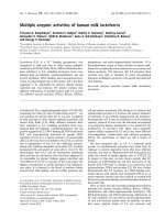
Fig. 1. (A) The partial nucleotide sequence of exon 1 (upper case), the 5¢ end of exon 2 (lower case) and the deduced amino acid sequences
of human trypsinogen 4. The numbering of the sequence is according to a previous publication [3]. (B) The 5¢ end of the human trypsino-
gen 4 cDNA, as determined by 5¢ rapid amplification of cDNA ends (5¢-RACE) PCR. (C) N-terminal amino acid sequence of human trypsino-
gen 4 isolated from a 71.0 g occipital cortex sample by a mAb 1 ⁄ B1 column. (D) N-terminal sequences of human trypsinogen 4 (isolated
from a 71.5 g frontal cortex sample by a mAb 1 ⁄ B1 column) after enterokinase digestion. The amount of N-terminal amino acids detected is
indicated below the sequences. (E) Western blot (with mAb 1 ⁄ B1) of human trypsinogen 4 isolated from an occipital cortex sample of
human brain by a mAb p28 column. Protein molecular weight markers are indicated on the left side. Lane 1, trypsinogen 4, isolated from
human brain, by mAb p28 immunoaffinity chromatography. Lane 2, recombinant tag-p72-trypsinogen 4. Lane 3, recombinant tag-p28-trypsi-
nogen 4. Lane 4, recombinant trypsin 4. (F) Searching for proteolytic activity in human brain samples. Protein molecular weight markers are
indicated on the left side. Lane 1, recombinant tag-p72 trypsinogen 4 incubated for 2 h at 37 °C with total brain extract. Lane 2, recombinant
tag-p72 trypsinogen 4, incubated for 2 h at 37 °C with brain extract previously passed through a mAb 1 ⁄ B1 immunoaffinity column. In both
cases, trypsinogen 4 was recovered by mAb 1 ⁄ B1 immunoaffinity chromatography. The blot was developed with mAb 1 ⁄ B1. Lane 3, recom-
binant tag-p72-trypsinogen 4. Lane 4, recombinant tag-p28-trypsinogen 4. Lane 5, recombinant trypsin 4.
A. L. Ne
´
meth et al. Translation initiation of trypsinogen 4
FEBS Journal 274 (2007) 1610–1620 ª 2007 The Authors Journal compilation ª 2007 FEBS 1611
genes, such as proto-oncogenes, genes for transcrip-
tion factor kinases and growth factors [5–10]. Origin-
ally, it was thought that regardless of the initiation
codon used, methionine should be the initiating resi-
due, and those few cases in which the reported initi-
ator amino acid was not a methionine were limited to
viral genes containing an internal ribosome entry site
upstream of the nonconventional start codon [11,12].
Recently, however, Schwab and coworkers [13,14]
have shown that leucine can also be translated as an
initiator amino acid by using a CUG codon in short
cryptic peptides in antigen-presenting cells. In this
case, the leucine start does not depend on an internal
ribosome entry site-like mRNA structure, and its
translational efficiency is enhanced by a nucleotide
context slightly different from the consensus Kozak
sequence [15].
As a biochemical approach to determine the exact
N-terminal sequence of trypsinogen 4, mAbs were
raised against human trypsin 4 (obtained by enterokin-
ase activation of recombinant human trypsinogen 4)
and a synthetic fragment of the N-terminal 28-amino
acid leader peptide of trypsinogen 4. We used these
mAbs to purify and chemically identify trypsinogen 4
from human brain and from transiently transfected
human cell lines. Our results show that Isoform B of
trypsinogen 4, with a leucine N terminus, is the pre-
dominant (if not the exclusive) form of the enzyme in
human brain, whereas both Isoform B and Isoform A
can be extracted from transfected cells. Here we report,
from amino acid sequencing, that although the N-ter-
minal residue of the longer Isoform A isolated from
the transiently transfected cells is methionine, as expec-
ted, the N-terminal amino acid of Isoform B, isolated
from human brain and transiently transfected cells, is
leucine.
Results
Determination of the 5¢-terminal sequence of
human trypsinogen 4 mRNA
In the original publication reporting the cloning of
trypsinogen 4 cDNA, no ATG codon was found, even
in the longest cDNA [3]. We repeated this experiment,
with several brain samples, under slightly different
conditions. We used C-tailing and an inosine contain-
ing abridged anchor primer, according to the 5¢-rapid
amplification of cDNA ends (5¢-RACE System; Gibco-
BRL), whereas in the original publication, G-tailing
was used. Nevertheless, we obtained practically the
same result, because the 5¢ end of the cDNA was only
four bases upstream from the putative CTG transla-
tion start codon (Fig. 1B) of Isoform B. Several
attempts to isolate a cDNA containing the first
upstream in-frame ATG codon were unsuccessful. It is
interesting to note that the longest transcript deposited
in the GenBank database (accession no. BI823946)
also lacks the ATG ()44) start codon and starts from
the third (G) nucleotide of the above-mentioned ATG
codon.
Isolation and chemical identification of
trypsinogen 4 from human brain
Different antihuman trypsinogen 4 mAbs were raised
separately against recombinant human trypsin 4
(mAb 1 ⁄ B1, mAb 6 ⁄ B7) and the 28-amino acid leader
peptide (mAb p28). Although all of these antibodies
react with the leader peptide containing forms of try-
psinogen, activated trypsin is only recognized by anti-
bodies 1 ⁄ B1 and 6 ⁄ B7. Two antibodies – mAb 1 ⁄ B1
and mAb p28 – were immobilized separately on cyano-
gen bromide activated Sepharose 4B. Pilot studies on
the isolation of trypsinogen 4 from extracts of four dif-
ferent regions of human brain (see the Experimental
procedures) showed that from all samples, and by both
immunoaffinity columns, proteins of the same molecu-
lar size were isolated. The size and immunoreactivity
of this protein corresponded to those of recombinant
tag-p28 trypsinogen 4. This is illustrated in lane 1 of
Fig. 1E, which shows a western blot (detected by
mAb 1 ⁄ B1) of trypsinogen 4, which was isolated via a
mAb p28 column from a sample of human occipital
cortex.
Affinity-purified proteins from three different brain
regions were sequenced. In each case, we identified leu-
cine as the only N-terminal amino acid of the isolated
protein, irrespective of the specificity of the immobi-
lized antibody (Fig. 1C). In order to prove the integ-
rity of the isolated protein, human trypsinogen 4,
isolated from a sample of the frontal cortex, was sub-
jected to enterokinase digestion; N-terminal sequencing
revealed the presence of both trypsin 4 with the N-ter-
minal isoleucine and intact Isoform B starting with
leucine (Fig. 1D).
Searching for a protease with potential
processing activity in brain extract
To demonstrate the absence of protease activity
capable of cleaving the trypsinogen leader sequence
during the isolation process, we added recombinant
Isoform A (tag-p72-trypsinogen 4) (Fig. 1F) to homo-
genized human brain samples and incubated them,
without inhibitors, at 37 °C for 2 h. Then, the sam-
Translation initiation of trypsinogen 4 A. L. Ne
´
meth et al.
1612 FEBS Journal 274 (2007) 1610–1620 ª 2007 The Authors Journal compilation ª 2007 FEBS
ples were centrifuged at 100 000 g for 20 min and the
supernatants were subjected to immunoaffinity chro-
matography using immobilized mAb 1 ⁄ B1. Western
blotting of the eluted material clearly showed that the
isolated protein was mostly intact (Fig. 1F, lane 1).
Although faint bands indicated some proteolytic
breakdown, no traces of a fragment, corresponding to
Isoform B of trypsinogen 4, was found. Similar results
were obtained when tag-p72-trypsinogen 4 was added
to a brain homogenate that had been previously
passed through an immunosorbent column (Fig. 1F,
lane 2).
Isolation and chemical identification of
trypsinogen 4 from transiently transfected HeLa
cells
Transfection experiments, using several constructs
(Fig. 2A,3A), were used in different cell lines. We tran-
siently transfected HeLa cells with p72
M
T4,
p72
M
p28
L(TTG)
T4 and p28
L
T4 constructs, and the cells
were stained with antihuman trypsinogen 4 p28
(mAb p28; data not shown). Cell lysates from trans-
fected cells were examined by western blotting
(Fig. 2B) and subjected to immunoaffinity chromato-
graphy on a mAb 1 ⁄ B1 column. Immunoreactive
proteins, eluted in single fractions from the immunoaf-
finity columns, were analyzed by N-terminal amino
acid sequencing (Fig. 2C,D). Western blots, together
with the N-terminal amino acid sequences of trypsino-
gens isolated from 6 · 10
6
cells transfected with the
p72
M
T4 construct, indicated that both Isoforms A and
B of human trypsinogen 4 were expressed. Cells trans-
fected with the p72
M
p28
L(TTG)
construct, however,
expressed only the longer isoform (Isoform A) and no
traces of the shorter isoform (Isoform B). By contrast,
the expression of only Isoform B was detected in the
cells transfected with the p28
L
T4 construct (Fig. 2B)
and leucine was identified as the sole N-terminal amino
acid of this protein (Fig. 2D).
A
B
C D
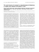
Fig. 2. (A) Schematic representation of the gene constructs used for expression of different isoforms of human trypsinogen 4
(p72
M
p28
L
(
TTG)
T4, p72
M
T4, p28
L
T4) in HeLa cells. The white box indicates nontranslated regions caused by the deletion of the AUG initiation
codon, active trypsin 4 is represented by the blue box. (B) Western blot of human trypsinogen 4, detected by using mAb p28. Protein
molecular weight markers are indicated on the left side. Lane 1, recombinant tag-p28-trypsinogen 4. Lane 2, recombinant tag-p72-trypsino-
gen 4. Lane 3, nontransfected, control HeLa cells 4. Lane 4, trypsinogen 4 detected from p72
M
p28
L
(
TTG)
T4 transfected HeLa cells. Lane 5,
trypsinogen 4 detected from p72
M
T4 transfected HeLa cells. Lane 6, trypsinogen 4 detected from p28
L
T4 transfected HeLa cells. (C) N-ter-
minal amino acid sequence of human trypsinogen 4 isolated from HeLa cells transiently transfected with p72
M
T4 plasmid. (D) N-terminal
amino acid sequence of human trypsinogen 4 isolated from HeLa cells transiently transfected with p28
L
T4 plasmid. The amount of N-ter-
minal amino acids detected is indicated below the sequences.
A. L. Ne
´
meth et al. Translation initiation of trypsinogen 4
FEBS Journal 274 (2007) 1610–1620 ª 2007 The Authors Journal compilation ª 2007 FEBS 1613
A
B
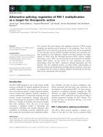
Fig. 3. (A) Green fluorescent protein (GFP)-fused plasmid constructs. Amino acid numbering and the indicated sequences are as described in
Fig. 1A. The white box indicates nontranslated regions caused by the deletion of the initiation AUG codon; active trypsin 4 is represented by
a blue box. p72
M
T4-GFP, p72 form of trypsinogen 4 with a C-terminal GFP fusion protein; GFP-p72
M
T4, p72 form of trypsinogen 4 with an
N-terminal GFP fusion protein; p28
L
T4-GFP, p28 form of trypsinogen 4 with a deleted ATG()44) codon, CTG(+1) coding for a leucine initiator
amino acid and a C-terminal GFP fusion protein; p28
M
T4-GFP, p28 form of trypsinogen 4 with a deleted ATG()44) codon, a mutated
ATG(+1) coding for methionine as the initiator amino acid and a C-terminal GFP fusion protein; p72
M
-GFP, p72 leader peptide with a C-ter-
minal GFP fusion protein without the trypsinogen 4 catalytic domain; p28
L
-GFP, p28 leader peptide with a deleted ATG()44) codon, CTG(+1)
coding for a leucine initiator amino acid and a C-terminal GFP fusion protein without the trypsinogen 4 catalytic domain; p28
M
-GFP, p28 lea-
der peptide with a deleted ATG()44) codon, a mutated ATG(+1) coding for methionine as the initiator amino acid and a C-terminal GFP
fusion protein without the trypsinogen 4 catalytic domain; p28
L
T4*-GFP, p28 form of trypsinogen 4 with a deleted 5¢-UTR sequence
between ATG()44) and GGG()3), leaving only a 7 bp upstream sequence before CTG(+1) coding for a leucine initiator amino acid and the
C-terminal GFP fusion protein; p28
M
T4*-GFP, p28 form of trypsinogen 4 with a deleted 5¢-UTR sequence between ATG()44) and GGG()3),
leaving only a 7 bp upstream sequence before a mutated ATG(+1) coding for a methionine initiator amino acid and C-terminal GFP fusion
protein. (B) Representative pictures from U87 human astroglioma cells, transiently transfected with different constructs, as indicated above
the pictures. In each case, single optical sections taken by confocal microscopy are presented. GFP labelling (green) always colocalized with
mAb p28 immunostaining (red). Cell nuclei were stained with Draq5 (blue). Depending on the constructs and the relative trypsinogen 4
expression levels, aggregation of GFP-labelled proteins were occasionally observed. Bars indicate 5 lm.
Translation initiation of trypsinogen 4 A. L. Ne
´
meth et al.
1614 FEBS Journal 274 (2007) 1610–1620 ª 2007 The Authors Journal compilation ª 2007 FEBS
Transient transfection of human U87
glioblastoma cells with trypsinogen 4 GFP-fused
constructs
We expressed human trypsinogen 4, fused with GFP
reporter protein, in the U87 human glioblastoma cell
line. Cells were transiently transfected with the con-
structs depicted in Fig. 3A and were immunostained,
24h post-transfection, with mAbs raised against the
activated protease, mAb 1 ⁄ B1 (data not shown), or
against the 28-residue leader peptide, mAb p28
(Fig. 3B). The GFP reporter protein always colocalized
with the immunostaining, with antibodies recognizing
either the p28 leader sequence (mAb p28) or the prote-
ase domain (mAb 1 ⁄ B1), indicating that trypsinogen 4
was localized mainly in an inactive form in the trans-
fected cells. The observed localization of trypsinogen 4
was the same when GFP was fused to the N- or the
C-terminal end of the molecule (data not shown).
We determined the number of cells showing GFP
fluorescence by visual inspection of pictures taken
from several microscopic fields. We consider the per-
centage of the GFP-positive cells as a measure of relat-
ive expression, because all experimental parameters,
number of cells transfected, amount of plasmid, incu-
bation time, etc., were essentially identical at each
transfection (see the Experimental procedures).
In the case of constructs using an AUG initiation
codon at site )44, the relative expression levels of
Isoform A (expressed together with Isoform B; Fig. 2B,
lane 5) were elevated compared with Isoform B with
an AUG start codon (p72
M
T4-GFP versus p28
M
T4-GFP or p72
M
-GFP versus p28
M
-GFP; Table 1).
The expression level of Isoform B with the wild-type
CUG initiation codon was lower than that of
Isoform A (expressed together with Isoform B)
(p72
M
T4-GFP versus p28
L
T4-GFP; Table 1) and was
dependent on the length of the wild-type upstream
sequence preceding the CUG codon (p28
L
T4-GFP ver-
sus p28
L
T4*-GFP; Table 1). Nevertheless, protein
expression was detected in all cases in which the CUG
initiation codon was employed. Analogous constructs
with the AUG initiation codon (p28
M
T4-GFP versus
p28
M
T4*-GFP) did not show dependence on the
length of the 5¢-UTR region.
Discussion
The occurrence of human trypsinogen 4 was first
revealed in human brain [3], but later it was also found
in human epithelial cells from prostate, colon and air-
way, and in several different tumors [16]. Trypsino-
gen 4 has two distinctive features: it contains an
unusual mutation (Gly193 to Arg), responsible for its
unique enzymatic properties [17–19]; and has an
unconventional leader sequence (Fig. 1A).
By sequencing trypsinogen 4 samples isolated from
human brain following only a short (2–5 h) post mor-
tem delay, we found no traces of Isoform A beginning
with Met()44) (Fig. 1A). This result contrasts with the
predictions of two isoforms based on the analysis of
the PRSS3 gene [3]. Instead, in each case we identified
only the sequence corresponding to Isoform B begin-
ning with leucine (Fig. 1C). We were unable to isolate
Isoform A from any parts of the human brain. How-
ever, we cannot exclude the presence of the longer
isoform in certain tissues; in addition, we found that
Isoform A was expressed in cells transfected with the
construct containing the full-length Isoform A gene
(p72
M
T4) (Fig. 2B). In accordance with previously
published data [3,16], we were unable to detect any
mRNA containing the upstream AUG codon for
Met()44), (Fig. 1B). As we were working with human
brain samples, degradation of RNA owing to the post
mortem delay is a possibility.
In theory, our finding that the zymogen form of
trypsinogen 4 possesses a leucine N terminus has two
explanations: either a hitherto-unknown proteolytic
processing mechanism is responsible for cleaving the
leader sequence, or leucine is, in fact, the initiator
amino acid. The first possibility appeared to be
Table 1. Relative expression levels in U87 cells transiently trans-
fected with green fluorescent protein (GFP)-fusion constructs. Plas-
mids used for transfections are as depicted in Fig. 3A, with the
exception of pAcGFP-N1, indicating the cloning vector without any
trypsinogen constructs. The percentage of GFP-positive cells was
determined by comparing the number of cells showing GFP fluores-
cence with the total number of 4’-6-diamidino-2-phenylindole-posit-
ive cell nuclei in each microscopic field. Averages were calculated
from three randomly chosen fields, and then the values of at least
three independent transfection experiments were averaged (per-
centage ± standard deviation). GFP-positive cells were identified by
visual inspection of pictures taken at identical exposure settings
and were verified by inspecting the number of cells immunostained
with mAb p28.
Construct
Percentage of
GFP-positive cells
pAcGFP-N1 27.8 ± 5.0
p72
M
T4-GFP 21.0 ± 2.5
p28
L
T4-GFP 7.6 ± 1.8
p28
M
T4-GFP 14.6 ± 2.9
p72
M
-GFP 23.0 ± 3.9
p28
L
-GFP 13.8 ± 2.4
p28
M
-GFP 15.1 ± 0.9
p28
L
T4*-GFP 2.0 ± 1.0
p28
M
T4*-GFP 17.2 ± 3.8
A. L. Ne
´
meth et al. Translation initiation of trypsinogen 4
FEBS Journal 274 (2007) 1610–1620 ª 2007 The Authors Journal compilation ª 2007 FEBS 1615
unlikely in the light of our in vitro experiments,
according to which extracts from post mortem human
brain samples did not convert the recombinant
Isoform A (tag-p72-trypsinogen 4) to an Isoform B-like
protein (Fig. 1F). Furthermore, expression of
Isoform B with a leucine N terminus was also detected
in HeLa cells transfected with a construct containing
the full-length Isoform A gene (p72
M
T4) (Fig. 2B,C).
Importantly, we also detected N-terminal leucine in
trypsinogen 4 isolated from cells transfected with a
gene construct that lacks the upstream AUG codon
for Met()44) (p28
L
T4) (Fig. 2B,D). All results, listed
above, support our proposal that CUG is the initiation
codon directing the incorporation of leucine, rather
than methionine, into Isoform B of trypsinogen 4. The
most convincing experimental evidence in favor of this
hypothesis, however, came from a comparison of the
expressed proteins from HeLa cells transfected with
constructs p72
M
p28
L(TTG)
T4 and p72
M
T4, respectively
(Fig. 2B, lane 4 versus lane 5). In cells transfected with
the former construct, in which CTG encoding Leu28
was replaced with TTG, another codon for Leu, only
Isoform A was formed, whereas in cells transfected
with the original construct containing the wild-type
DNA sequence for trypsinogen 4, both Isoforms A and
B were detected. The exclusive interpretation of this
experiment is that the generation of Isoform B of try-
psinogen 4 occurs at the level of translation and not
post-translationally.
Recently, Schwab and coworkers [13,14] presented a
similar case in eukaryotes: during the antigen presen-
tation by Class I major histocompatibility complex
molecules, the synthesis of a cryptic peptide was initi-
ated with leucine by using CUG as the initiation
codon without an upstream internal ribosomal entry
site. The contextual sequence requirements for non-
AUG initiation are not fully understood, and the
critical nature of nucleotides that surround the non-
AUG triplet is controversial [6]. The Kozak context
of the PRSS3 Isoform B CUG initiation codon
(GCGGGCcugG) resembles the optimal context of an
AUG codon (GCCRCCaugG) [15]. In the experiment
of Schwab and coworkers, however, the optimal con-
text of CUG initiation was TCCACCcugG, different
from that of PRSS3. In the present study, shortening
of the wild-type 5¢-UTR region upstream of the CUG
initiation codon in the p28
L
T4*-GFP construct to
only seven nucleotides led to significantly decreased,
but not abolished, expression of the GFP-fused
enzyme (Table 1). A decrease in the expression level
was not observed when the 5¢-UTR region was
removed preceding the AUG initiation codon. This
finding indicates the important, but not exclusive, role
of the 5¢-UTR region beyond the Kozak region in
translation initiation from the CUG initiation codon.
There is an 30-nucleotide-long GC-rich region pre-
ceding the CUG start codon that might have a role in
recognition of the suboptimal translational initiation
site. Irrespective of the length of the 5¢-UTR region,
we found that the relative expression levels were lower
in both U87 and HeLa cells when leucine was used as
the initiator amino acid (Table 1); this suggests that
CUG translational initiation may control the expres-
sion level. Thus, one is tempted to speculate that
under physiological conditions, the translational initi-
ation of human trypsinogen 4 with a Leu N terminus
may function to keep the expression of the protein at
a relatively low level.
The first exon of trypsinogen 4 is derived from the
noncoding first exon of LOC120224, a chromosome-11
gene [2]. LOC120224 codes for a widely conserved
transmembrane protein of unknown function. The
missing upstream AUG initiation codon in the
LOC120224 transmembrane protein does not necessar-
ily mean that translation starts from a downstream
AUG, as predicted by genome and mRNA analysis,
but raises the possibility that the translated form may
have used a CUG start codon with an N-terminal leu-
cine amino acid. Our present study indicates that non-
AUG translation initiation may be operable more
often than anticipated. This may have a great impact
on the analysis of genes on the basis of genome
sequencing.
It has been suggested that human trypsinogen 4
plays functional roles in human cancer and metastasis
[20–22], amyloid fragment production in aged astro-
cytes [23], or in epithelial tissues modulating protease-
activated receptor-2 and -4 activity [16,24]. More
recently, in an in vitro study we have shown that
recombinant human trypsin 4 selectively clips residues
80–97 from human myelin basic protein [25], indicating
a possible link to the development of multiple sclerosis
[26,27]. It is a possibility to consider, that a significant
release and activation of trypsinogen 4 would occur
only under pathological conditions when the trypsino-
gen 4-expressing cells undergo damage in the human
brain. Until clinical experiments support or deny this
hypothesis, the biological function of human trypsino-
gen 4 remains in doubt.
Experimental procedures
Human brain samples
Tissue samples were obtained from the Human Brain
Tissue Bank, Budapest. Brains were removed from the
Translation initiation of trypsinogen 4 A. L. Ne
´
meth et al.
1616 FEBS Journal 274 (2007) 1610–1620 ª 2007 The Authors Journal compilation ª 2007 FEBS
skull, rapidly frozen on dry ice and stored at ) 70 °C until
dissection. Samples from four different human brains were
used for human trypsinogen 4 isolation in separate experi-
ments. These were as follows: 77.3 and 76.9 g samples of
the occipital and temporal cortex, respectively, from an
81-year-old woman, a 71.5 g sample of frontal cortex from
an 83-year-old man and a 71.0 g sample of occipital cortex
from a 85-year-old man with a short (2–5 h) post mortem
delay.
RNA isolation, reverse transcription and 5¢-RACE
Total RNA was isolated from 30 to 100 mg of human frontal
cortex tissue samples, using TRI Reagent (Sigma, Budapest,
Hungary) according to the manufacturer’s instructions.
First-strand cDNA was synthesized by priming with gene-
specific primer 1 (5¢- GGCTTTACACTCAGCCTGGG-3¢).
Reverse transcription was performed by the RevertAid H
Minus First Strand cDNA Synthesis Kit (Fermentas, Vilnius,
Lithuania). The synthesized cDNA was subjected to homo-
polymeric tailing to create a binding site for the abridged
anchor primer on the 3¢ end of the cDNA. PCR amplification
of the C-tailed cDNA was performed with the abridged
anchor primer (5¢-GGCCACGCGTCGACTAGTACGGGII
GGGIIGGGIIG-3¢,5¢-RACE System; Gibco-BRL, Grand
Island, NY, USA) and a nested gene-specific primer 2
(5¢-GGAGAGTTTGATCAGCATGATGTC-3¢) using Taq
polymerase. The PCR products were cloned into pBluescript
vector, via TA ligation, and then sequenced.
Cloning and expression of the PRSS3 gene
The gene sequence coding for Isoform B of human trypsi-
nogen 4 was cloned from human brain cDNA with the
primers FP1 (5¢-CGCATATGGAGCTGCACCCGCTTC
TG-3¢) and RP1 (5¢-GACTGCAGGGATCCCGGGGG
CTTTAGC-3¢). The PCR product was subcloned into vec-
tor pET-15b (Novagen, Madison, WI, USA). This construct
resulted in a fusion protein with a histidine tag at its N ter-
minus (tag-p28-trypsinogen 4). As the mRNA correspond-
ing to the Isoform A of human trypsinogen 4 could not be
found with the 5¢-RACE technique, the DNA sequence
encoding the first exon was PCR amplified from genomic
DNA using the forward and reverse primers FP2 (5¢-
CTGCATATGTGCGGACCTGACGACAGATGC-3¢) and
RP2 (5¢-CTGCAGCAACTGTGCCCAGCGCCTCGC-3¢),
and then fused with the cloned Isoform B coding sequence
using the naturally occurring AlwNI site. The gene was
subcloned into the expression vector pET-15b (tag-p72-
trypsinogen 4).
To express the splice Isoforms A and B of human trypsi-
nogen 4 in Escherichia coli, 500 mL cultures of Rosettaä
(DE3)pLysS cells (Novagen), transformed with the con-
structs, were grown at 37 °C in Luria–Bertani medium con-
taining ampicillin. Cells were harvested, and the isolation
and refolding of the inclusion bodies were carried out, as
described previously [17,28], with minor modification.
The full-length Isoform A gene was used as template in a
PCR reaction, with Hu4-F1 (5¢-GCGCAAGCTTCCTGGA
GGATGTGCGGACCTGACGAC-3¢) and Hu4-R1 (5¢-GC
CTGGATCCGAGCTGTTGGCAGCGATGG-3¢) primers,
for subcloning the PRSS3 gene into pcDNA3 (Invitrogen,
Carlsbad, CA, USA), pAcGFP1-N1 and pAcGFP1-C1 (BD
Biosciences, Clontech, Mountain View, CA, USA) vectors at
HindIII and BamHI sites, resulting in p72
M
T4, p72
M
T4-GFP
and GFP-p72
M
T4 constructs, respectively. Hu4-F2 (5¢-GCG
CAAGCTTGCGGACCTGACGACAGATGC-3¢)and
Hu4R1 primers were used to amplify the PRSS3 gene
sequence lacking the initial ATG codon, which was sub-
cloned into pcDNA3 and pAcGFP1-N1 vectors, resulting in
p28
L
T4 and p28
L
T4-GFP constructs, respectively. The muta-
tion Leu1 to Met was introduced by the megaprimer PCR
reaction in p28
L
T4-GFP, by using the mutagenic primer
p28ATG (5¢-GAGCTCCATGCCCGCCC-3¢). The resulting
construct (p28
M
T4-GFP) lacked the initial ATG codon of
Isoform A, and the initial CTG codon of Isoform B was
mutated to ATG. The corresponding constructs were made
lacking the trypsin catalytic domain using p72GFP pri-
mer (5¢-GTCGGATCCTTGTCATCATCGTCAAAGG-3¢),
resulting in p72
M
-GFP, p28
L
-GFP and p28
M
-GFP con-
structs in the pAcGFP-N1 expression vector. The silent
mutation of the CTG initiation codon to TTG was intro-
duced by the mutagenic primer, p28TTG (5¢-GTGCAG
CTCCAAGCCCGCCCC-3¢), by using the megaprimer PCR
method, and the gene harboring the mutation was cloned
into the pcDNA3 vector. This construct is designated as
p72
M
p28
L(TTG)
T4. The p28
L
T4*-GFP and p28
M
T4*-GFP
constructs were made by removing the 5¢-UTR region of Iso-
form B (that is part of the coding region of Isoform A) from
the p28
M
T4-GFP and p28
L
T4-GFP constructs, using the
p28Hind primer (5¢-CGCAGCGAAGCTT
GGCGGGC-3¢).
Only a seven-nucleotide-long sequence was left before the
putative CUG initiator codon (underlined) to ensure the
wild-type Kozak sequence was maintained.
Antibodies
Recombinant human trypsin 4, and the synthetic 28-amino
acid leader peptide, were used to immunize female BALB ⁄ c
mice (Charles River Laboratories, Raleigh, NC, USA).
Antigen-specific B lymphocytes, prepared from the spleens
of high-responder animals, were preselected using a method
developed in our laboratory and published previously [29].
The fusion partner was the Sp-2 ⁄ 0 Ag14 (ATCC, Mana-
ssas, VA, USA) mouse myeloma cell line, and the hybri-
doma cells were prepared and cloned as described
previously [30]. The selected clones of hybridomas were cul-
tured in DMEM (Sigma) containing 10% fetal bovine
serum (Gibco-BRL). The mass production of antibodies
was performed by hybridoma fermentation (Harvest
A. L. Ne
´
meth et al. Translation initiation of trypsinogen 4
FEBS Journal 274 (2007) 1610–1620 ª 2007 The Authors Journal compilation ª 2007 FEBS 1617
Mouse; Serotec, Oxford, UK). mAbs were purified by Pro-
tein-G based Sepharose 4B affinity chromatography (Phar-
macia, Upsalla, Sweden) and then concentrated by Amicon
ultrafiltration (Millipore, Billerica, MA, USA). Different
antigens were used to characterize immunoserologically the
mAbs raised against the protease domain (mAb 1 ⁄ B1 and
mAb 6 ⁄ B7) and the 28-amino acid leader peptide
(mAb p28) of human trypsinogen 4 (data not shown).
Immunoaffinity media preparation
mAbs 1 ⁄ B1 and p28 were immobilized separately on cyano-
gen bromide-activated Sepharose 4B (Pharmacia). Antibod-
ies were dialyzed against the coupling buffer (0.1 m
NaHCO
3
, pH 8.3, containing 0.5 m NaCl) and mixed with
the resin. Coupling efficiency proved to be > 90%. The sta-
bility of coupling was tested by washing the resin with the
elution buffer of the chromatography (50 mm HCl). Under
these conditions, the coupled antibody was not eluted from
the column.
Isolation of trypsinogen 4 from human brain
Samples were homogenized in five volumes of NaCl ⁄ P
i
,
pH 7.4 and the homogenate was centrifuged at 100 000 g
for 20 min. The pellet was then rehomogenized in five vol-
umes of NaCl ⁄ P
i
, pH 7.4, containing 1% (v ⁄ v) Tween-20,
1mm phenylmethanesulfonyl fluoride, 1 mm cystatin,
1mm leupeptin and 1 mm EDTA as protease inhibitors.
After centrifugation of the homogenate at 100 000 g for
20 min, the supernatant was immediately used for immu-
noaffinity chromatography. The total protein concentra-
tion was determined by the bicinchoninic acid method
(Sigma).
Cell lines and transfections
HeLa and U87 were used for transfection assays. HeLa
cells were grown in DMEM ⁄ F-12 medium supplemented
with 10% fetal bovine serum, 1 mm sodium pyruvate,
100 UÆmL
)1
of streptomycin and 100 lgÆmL
)1
of penicillin,
whereas U87 human glioblastoma cells were cultured in
DMEM containing 10% fetal bovine serum, 4500 mgÆmL
)1
of glucose and 40 lgÆmL
)1
of gentamycin (all Sigma), in a
humidified 37 °C incubator with 5% CO
2
. For transfection
assays, 10
5
HeLa or U87 cells were seeded onto poly
l-lysine (Sigma)-coated 13 mm diameter glass coverslips in
24-well plates, transfected either with Fugene 6 (Roche,
Mannheim, Germany; HeLa cells) or Lipofectamine 2000
(Gibco; U87 cells) transfection reagents, according to
the manufacturers’ instructions, and were processed 24 h
after transfection. For immunofluorescence cell studies in
HeLa or U87 cells, 250 ng or 1 lg of DNA was used,
respectively.
Isolation of trypsinogen 4 from HeLa cells
A total of 6 · 10
6
HeLa cells, seeded into 60-mm Petri dishes
and transfected with p72
M
T4 or p28
L
T4 plasmid constructs,
were used for trypsinogen isolation in separate parallel
experiments. Cells were homogenized in 5 mL of lysis buffer
(1% Tween-20, 50 mm Tris ⁄ HCl, 150 mm NaCl, pH 8, con-
taining 1 mm phenylmethanesulfonyl fluoride, 1 mm benz-
amidine, 1 mm cystatine, 1 mm leupeptin and 1 mm EDTA).
Homogenized samples were incubated for 1 h on ice and
then, after centrifugation (14 000 g, 20 min, 4 °C) the sup-
ernatants were used for immunoaffinity chromatography.
Immunoaffinity chromatography
Supernatant fractions of human brain and transfected
HeLa cell homogenates were passed through the 1.5–
0.5 mL immunoaffinity column. The columns were washed
three times with 10 mL of NaCl ⁄ P
i
, pH 7.4, containing 1%
Tween-20 and 150 mm NaCl. Elution was carried out with
50 mm HCl, and 1–0.5 mL fractions were collected. Frac-
tions were screened for trypsin immunoreactivity by gel
electrophoresis and western blotting.
Western blot analysis
Proteins were separated by SDS–PAGE (15% gel) and were
transferred to nitrocellulose membranes (Pharmacia). Blots
were blocked in NaCl ⁄ Tris-Tween buffer (20 mm Tris,
pH 8.0, 150 mm NaCl, 0.05% Tween-20) at room tempera-
ture and then incubated with mAb 1 ⁄ B1 or p28 (1 : 3000)
overnight at 4 °C. After being washed for 3 · 5 min with
NaCl ⁄ Tris-Tween, blots were incubated with biotin-conju-
gated anti-mouse secondary serum (B-7151; Sigma), at a
1 : 5000 dilution, in NaCl ⁄ Tris-Tween, for 1 h at room
temperature. After washing, the blots were incubated with
ExtrAvidin peroxidase conjugate (E-2886; Sigma), at a
1 : 3000 dilution, for 1 h at room temperature followed by a
5 min wash in NaCl ⁄ Tris. The color development reaction
was carried out using diaminobenzidine (Sigma), in NaCl ⁄
Tris, in the presence of 0.4 mm NiCl
2
and 1.25% H
2
O
2
.
Amino acid sequence determination
Fractions containing human trypsinogen 4 immunoreactivity
were freeze-dried, dissolved in 10 m m NH
4
HCO
3
and subjec-
ted to N-terminal amino acid analysis in a Procise sequencer
(ABI 494; Applied Biosystems, Foster City, CA, USA)
employing an edman degradation sequenator program.
Immunostaining
Transfected HeLa cells were fixed with cold methanol for
15 min, or with 4% paraformaldehyde for 20 min, at
Translation initiation of trypsinogen 4 A. L. Ne
´
meth et al.
1618 FEBS Journal 274 (2007) 1610–1620 ª 2007 The Authors Journal compilation ª 2007 FEBS
room temperature. The staining patterns were similar with
the different fixatives used. The cells were washed in
NaCl ⁄ P
i
containing 0.1% Triton-X-100, then blocked for
30 min in NaCl ⁄ P
i
containing 0.1% Triton-X-100 and 5%
goat serum (Sigma). Subsequently, the cells were stained
with mAb p28 (1 : 1000), followed by fluorescein iso-
thiocyanate or Texas-Red conjugated anti-mouse sera
(Jackson Laboratories, Bar Harbor, ME, USA), all dilu-
ted in 0.1% Triton-X-100 containing 5% goat serum.
After washing in NaCl ⁄ P
i
, nuclei were counterstained with
4¢,6-diamidino-2-phenylindole, and the coverslips were
mounted on Crystal Mount medium (Biomeda Corp.,
Foster City, CA, USA). Transfected U87 cells were fixed
by 4% paraformaldehyde in NaCl ⁄ P
i
(pH 7.4) for 20 min
at room temperature, permeabilized with 0.1% Triton-X-
100 for 5 min and blocked by 2% BSA-NaCl ⁄ P
i
-0.1% Na
azide (blocking solution) for 1 h at room temperature.
Cells were incubated with mAb p28 (1 : 1000), at 4 ° C
overnight, followed by anti-mouse biotin (1 : 1000; goat
IgG; Jackson Laboratories), for 1.5 h at room tempera-
ture, and Extravidin-TRITC (1 : 1000; Sigma) for 1 h at
room temperature. All antibodies were diluted in blocking
solution. Nuclei were labeled by incubation with 4¢,6-
diamidino-2-phenylindole or DRAQ5 (fluorescent dies) for
10 min at room temperature (1 : 2000; BioStatus Ltd,
Shepshed, UK), then the coverslips were washed and
mounted using Mowiol 4.88 (Polysciences Gmbh, Eppel-
heim, Germany).
Fluorescence microscopy
Confocal microscopy was carried out by a 488 nm Argon
laser, and by 546 nm and 633 nm Helium-Neon lasers,
using the ·60 oil-immersion objective of an Olympus
IX71 microscope equipped with fluoview500 software
(Olympus, Tokyo, Japan). The sequential scanning mode
was used during recordings to exclude potential cross-talk
completely between different channels. For wide-field
observations in HeLa cells, a Leica DMLS microscope
(Leica Microsystems, Wetzlar, Germany), equipped with
appropriate filter sets and a cooled CCD camera
(spot; Digital Instruments, Buffalo, NY, USA), and a
C-PLAN ·100 immersion objective, was used, and digital
images were recorded with spot 4.0.2. To estimate the
relative expression levels for different GFP-tagged con-
structs in U87 cells, the number of GFP-positive cells
was compared with the total number of 4¢-6-diamidino-2-
phenylindole-positive cell nuclei. Digital images from at
least three randomly chosen microscopic fields from
transfected U87 cells were recorded with a ·20 objec-
tive on an Olympus BX-51 microscope fitted with a
fluoview2 camera, and the numbers obtained were aver-
aged. These values were determined in three independent
transfection experiments, and the averages are shown in
Table 1.
Acknowledgements
This study was supported by Hungarian Research
Grants OTKA to L. Gra
´
f (T047154, TS 049812),
L. Szila
´
gyi (T037568) and J. Gergely (TS 0044711).
References
1 Roach JC, Wang K, Gan L & Hood L (1997) The
molecular evolution of the vertebrate trypsinogens.
J Mol Evol 45, 640–652.
2 Rowen L, Williams E, Glusman G, Linardopoulou E,
Friedman C, Ahearn ME, Seto J, Boysen C, Qin S,
Wang K et al. (2005) Interchromosomal segmental
duplications explain the unusual structure of PRSS3,
the gene for an inhibitor-resistant trypsinogen. Mol Biol
Evol 22, 1712–1720.
3 Wiegand U, Corbach S, Minn A, Kang J & Mu
¨
ller-Hill
B (1993) Cloning of the cDNA encoding human brain
trypsinogen and characterization of its product. Gene
136, 167–175.
4 Becerra SP, Rose JA, Hardy M, Baroudy BM &
Anderson CW (1985) Direct mapping of adeno-asso-
ciated virus capsid proteins B and C: a possible ACG
initiation codon. Proc Natl Acad Sci USA 82, 7919–7923.
5 Acland P, Dixon M, Peters G & Dickson C (1990) Sub-
cellular fate of the int-2 oncoprotein is determined by
choice of initiation codon. Nature 343, 662–665.
6 Kozak M (1997) Recognition of AUG and alternative
initiator codons is augmented by G in position +4 but
is not generally affected by the nucleotides in positions
+5 and +6. EMBO J 16, 2482–2492.
7 Peabody DS (1989) Translation initiation at non-AUG
triplets in mammalian cells. J Biol Chem 264, 5031–
5035.
8 Hann SR, King MW, Bentley DL, Anderson CW &
Eisenman RN (1988) A non-AUG translational initia-
tion in c-myc exon 1 generates an N-terminally distinct
protein whose synthesis is disrupted in Burkitt’s
lymphomas. Cell 52, 185–195.
9 Saris CJ, Domen J & Berns A (1991) The pim-1 onco-
gene encodes two related protein-serine ⁄ threonine kina-
ses by alternative initiation at AUG and CUG. EMBO
J 10, 655–664.
10 Touriol C, Bornes S, Bonnal S, Audigier S, Prats H,
Prats AC & Vagner S (2003) Generation of protein
isoform diversity by alternative initiation of translation
at non-AUG codons. Biol Cell 95, 169–178.
11 Sasaki J & Nakashima N (2000) Methionine-indepen-
dent initiation of translation in the capsid protein of an
insect RNA virus. Proc Natl Acad Sci USA 97, 1512–
1515.
12 Wilson JE, Pestova TV, Hellen CU & Sarnow P (2000)
Initiation of protein synthesis from the A site of the
ribosome. Cell 102, 511–520.
A. L. Ne
´
meth et al. Translation initiation of trypsinogen 4
FEBS Journal 274 (2007) 1610–1620 ª 2007 The Authors Journal compilation ª 2007 FEBS 1619
13 Schwab SR, Li KC, Kang C & Shastri N (2003) Consti-
tutive display of cryptic translation products by MHC
class I molecules. Science 301 , 1367–1371.
14 Schwab SR, Shugart JA, Horng T, Malarkannan S &
Shastri N (2004) Unanticipated antigens: translation
initiation at CUG with leucine. PLoS Biol 2, e366.
15 Kozak M (1986) Point mutations define a sequence
flanking the AUG initiator codon that modulates trans-
lation by eukaryotic ribosomes. Cell 44, 283–292.
16 Cottrell GS, Amadesi S, Grady EF & Bunnett NW
(2004) Trypsin IV, a novel agonist of protease-activated
receptors 2 and 4. J Biol Chem 279, 13532–13539.
17 Katona G, Berglund GI, Hajdu´ J, Gra
´
f L & Szila
´
gyi L
(2002) Crystal structure reveals basis for the inhibitor
resistance of human brain trypsin. J Mol Biol 315,
1209–1218.
18 Szmola R, Kukor Z & Sahin-To
´
th M (2003) Human
mesotrypsin is a unique digestive protease specialized
for the degradation of trypsin inhibitors. J Biol Chem
278, 48580–48589.
19 To
´
th J, Gombos L, Simon Z, Medveczky P, Szila
´
gyi L,
Gra
´
fL&Ma
´
lna
´
si-Csizmadia A (2006) Thermodynamic
analysis reveals structural rearrangement during the
acylation step in human trypsin 4 on 4-methylumbelli-
feryl 4-guanidinobenzoate substrate analogue. J Biol
Chem 281, 12596–12602.
20 Diederichs S, Bulk E, Steffen B, Ji P, Tickenbrock L,
Lang K, Zanker KS, Metzger R, Schneider PM, Gerke
V et al. (2004) S100 family members and trypsinogens
are predictors of distant metastasis and survival in
early-stage non-small cell lung cancer. Cancer Res 64,
5564–5569.
21 Marsit CJ, Karagas MR, Danaee H, Liu M, Andrew A,
Schned A, Nelson HH & Kelsey KT (2006) Carcinogen
exposure and gene promoter hypermethylation in blad-
der cancer. Carcinogenesis 27, 112–116.
22 Yamashita K, Mimori K, Inoue H, Mori M & Sidransky
D (2003) A tumor-suppressive role for trypsin in human
cancer progression. Cancer Res 63, 6575–6578.
23 Minn A, Schubert M, Neiss WF & Mu
¨
ller-Hill B (1998)
Enhanced GFAP expression in astrocytes of transgenic
mice expressing the human brain-specific trypsinogen
IV. Glia 22, 338–347.
24 Grishina Z, Ostrowska E, Halangk W, Sahin-To
´
th M &
Reiser G (2005) Activity of recombinant trypsin iso-
forms on human proteinase-activated receptors (PAR):
mesotrypsin cannot activate epithelial PAR-1-2, but
weakly activates brain PAR-1. Br J Pharmacol 146,
990–999.
25 Medveczky P, Antal J, Patthy A, Ke
´
kesi K, Juha
´
sz G,
Szila
´
gyi L & Gra
´
f L (2006) Myelin basic protein, an
autoantigen in multiple sclerosis, is selectively processed
by human trypsin 4. FEBS Lett 580, 545–552.
26 O’Connor KC, Bar-Or A & Hafler DA (2001) The
neuroimmunology of multiple sclerosis: possible roles of
T and B lymphocytes in immunopathogenesis. J Clin
Immunol 21, 81–92.
27 Steinman L (1996) Multiple sclerosis: a coordinated
immunological attack against myelin in the central
nervous system. Cell 85, 299–302.
28 Szila
´
gyi L, Ke
´
nesi E, Katona G, Kaslik G, Juha
´
sz G &
Gra
´
f L (2001) Comparative in vitro studies on native
and recombinant human cationic trypsins. Cathepsin B
is a possible pathological activator of trypsinogen in
pancreatitis. J Biol Chem 276 , 24574–24580.
29 Najbauer J, Tigyi GJ & Ne
´
meth P (1986) Antigen-speci-
fic cell adherence assay: a new method for separation of
antigen-specific hybridoma cells. Hybridoma 5, 361–370.
30 Kohler G & Milstein C (1975) Continuous cultures of
fused cells secreting antibody of predefined specificity.
Nature 256, 495–497.
Translation initiation of trypsinogen 4 A. L. Ne
´
meth et al.
1620 FEBS Journal 274 (2007) 1610–1620 ª 2007 The Authors Journal compilation ª 2007 FEBS