Báo cáo khoa học: Branched chain mechanism of polymerization and ultrastructure of prion protein amyloid fibrils ppt
Bạn đang xem bản rút gọn của tài liệu. Xem và tải ngay bản đầy đủ của tài liệu tại đây (666.41 KB, 10 trang )
MINIREVIEW
Branched chain mechanism of polymerization and
ultrastructure of prion protein amyloid fibrils
Ilia V. Baskakov
1,2
1 Medical Biotechnology Center, University of Maryland Biotechnology Institute, Baltimore, MD, USA
2 Department of Biochemistry and Molecular Biology, University of Maryland School of Medicine, Baltimore, MD, USA
Prion diseases are a group of fatal neurodegenerative
maladies that can arise spontaneously or be inherited,
and that can also be infectious [1]. Despite enormous
investments over the last 30 years in searching for a
prion virus or virion [2–5], no prion-specific nucleic
acids associated with infectious prion particles have
ever been identified [6]. A notable shift has occurred in
the last few years from debating the question of whe-
ther a protein can be infectious to what makes a pro-
tein infectious and how many proteins are infectious
[7–9]. Elucidating the polymerization mechanisms and
structure of misfolded and aggregated isoforms of the
prion protein (PrP) will help solving these long-stand-
ing research problems.
Prion polymerization is a branched-
chain reaction
To model prion conversion, two kinetic models has
been exploited: the nucleation-polymerization [10] and
the template assisted [11]. These models have been
previously discussed in numerous review articles
[12–14] and therefore will not be presented here.
Although these two models have played an important
role in the evolution of our ideas regarding the
mechanism of prion conversion, neither of them
emphasize the importance of multiplication of the
active centers of prion conversion, a key step in
prion replication. When studying the kinetics of the
in vitro fibril formation, we were surprised to discover
that fibrillization of recombinant PrP (rPrP) displays
several kinetic features that can not be explained by
the nucleation-polymerization or the template assisted
models [15,16]. These ‘atypical’ features include: (a)
the dramatic effect of reaction volume on the length
of the lag-phase; (b) a volume-dependent threshold
effect; and (c) the highly cooperative sigmoidal kine-
tics of polymerization [15,16]. Although these features
could not be rationalized within nucleation-polymer-
ization or the template assisted models, they are
Keywords
amyloid fibrils; branched-chain mechanism;
in vitro conversion; polymerization kinetics;
prion diseases; prion protein
Correspondence
I. V. Baskakov, 725 West Lombard Street,
Baltimore, MD 21201, USA
Fax: +1 410 706 8184
Tel: +1 410 706 4562
E-mail:
(Received 9 March 2007, accepted 31 May
2007)
doi:10.1111/j.1742-4658.2007.05916.x
The discovery of prion disease and the establishment of the protein only
hypothesis of prion propagation raised substantial interest in the class of
maladies referred to as conformational diseases. Although significant pro-
gress has been made in elucidating the mechanisms of polymerization for
several amyloidogenic proteins and peptides linked to conformational dis-
orders and solving their fibrillar 3D structures, studies of prion protein
amyloid fibrils and their polymerization mechanism have proven to be very
difficult. The present minireview introduces the mechanism of branched-
chain reaction for describing the peculiar kinetics of prion polymerization
and summarizes our current knowledge about the substructure of prion
protein amyloid fibrils.
Abbreviations
AFM, atomic force microscopy; GdnHCl, guanidine hydrochloride; PK, proteinase K; PrP, prion protein; rPrP, recombinant prion protein.
3756 FEBS Journal 274 (2007) 3756–3765 ª 2007 The Author Journal compilation ª 2007 FEBS
consistent with the mechanism of branched-chain
reactions.
Employing the theory of branched-chain reactions
will greatly benefit our understanding of the prion rep-
lication mechanism. The first branched-chain processes
were described at the beginning of twentieth century
and the branched-chain theory was developed shortly
afterward in the 1920s by Nikolay Semenov [17].
Although this theory had enormous impact on the
developing chemical industry and nuclear sources of
energy, the Nobel Prize for this amazing discovery was
not awarded until 1956, almost half a century later
[18]. A number of odd features including a strong
dependence of the reaction rate on the volume or the
shape of reaction vessel, the presence of a lag-phase,
threshold effects and a strong dependence of the reac-
tion rate on microimpurities observed for this type of
reactions raised serious cautions and even jokes among
conventional chemists. It took more than 30 years for
the chemical community to be convinced that this
theory was not heretical. Certainly, the history of
developing the branched-chain mechanism and the
‘protein-only’ hypothesis of prion replication share
many things in common.
What is more surprising, the theory of branched-
chain reactions explains equally well such diverse pro-
cesses as an atomic explosion or prion replication.
Among key characteristics of the branched-chain
mechanism is the multiplication of active or catalytic
centers in the time course of the reactions, a feature
that makes these processes similar to the autocatalytic
reactions (Fig. 1). In a simplified expression, the reac-
tion rate is determined by the multiplication coefficient
(r), which is proportional to the probability of gener-
ating new active ⁄ catalytic centers divided by the prob-
ability of their loss or quenching. Depending upon the
rate of multiplication versus quenching, the reactions
may switch between auto-acceleration and decay modes.
When multiplication exceeds quenching (r > 1), the
reaction proceeds with self-acceleration. If the rate of
quenching is higher than the rate of multiplication
(r < 1), the reaction decays. When r is equal to 1.00,
the number of active centers remains constant during
the reaction time; therefore, the kinetics of such reac-
tions follow the formal mechanism of enzyme catalysis
(Fig. 1). However, apparently negligible changes in
experimental parameters, such as the presence of
microimpurities or a change in the shape of the reac-
tion vessel, may alter the r-value and switch the reac-
tion to decay mode or to auto-acceleration mode. The
branched-chain reactions have been known to be
unusually ‘sensitive’ to slight changes in experimental
parameters that might be seen as stochastic behavior,
in which the reaction follows the ‘all or nothing’ rule.
It is important to indicate that the branched-chain
mechanism is consistent with the sigmoidal kinetics of
fibrillation, which has been previously referred to as
‘nucleation-elongation’ kinetics (Fig. 2). According to
the nucleation-polymerization model, the lag-phase in
the fibrillation process corresponds to the nucleation
step, a stage when mature fibrils are not yet formed
(Fig. 2A). By contrast to this prediction, we found that
mature fibril were present at the lag-phase of rPrP
fibrillation [16,19]. This observation is consistent with
the branched-chain mechanism that attributes the lack
of an observable signal during the second part of the
lag-phase to the limitations in detecting small amounts
of the final reaction products (i.e. in this case, fibrils)
(Fig. 2B). As soon as the final reaction products are
formed even in miniscule amounts, the reaction rate
is accelerated due to the branched-chain mechanism
of multiplication of active centers. Therefore, in a
Branched chain reactions are
similar to autocatalytic processes
(multiplication
coefficient)
probability of formation of new active centers
probability of loss of active centers
r
~
reaction time
r = 1
r > 1
r >> 1
fibril elongation
The kinetics is similar to
catalytic processes
Fig. 1. Schematic representation of the
branched-chain mechanism. If no fibril frag-
mentation occurs, the fibril elongation reac-
tion follows the formal kinetics of enzyme
catalysis. Branched chain reactions are
accompanied by multiplication of active
centers (r >> 1). In prion polymerization,
multiplication of active centers occurs, pre-
sumably, as a result of fibril fragmentation.
Quenching or clearance of active centers
could partially counteract the process of
their multiplication (r > 1).
I. V. Baskakov Branched chain mechanism of polymerization
FEBS Journal 274 (2007) 3756–3765 ª 2007 The Author Journal compilation ª 2007 FEBS 3757
branched-chain mechanism, the length of the lag-phase
is regulated by the rate of multiplication of active cen-
ters. The higher the rate of multiplication, the shorter
is the lag-phase (Fig. 2C). The branched-chain mech-
anism predicts that the rate of fibril fragmentation
controls the length of lag-phase and the cooperativity
of sigmoidal kinetics (Fig. 2C). In our yet unpublished
studies, we, indeed, observed substantial differences in
the length of the lag-phase and polymerization rate of
PrP fibrillation reactions that were carried out under
identical solvent conditions, but subjected to different
fragmentation intensities (O. V. Bocharova & I. V.
Baskakov, unpublished results).
The mechanism of the branched-chain reaction pre-
dicts three potential outcomes for prion disease.
Depending on the dynamic balance between the rate of
multiplication versus clearance, prion disease could:
(a) progress very quickly to the clinical form (if >>1,
the kinetics of PrP
Sc
(Sc-scrapie) accumulation follow
the formal mechanism of branched-chain reactions);
(b) develop very slowly and exist at subclinical level
for a long period of time (r ¼ 1, the kinetics of PrP
Sc
formation follow the formal mechanism of enzyme cata-
lysis), or (c) never progress (r < 1, PrP
Sc
is cleared, the
rate of clearance follow apparent first order kinetics). It
has been shown that the concentration of PrP
Sc
in the
brain of experimental animals drops substantially in the
first week after intracerebral inoculation [20,21], indica-
ting that the rate of clearance may exceed the rate of
multiplication during the initial stage of prion transmis-
sion. Despite substantial resistance to proteolytic diges-
tion, the life-time of PrP
Sc
was found to be relatively
short (only 28 h) [22,23]. Therefore, for the disease to
progress to the clinical stage, the rate of PrP
Sc
multipli-
cation should eventually exceed the rate of clearance. If
the process of multiplication of the active PrP
Sc
form is
slower than the degradation, PrP
Sc
will be cleared
throughout an animal’s lifetime.
The critical role of the multiplication of active cen-
ters is reflected by the history of the development of
an experimental procedure for cell-free prion repli-
cation. Successful amplification of prion infectivity
in vitro was not achieved until the repetitive steps of
fibril fragmentation were introduced as a part of the
experimental protocol. In 1995, Caughey and coworkers
demonstrated that PrP
C
(C-cellular) can be converted
into the proteinase K (PK)-resistant form, referred to
as PrP-res, in the presence of PrP
Sc
in a cell-free sys-
tem [24,25]. In these studies, however, only small
amounts (approximately 20%) of PrP
C
supplied to the
reaction mixtures were converted into the PrP-res form
despite a 50-fold molar excess of PrP
Sc
used as a seed.
In subsequent studies, unlimited amplification of PrP
Sc
was achieved in the conversion reactions referred
to as misfolding cyclic amplification by introducing
repetitive cycles of elongation and fragmentation,
ThT lluorescence
The branched chain mechanism
nucle
-ation
elongation and
fragmentation
Time
Time
A
B
C
ThT lluorescence
nucleation
elongation
The nucleation-polymerization model
Time
r >>>1
r >>1
r >1
r = 1
ThT fluorescence
Fig. 2. Sigmoidal kinetics of rPrP polymerization. (A) The nuclea-
tion-polymerization model postulates that fibrillation consists of two
consecutive stages: nucleation that accounts for a lag-phase and
elongation. (B) The branched-chain mechanism predicts that the for-
mation of mature fibrils has already taken place during so-called
‘lag-phase’. However, only a small fraction of the rPrP monomer
converts into fibrils. Two parallel processes of fibril elongation and
fragmentation occur during the second part of a lag-phase and a
subsequent stage that has been referred to as ‘elongation’. Arrows
indicate the time point where the mature fibrils could be detected
according to the branched-chain mechanisms. (C) The branched-
chain mechanism predicts that the length of the lag-phase and the
polymerization rate are controlled by the r-value. Schematic repre-
sentation of four polymerization reactions that are carried out under
identical solvent conditions, but showed different lag-phase and
polymerization rates as a result of differences in fragmentation con-
ditions (I. V. Baskakov, unpublished data).
Branched chain mechanism of polymerization I. V. Baskakov
3758 FEBS Journal 274 (2007) 3756–3765 ª 2007 The Author Journal compilation ª 2007 FEBS
where fragmentation was induced by short pulses of
sonication [26–28]. Without sonication, substantially
lower levels of PrP
Sc
amplification were reported, illus-
trating that sonication is critical for multiplication of
active replication centers [29,30].
What factors regulate the clearance and multiplica-
tion of active PrP
Sc
centers? Multiple effects may
contribute to the clearance of PrP
Sc
: strain-specific
intrinsic stability of PrP
Sc
[31,32]; species and tissue-
specific variations in proteolytic activity [33,34];
interactions of PrP
Sc
with cellular cofactors such as
glycosaminoglycans [35–37] or polysaccharides [38]
that stabilize PrP
Sc
. Removal of active PrP
Sc
centers
could also occur due to aggregation of PrP
Sc
into large
plaques or oxidative modification of amino acid resi-
dues on the PrP
Sc
surface that are involved in prion
replication. Our previous studies revealed that sorption
of self-propagating amyloid fibrils to walls of reaction
vessels may account for deactivation of active seeds
in vitro, resulting in dramatic volume-dependent
threshold effects [15,16]. For the majority of branched-
chain reactions, the multiplication coefficient r depends
on the ratio of surface to volume of the reaction vessel
[18]. Vessel surfaces may either catalyze or deactivate
active centers, thus having a significant impact on the
lag-phase and final yield of the reactions. The volume-
dependent threshold is consistent with the scenario
that self-propagating forms of rPrP are adsorbed and
deactivated by the vessel surface. As the reaction
volume decreases, the surface-to-volume ratio grows.
Therefore, the threshold may be reached when the rate
of surface-dependent deactivation exceeds the rate of
multiplication of self-propagating forms. Indeed, we
found that amyloid fibrils have high propensity to
adsorb to walls of the reaction tubes made from differ-
ent materials [16]. Binding of fibrillar rPrP to surfaces
is reminiscent of that of PrP
Sc
. It is known that prion
diseases can be efficiently transmitted through wires
and surgical instruments contaminated with PrP
Sc
[39–42]. Although sorption of the active amyloid seeds
seems to be a peculiar property of in vitro fibrillization,
it may, in fact, mimic the clearance of the PrP
Sc
in vivo, and therefore provide mechanistic insight into
prion replication mechanisms.
With regards to the multiplication of active centers,
both external cofactors and the intrinsic fragility of
PrP
Sc
fibrils should control the rate of multiplication.
It is important to note that the fibril elongation does
not result in multiplication of the active or catalytic
centers, unless fibril fragmentation occurs (Fig. 1). Cel-
lular chaperones were found to be involved in frag-
mentation of yeast prion fibrils [43]. Cellular cofactors
participating in fragmentation of mammalian prion
fibrils have yet to be identified. The intrinsic fragility
(i.e. the ability of fibrils to fragment into shorter
pieces) seems to be controlled by the conformational
stability of amyloid fibrils and, specifically, by the
stability of the cross-b-fibrillar structure [8] (Y. Sun &
I. V. Baskakov, unpublished data). Recent studies have
revealed a strong link between conformational stability
and the intrinsic infectivity of fibrils formed by the
yeast prion protein Sup35 [44]. The amyloid fibrils that
displayed low conformational stability exhibited a high
efficiency of infection with the large majority of colon-
ies showing a strong phenotype. Vice versa, fibrils that
had high conformational stability displayed low infec-
tivity and produced ‘weak’ strains that disappeared
fast or that could be easily cured. A similar correlation
between conformational stability and infectivity was
observed for synthetic mammalian prions [45,46]. Both
yeast and mammalian prion studies indicated that the
intrinsic infectivity of fibrils might be controlled, at
least in part, by the conformational stability of the
cross-b-sheet core, an unexpected lesson that we have
learned [8]. If the intrinsic fragility of PrP
Sc
aggregates
does dictate the rate of prion propagation, this prop-
erty could account for substantial differences in the
incubation times produces by different strains of PrP
Sc
.
Future studies will determine whether conformational
stability proves to be the missing link in our search for
the physical determinants of prion fibril infectivity.
Elucidating the relationship between conformational
stability and infectivity may help us to answer the
intriguing questions as to why are some but not all
amyloidogenic proteins capable of forming infectious
fibrils, and why are some but not all types of amyloid
fibrils made of the same protein infectious.
Ulstrastructure of PrP amyloid fibrils
In recent years, there has been considerable debate as
to whether small nonfibrilar oligomeric particles are
more pathogenic or infectious than amyloid fibrils
[47,48]. A discussion regarding a plausible role for
fibrillar or nonfibrillar PrP aggregates in the pathologi-
cal process is meaningless unless the physical proper-
ties of b-structures and their origin are specified. The
key criterion in our classification of variable b-sheet
rich forms should be their substructure, and not size.
Our judgment as to whether PrP aggregates are fibril-
lar or nonfibrillar is often made solely base on tech-
niques with poor spatial resolution such as light
microscopy. Light microscopy has been utilized histor-
ically for neuropathological studies and used often for
classification of prion aggregates. Using light micro-
scopy only, it is easy to confuse nonfibrillar oligomers
I. V. Baskakov Branched chain mechanism of polymerization
FEBS Journal 274 (2007) 3756–3765 ª 2007 The Author Journal compilation ª 2007 FEBS 3759
with small fibrillar fragments (Fig. 3). In fact, the size
distribution of fibrils is very broad and, at any given
time, includes very small or short fibrillar fragments.
Short fibrils or their fragments can be generated at the
initial stages of fibril elongation, but also produced as
a result of fibril fragmentation. In addition to small
C
A
B
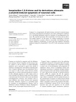
Fig. 3. Fluorescence and electron microscopy of rPrP amyloid fibrils. Amyloid fibrils were produced as described by Bocharova et al. [55] and
(A) stored in Na-acetate buffer, pH 5.5; (B) stored in Na-acetate buffer, pH 5.5, and sonicated for 1 min prior to imaging; and (C) stored in
Tris ⁄ HCl buffer, pH 7.4. All three samples were analyzed in parallel by thioflavine T-fluorescence microscopy (left panels) and by electron
microscopy (right panels). When observed by fluorescence microscopy, the fibrils subjected to 1 min of sonication (B) appeared as small
nonfibrillar oligomers. (A,B) Scale bars ¼ 1 lm; (C) scale bar ¼ 10 lm.
Branched chain mechanism of polymerization I. V. Baskakov
3760 FEBS Journal 274 (2007) 3756–3765 ª 2007 The Author Journal compilation ª 2007 FEBS
fragments, fibrils might form aggregates of various
shapes and densities (Fig. 3). Although fibrillar aggre-
gates or plaques are believed to be less pathogenic,
they might serve as repositories of more pathogenic
small fibrillar fragments and therefore are equally
important. Regardless of the fibril size and shape, the
key feature of fibrils is cross- b-sheet structure, which is
essential for the prion self-propagating activity. More-
over, the cross- b-sheet structure of amyloid fibrils is
substantially more stable kinetically and thermody-
namically than the structure of nonfibrillar oligomeric
species, ensuring that fibrils remain assembled and pre-
serving their physical properties even at low biologi-
cally relevant concentrations of PrP.
Because the infectious form of PrP has been often
referred to as nonfibrillar in nature, it is important to
evaluate the validity of such claims. First, if infectious
prions are indeed nonfibrillar, the question of how
could oligomeric nonfibrillar species be infectious in
the absence of the self-propagating cross-b structure
needs to be answered. Second, the vast majority of
experimental procedures used for extraction and purifi-
cation of PrP
Sc
involved sonication, treatment with
detergents and, sometimes, freezing and thawing
[49–51]. All of these steps cause severe fragmentation
of fibrils. In our experience, sonication for only 1 min
is sufficient to fragment fibrils into small fibrillar frag-
ments that could easily be confused with nonfibrillar
particles (Fig. 3B).
In our search for physical properties that are essen-
tial for prion infectivity it is important to gain infor-
mation about the substructure of PrP fibrils. What
regions of PrP molecule adopt cross-b-sheet conforma-
tion within amyloid fibrils? Can we control the con-
formational stability of cross-b-sheet core?
The large size of PrP molecules in combination with
the highly aggregated, heterogeneous and insoluble
nature of PrP fibrils precluded application of NMR
and other high-resolution techniques. In the absence of
methods to solve structure of PrP fibrils in the near
future, we employed several alternative approaches for
elucidating ultrastructure of fibrils. High resolution
atomic force microscopy revealed that fibrils produced
in vitro from the full-length rPrP consisted of several
laterally assembled filaments [52]. In our recent studies,
we found that the fibrils produced under single growth
conditions varied with respect to the number of consti-
tutive filaments and the manner in which the filaments
were assembled. The high-order fibrils formed through
a highly hierarchical mechanism of lateral assembly.
At each step, filaments were found to associate in pairs
in a pattern resembling dichotomous coalescence
(Fig. 4) [19,52]. Because of alternative modes of lateral
assembly, fibrils produced under a single growth condi-
tion were heterogeneous with respect to the width,
height and twisting morphology.
How many PrP molecules are packed per 1 nm
within an amyloid fibril? As judged from atomic force
microscopy (AFM) measurements and atomic volume
calculations, a single full-length rPrP polypeptide occu-
pied a distance of approximately 1.2 nm within a
single filament (Fig. 5A) [52]. The amyloid fibrils are
Dichotomous mechanism
of lateral assembly
Width (nm)
20 40 60 80
Height, nm
0
5
10
15
AB
Fig. 4. Hierarchical mechanism of lateral
assembly. (A) Electron microscopy image of
an amyloid fibril taken at the intermediate
stage of lateral assembly. Several ‘coales-
cent forks’ (marked by arrows) could be
observed within an individual fibril. Sche-
matic representation of the mechanism of
dichotomous assembly is shown in inset.
Based on data from [19]. (B) Height–width
profiles of fibrils grown under single growth
conditions illustrate polymorphism in fibril
dimension that occurred as a result of the
hierarchical mechanism of lateral assembly.
Based on data from [52].
I. V. Baskakov Branched chain mechanism of polymerization
FEBS Journal 274 (2007) 3756–3765 ª 2007 The Author Journal compilation ª 2007 FEBS 3761
known to be build of b-strands oriented perpendicular
to the fibrillar axis with the distance between two
neighboring b-strands of approximately 4.8 A
˚
. There-
fore, the axial distance occupied by one rPrP molecule
should be equivalent to approximately 2.5 layers of
b-strands. Our studies using PK digestion assay
revealed that the PK resistant core of the amyloid
fibrils consisted of residues 138 ⁄ 141–230, 152 ⁄ 153–230
and 162–230, where the fragment 162–230 was the
most resistant to PK digestion (Fig. 5) [53,54]. Upon
treatment with PK, the 152 ⁄ 153–230 and 162–230
PK-resistant fragments maintained fibrillar structure
and preserved a high b-sheet context with strong inter-
molecular hydrogen bonds. Remarkably, the b-sheet
rich fibrillar cores encompassed by residues 152 ⁄ 153–
230 and 162–230 were found to maintain high seeding
activity in vitro despite cleavage of the N-terminal and
central regions [53,55]. Consistent with these studies,
the rPrP regions 159–174 and 224–230 were observed
to be buried in the fibril interior and were the most
resistant to GdnHCl-induced denaturation as judged
from the newly developed dual color immunofluores-
Fig. 5. (A) Three-dimensional AFM image of amyloid fibril. The fibril consists of several filaments assembled laterally in horizontal and vertical
dimensions as seen by a stepwise increase in fibrillar height. Although atomic volume calculations predicted that single PrP molecule occu-
pies the distance of approximately 1.2 nm (52), the precise 3D structure of PrP within amyloid fibrils has yet to be determined. Despite
changes in the shape of the PrP molecule upon conversion from the native a-helical form (inset) into the fibrillar form, the atomic volume
occupied by a single PrP polypeptide chain does not change substantially. (B) Schematic diagram illustrating mapping of PrP regions within
amyloid fibrils. The PK-resistant b-sheet rich core of amyloid fibrils composed of residues 152–230 and 162–230; PK-cleavage sites are
indicated by red arrows. Based on data from [55]. The epitopes to antibodies AH6 and R1 were solvent unaccessible and were the most
resistant to GdnHCl-induced denaturation (highlighted in red); the epitope to antibody D18 was found to be cryptic under native conditions
and solvent exposed under partially denaturing conditions (highlighted in orange), whereas the epitopes to antibodies D13 and AG4 were
solvent-accessible regardless of the solvent conditions (highlighted in green); based on data from [56]. Residues 98, 127, 144, 196 and 230
(blue) showed cooperative unfolding, whereas unfolding at residue 88 (green) was noncooperative; based on data from [58].
Branched chain mechanism of polymerization I. V. Baskakov
3762 FEBS Journal 274 (2007) 3756–3765 ª 2007 The Author Journal compilation ª 2007 FEBS
cence microscopy assay (Fig. 5) [56]. The 132–156
segment was cryptic under native conditions and
solvent-exposed under partially denaturing conditions,
whereas region 95–105 was solvent-accessible regard-
less of the solvent conditions [56]. In fibrils formed
from truncated rPrP 90–230, the residues 169–230
showed the slowest hydrogen exchange rate confirming
that the C-terminal part is involved in the b-sheet
structure [57]. Site-specific conformational studies
revealed that the C-terminal region accounts for the
high conformational stability of amyloid fibrils [58]. As
judged from the C
1 ⁄ 2
values, the conformational stabil-
ity of the residues within the region 127–230 were
found to be similar to the global stability of the amy-
loid structure, whereas the stability of residue 98 was
substantially lower than the global stability, but
approached that of natively folded proteins [58].
Taken together, the data accumulated to date have
indicated that the C-terminal part of the rPrP molecule
encompassing residues 152–230 and 162–230, and poss-
ibly 169–230, acquires cross-b-sheet self-propagating
core in amyloid fibrils [53,54,56–58]. These regions
account for the high conformational stability and
structural integrity of fibrils. The central regions
encompassing residues 90–150 are likely to be involved
in forming the fibrillar interface that participates in
lateral interactions between filaments within mature
fibrils. Whether the PrP
Sc
infectious particle has a
substructure similar to that of rPrP fibrils generated
in vitro remains to be determined in future studies.
Acknowledgements
I.V.B. is supported by a National Institute of Health
grant, NS045585.
References
1 Prusiner SB (1998) Prions (Les Prix Nobel Lecture). In
Les Prix Nobel (Fra
¨
ngsmyr T, ed.), pp. 268–323. Almq-
vist & Wiksell International, Stockholm.
2 Manuelidis L (2006) A 25 nm virion is the likely cause
of transmnissible spongiform encephalopathies. J Cell
Biochem 100, 897–915.
3 Chesebro B (1998) Prion diseases: BSE and prions:
uncertainties about the agent. Science 279, 42–43.
4 Merz PA, Rohwer RG, Kascsak R, Wisniewski HM,
Somerville RA, Gibbs CJ Jr & Gajdusek DC (1984)
Infection-specific particle from the unconventional slow
virus diseases. Science 225, 437–440.
5 Rohwer RG (1984) Scrapie infectious agent is virus-like
in size and susceptibility to inactivation. Nature 308,
658–662.
6 Safar JG, Kellings K, Serban A, Groth D, Cleaver JE,
Prusiner SB & Riesner D (2005) Search for a prion-
specific nucleic acid. J Virol 79, 10796–10806.
7 Soto C, Estrada L & Castilla J (2006) Amyloids, prions
and the inherent infectious nature of misfolded protein
aggregates. Trends Biochem Sci 31, 150–155.
8 Baskakov IV & Breydo L (2007) Converting the prion
protein: what makes the protein infectious. Biochim
Biophys Acta 1772, 692–703.
9 Chien P, Weissman JS & DePace AH (2004) Emerging
principles of conformation-based prion inheritance.
Annu Rev Biochem 73, 617–656.
10 Harper JD & Lansbury PT Jr (1997) Models of amyloid
seeding in Alzheimer’s disease and scrapie: mechanistic
truths and physiological consequences of the time-
dependent solubility of amyloid proteins. Annu Rev
Biochem 66, 385–407.
11 Cohen FE, Pan K-M, Huang Z, Baldwin M, Fletterick
RJ & Prusiner SB (1994) Structural clues to prion repli-
cation. Science 264, 530–531.
12 Cohen FE & Prusiner SB (1998) Pathologic conforma-
tions of prion proteins. Annu Rev Biochem 67, 793–
819.
13 Aguzzi A & Polymenidou M (2004) Mammalian prion
biology: one century of evolving concepts. Cell 116,
313–327.
14 Lansbury PT & Caughey B (1995) The chemistry of
scrapie infection: implications of the ‘ice 9’ metaphor.
Curr Biol 2, 1–5.
15 Baskakov IV (2004) Autocatalytic conversion of recom-
binant prion proteins displays a species barrier. J Biol
Chem 279, 586–595.
16 Baskakov IV & Bocharova OV (2005) In vitro conver-
sion of mammalian prion protein into amyloid fibrils
displays unusual features. Biochemistry 44, 2339–2348.
17 Semenoff NN (1929) Chem Rev 6, 347–379.
18 Semenov NN (1957) The Nobel Prize Lecture: einige
probleme der kettenreaktionen und der verbrennungs-
theorie. Angew Chem 69, 767–777.
19 Makarava N, Bocharova OV, Salnikov VV, Breydo L,
Anderson M & Baskakov IV (2006) Dichotomous
versus palm-type mechanisms of lateral assembly of
amyloid fibrils. Protein Sci
15, 1334–1341.
20 Bolton DC, Seligman SJ, Bablanian G, Windsor D,
Scala LJ, Kim KS, Chen CJ, Kascsak RJ & Bendheim
PE (1991) Molecular location of a species-specific epi-
tope on the hamster scrapie agent protein. J Virol 65,
3667–3675.
21 Bueler H, Aguzzi A, Sailer A, Greiner RA, Autenried
P, Aguet M & Weissmann C (1993) Mice devoid of PrP
are resistant to scrapie. Cell 73, 1339–1347.
22 Peretz D, Williamson RA, Kaneko K, Vergara J, Lecl-
erc E, Schmitt-Ulms G, Mehlhorn IR, Legname G,
Wormald MR et al. (2001) Antibodies inhibit prion
I. V. Baskakov Branched chain mechanism of polymerization
FEBS Journal 274 (2007) 3756–3765 ª 2007 The Author Journal compilation ª 2007 FEBS 3763
propagation and clear cell cultures of prion infectivity.
Nature 412, 739–743.
23 Enari M, Flechsig E & Weissmann C (2001) Scrapie
prion protein accuulation by scrapie-infected neuro-
blastoma cells abrogated by exosure to a prion protein
antibody. Proc Acad Natl Sci USA 98, 9295–9299.
24 Bessen RA, Kocisko DA, Raymond GJ, Nandan S,
Lansbury PT & Caughey B (1995) Non-genetic propa-
gation of strain-specific properties of scrapie prion pro-
tein. Nature 375, 698–700.
25 Caughey B, Kocisko DA, Raymond GJ & Lansbury PT
Jr (1995) Aggregates of scrapie-associated prion protein
induce the cell-free conversion of protease-sensitive
prion protein to the protease-resistant state. Chem Biol
2, 807–817.
26 Saborio GP, Permanne B & Soto C (2001) Sensitive
detection of pathological prion protein by cyclic amplifi-
cation of protein misfolding. Nature 411, 810–813.
27 Castilla J, Saa P, Hetz C & Soto C (2005) In vitro gen-
eration of infectious scrapie prions. Cell 121, 195–206.
28 Weber P, Giese A, Piening N, Mitteregger G, Thomzig
A, Beekes M & Kretzschmar HA (2006) Cell-free for-
mation of misfolded prion protein with authentic prion
infectivity. Proc Acad Natl Sci USA 103, 15823.
29 Deleault NR, Lucassen RW & Supattapone S (2003)
RNA molecules stimulate prion protein conversion.
Nature 425, 717–720.
30 Lucassen R, Nishina K & Supattapone S (2003) In vitro
amplification of protease-resistant prion protein requires
free sulfhydryl groups. Biochemistry 42, 4127–4135.
31 Peretz D, Scott M, Groth D, Williamson A, Burton D,
Cohen FE & Prusiner SB (2001) Strain-specified relative
conformational stability of the scrapie prion protein.
Protein Sci 10, 854–863.
32 Kuczius T & Groschup MH (1999) Differences in prote-
inase K resistance and neuronal deposition of abnormal
prion proteins characterize bovine spongioform
encephalopathy (BSE) and scrapie strains. Mol Med 5,
406–418.
33 Luhr KM, Nordstromm EK, Low P, Ljunggren HG,
Taraboulos A & Kristensson K (2004) Scrapie protein
degradation by cysteine protease in CD11c+ dendritic
cells and GT1-1 neuronal cells. J Virol 78,
4776–4782.
34 Yadavalli R, Guttmann RP, Seward T, Centers AP,
Williamson RA & Telling GC (2004) Calpain-dependent
endoproteolytic cleavage of PrPSc modulates scrapie
prion. J Biol Chem 279, 21948–21956.
35 Wong C, Xiong LW, Horiuchi M, Raymond L, Wehrly
K, Chesebro B & Caughey B (2001) Sulfated glycans
and elevated temperature stimulate PrP(Sc)-dependent
cell-free formation of protease-resistant prion protein.
EMBO J 20, 377–386.
36 Shaked GM, Meiner Z, Avraham I, Taraboulos A &
Gabizon R (2001) Reconstitution of prion infectivity
from solubolozed protease-resistant PrP and nonprotein
components of prion rods. J Biol Chem 276, 14324–
14328.
37 Ben-Zaken O, Tzaban S, Tal Y, Horonchik. L, Esko
JD, Vlodavsky I & Taraboulos A (2003) Cellular hepa-
ran sulfate participates in the metabolism of prions.
J Biol Chem 41, 40041–40049.
38 Dumpitak C, Beekes M, Weinmann N, Metzger S,
Winklhofer KF, Tatzelt J & Riesner D (2006) The poly-
saccharide scaffold of PrP 27–30 is a common com-
pound of natural prions and consists of a-linked
polyglucose. Biol Chem 386, 1149–1155.
39 Bernouilli C, Siegfried J, Baumgartner G, Regli F,
Rabinowicz T, Gajdusek DC & Gibbs CJ Jr (1977)
Danger of accidental person to person transmission of
Creutzfeldt–Jakob disease by surgery. Lancet 1, 478–
479.
40 Gibbs CJ Jr, Asher DM, Kobrine A, Amyx HL, Sulima
MP & Gajdusek DC (1994) Transmission of Creutz-
feldt–Jakob disease to a chimpanzee by electrodes con-
taminated during neurosurgery. J Neurol Neurosurg
Psychiatry 57, 757–758.
41 Zobeley E, Flechsig E, Cozzio A, Enari M & Weiss-
mann C (1999) Infectivity of scrapie prions bound to a
stainless steel surface. Mol Med 5, 240–243.
42 Weissmann C, Enari M, Klohn PC, Rossi D & Flechsig
E (2002) Transmission of prions. J Infect Dis 186 (Sup-
pl. 2), S157–S165.
43 Shorter J & Lindquist S (2004) Hsp104 catalyzes forma-
tion and elimination of self-replicating Sup35 prion con-
formers. Science 304, 1793–1797.
44 Tanaka M, Chien P, Naber N, Cooke R & Weissman
JS (2004) Conformational variations in an infectious
protein determine prion strain differences. Nature 6980,
323–328.
45 Legname G, Nguyen H-OB, Baskakov IV, Cohen FE,
DeArmond SJ & Prusiner SB (2005) Strain-specified
characteristics of mouse synthetic prions. Proc Natl
Acad Sci USA 102, 2168–2173.
46 Legname G, Nguyen H-OB, Peretz D, Cohen FE,
DeArmond SJ & Prusiner SB (2006) Continuum of pri-
on protein structures enciphers a multitude of prion iso-
late-specified phenotypes. Proc Acad Natl Sci USA 103,
19105–19110.
47 Caughey B & Lansbury PT (2003) Protofibrils, pores,
fibrils, and neurodegeneration: separating the respon-
sible protein aggregates from the innocent bystanders.
Annu Rev Neurosci 26, 267–298.
48 Chiesa R & Harris DA (2001) Prion diseases: what is
the neurotoxic molecule? Neurobiol Dis 8, 743–763.
49 Silveira JR, Raymond GJ, Hughson A, Race RE, Sim
VL, Hayes SF & Caughey B (2005) The most infectious
prion protein particles. Nature 437, 257–261.
50 Liberski PP, Brown P, Xiao S-Y & Gajdusek DC (1991)
The ultrastructural diversity of scrapie-associated fibrils
Branched chain mechanism of polymerization I. V. Baskakov
3764 FEBS Journal 274 (2007) 3756–3765 ª 2007 The Author Journal compilation ª 2007 FEBS
isolated from experimental scrapie and Creutzfeldt–
Jakob disease. J Comp Pathol 105, 377–386.
51 Kascsak RJ, Rubenstein R, Merz PA, Carp RI, Wis-
niewski HM & Diringer H (1985) Biochemical differ-
ences among scrapie-associated fibrils support the
biological diversity of scrapie agents. J Gen Virol 66,
1715–1722.
52 Anderson M, Bocharova OV, Makarava N, Breydo L,
Salnikov VV & Baskakov IV (2006) Polymorphysm and
ultrastructural organization of prion protein amyloid
fibrils: an insight from high resolution atomic force
microscopy. J Mol Biol 358, 580–596.
53 Bocharova OV, Breydo L, Salnikov VV, Gill AC &
Baskakov IV (2005) Synthetic prions generated in vitro
are similar to a newly identified subpopulation of PrPSc
from sporadic Creutzfeldt–Jakob Disease PrP
Sc
. Protein
Sci 14, 1222–1232.
54 Bocharova OV, Makarava N, Breydo L, Anderson M,
Salnikov VV & Baskakov IV (2006) Annealing PrP
amyloid firbils at high temperature results in extension
of a proteinase K resistant core. J Biol Chem 281, 2373–
2379.
55 Bocharova OV, Breydo L, Parfenov AS, Salnikov VV &
Baskakov IV (2005) In vitro conversion of full length
mammalian prion protein produces amyloid form with
physical property of PrPSc. J Mol Biol 346, 645–659.
56 Novitskaya V, Makarava N, Bellon A, Bocharova OV,
Bronstein IB, Williamson RA & Baskakov IV (2006)
Probing the conformation of the prion protein within a
single amyloid fibril using a novel immunoconforma-
tional assay. J Biol Chem 281, 15536–15545.
57 Lu X, Wintrode PL & Surewicz WK (2007) Beta-sheet
core of human prion protein amyloid fibrils as deter-
mined by hydrogen ⁄ deuterium exchange. Proc Acad
Natl Sci USA 104, 1510–1515.
58 Sun Y, Breydo L, Makarava N, Yang Q, Bocharova
OV & Baskakov IV (2007) Site-specific conformational
studies of PrP amyloid fibrils revealed two cooperative
folding domain within amyloid structure. J Biol Chem
282, 9090–9097.
I. V. Baskakov Branched chain mechanism of polymerization
FEBS Journal 274 (2007) 3756–3765 ª 2007 The Author Journal compilation ª 2007 FEBS 3765