Báo cáo khoa học: The modulation of metal bio-availability as a therapeutic strategy for the treatment of Alzheimer’s disease pptx
Bạn đang xem bản rút gọn của tài liệu. Xem và tải ngay bản đầy đủ của tài liệu tại đây (137.78 KB, 9 trang )
MINIREVIEW
The modulation of metal bio-availability as a therapeutic
strategy for the treatment of Alzheimer’s disease
Peter J. Crouch
1
, Anthony R. White
1
and Ashley I. Bush
2,3
1 Department of Pathology and Centre for Neuroscience, The University of Melbourne, Australia
2 The Mental Health Research Institute of Victoria, Parkville, Australia
3 Department of Psychiatry, Massachusetts General Hospital, Charlestown, MA, USA
Introduction
Alzheimer’s disease (AD) destroys the mental health of
millions of people worldwide. Sufferers lose their inde-
pendence and require dedicated on-going care, usually
from members of their own family and at enormous
economic and social cost. There is no cure for AD,
and the development of effective therapeutic strategies
is hampered by a paucity of information on the biolo-
gical mechanisms underlying the disease. The severity
of AD relates to a multitude of age-related cellular
processes culminating in neuronal and synaptic dys-
function. Successful therapeutic strategies that target
the causative neuropathological events will have an
enormous impact on treatment of AD patients.
Four consistent features characterize the AD brain:
(a) the presence of extracellular amyloid plaques com-
prised mainly of aggregated, insoluble amyloid-b (Ab)
peptide; (b) the presence of intracellular neurofibrillary
tangles (NFTs) containing hyperphosphorylated tau;
(c) increased oxidative damage to lipids, proteins and
nucleic acids; and (d) a loss of biometal homeostasis.
The deposition of aggregated Ab and the hyperphos-
porylation of tau have been shown to cause neuronal
damage and contribute substantially to the pathology
of AD [1]. Therefore, both have received considerable
Keywords
Alzheimer’s disease; amyloid-b; biometals;
copper; iron; oxidative stress; tau; zinc
Correspondence
A. I. Bush, The Mental Health Research
Institute of Victoria, 155 Oak Street,
Parkville, Victoria 3052, Australia
Fax: +61 39387 5061
Tel: +61 39389 2962
E-mail:
(Received 9 March 2007, revised 17 May
2007, accepted 18 May 2007)
doi:10.1111/j.1742-4658.2007.05918.x
The postmortem Alzheimer’s disease brain is characterized histochemically
by the presence of extracellular amyloid plaques and neurofibrillary tangles.
Also consistent with the disease is evidence for chronic oxidative damage
within the brain. Considerable research data indicates that these three crit-
ical aspects of Alzheimer’s disease are interdependent, raising the possibility
that they share some commonality with respect to the ever elusive initial
factor(s) that triggers the development of Alzheimer’s disease. Here, we
discuss reports that show a loss of metal homeostasis is also an important
event in Alzheimer’s disease, and we identify how metal dyshomeostasis may
contribute to development of the amyloid-b, tau and oxidative stress biology
of Alzheimer’s disease. We propose that therapeutic agents designed to
modulate metal bio-availability have the potential to ameliorate several of
the dysfunctional events characteristic of Alzheimer’s disease. Metal-based
therapeutics have already provided promising results for the treatment of
Alzheimer’s disease, and new generations of pharmaceuticals are being
developed. In this review, we focus on copper dyshomeostasis in Alzheimer’s
disease, but we also discuss zinc and iron.
Abbreviations
AD, Alzheimer’s disease; Ab, amyloid-b; APP, amyloid-b A4 precursor protein; CHO, Chinese Hamster Ovary; Cp, ceruloplasmin; CQ,
clioquinol; CNS, central nervous system; COX, cytochrome c oxidase; CSF, cerebrospinal fluid; Cu ⁄ Zn-SOD, copper ⁄ zinc superoxide
dismutase; GSK3, glycogen synthase kinase-3; MMP, matrix metalloproteinase; NFT, neurofibrillary tangles; NMDA, N-methyl-
D-aspartate;
PDTC, pyrrolidine dithiocarbamate; ROS, reactive oxygen species; SOD, superoxide dismutase; Tg, transgenic; ZnT3, Zn transporter-3.
FEBS Journal 274 (2007) 3775–3783 ª 2007 The Authors Journal compilation ª 2007 FEBS 3775
research attention as potential therapeutic targets. Pla-
ques and NFTs, however, cannot be regarded as ‘up-
stream’ causative factors in the development of AD.
While aggregated, insoluble Ab found within plaques
can cause neurotoxicity, soluble intermediate Ab oligo-
mers are substantially more toxic [2]. Similarly, NFTs
can also contribute to neurodegeneration, but their
development is the result of an aberrant shift in activ-
ity of tau kinases and phosphatases [3]. Thus, although
targeting plaques and NFTs may ameliorate some of
the consequences of AD and no doubt lessen the bur-
den of the disease, the biological mechanisms that
caused them to develop will remain unchecked. The
fundamental research aim in AD research is to identify
the up-stream biological mechanisms that trigger the
development of AD. Identifying these mechanisms will
substantially facilitate the development of more effect-
ive therapeutic strategies.
Metal dyshomeostasis and AD
Transition metals such as Cu and Fe are essential for
normal cell functionality. Due to their capacity to
move between transition states, they are most abun-
dant within redox enzymes such as Cu ⁄ Zn-superoxide
dismutase (Cu ⁄ Zn-SOD), tyrosinase, and cytochrome c
oxidase (COX). In these enzymes, the redox potential
of the metals is harnessed to provide the enzymes with
their electron transfer capabilities. Paradoxically, it is
the same redox potential of the metals that makes
them potentially toxic to the cell. Under conditions
where they are allowed to accumulate freely, redox act-
ive metals contribute directly to cellular oxidative dam-
age by generating the highly reactive and toxic OH
•
via Fenton and Haber–Weiss reactions [4]. Cells have
therefore developed sophisticated regulation and trans-
fer systems to ensure tight control of metals within the
cell [5,6]. The toxic effects of Fe and Cu and their role
in numerous neurodegenerative and age-related dis-
eases have been reviewed recently [7].
Equally detrimental to the cell is metal deficiency.
Deficient metal bio-availability causes decreased activ-
ity of critical enzymes because activity of the enzymes
is dependent on optimal metal loading. Cu deficiency
is central to the recessive Menkes and Wilson diseases,
and has been implicated in AD. Just as excess Cu can
contribute to oxidative damage by catalysing the pro-
duction of OH
•
, so too can deficient Cu by preventing
normal activity of cuproenzymes important in main-
taining cellular oxidative homeostasis.
The cortical glutamatergic synapse, where amyloid
pathology first commences in AD, contains exception-
ally high concentrations of Zn and Cu, which are
released during neurotransmission. Zn
2+
is released
together with glutamate from presynaptic terminals to
achieve concentrations in the order of 300 lm [8]. The
Zn
2+
is concentrated into glutamate vesicles by Zn
transporter-3 (ZnT3), which is only expressed in gluta-
matergic neurons [9]. Ionic Cu is released into the cleft
following postsynaptic stimulation of the N-methyl-
d-aspartate (NMDA) receptor [10,11] and is concen-
trated into postsynaptic vesicles by the Menkes
Cu7aATPase [11].
Cu dyshomeostasis is evident within the brain during
normal aging [12–15], but is substantially more pro-
nounced within the aged AD-affected brain [16,17].
Lovell et al. [17] demonstrated that Cu levels in the
unaffected (i.e. plaque-free) neuropil of the AD brain
are approximately 400% higher than in the neuropil of
the healthy brain, and that within the AD brain itself
Cu levels are approximately 30% higher within the
amyloid plaques compared to plaque-free regions.
These data indicate that an accumulation of Cu within
the brain, be it a cause or consequence, is consistent
with the development of AD. Despite these gross
increases in cerebral extracellular Cu, intracellular Cu
appears to be deficient in the AD brain [18]. The cupro-
enzymes COX and peptidylglycine a-amidating mono-
oxygenase have significantly decreased activities in AD
brain and cerebrospinal fluid (CSF), respectively
[19,20], and deficiencies in COX activity may be
responsible for the deficit in energy metabolism charac-
teristic of AD brain [21]. There are conflicting reports
in the literature about levels of the cuproprotein cerulo-
plasmin (Cp) in AD brain [22,23], but Cp activity has
been reported to be decreased in plasma [24], despite
being elevated in CSF [25] and in plasma [26]. Further-
more, the antioxidant Cu ⁄ Zn-SOD shows decreased
activity in both AD brain and transgenic (Tg) animal
models of AD despite increased protein expression [27].
The loss of Cu ⁄ Zn-SOD activity likely results from a
deficiency in active site Cu because activity can be
restored by dietary Cu supplementation [27].
Homeostasis of the transition metal Fe is also altered
in the AD brain. It accumulates within extracellular
amyloid plaques [17,28] and localizes within NFTs [29].
Like Cu, Fe is essential within the cell because of its
redox potential, and Fe dyshomeostasis can similarly
contribute to cellular dysfunction when in excess (by
catalysing OH
•
production) and when deficient
(decreased enzyme activity). Of particular relevance to
Fe dyshomeostasis in AD is Cp. As described above, Cp
is a cuproprotein, but its functional role within the cell
is to detoxify and remove excess Fe. Decreased Cp levels
in the brain [22], possibly due to decreased Cu homeo-
stasis, may contribute to Fe accumulation in AD [30]
Modulation of metal availability for treating AD P. J. Crouch et al.
3776 FEBS Journal 274 (2007) 3775–3783 ª 2007 The Authors Journal compilation ª 2007 FEBS
because Jeong and David [31] have recently shown that
Cp deficiency leads to increased Fe levels in the CNS.
By contrast to Cu and Fe, Zn is redox-silent, and
therefore does not contribute directly to redox reac-
tions. Its role within the brain, however, is essential
nonetheless. It is required for the activity of enzymes
such as Cu ⁄ Zn-SOD [32] and matrix metalloproteinas-
es [33] where it is required in a structural role rather
than a redox-active role. Perhaps the most critical role
for Zn within the brain is in neurotransmission across
the glutamatergic synapse [34]. Within the synaptic
cleft, Zn concentrations can reach approximately
300 lm [35] where it is believed to function as a coun-
ter ion for the high concentrations of glutamate
present and quenches the response of the NMDA
receptor [36]. Like Cu and Fe, considerable data indi-
cates a loss of Zn homeostasis in AD. Abnormally
high concentrations of Zn are associated with amyloid
plaques in AD brain [17,37,38] and AD Tg mice [39].
In a pertinent study performed in vivo, Lee et al. [40]
crossed AD Tg mice (Tg2576) with mice deficient in
ZnT3, the protein responsible for loading Zn into syn-
aptic vesicles for release into the synaptic cleft. These
mice exhibited a 50% decrease in amyloid plaque bur-
den compared to Tg2576 littermates, indicating that
the pool of Zn essential for glutamatergic neurotrans-
mission may contribute to plaque formation. The syn-
aptic Zn has recently been demonstrated to be in
communication with the plasma, and contributes to
amyloid congophilic angiopathy, which is abolished in
Tg2576 mice where the gene for ZnT3 is ablated [41].
Amyloid-b
After the 4.5 kDa Ab peptide was identified as a
major component of the amyloid plaques in AD brain
[42,43], global AD research focused on this peptide as
a causative agent in the disease. The 39–43 amino acid
cleavage product of the Ab A4 precursor protein
(APP) is initially present as a soluble, unaggregated
species, and it is only through the processes of olig-
omerization, aggregation and fibrilization that Ab
forms amyloid plaques. As amyloid plaques are prom-
inent in the postmortem AD brain, early research the-
ories placed the accumulation of extracellular,
insoluble forms of Ab as central to the disease process
[44]. However, as studies emerged reporting that sol-
uble, intermediate Ab oligomers were more toxic than
fibrillar Ab [2,45–47], it became evident that, although
amyloid plaques no doubt contribute to neuronal dys-
function, the occurrence of plaques may be several
steps downstream from the more critical causes of
AD.
Ab readily binds Cu and Zn via its three N-terminal
histidine residues [48–51], and several compelling stud-
ies have shown that the interaction between Ab and
these metals can promote the formation of Ab oligo-
mers, aggregates and fibrils [48,52–55]. Metal mediated
oligomerization of Ab may therefore contribute to the
potent inhibition of synaptic transmission mediated by
Ab. Several studies have now shown that Ab mediated
inhibition of synaptic transmission is dependent on the
presence of Ab oligomers, and that Ab monomers are
relatively nontoxic in these assays [56,57]. As described
above, synaptically released Zn is required for the for-
mation of amyloid plaques in Tg mice [41]. Although
amyloid plaques contain predominantly higher order
Ab aggregates, an initial formation of toxic Ab oligo-
mers within the synaptic cleft may be determined by
Zn released from the presynaptic terminus. Similarly,
Cu released into the synaptic cleft following activation
of postsynaptic NMDA receptors [10,11] may also
facilitate extracellular Ab oligomerization. Shankar
et al. [58] have reported that the loss of hippocampal
synapses in rat organotypic slices is mediated by Ab
oligomers and is dependent on the activity of NMDA-
type glutamate receptors.
The concentrations of metal required to induce Ab
oligomerization and aggregation are relatively low, and
well within the physiological ranges that could be
expected within the brain. The capacity for metals to
facilitate this process may therefore be a critical factor
in the Ab mediated pathology of the AD brain. Subse-
quent to an early report demonstrating that the
Cu- and Zn-induced aggregation of Ab could be pre-
vented by EDTA [53], disrupting Ab–metal interac-
tions has been an attractive therapeutic target. In this
regard, a salient study demonstrated that the amyloid
plaque burden in brains decreased by 49% when
Tg2576 mice were treated orally with the 8-hydroxy-
quinoline derivative clioquinol (CQ) [59]. CQ is a
moderate metal chelator capable of crossing the
blood–brain barrier, and it was believed that CQ solu-
bilized the Ab plaques by stripping them of their metal
content. This supported the in vitro work previously
reported [53], and was consistent with the notion that
excess extracellular metals contribute to the amyloid
pathology of AD.
Relative to the soluble Ab burden of the AD brain,
a recent study described a mechanism by which
decreased intracellular metal bioavailability may con-
tribute to the accumulation of soluble Ab outside the
cell. White et al. [60] treated Chinese Hamster Ovary
cells over-expressing human APP (CHO-APP) with
CQ complexed to Cu or Zn, and found that the
metal-CQ treatment substantially decreased the levels
P. J. Crouch et al. Modulation of metal availability for treating AD
FEBS Journal 274 (2007) 3775–3783 ª 2007 The Authors Journal compilation ª 2007 FEBS 3777
of soluble Ab present in the cell culture medium.
Metal-CQ treatment decreased the levels of extracellu-
lar Ab not by preventing an Ab–metal interaction
outside the cell, but by facilitating the delivery of
metals into the cell. White et al. [60] demonstrated
that CQ facilitated the delivery of Cu and Zn across
the plasma membrane, as determined by inductively
coupled plasma mass spectrometry. Once inside the
cell, Cu and Zn, but not Fe, activated phosphoinosi-
tol 3-kinase mediated protein kinase pathways, which
ultimately led to an increase in the secretion of matrix
metalloproteinases (MMPs). The capacity for MMPs
to degrade Ab has been reported by several groups
[61–64]. Treatment with CQ may therefore have a
two-fold effect on Ab; by binding extracellular metals,
it prevents metal mediated Ab aggregation and toxicity
and, by then delivering the bound metals into the cell,
it activates specific protein kinases that induce an
increase in the production of Ab-degrading MMPs.
Neurotoxicity generated by the interaction between
Ab and metals may be more complex than the cata-
lysis of Ab aggregation. Numerous studies have now
shown that several potential mechanisms of neurotox-
icity for soluble Ab are exacerbated by, if not depend-
ent on, the presence of metals. This indicates that
Ab–metal interactions, possibly occurring within the
cell, may induce mechanisms of neurotoxicity that
involve soluble Ab oligomers, and that the mechanisms
of toxicity precede Ab aggregation and accumulation.
Curtain et al. [49,65] demonstrated that the capacity
for Ab to bind Zn and Cu determined its ability to
penetrate and disrupt membranes; Crouch et al .
demonstrated that Ab-mediated inhibition of cyto-
chrome c oxidase requires the presence of at least
equimolar concentrations of Cu [66], and that the
inhibition was not supported by Zn or Fe [67]; and
Huang et al. [68] demonstrated that the potential for
Ab to generate neurotoxic H
2
O
2
is dependent on the
presence of Cu. These metal-mediated toxic effects of
Ab were abolished by preventing the Ab–metal interac-
tion with chelators such as EDTA, raising the possibil-
ity that therapeutics designed to disrupt Ab–metal
interactions may prevent more than Ab aggregation
and plaque formation.
Tau
The native function of the microtubule-associated pro-
tein tau is to maintain integrity of the cytoskeleton by
promoting assembly and stability of microtubules. Tau
isolated from a healthy brain is partially phosphorylat-
ed [69,70], indicating that the normal function of tau
requires some phosphorylation. However, in AD, tau
is hyperphosphorylated, and hyperphosphorylated tau
is the form that aggregates in NFTs [71]. The loss of
functional tau from the microtubule network can be
compensated for by the other microtubule-associated
proteins, MAP1A ⁄ MAP1B and MAP2. It is the toxic
gain of function exhibited by hyperphosphorylated tau
that renders it most harmful towards the cell. Hyper-
phosphorylated tau is capable of sequestering normal
tau as well as MAP1A ⁄ MAP1B and MAP2 [72,73],
and this compounding loss of essential proteins desta-
bilizes the microtubule network, contributing to neuro-
fibrillary degeneration.
Tau hyperphosphorylation occurs because of an
imbalance in the activity of tau kinases and phospha-
tases [3]. One particular tau kinase pertinent to metal
dyshomeostasis in AD is glycogen synthase kinase-3
(GSK3). GSK3 has recently been implicated as a crit-
ical kinase involved in the hyperphosphorylation of
tau [74]. Only active (nonphosphorylated) GSK3 con-
tributes to tau hyperphosphorylation, and Plattner
et al. [74] demonstrated that the negative regulation of
GSK3 (i.e. its phosphorylation) is lost in aged, but not
young, Tg p25 mice. This loss of regulation resulted in
an increase in GSK3 activity and tau hyperphosphory-
lation. Whether the change in GSK3 regulation in
these mice occurred in response to an age-related
decline in intracellular metals was not examined, but
the study by White et al. [60], described above, provi-
ded evidence for a possible connection. When the
bio-availability of intracellular Cu and Zn was
increased in CHO-APP cells by treating with CuCQ or
ZnCQ complexes, a downstream target of the activated
protein kinase pathways was GSK3. By contributing
to an increase in GSK3 phosphorylation, this metal
mediated effect therefore decreased potential phos-
phorylation of tau by GSK3. The study of White et al.
[60] did not present data on tau phosphorylation, but
the possibility that metal mediated modulation of
GSK3 represents a strong candidate therapeutic target
for preventing tau hyperhosphorylation has been
strengthened by a recent study. Malm et al. [75]
treated AD Tg mice with the Cu ligand pyrrolidine
dithiocarbamate (PDTC) and reported that the treat-
ment increased brain Cu levels and activated the same
protein kinases previously reported by White et al.
[60]. Treatment with PDTC led to an increase in
GSK3 phosphorylation and a substantial decrease in
tau phosphorylation [75]. Therapeutic modulation of
metal bio-availability, such as that described by White
et al. [60] and Malm et al. [75], may therefore repre-
sent a potential therapeutic strategy for preventing
the tau hyperphosphorylation and NFT formation
characteristic of AD.
Modulation of metal availability for treating AD P. J. Crouch et al.
3778 FEBS Journal 274 (2007) 3775–3783 ª 2007 The Authors Journal compilation ª 2007 FEBS
Oxidative stress in AD
A significant, consistent feature of AD is that the
affected brain is under chronic oxidative stress. An
early report in 1986 [76] described an increase in activ-
ity of enzymes from the hexose monophosphate path-
ways in postmortem AD brain samples compared to
age-matched controls, and proposed that this reflected
increased oxidative stress in the AD brain. Numerous
reports have since provided direct data to show exten-
sive oxidative damage in the AD brain [77]. Oxidative-
ly damaged lipids, proteins and nucleic acids have all
been reported [78–80].
A critical factor in oxidative stress within the AD
brain is intracellular Cu. Insufficient intracellular Cu
can contribute to an increase in oxidative stress (des-
cribed above), and several lines of evidence indicate
that intracellular Cu deficiency in AD may involve Ab
and APP. Ab and its precursor APP both bind Cu,
and over-expression of a C-terminal fragment of APP
or full length APP, both containing the Ab domain,
results in an overall decrease in Cu within the brain of
Tg mice [81]. Conversely, APP knockout mice show a
40% increase in Cu levels within the cerebral cortex
[82]. Furthermore, APP gene expression is down-regu-
lated by decreased availability of intracellular Cu [83]
and up-regulated by increased availability of Cu [84].
Collectively, these data present a strong case for
the native role of APP ⁄ Ab in regulating intracellular
Cu: Cu alters APP gene expression [83,84], and the
APP ⁄ Ab produced binds then transports Cu out of the
cell. However, once intracellular Cu levels become too
low, possibly because of the aberrant increase in Ab
production consistent with AD, the antioxidant capa-
city of the cell may be compromised, leading to an
increase in oxidative stress. The study by Busciglio
et al. [85] in this regard is of particular relevance.
These authors demonstrated that an increase in oxida-
tive stress alters APP processing and generates an
increase in Ab production. If an oxidative stress-
induced increase in Ab production promotes excess Cu
transport out of the cell, further oxidative stress due to
deficient cellular Cu may be created, therefore creating
a vicious cycle. Support for this possibility was presen-
ted in a recent review [86].
In addition to promoting oligomerization and aggre-
gation, interactions between Cu and Ab result in free
radical generation in vitro . Synthetic Ab reduces Cu(II)
to Cu(I) with subsequent reduction of O
2
giving rise to
H
2
O
2
[14]. H
2
O
2
is itself toxic and can diffuse through
the cell membrane to oxidize lipids and intracellular pro-
teins. However, greater oxidative damage is induced
when H
2
O
2
interacts with Ab-bound Cu(I) resulting in
OH
•
[15]. OH
•
reacts with lipids, proteins and nucleic
acids, resulting in extensive modifications that are often
irreversible and impede normal cellular turnover of
these components. Furthermore, OH
•
interaction with
Ab itself can increase Ab aggregation through the
di-tyrosine-mediated cross-linking of Ab peptides
[16–18]. This is consistent with the high di-tyrosine con-
tent observed in AD brain tissue [17].
Oxidative stress within the AD brain is also closely
related to tau hyperphosphorylation. For example,
Gomez-Ramos et al. [87] demonstrated that the pres-
ence of acrolein, a peroxidation product from arachi-
donic acid, induces considerable tau phosphorylation
and, in a subsequent review article, this group proposed
that tau hyperphosphorylation and the formation of
NFTs may even represent a normal, protective cellular
response to increased oxidative stress [88]. Further-
more, protein kinase signalling pathways sensitive to
oxidative stress and known to be altered in AD have
been implicated in the phosphorylation of tau [89].
Such data indicate that an increase in cellular oxidative
stress, be it through the generation of products of oxi-
dative damage or the activation of specific cell signal-
ling pathways, leads to tau hyperphosphorylation and
NFT formation.
Summary
Oxidative stress, tau hyperphosphorylation and the Ab
biology of AD are all intricately linked, and consider-
able research data now exist to indicate that they inter-
act in a series of dysfunctional mechanisms that can
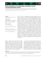
ultimately lead to cognitive decline. The early event(s)
that initiates this neurodegenerative cycle has not been
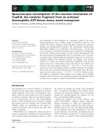
Fig. 1. Potential relationship between decreased intracellular metal
bio-availability and the oxidative stress, tau hyperphosphorylation
and extracellular Ab accumulation characteristic of AD.
P. J. Crouch et al. Modulation of metal availability for treating AD
FEBS Journal 274 (2007) 3775–3783 ª 2007 The Authors Journal compilation ª 2007 FEBS 3779
established, but the role for metal dyshomeostasis in
all aspects is clear. In the AD affected brain, metal
dyshomeostasis is evident in the form of a substantial
increase in the levels of extracellular metals and a
decrease in the levels of intracellular metals. Here, we
have presented evidence to show that decreased metal
bio-availability within the cell is consistent with
increased oxidative stress, a loss of regulation of Ab
production, and an increase in tau hyperphosphoryla-
tion. Furthermore, an increase in extracellular metals
can catalyse Ab oligomerization and aggregation, and
the amyloid plaques that subsequently form may then
exacerbate intracellular metal deficiency by sequester-
ing metals outside the cell. Figure 1 summarizes these
interdependent dysfunctional events. With the loss of
biometal homeostasis placed central to all of these
AD-related neurodegenerative mechanisms, the modu-
lation of metal bio-availability has strong potential in
the therapeutic treatment of AD.
References
1 Goedert M & Spillantini MG (2006) A century of
Alzheimer’s disease. Science 314, 777–781.
2 Dahlgren KN, Manelli AM, Stine WBJ, Baker LK,
Krafft GA & LaDu MJ (2002) Oligomeric and fibrillar
species of amyloid-b peptides differentially affect neur-
onal viability. J Biol Chem 277, 32046–32053.
3 Iqbal K, Alonso Adel C, Chen S, Chohan MO, El-
Akkad E, Gong CX, Khatoon S, Li B, Liu F, Rahman
A et al. (2005) Tau pathology in Alzheimer disease and
other tauopathies. Biochim Biophys Acta 1739, 198–210.
4 Barnham KJ, Masters CL & Bush AI (2004) Neurode-
generative diseases and oxidative stress. Nat Rev Drug
Discov 3, 205–214.
5 Arredondo M & Nunez MT (2005) Iron and copper
metabolism. Mol Aspects Med 26, 313–327.
6 Prohaska JR & Gybina AA (2004) Intracellular copper
transport in mammals. J Nutr 134 , 1003–1006.
7 Brewer GJ (2007) Iron and copper toxicity in diseases
of aging, particularly atherosclerosis and Alzheimer’s
disease. Exp Biol Med (Maywood) 232, 323–335.
8 Danscher G & Stoltenberg M (2005) Zinc-specific auto-
metallographic in vivo selenium methods: tracing of
zinc-enriched (ZEN) terminals, ZEN pathways, and
pools of zinc ions in a multitude of other ZEN cells.
J Histochem Cytochem 53 , 141–153.
9 Palmiter RD, Cole TB, Quaife CJ & Findley SD (1996)
ZnT-3, a putative transporter of zinc into synaptic vesi-
cles. Proc Natl Acad Sci USA 93, 14934–14939.
10 Schlief ML, Craig AM & Gitlin JD (2005) NMDA
receptor activation mediates copper homeostasis in hip-
pocampal neurons. J Neurosci 25, 239–246.
11 Schlief ML, West T, Craig AM, Holtzman DM & Git-
lin JD (2006) Role of the Menkes copper-transporting
ATPase in NMDA receptor-mediated neuronal toxicity.
Proc Natl Acad Sci USA 103, 14919–14924.
12 Massie HR, Aiello VR & Iodice AA (1979) Changes
with age in copper and superoxide dismutase levels in
brains of C57BL ⁄ 6J mice. Mech Ageing Dev 10,
93–99.
13 Maynard CJ, Cappai R, Volitakis I, Cherny RA, White
AR, Beyreuther K, Masters CL, Bush AI & Li QX
(2002) Overexpression of Alzheimer’s disease amyloid-b
opposes the age-dependent elevations of brain copper
and iron. J Biol Chem 277 , 44670–44676.
14 Morita A, Kimura M & Itokawa Y (1994) The effect of
aging on the mineral status of female mice. Biol Trace
Elem Res 42, 165–177.
15 Takahashi S, Takahashi I, Sato H, Kubota Y, Yoshida
S & Muramatsu Y (2001) Age-related changes in the
concentrations of major and trace elements in the brain
of rats and mice. Biol Trace Elem Res 80, 145–158.
16 Connor JR, Milward EA, Moalem S, Sampietro M,
Boyer P, Percy ME, Vergani C, Scott RJ & Chorney M
(2001) Is hemochromatosis a risk factor for Alzheimer’s
disease? J Alzheimers Dis 3, 471–477.
17 Lovell MA, Robertson JD, Teesdale WJ, Campbell JL
& Markesbery WR (1998) Copper, iron and zinc in Alz-
heimer’s disease senile plaques. J Neurol Sci 158, 47–52.
18 Maynard CJ, Bush AI, Masters CL, Cappai R & Li QX
(2005) Metals and amyloid-b in Alzheimer’s disease. Int
J Exp Pathol 86, 147–159.
19 Cottrell DA, Blakely EL, Johnson MA, Ince PG &
Turnbull DM (2001) Mitochondrial enzyme-deficient
hippocampal neurons and choroidal cells in AD. Neur-
ology 57, 260–264.
20 Maurer I, Zierz S & Moller HJ (2000) A selective defect
of cytochrome c oxidase is present in brain of Alzheimer
disease patients. Neurobiol Aging 21, 455–462.
21 McGeer EG, McGeer PL, Harrop R, Akiyama H &
Kamo H (1990) Correlations of regional postmortem
enzyme activities with premortem local glucose meta-
bolic rates in Alzheimer’s disease. J Neurosci Res 27,
612–619.
22 Connor JR, Tucker P, Johnson M & Snyder B (1993)
Ceruloplasmin levels in the human superior temporal
gyrus in aging and Alzheimer’s disease. Neurosci Lett
159, 88–90.
23 Loeffler DA, LeWitt PA, Juneau PL, Sima AA, Nguyen
HU, DeMaggio AJ, Brickman CM, Brewer GJ, Dick
RD, Troyer MD et al. (1996) Increased regional brain
concentrations of ceruloplasmin in neurodegenerative
disorders. Brain Res 738, 265–274.
24 Snaedal J, Kristinsson J, Gunnarsdottir S, Olafsdottir
Baldvinsson M & Johannesson T (1998) Copper, cerulo-
plasmin and superoxide dismutase in patients with
Modulation of metal availability for treating AD P. J. Crouch et al.
3780 FEBS Journal 274 (2007) 3775–3783 ª 2007 The Authors Journal compilation ª 2007 FEBS
Alzheimer’s disease: a case–control study. Dement
Geriatr Cogn Disord 9, 239–242.
25 Loeffler DA, DeMaggio AJ, Juneau PL, Brickman CM,
Mashour GA, Finkelman JH, Pomara N & LeWitt PA
(1994) Ceruloplasmin is increased in cerebrospinal fluid
in Alzheimer’s disease but not Parkinson’s disease.
Alzheimer Dis Assoc Disord 8, 190–197.
26 Hye A, Lynham S, Thambisetty M, Causevic M, Camp-
bell J, Byers HL, Hooper C, Rijsdijk F, Tabrizi SJ,
Banner S et al. (2006) Proteome-based plasma biomark-
ers for Alzheimer’s disease. Brain 129, 3042–3050.
27 Bayer TA, Schafer S, Simons A, Kemmling A, Kamer
T, Tepest R, Eckert A, Schussel K, Eikenberg O, Stur-
chler-Pierrat C et al. (2003) Dietary Cu stabilizes brain
superoxide dismutase 1 activity and reduces amyloid Ab
production in APP23 transgenic mice. Proc Natl Acad
Sci USA 100, 14187–14192.
28 Grundke-Iqbal I, Fleming J, Tung YC, Lassmann H,
Iqbal K & Joshi JG (1990) Ferritin is a component of
the neuritic (senile) plaque in Alzheimer dementia. Acta
Neuropathol (Berl) 81, 105–110.
29 Sayre LM, Perry G, Harris PL, Liu Y, Schubert KA &
Smith MA (2000) In situ oxidative catalysis by neuro-
fibrillary tangles and senile plaques in Alzheimer’s dis-
ease: a central role for bound transition metals.
J Neurochem 74, 270–279.
30 Schenck JF, Zimmerman EA, Li Z, Adak S, Saha A,
Tandon R, Fish KM, Belden C, Gillen RW, Barba A
et al. (2006) High-field magnetic resonance imaging of
brain iron in Alzheimer disease. Top Magn Reson Ima-
ging 17, 41–50.
31 Jeong SY & David S (2006) Age-related changes in iron
homeostasis and cell death in the cerebellum of cerulo-
plasmin-deficient mice. J Neurosci 26, 9810–9819.
32 Johnson F & Giulivi C (2005) Superoxide dismutases
and their impact upon human health. Mol Aspects Med
26, 340–352.
33 Birkedal-Hansen H, Moore WG, Bodden MK, Windsor
LJ, Birkedal-Hansen B, DeCarlo A & Engler JA (1993)
Matrix metalloproteinases: a review. Crit Rev Oral Biol
Medical 4, 197–250.
34 Wall MJ (2005) A role for zinc in cerebellar synaptic
transmission? Cerebellum 4, 224–229.
35 Frederickson CJ, Suh SW, Silva D, Frederickson CJ &
Thompson RB (2000) Importance of zinc in the central
nervous system: the zinc-containing neuron. J Nutr 130,
1471S–1483S.
36 Frederickson CJ, Koh JY & Bush AI (2005) The neuro-
biology of zinc in health and disease. Nat Rev Neurosci
6, 449–462.
37 Dong J, Atwood CS, Anderson VE, Siedlak SL, Smith
MA, Perry G & Carey PR (2003) Metal binding and
oxidation of amyloid-b within isolated senile plaque
cores: Raman microscopic evidence. Biochemistry 42,
2768–2773.
38 Stoltenberg M, Bruhn M, Sondergaard C, Doering P,
West MJ, Larsen A, Troncoso JC & Danscher G (2005)
Immersion autometallographic tracing of zinc ions in
Alzheimer b-amyloid plaques. Histochem Cell Biol 123,
605–611.
39 Lee JY, Mook-Jung I & Koh JY (1999) Histochemically
reactive zinc in plaques of the Swedish mutant
b-amy-
loid precursor protein transgenic mice. J Neurosci 19,
RC10.
40 Lee JY, Cole TB, Palmiter RD, Suh SW & Koh JY
(2002) Contribution by synaptic zinc to the gender-dis-
parate plaque formation in human Swedish mutant APP
transgenic mice. Proc Natl Acad Sci USA 99, 7705–
7710.
41 Friedlich AL, Lee JY, van Groen T, Cherny RA, Volit-
akis I, Cole TB, Palmiter RD, Koh JY & Bush AI
(2004) Neuronal zinc exchange with the blood vessel
wall promotes cerebral amyloid angiopathy in an animal
model of Alzheimer’s disease. J Neurosci 24, 3453–3459.
42 Masters CL, Simms G, Weinman NA, Multhaup G,
McDonald BL & Beyreuther K (1985) Amyloid plaque
core protein in Alzheimer disease and Down syndrome.
Proc Natl Acad Sci USA 82, 4245–4249.
43 Selkoe DJ, Abraham CR, Podlisny MB & Duffy LK
(1986) Isolation of low-molecular-weight proteins from
amyloid plaque fibers in Alzheimer’s disease. J Neuro-
chem 46, 1820–1834.
44 Hardy JA & Higgins GA (1992) Alzheimer’s disease:
the amyloid cascade hypothesis. Science 256, 184–185.
45 Hartley DM, Walsh DM, Ye CP, Diehl T, Vasquez S,
Vassilev PM, Teplow DB & Selkoe DJ (1999) Protofi-
brillar intermediates of amyloid b-protein induce acute
electrophysiological changes and progressive neurotoxic-
ity in cortical neurons. J Neurosci 19, 8876–8884.
46 Hoshi M, Sato M, Matsumoto S, Noguchi A, Yasutake K,
Yoshida N & Sato K (2003) Spherical aggregates of
b-amyloid (amylospheroid) show high neurotoxicity and
activate tau protein kinase I ⁄ glycogen synthase kinase-
3b. Proc Natl Acad Sci USA 100, 6370–6375.
47 Kayed R, Head E, Thompson JL, McIntire TM, Milton
SC, Cotman CW & Glabe CG (2003) Common struc-
ture of soluble amyloid oligomers implies common
mechanism of pathogenesis. Science 300, 486–489.
48 Atwood CS, Scarpa RC, Huang X, Moir RD, Jones
WD, Fairlie DP, Tanzi RE & Bush AI (2000) Charac-
terization of copper interactions with Alzheimer amyloid
b peptides: identification of an attomolar-affinity copper
binding site on amyloid b1–42. J Neurochem 75, 1219–
1233.
49 Curtain CC, Ali F, Volitakis I, Cherny RA, Norton RS,
Beyreuther K, Barrow CJ, Masters CL, Bush AI &
Barnham KJ (2001) Alzheimer’s disease amyloid-b binds
copper and zinc to generate an allosterically ordered
membrane-penetrating structure containing superoxide
dismutase-like subunits. J Biol Chem 276, 20466–20473.
P. J. Crouch et al. Modulation of metal availability for treating AD
FEBS Journal 274 (2007) 3775–3783 ª 2007 The Authors Journal compilation ª 2007 FEBS 3781
50 Syme CD, Nadal RC, Rigby SE & Viles JH (2004) Cop-
per binding to the amyloid-beta (Abeta) peptide associ-
ated with Alzheimer’s disease: folding, coordination
geometry, pH dependence, stoichiometry, and affinity of
Ab-(1–28): insights from a range of complementary
spectroscopic techniques. J Biol Chem 279, 18169–
18177.
51 Danielsson J, Pierattelli R, Banci L & Graslund A
(2007) High-resolution NMR studies of the zinc-binding
site of the Alzheimer’s amyloid b-peptide. FEBS J 274,
46–59.
52 Atwood CS, Moir RD, Huang X, Scarpa RC, Bacarra
NME, Romano DM, Hartshorn MA, Tanzi RE & Bush
AI (1998) Dramatic aggregation of Alzheimer Ab by
Cu(II) is induced by conditions representing physiologi-
cal acidosis. J Biol Chem 273, 12817–12826.
53 Bush AI, Pettingall WH, Multhaup G, Paradis M, Von-
sattel J-P, Gusella JF, Beyreuther K, Masters CL &
Tanzi RE (1994) Rapid induction of Alzheimer Ab
amyloid formation by zinc. Science 265, 1464–1467.
54 Huang X, Atwood CS, Moir RD, Hartshorn MA,
Vonsattel JP, Tanzi RE & Bush AI (1997) Zinc-induced
Alzheimer’s Ab1–40 aggregation is mediated by con-
formational factors. J Biol Chem 272, 26464–26470.
55 Smith DP, Smith DG, Curtain CC, Boas JF, Pilbrow
JR, Ciccotosto GD, Lau TL, Tew DJ, Perez K, Wade
JD et al. (2006) Copper mediated amyloid-b toxicity is
associated with an intermolecular histidine bridge. J Biol
Chem 281, 15145–15154.
56 Walsh DM, Klyubin I, Fadeeva JV, Cullen WK, Anwyl
R, Wolfe MS, Rowan MJ & Selkoe DJ (2002) Naturally
secreted oligomers of amyloidb protein potently inhibit
hippocampal long-term potentiation in vivo. Nature 416,
535–539.
57 Walsh DM, Townsend M, Podlisny MB, Shankar GM,
Fadeeva JV, El Agnaf O, Hartley DM & Selkoe DJ
(2005) Certain inhibitors of synthetic amyloid b-peptide
(Ab) fibrillogenesis block oligomerization of natural Ab
and thereby rescue long-term potentiation. J Neurosci
25, 2455–2462.
58 Shankar GM, Bloodgood BL, Townsend M, Walsh
DM, Selkoe DJ & Sabatini BL (2007) Natural oligo-
mers of the Alzheimer amyloid-b protein induce reversi-
ble synapse loss by modulating an NMDA-type
glutamate receptor-dependent signaling pathway. J Neu-
rosci 27, 2866–2875.
59 Cherny RA, Atwood CS, Xilinas ME, Gray DN, Jones
WD, McLean CA, Barnham KJ, Volitakis I, Fraser
FW, Kim Y et al. (2001) Treatment with a copper-zinc
chelator markedly and rapidly inhibits b-amyloid accu-
mulation in Alzheimer’s disease transgenic mice. Neuron
30, 665–676.
60 White ART, Laughton KM, Volitakis I, Sharples RA,
Xilinas ME, Hoke DE, Holsinger RM, Evin G, Cherny
RA, Hill AF et al. (2006) Degradation of the Alzheimer
disease amyloid b-peptide by metal-dependent up-regu-
lation of metalloprotease activity. J Biol Chem 281,
17670–17680.
61 Backstrom JR, Lim GP, Cullen MJ & Tokes ZA (1996)
Matrix metalloproteinase-9 (MMP-9) is synthesized in
neurons of the human hippocampus and is capable of
degrading the amyloid-b peptide (1–40). J Neurosci 16,
7910–7919.
62 Stix B, Kahne T, Sletten K, Raynes J, Roessner A &
Rocken C (2001) Proteolysis of AA amyloid fibril pro-
teins by matrix metalloproteinases-1, -2, and -3. Am J
Pathol 159, 561–570.
63 Yan P, Hu X, Song H, Yin K, Bateman RJ, Cirrito JR,
Xiao Q, Hsu FF, Turk JW, Xu J et al. (2006) Matrix
metalloproteinase-9 degrades amyloid-b fibrils in vitro
and compact plaques in situ. J Biol Chem 281, 24566–
24574.
64 Yin KJ, Cirrito JR, Yan P, Hu X, Xiao Q, Pan X, Bat-
eman R, Song H, Hsu FF, Turk J et al. (2006) Matrix
metalloproteinases expressed by astrocytes mediate
extracellular amyloid-b peptide catabolism. J Neurosci
26, 10939–10948.
65 Curtain CC, Ali FE, Smith DG, Bush AI, Masters CL
& Barnham KJ (2003) Metal ions, pH, and cholesterol
regulate the interactions of Alzheimer’s disease amyloid-
b peptide with membrane lipid. J Biol Chem 278, 2977–
2982.
66 Crouch PJ, Barnham KJ, Duce JA, Blake RE, Masters
CL & Trounce IA (2006) Copper-dependent inhibition
of cytochrome c oxidase by Ab(1–42) requires reduced
methionine at residue 35 of the Ab peptide. J Neuro-
chem 99, 226–236.
67 Crouch PJ, Blake R, Duce JA, Ciccotosto GD, Li QX,
Barnham KJ, Curtain CC, Cherny RA, Cappai R,
Dyrks T et al. (2005) Copper-dependent inhibition of
human cytochrome c oxidase by a dimeric conformer of
amyloid-b
1)42
. J Neurosci 25, 672–679.
68 Huang X, Atwood CS, Hartshorn MA, Multhaup G,
Goldstein LE, Scarpa RC, Cuajungco MP, Gray DN,
Lim J, Moir RD et al. (1999) The Ab peptide of
Alzheimer’s disease directly produces hydrogen
peroxide through metal ion reduction. Biochemistry
38, 7609–7616.
69 Ksiezak-Reding H, Liu WK & Yen SH (1992) Phos-
phate analysis and dephosphorylation of modified tau
associated with paired helical filaments. Brain Res 597,
209–219.
70 Kopke E, Tung YC, Shaikh S, Alonso AC, Iqbal K &
Grundke-Iqbal I (1993) Microtubule-associated protein
tau. Abnormal phosphorylation of a non-paired helical
filament pool in Alzheimer disease. J Biol Chem 268,
24374–24384.
71 Grundke-Iqbal I, Iqbal K, Tung YC, Quinlan M,
Wisniewski HM & Binder LI (1986) Abnormal phos-
phorylation of the microtubule-associated protein tau
Modulation of metal availability for treating AD P. J. Crouch et al.
3782 FEBS Journal 274 (2007) 3775–3783 ª 2007 The Authors Journal compilation ª 2007 FEBS
(tau) in Alzheimer cytoskeletal pathology. Proc Natl
Acad Sci USA 83, 4913–4917.
72 Alonso AC, Grundke-Iqbal I & Iqbal K (1996) Alzhei-
mer’s disease hyperphosphorylated tau sequesters nor-
mal tau into tangles of filaments and disassembles
microtubules. Nat Med 2, 783–787.
73 Alonso AD, Grundke-Iqbal I, Barra HS & Iqbal K
(1997) Abnormal phosphorylation of tau and the mech-
anism of Alzheimer neurofibrillary degeneration: seques-
tration of microtubule-associated proteins 1 and 2 and
the disassembly of microtubules by the abnormal tau.
Proc Natl Acad Sci USA 94, 298–303.
74 Plattner F, Angelo M & Giese KP (2006) The roles of
cyclin-dependent kinase 5 and glycogen synthase kinase
3 in tau hyperphosphorylation. J Biol Chem 281, 25457–
25465.
75 Malm TM, Iivonen H, Goldsteins G, Keksa-Goldsteine
V, Ahtoniemi T, Kanninen K, Salminen A, Auriola S,
Van Groen T, Tanila H et al. (2007) Pyrrolidine dithio-
carbamate activates Akt and improves spatial learning
in APP ⁄ PS1 mice without affecting b-amyloid burden.
J Neurosci 27, 3712–3721.
76 Martins RN, Harper CG, Stokes GB & Masters CL
(1986) Increased cerebral glucose-6-phosphate dehydro-
genase activity in Alzheimer’s disease may reflect oxida-
tive stress. J Neurochem 46, 1042–1045.
77 Butterfield DA, Drake J, Pocernich C & Castegna A
(2001) Evidence of oxidative damage in Alzheimer’s dis-
ease brain: central role for amyloid beta-peptide. Trends
Mol Med 7, 548–554.
78 Castegna A, Aksenov M, Aksenova M, Thongboonkerd
V, Klein JB, Pierce WM, Booze R, Markesbery WR &
Butterfield DA (2002) Proteomic identification of oxida-
tively modified proteins in Alzheimer’s disease brain.
Part I: creatine kinase BB, glutamine synthase, and
ubiquitin carboxy-terminal hydrolase L-1. Free Radic
Biol Med 33, 562–571.
79 Markesbery WR & Lovell MA (1998) Four-hydroxy-
nonenal, a product of lipid peroxidation, is increased in
the brain in Alzheimer’s disease. Neurobiol Aging 19,
33–36.
80 Mecocci P, MacGarvey U & Beal MF (1994) Oxidative
damage to mitochondrial DNA is increased in Alzhei-
mer’s disease. Ann Neurol 36, 747–751.
81 Maynard CJ, Cappai R, Volitakis I, Cherny RA, White
AR, Beyreuther K, Masters CL, Bush AI & Li QX
(2002) Overexpression of Alzheimer’s disease amyloid-b
opposes the age-dependent elevations of brain copper
and iron. J Biol Chem 277 , 44670–44676.
82 White AR, Reyes R, Mercer JF, Camakaris J, Zheng H,
Bush AI, Multhaup G, Beyreuther K, Masters CL &
Cappai R (1999) Copper levels are increased in the
cerebral cortex and liver of APP and APLP2 knockout
mice. Brain Res 842, 439–444.
83 Bellingham SA, Lahiri DK, Maloney B, La Fontaine S,
Multhaup G & Camakaris J (2004) Copper depletion
down-regulates expression of the Alzheimer’s disease
amyloid-b precursor protein gene. J Biol Chem 279,
20378–20386.
84 Armendariz AD, Gonzalez M, Loguinov AV & Vulpe
CD (2004) Gene expression profiling in chronic copper
overload reveals upregulation of Prnp and App. Physiol
Genomics 20, 45–54.
85 Busciglio J, Pelsman A, Wong C, Pigino G, Yuan M,
Mori H & Yankner BA (2002) Altered metabolism of
the amyloid b precursor protein is associated with mito-
chondrial dysfunction in Down’s syndrome. Neuron 33,
677–688.
86 Bayer TA, Schafer S, Breyhan H, Wirths O, Treiber C
& Multhaup G (2006) A vicious circle: role of oxidative
stress, intraneuronal Ab and Cu in Alzheimer’s disease.
Clin Neuropathol 25, 163–171.
87 Gomez-Ramos A, Diaz-Nido J, Smith MA, Perry G &
Avila J (2003) Effect of the lipid peroxidation product
acrolein on tau phosphorylation in neural cells. J Neu-
rosci Res 71, 863–870.
88 Lee HG, Perry G, Moreira PI, Garrett MR, Liu Q,
Zhu X, Takeda A, Nunomura A & Smith MA (2005)
Tau phosphorylation in Alzheimer’s disease: pathogen
or protector? Trends Mol Med 11 , 164–169.
89 Reynolds CH, Betts JC, Blackstock WP, Nebreda AR
& Anderton BH (2000) Phosphorylation sites on tau
identified by nanoelectrospray mass spectrometry: differ-
ences in vitro between the mitogen-activated protein
kinases ERK2, c-Jun N-terminal kinase and P38, and
glycogen synthase kinase-3b. J Neurochem 74, 1587–
1595.
P. J. Crouch et al. Modulation of metal availability for treating AD
FEBS Journal 274 (2007) 3775–3783 ª 2007 The Authors Journal compilation ª 2007 FEBS 3783