Báo cáo khoa học: Fowlicidin-3 is an a-helical cationic host defense peptide with potent antibacterial and lipopolysaccharideneutralizing activities ppt
Bạn đang xem bản rút gọn của tài liệu. Xem và tải ngay bản đầy đủ của tài liệu tại đây (895.39 KB, 11 trang )
Fowlicidin-3 is an a-helical cationic host defense peptide
with potent antibacterial and lipopolysaccharide-
neutralizing activities
Yugendar R. Bommineni
1
*, Huaien Dai
2
*, Yu-Xi Gong
2
, Jose L. Soulages
3
, Samodha C. Fernando
1
,
Udaya DeSilva
1
, Om Prakash
2
and Guolong Zhang
1
1 Department of Animal Science, Oklahoma State University, Stillwater, OK, USA
2 Department of Biochemistry, Kansas State University, Manhattan, KS, USA
3 Department of Biochemistry and Molecular Biology, Oklahoma State University, Stillwater, OK, USA
Cationic antimicrobial peptides comprise a large group
of small peptides with extremely diverse amino-acid
sequences but with conserved features in each family
[1,2]. Acting as an important first line of defense, these
peptides are mostly produced by innate immune cells
such as phagocytes, mucosal epithelial cells, and skin
keratinocytes in vertebrates, capable of killing a broad
range of bacteria, fungi, and viruses, including resist-
ant strains [1,2]. Because of nonspecific membrane-lytic
activities, antimicrobial peptides have a low tendency
to develop resistance, a desirable feature as a new class
of antimicrobial agents [1,3,4].
Keywords
antibiotic resistance; antimicrobial peptide;
cathelicidin; host defense; structure–activity
relationship
Correspondence
O. Prakash, Department of Biochemistry,
Kansas State University, Manhattan,
KS 66506, USA
Fax: +1785 532 7278
Tel: +1785 532 2345
E-mail:
G. Zhang, Department of Animal Science,
Oklahoma State University, Stillwater,
OK 74078, USA
Fax: +1405 744 7390
Tel: +1405 744 6619
E-mail:
*These authors contributed equally to this
paper
(Received 13 October 2006, revised 7
November 2006, accepted 10 November
2006)
doi:10.1111/j.1742-4658.2006.05589.x
Cathelicidins are an important family of cationic host defense peptides in
vertebrates with both antimicrobial and immunomodulatory activities.
Fowlicidin-1 and fowlicidin-2 are two newly identified chicken cathelicidins
with potent antibacterial activities. Here we report structural and func-
tional characterization of the putatively mature form of the third chicken
cathelicidin, fowlicidin-3, for exploration of its therapeutic potential. NMR
spectroscopy revealed that fowlicidin-3 comprises 27 amino-acid residues
and adopts a predominantly a-helical structure extending from residue 9 to
25 with a slight kink induced by a glycine at position 17. It is highly
potent against a broad range of Gram-negative and Gram-positive bacteria
in vitro, including antibiotic-resistant strains, with minimum inhibitory con-
centrations in the range 1–2 lm. It kills bacteria quickly, permeabilizing
cytoplasmic membranes immediately on coming into contact with them.
Unlike many other host defense peptides with antimicrobial activities that
are diminished by serum or salt, fowlicidin-3 retains bacteria-killing activit-
ies in the presence of 50% serum or physiological concentrations of salt.
Furthermore, it is capable of suppressing lipopolysaccharide-induced
expression of proinflammatory genes in mouse macrophage RAW264.7
cells, with nearly complete blockage at 10 lm. Fowlicidin-3 appears to be
an excellent candidate for future development as a novel antimicrobial and
antisepsis agent, particularly against antibiotic-resistant pathogens.
Abbreviations
CCL, CC chemokine ligand; CFU, colony forming unit; EC
50
, 50% effective concentration; LPS, lipopolysaccharide; MCP-1, monocyte
chemotactic protein-1; MIC, minimum inhibitory concentration; MIP-1a, monocyte inflammatory protein-1a; MDCK, Madin–Darby canine
kidney cells; NOE, nuclear Overhauser effect.
418 FEBS Journal 274 (2007) 418–428 ª 2006 The Authors Journal compilation ª 2006 FEBS
Besides having direct microbicidal activities, anti-
microbial peptides have increasingly been appreciated
to play a profound role in regulating host immune
responses to infections. Many peptides have been
shown to be actively involved in binding and neutral-
ization of lipopolysaccharide (LPS), chemotaxis of
immune cells, regulation of dendritic cell differenti-
ation, induction of angiogenesis and re-epithelializa-
tion, and modulation of cytokine and chemokine gene
expression [5–7]. To better reflect the pleiotropic effects
of antimicrobial peptides on various aspects of innate
and adaptive immunity, these peptides have been pro-
posed to be renamed as host defense peptides [6,7].
Both antimicrobial and immunomodulatory activities
of these peptides are being harnessed and manipulated
for therapeutic benefit. It is possible to use these pep-
tides for antimicrobial therapy without provoking
detrimental proinflammatory responses [6–8].
Cathelicidins represent a major family of host def-
ense peptides that have been identified in fish, birds,
and mammals [9–11]. All cathelicidins share a highly
conserved cathelin pro-sequence at the N-terminus,
with extremely variable C-terminal sequences having
antimicrobial and immune regulatory activities [9–11].
We recently identified three chicken cathelicidins, fow-
licidins-1–3 [12]. On the basis of the conserved ela-
stase cleavage site present in the precursor sequences,
we predicted that mature forms of fowlicidins-1–3 are
likely to consist of 26, 31, and 27 amino-acid residues
in the C-terminal regions, respectively [12]. We fur-
ther found that putatively mature fowlicidin-1 and
fowlicidin-2 are among the most efficacious cathelici-
dins that have been reported, with fowlicidin-1 being
slightly more potent than fowlicidin-2 in killing bac-
teria [12].
To evaluate the potential of putatively mature fow-
licidin-3 as a model for the design of antimicrobial
agents, here we report structural and functional char-
acterization of fowlicidin-3, a third chicken cathelici-
din that is likely to have evolved from fowlicidin-1 by
gene duplication [12]. Similar to fowlicidin-1, puta-
tively mature fowlicidin-3 peptide was found to be
largely a-helical with a kink in the central region and
a relatively flexible unstructured segment in the N-ter-
minal region. Fowlicidin-3 is highly active against a
broad range of bacteria in vitro, including antibiotic-
resistant strains, but 4–6-fold less toxic to mammalian
host cells than fowlicidin-1. Moreover, fowlicidin-3 is
more potent than fowlicidin-1 in blocking LPS-
induced proinflammatory responses. Collectively, fow-
licidin-3 represents an attractive antibacterial and
antisepsis drug candidate for further clinical develop-
ment.
Results
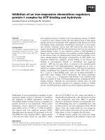
Structural characterization of fowlicidin-3
Putatively mature fowlicidin-3 comprising 27 amino-
acid residues was synthesized and purified to > 95%
purity, and its mass was confirmed by MS to be
3095.1 Da, consistent with the calculated value
(3094.8 Da). Putatively mature fowlicidin-1 comprising
26 amino acids was similarly synthesized and purified as
a reference peptide with an observed mass of 3141.6 Da
and calculated mass of 3141.9 Da, as described [13].
CD spectroscopy was first performed to determine
the secondary structure of fowlicidin-3 in the presence
of different concentrations of trifluoroethanol and
sodium dodecyl sulfate (SDS). As shown in Fig. 1A,
Fig. 1. CD spectra of fowlicidin-3 in different concentrations of tri-
fluoroethanol (TFE) (A) and SDS (B) with or without 0.15
M NaCl.
Y. R. Bommineni et al. Structure and functions of fowlicidin-3
FEBS Journal 274 (2007) 418–428 ª 2006 The Authors Journal compilation ª 2006 FEBS 419
fowlicidin-3 was largely unstructured in phosphate
buffer and began to transform into a typical a-helical
conformation after the addition of trifluoroethanol in
a dose-dependent manner. Significant a-helical content
(86%) with virtually no b-sheet structure was observed
in fowlicidin-3 in 50–60% trifluoroethanol (Fig. 1A).
Similarly, fowlicidin-3 exhibited 53% a-helical content
in the presence of 0.25% SDS, and the a-helical con-
tent remained largely unaltered in 0.5% or 2.0% SDS
micelles (Fig. 1B). These results suggest that fowlici-
din-3 is likely to adopt a predominantly a-helical con-
formation when interacting with bacterial membranes.
To further determine the tertiary structure of fowlic-
idin-3, 2D NMR spectroscopy was used. Because the
a-helical content of fowlicidin-3 peaked in 50% tri-
fluoroethanol (Fig. 1A) and NMR signals in trifluoro-
ethanol were much sharper and more intense than in
SDS micelles, fowlicidin-3 (4 mm) prepared in 50%
deuterated trifluoroethanol (trifluoroethanol-d3) ⁄ 50%
water (v ⁄ v) was selected for detailed NMR studies as
described [13]. Complete proton resonance assignments
were obtained using spin system identification and
sequential assignments from NMR spectra recorded at
25 °C (Supplementary Figs. S1 and S2). Consistent
with the CD results, the Ca-proton chemical shift
index, together with the presence of a number of
sequential d
NN
(i, i +1), nonsequential d
aN
(i, i +3),
and d
ab
(i, i +3) nuclear Overhauser effect (NOE)
peaks (Fig. 2), clearly indicates an a-helical conforma-
tion for fowlicidin-3.
A total of 205 NOE constraints, including 68 intra-
residue, 86 sequential, and 51 medium-range con-
straints, were used to calculate the tertiary structure of
fowlicidin-3 (Table 1). From 100 calculated structures
that satisfied the experimental restraints, 20 structures
with the lowest total energy were selected for further
analysis. A Ramachandran plot, produced by pro-
check-nmr [14], showed that 64.8% residues are in
the most favored region and 33.4% are in additional
allowed regions (Table 1). A superimposition of the 20
lowest-energy structures showed a considerable degree
of flexibility, with a pairwise rmsd of the backbone of
3.03 A
˚
(Table 1). However, alignments along residues
V9–A16 (Fig. 3A) and N19–R25 (Fig. 3B) of the 20
structures resulted in backbone rmsd values of
< 0.4 A
˚
in both cases (Table 1), suggesting relative
rigidity of these two a-helical segments.
The energy-minimized average structure of fowlici-
din-3 was further calculated, showing a predominantly
a-helical structure extending from V9 to R25 with a
relatively flexible N-terminal segment (Fig. 3C). A clo-
ser examination of the NMR structure revealed a kink
within the long a-helix between residues 16 and 19,
due to the presence of a glycine at position 17. This
was indicated by the fact that the C
aH
chemical index
showed no shift for G17 and I18 (Fig. 2), consistent
with the notion that glycine usually allows peptide
backbone flexibility. As evidenced by a lack of NOEs
(Fig. 2), such a kink indeed provides conformational
flexibility between two short a-helical segments (com-
pare Fig. 3A,B), reminiscent of fowlicidin-1 [13].
Superimposition of fowlicidin-1 on fowlicidin-3 indeed
revealed substantial overlapping, except for the flexible
N-sequences (Fig. 3D). This is perhaps not surprising,
given the fact that both peptides are likely to have
evolved by duplication and share > 60% identity in
Fig. 2. Schematic diagram of C
aH
chemical-shift index as well as
sequential and medium distance NOE connectivities for fowlicidin-3.
The thickness of the bar reflects the strength of the NOE connectiv-
ities.
Table 1. Structural statistics of the 20 lowest-energy structures of
fowlicidin-3.
NOE constraints
Total 205
Intraresidue (n ¼ 0) 68
Sequential (n ¼ 1) 86
Medium range (n ¼ 2,3,4) 51
Constraints ⁄ residue 7.6
Energies (kcalÆmol
)1
)
Total 18.64 ± 5.30
Bonds 2.82 ± 0.17
Angles 18.58 ± 0.69
van der Waals ) 14.67 ± 5.00
NOE 11.27 ± 0.65
Pairwise rmsds for residues 1–27 (A
˚
)
Backbone atoms 3.03 ± 0.82
Heavy (nonhydrogen) atoms 4.44 ± 0.87
Rmsds to mean structure (A
˚
) (residues 9–16)
Backbone atoms 0.36 ± 0.14
Heavy (nonhydrogen) atoms 0.90 ± 0.24
Rmsds to mean structure (A
˚
) (residues 19–25)
Backbone atoms 0.34 ± 0.13
Heavy (nonhydrogen) atoms 1.54 ± 0.39
Percentage of residues in regions of /–w space
Core 64.8%
Allowed 33.4%
Generously allowed 1.8%
Disallowed 0.0%
Structure and functions of fowlicidin-3 Y. R. Bommineni et al.
420 FEBS Journal 274 (2007) 418–428 ª 2006 The Authors Journal compilation ª 2006 FEBS
amino-acid sequence in the putatively mature region
(Fig. 3E). Despite structural similarities, it will be
interesting to study whether the two fowlicidins differ
in functional properties.
Evaluation of antibacterial properties of
fowlicidin-3
Fowlicidin-1 was found to be among the most potent
cathelicidins in killing bacteria [12]. To evaluate the
antibacterial spectrum and efficacy of fowlicidin-3, we
performed standard broth microdilution assays in
100% Muller-Hinton broth as recommended by the
Clinical and Laboratory Standards Institute [15] using
fowlicidin-1 as a reference peptide. As shown in
Table 2, fowlicidin-3 was active against a wide range
of Gram-negative and Gram-positive bacteria with
minimum inhibitory concentrations (MICs) in the
range 1–2 lm, often showing slightly higher potency
than fowlicidin-1. Moreover, fowlicidin-3 exhibited
no diminished efficiency against antibiotic-resistant
strains, including multidrug-resistant Salmonella enteri-
ca serovar Typhimurium DT104 and two methicillin-
resistant Staphylococcus aureus strains tested.
Most cationic host defense peptides, including cathe-
licidins, are membrane-lytic agents, killing bacteria by
physical interaction with and disruption of bacterial
cell membranes, although increasing evidence suggests
the presence of intracellular targets for certain peptides
[1,16]. To examine the mechanism of action and bac-
terial killing kinetics of fowlicidin-3, Escherichia coli
ML-35p, a strain that contains a plasmid giving consti-
tutive expression of b-galactosidase in the cytosol, was
incubated with different concentrations of peptides for
1 h in the presence of a chromogenic substrate, o-ni-
trophenyl-b-d-galactopyranoside [17–19]. It is conceiv-
able that the amount of b-galactosidase released, as
indicated by color change, is proportional to the
degree of permeabilization of bacterial cytosolic mem-
branes by fowlicidins. As shown in Fig. 4, membrane
permeabilization began almost immediately upon the
addition of 1 lm fowlicidin-3 or fowlicidin-1 to bac-
teria, reaching a plateau at 30–40 min, entirely
consistent with earlier colony counting assays with
fowlicidin-1, in which bacteria were killed quickly with
the maximum killing occurring 30 min after incubation
of the peptide with bacteria [12]. Identical trends were
Fig. 3. Tertiary structure of fowlicidin-3 in 50% trifluoroethanol. (A)
Superimposition of the backbones of the 20 lowest-energy struc-
tures of fowlicidin-3 best-fitted to residues 9–16. (B) Superimposi-
tion of the backbones of the 20 lowest-energy structures of
fowlicidin-3 best-fitted to residues 19–25. (C) Ribbon diagram of the
minimized average structure of fowlicidin-3. (D) Superimposition of
the average structures of fowlicidin-3 with fowlicidin-1. The struc-
tures were generated by using
MOLMOL. (E) Sequence alignment of
fowlicidin-3 and fowlicidin-1. Dashes are created to maximize the
alignment, and the total amino-acid residue numbers are also indi-
cated. Vertical bars connecting sequences denote identities, and
colons mean similarities. The conserved glycine is boxed.
Table 2. MICs of fowlicidin-3 (Fowl-3) in comparison with fowlici-
din-1 (Fowl-1). MICs were determined as the lowest peptide con-
centration that gave no visible bacterial growth after overnight
incubation in a standard broth microdilution assay using 100%
Muller–Hinton broth. The experiments were repeated at least twice
for each bacterial strain with similar values. MRSA, Methicillin-
resistant Staph. aureus.
Bacteria ATCC No. Fowl-3 (l
M) Fowl-1 (lM)
Gram-negative
E. coli 25922 2 2
S. typhimurium 14028 2 2
S. enteritidis 13076 2 2
K. pneumoniae 13883 1 1
S. typhimurium DT104 700408 2 2
Gram-positive
L. monocytogenes 19115 2 2
Staph. aureus 25923 1 2
Staph. aureus (MRSA) 43300 1 2
Staph. aureus (MRSA) BAA-39 1 1
Y. R. Bommineni et al. Structure and functions of fowlicidin-3
FEBS Journal 274 (2007) 418–428 ª 2006 The Authors Journal compilation ª 2006 FEBS 421
also observed with 0.5 and 2 lm fowlicidin concentra-
tions (data not shown). These results imply that, as
with most other host defense peptides, physical mem-
brane disruption appears to be a major mechanism of
killing bacteria for fowlicidin-3 and fowlicidin-1.
Physiological concentrations of salt prove to be
inhibitory to the antibacterial activities of many anti-
microbial peptides, such as human cathelicidin LL-37
[18] and a-defensin and b-defensin [20,21]. However,
the presence of 100 mm NaCl had little impact on
membrane permeabilization, with only a minimal delay
in killing kinetics for fowlicidin-3 (Fig. 4), consistent
with our direct colony counting assay (data not
shown). Indeed, the presence of physiological concen-
trations of NaCl did not affect the structure of fowlici-
din-3 in membrane mimetic environments (Fig. 1A,B).
These data suggest that, similar to fowlicidin-1 and
fowlicidin-2 [12], fowlicidin-3 kills bacteria in a salt-
independent manner, in contrast with many other pep-
tides, the activities of which are severely suppressed in
the presence of salt [18,20,21].
Serum has been found to be another important
inhibitory factor in bactericidal activities of many host
defense peptides, probably because of the presence of
certain salts, bivalent cations, and peptide-binding pro-
teins. To examine the effect of serum on antibacterial
efficacy of fowlicidin-3, a radial diffusion assay [22]
was performed with E. coli O157:H7 ATCC 700728
and Staph. aureus ATCC 25923 and peptides diluted
with and without 50% human or chicken serum. The
results revealed that both fowlicidin-3 and fowlicidin-1
retained > 80% activity against Gram-negative E. coli
O157:H7 in either serum (Fig. 5). The same trend was
also true with Gram-positive Staph. aureus (data not
shown). These results imply in vivo therapeutic poten-
tial for fowlcidin-3 and fowlcidin-1 for systemic appli-
cations.
Evaluation of the toxicity of fowlicidin-3
to mammalian cells
As compared with b-sheet defensins, a considerably
higher degree of toxicity to mammalian cells occurs
with a-helical cathelicidins, limiting their potential as
antimicrobial agents. To study the toxicity of fowlici-
din-3, Madin-Darby canine kidney (MDCK) epithelial
cells were first incubated with different concentrations
0 10 20 30 40 50 60
0.0
0.5
1.0
1.5
No Peptide
No Peptide + Salt
Fowl-3
Fowl-1
Fowl-3 + Salt
Fowl-1+ Salt
Time (min)
A
504
Fig. 4. Peameabilization of bacterial cytoplasmic membrane by fow-
licidins. E. coli ML-35p was diluted to (2.5–5) · 10
7
CFUÆmL
)1
and
incubated with 1 l
M fowlicidin-3 or fowlcidin-1 in 10 mM sodium
phosphate, pH 7.4, in the presence and absence of 100 m
M NaCl
at 37 °C. A chromogenic substrate for b-galactosidase, o-nitrophe-
nyl-b-
D-galactopyranoside, was also added to a final concentration
of 1.5 m
M. The absorbance at 405 nm was monitored every 2 min
for the production of p-nitrophenol for up to 1 h. Data shown are
representative of two independent experiments with highly similar
results.
Fowl-3
Fowl-1
Chicken
Serum
Human
Serum
No
Serum
0
20
40
60
80
100
)%( ytivitcA evitaleR
Fowlicidin-3 Fowlicidin-1
A
B
Fig. 5. Effect of serum on the antibacterial activity of fowlicidins by
radial diffusion assay. Fowlicidin-3 or fowlcidin-1, 1 lg diluted in
0.01% acetic acid with and without 50% human or chicken serum,
was added to the wells of the underlay gel containing E. coli
O157:H7 ATCC 700728 (4 · 10
5
CFUÆmL
)1
). After overnight incuba-
tion, bacterial clearance zones were recorded, and antibacterial
activities (%) in the presence of serum were calculated relative to
the activities without serum. In (B), open bars represent no serum
controls, and striped and solid bars are 50% human and chicken
serum, respectively. Data shown are mean ± (SEM) from two inde-
pendent experiments.
Structure and functions of fowlicidin-3 Y. R. Bommineni et al.
422 FEBS Journal 274 (2007) 418–428 ª 2006 The Authors Journal compilation ª 2006 FEBS
of fowlicidins in the presence or absence of 10% fetal
bovine serum, and then a cell viability assay was per-
formed as described [12]. As compared with fowlicidin-1
with a 50% effective concentration (EC
50
)of 2 lm,
fowlicidin-3 killed 50% MDCK cells at 12 lm
(Fig. 6A). Moreover, the presence of 10% serum further
reduced the toxicity of fowlicidin-3 by twofold
(Fig. 6A).
To test the hemolytic activity of fowlicidin-3 further,
freshly isolated human erythrocytes were incubated
with fowlicidins with and without 10% fetal bovine
serum, and erythrocyte lysis was measured according
to the release of hemoglobin [12]. In the absence
of serum, 50% hemolysis occurred at 9 lm for fowlici-
din-3, whereas fowlicidin-1 was considerably more
toxic with an EC
50
of 1.5 lm (Fig. 6B). Serum sub-
stantially reduced hemolysis of both peptides, with
EC
50
values of 80 lm for fowlicidin-3 and 13 lm for
fowlicidin-1 in 10% fetal bovine serum. Taking the
results together, fowlicidin-3 is slightly more potent
than fowlicidin-1 in killing many bacterial strains tes-
ted, but is 4–6-fold less toxic to mammalian cells than
fowlicidin-1, indicating higher therapeutic potential for
fowlicidin-3.
Inhibition of LPS-induced proinflammatory gene
expression by fowlicidin-3
Because fowlicidin-1 and fowlicidin-2 were found to be
able to bind LPS directly and suppressed LPS-induced
cytokine gene expression [12], we sought to determine
whether fowlicidin-3 has a similar LPS-neutralizing
activity. Mouse macrophage RAW264.7 cells were sti-
mulated for 4 h with 100 ngÆmL
)1
LPS in the presence
and absence of different concentrations of fowlicidins,
followed by real-time RT-PCR analysis of the expres-
sions of three common proinflammatory genes, inclu-
ding interleukin-1b, CC chemokine ligand 2 (CCL2) ⁄
monocyte chemotactic protein-1 (MCP-1), and
CCL3 ⁄ monocyte inflammatory protein-1a (MIP-1a).
As shown in Fig. 7, fowlicidin-3 dose-dependently
inhibited the expression of interleukin-1b or CCL3 ⁄
MIP-1a genes, with a concentration of 10 lm reducing
0 10 20 30 40 50
0
20
40
60
80
100
A
B
Fowl-3
Fowl-3 + FBS
Fowl-1
Fowl-1 + FBS
Peptide (µM)
)%( htaeD lleC
0 20 40 60 80 100
0
20
40
60
80
100
Fowl-3 + FBS
Fowl-3
Fowl-1 + FBS
Fowl-1
Peptide (µM)
)%( sisylomeH
Fig. 6. Toxicity of fowlicidins to MDCK cells (A) and human erythro-
cytes (B) in the presence and absence of 10% fetal bovine serum
(FBS). EC
50
is indicated as dotted lines in both panels. Data shown
are mean ± SEM from two to four independent experiments.
0
5
10
15
20
25
30
Control Fowlicidin-3 Fowlicidin-1
LPS -+ + + + + +
Peptide
0
1000
2000
3000
4000
5000
6000
7000
8000
CCL3/MIP-1α
IL-1β
egnahC dloF evitaleR
Fig. 7. Inhibition of LPS-induced expression of interleukin-1b and
CCL3 ⁄ MIP-1a in RAW264.7 cells. Cells were pretreated for 30 min
with and without fowlicidin-3 (0.5, 2.5, and 10 l
M) or fowlicidin-1
(2.5 and 10 l
M) in duplicate, followed by stimulation for another 4 h
with 100 ngÆmL
)1
LPS. Total RNA was then isolated and subjected
to real-time RT-PCR analysis. Data shown are mean ± SEM from
two independent experiments.
Y. R. Bommineni et al. Structure and functions of fowlicidin-3
FEBS Journal 274 (2007) 418–428 ª 2006 The Authors Journal compilation ª 2006 FEBS 423
expression of both genes by > 95%. A similar block-
age of CCL2 ⁄ MCP-1 expression was also observed
(data not shown). Because treating cells with fowlici-
din-3 alone had no effect on gene expression (Fig. 7),
such LPS-neutralizing activity was specific. It is note-
worthy that, compared with fowlicidin-1, fowlicidin-3
is more potent in inhibiting LPS-induced gene expres-
sion (Fig. 7), suggesting that fowlicidin-3 may be more
effective in antisepsis therapy.
Discussion
Our previous analyses of genomic sequences have
revealed that the genes for fowlicidin-1 and fowlicidin-
3 are almost identical in the first three exons and first
three introns [12]. The fourth exon, which primarily
encodes biologically active, mature sequences, also
shares > 60% identity between the two peptides
(Fig. 3E). Therefore, these two fowlicidins were most
likely to be duplicated from each other during evolu-
tion. The putatively mature fowlicidin-3 peptide con-
sists of 27 amino acid residues with a charge of +6
and no anionic residues, whereas fowlicidin-1 is com-
posed of 26 amino acids with a net charge of +8. Evo-
lution of two highly similar antimicrobial peptides
with potent antibacterial activities may represent an
enforcement of innate host defense. It is also plausible
that fowlicidin-1 and fowlicidin-3 may have some non-
overlapping biological functions yet to be discovered.
Because of a similarity in primary sequence, it is not
surprising that the two fowlicidins adopt a similar
a-helical conformation in membrane-mimicking envi-
ronments (Fig. 3D). Moreover, both peptides contain
a kink near the central helical region due to the pres-
ence of a conserved glycine residue (Fig. 3E). Interest-
ingly, such a glycine-induced hinge is not unique to
fowlicidins, but appears to be a common feature for
many a-helical cationic host defense peptides
[13,23,24]. The presence of a hinge structure has been
shown to be beneficial in enhancing molecular flexibil-
ity while reducing the toxicity of otherwise rigid pep-
tides to mammalian cells [23,24].
Amphipathicity is another hallmark of most a-heli-
cal cationic peptides [23,24]. However, unlike typical
a-helical peptides, the long helices of fowlicidin-1 and
fowlicidin-3 are much less amphipathic, with no obvi-
ous segregation of hydrophobic residues from hydro-
philic residues (Fig. 8). Furthermore, the a-helical
region is highly hydrophobic (Fig. 8) in that fowlici-
din-3 is composed of only one cationic (K22) and three
polar uncharged residues (N12, T13 and N19), whereas
fowlicidin-1 consists of only two cationic (R11 and
R21) and two polar uncharged residues (T12 and N18)
(Fig. 3E). Instead, positively charged residues are
mostly concentrated at both tails (Figs 3E and 8).
A series of antibacterial tests revealed that, similar
to fowlicidin-1, fowlicidin-3 possesses potent, broad-
spectrum, and fast-acting bactericidal activities with
similar efficiency against both antibiotic-susceptible
and antibiotic-resistant bacterial strains. Killing of bac-
teria by fowlicidins starts immediately on contact with
bacteria, in sharp contrast with human cathelicidin
LL-37, which takes up to 20–30 min before permeabili-
zation of bacterial inner membranes occurs [17,18].
More significantly, bacterial killing activity is largely
unaffected by salt or serum, making fowlicidins
attractive therapeutic candidates for potential in vivo
systemic applications.
In spite of similarities in structural and antibacterial
properties, fowlicidin-3 is much less toxic to mamma-
lian cells than fowlicidin-1. Because the cytotoxicity
(EC
50
) of fowlicidin-3 is at least 10–40-fold (in the
presence of serum) higher than MICs against all bac-
terial strains tested, a therapeutic window clearly exists
for fowlicidin-3, particularly for systemic applications.
Fig. 8. Surface accessibilities of fowlicidins. (A) Front view of the
solvent-accessible surface of fowlicidin-3. (B) Back view of the sol-
vent-accessible surface of fowlicidin-3. (C) Front view of the sol-
vent-accessible surface of fowlicidin-1. (D) Back view of the
solvent-accessible surface of fowlicidin-1. Positively charged resi-
dues are in blue, polar uncharged residues are in pink, and hydro-
phobic residues are in yellow. The N-terminus is on the top. The
figures were generated using
PYMOL ().
Structure and functions of fowlicidin-3 Y. R. Bommineni et al.
424 FEBS Journal 274 (2007) 418–428 ª 2006 The Authors Journal compilation ª 2006 FEBS
More desirably, fowlicidin-3 is highly potent in block-
ing LPS-induced proinflammatory gene expression.
Collectively, fowlicidin-3 appears to have promising
therapeutic potential for further development as a
novel antimicrobial and antisepsis agent.
It is interesting to note that the higher toxicity asso-
ciated with fowlicidin-1 is probably due to limited flexi-
bility of the a-helix, which is a result of the physical
hindrance caused by the side chain of a nearby tyrosine
[13]. Although fowlicidin-3 is devoid of aromatic resi-
dues adjacent to the conserved glycine (Fig. 3E), it will
be important to examine the impact of further enhan-
cing its flexibility on the functional properties. In fact,
the flexibility of the hinge region has often been found
to be positively correlated with a decrease in the toxic-
ity of many a-helical peptides [23,24]. Because amphi-
pathicity, hydrophobicity, and helicity are among the
most important factors that influence the antibacterial
and toxicity of a-helical cationic peptides [23,24],
rational changes of these structural and physicochemi-
cal parameters are likely to further improve the thera-
peutic potential of fowlicidin-3.
Experimental procedures
Peptide synthesis
Putatively mature fowlicidin-1 (RVKRVWPLVIRTVIA
GYNLYRAIKKK) and fowlicidin-3 (KRFWPLVPVAIN
TVAAGIN LYKAIRRK) were chemically synthesized
using the standard solid-phase method of SynPep (Dublin,
CA, USA) and Bio-Synthesis (Lewisville, TX, USA),
respectively. Both peptides were purified to > 95% purity
by RP-HPLC. The mass and purity of each peptide were
further confirmed by MS using the Voyager DE-PRO
instrument (Applied Biosystems, Foster City, CA, USA)
housed in the Recombinant DNA ⁄ Protein Core Facility
of Oklahoma State University. Lyophilized peptides were
reconstituted in 0.01% acetic acid, and concentrations were
measured by UV absorbance at 280 nm in the presence of
6 m guanidine hydrochloride [25], based on the absorption
coefficients for aromatic tryptophan and tyrosine residues
present in both peptides.
CD spectroscopy and secondary-structure
determination
The secondary structure of fowlicidin-3 was determined on
a Jasco-715 spectropolarimeter (JASCO, Tokyo, Japan)
using a 0.1-cm path length cell over the 180–260 nm range
as described [13]. The CD spectra were acquired at 25 °C
every 1 nm with a 2-s averaging time per point and a 1-nm
band pass. Fowlicidin-3 (10 lm) was measured in 50 mm
potassium phosphate buffer, pH 7.4, with or without differ-
ent concentrations of trifluoroethanol (0%, 10%, 20%,
40%, 50% and 60%) or SDS micelles (0.25%, 0.5% and
2.0%). CD analyses were also performed in 50% trifluoro-
ethanol and 2.0% SDS in phosphate buffer with addition
of 150 mm NaCl. Mean residue ellipticity (MRE) was
expressed as [h]
MRE
(degreesÆcm
2
Ædmol
)1
). The contents of
the secondary-structural elements, including regular and
distorted a-helix, regular and distorted b-sheet, turns, and
unordered structures, were analyzed using the program
selcon3 [26].
NMR spectroscopy and tertiary structure
calculations
The NMR experiments were performed with 500-MHz
Varian UNITY plus NMR spectrometer (Varian, Palo
Alto, CA, USA) as previously described [13]. Because
NMR signals in 50% trifluoroethanol-d
3
⁄ 50% water mix-
ture were much sharper and intense than in SDS micelles,
fowlicidin-3 (4 mm) prepared in trifluoroethanol ⁄ water
(1 : 1, v ⁄ v) was selected for detailed NMR studies. The
data sets were acquired at different temperatures ranging
from 10 to 35 °C. The 2D
1
H-
1
H TOCSY spectra with an
isotropic mixing time of 100 ms at a B
1
field strength of
8 kHz and 2D
1
H-
1
H NOESY spectra with mixing times of
100, 200, 300, 400 and 500 ms were collected. The trifluoro-
ethanol peak (3.88 p.p.m. at 25 °C) was considered as the
reference for chemical shift assignments. A mixing time of
300 ms was initially used for distance constraint measure-
ments, and the assigned NOE peaks were then checked with
the spectra obtained with a 100-ms mixing time. For
molecular modeling calculations, only NOE peaks present
in the NOESY spectra obtained with a mixing time of
100 ms were used to rule out the peaks due to spin diffu-
sion. The intensities of the cross-peaks in NOESY spectra
were classified as strong, medium, and weak, corresponding
to distance restraints of 1.8–2.8, 1.8–4.0, and 1.8–5.0 A
˚
,
respectively. The distance restraints were then used to cal-
culate structures using the program cns (version 1.1) [27],
using a simulated annealing protocol for torsion angle
dynamics. From all 100 calculated structures accepted, 20
structures with the lowest total energy were selected and an-
alyzed with molmol [28] and procheck- nmr [14]. The
atomic co-ordinates and structure factors of putatively
mature fowlicidin-3 have been deposited under accession
code 2HFR in the Protein Data Bank, Research Collabora-
tory for Structural Bioinformatics, Rutgers University, New
Brunswick, NJ, USA ( />Bacterial culture and antibacterial testing
Gram-negative bacteria (E. coli ATCC 25922, S. enterica
serovar Typhimurium ATCC 14028, S. enterica serovar
Y. R. Bommineni et al. Structure and functions of fowlicidin-3
FEBS Journal 274 (2007) 418–428 ª 2006 The Authors Journal compilation ª 2006 FEBS 425
Typhimurium DT104 ATCC 700408, and Klebsiella pneu-
moniae ATCC 13883), and Gram-positive bacteria (Listeria
monocytogenes ATCC 19115, Staph. aureus ATCC 25923,
Staph. aureus ATCC BAA-39, and Staph. aureus ATCC
43300) were purchased from either ATCC (Manassas, VA,
USA) or MicroBiologics (St Cloud, MN, USA) and tested
individually against fowlicidin-1 and fowlicidin-3. The
MICs were determined by a standard broth microdilution
assay as recommended by the Clinical and Laboratory
Standards Institute [15]. Briefly, overnight bacterial culture
was subcultured in fresh trypticase soy broth with shaking
at 250 r.p.m. at 37 °C for 3 h to reach the mid-exponential
phase of growth. Bacteria were then washed twice in
10 mm sodium phosphate buffer, pH 7.4, and diluted to
5 · 10
5
CFUÆmL
)1
in Muller-Hinton broth (BBL, Cockeys-
ville, MD, USA). After 90 lL bacteria had been dispensed
into 96-well cell culture plates, 10 lL peptides in serial two-
fold dilutions were added in duplicate. The MIC value of
each peptide was determined as the lowest peptide concen-
tration that gave no visible bacterial growth after overnight
incubation at 37 °C.
Assay of cytoplasmic membrane
permeabilization
E. coli ML-35p was kindly provided by R Gallo (UCSD, La
Jolla, CA, USA) and used as described [17–19]. Briefly, mid-
exponential phase bacteria were washed twice in 10 mm
sodium phosphate buffer, pH 7.4, diluted to 0.03 A
600
[equiv-
alent to (2.5–5) · 10
7
CFUÆmL
)1
) in the same phosphate
buffer containing 1% trypticase soy broth with and without
100 mm NaCl. After 80 lL bacteria had been dispensed into
each well of a 96-well tissue culture plate, different concen-
trations of fowlicidins and 1.5 mm o-nitrophenyl-b-d-gal-
actopyranoside (Sigma, St Louis, MO, USA) were added to
a total volume of 100 lL per well. The production of p-nitro-
phenol was monitored spectrophotometrically at 37 °C and
405 nm every 2 min for up to 1 h with periodic shaking.
Serum effect on the antibacterial activity
of fowlicidin-3
The radial diffusion assay [22] was used to study the effect
of serum on the antibacterial activity of fowlicidins. Briefly,
after solidification of the underlay gel containing
4 · 10
5
CFUÆmL
)1
Staph. aureus ATCC 25923 or E. coli
O157:H7 ATCC 700728, small wells ( 3 mm in diameter)
were punched. Then 1 lg fowlicidin-1 or fowlicidin-3 was
diluted to a total of volume of 4 lL in 0.01% acetic acid
with or without 50% chicken or human serum and added
separately to the wells. After 3 h of diffusion at 37 °C, the
nutrient-rich overlay gel was poured and incubated at
37 °C overnight. The diameters of the bacterial clearance
zones were measured.
Cytotoxicity assay
The toxicity of fowlicidin-3 toward mammalian epithelial
cells was evaluated by using MDCK cells (ATCC) and an
Alamar Blue dye (Biosource, Camarillo, CA, USA) as des-
cribed [12]. Briefly, MDCK cells were seeded in 96-well
plates with 1.5 · 10
5
cells ⁄ well and allowed to grow over-
night in Dulbecco’s modified Eagle medium (DMEM), con-
taining 10% fetal bovine serum to 80–90% confluence. After
cells had been washed with serum-free DMEM, 90 lL fresh
DMEM with or without 10% fetal bovine serum was added
to each well, followed by the addition of 10 lL serially dilu-
ted peptides in duplicate. After 18 h of incubation at 37 °C
under 5% CO
2
,10lL Alamar Blue dye was added to each
well and incubated for another 6 h. The fluorescence was
read with excitation at 545 nm and emission at 590 nm. Per-
centage cell death (%) was calculated as [1 ) (F
peptide
)
F
background
) ⁄ (F
acetic acid
) F
background
)] · 100, where F
peptide
is
the fluorescence of cells exposed to different concentrations
of peptides, F
acetic acid
is the fluorescence of cells exposed to
0.01% acetic acid only, and F
background
is the background
fluorescence of 10% AlamarBlue dye in cell culture medium
without cells. Cytotoxicity (EC
50
) was defined as the peptide
concentration that caused 50% cell death.
Hemolysis assay
Freshly collected chicken and human blood were used for
evaluating hemolytic activity as described [12,13]. The pro-
tocols for collection of human and chicken blood were
approved by the Institutional Review Board and Institu-
tional Animal Care and Use Committee of Oklahoma
State University, respectively. Briefly, EDTA-anticoagulated
blood was washed twice in NaCl ⁄ P
i
and diluted to 0.5% in
NaCl ⁄ P
i
with or without 10% fetal bovine serum. Erythro-
cytes (90 lL aliquots) were then dispensed into a 96-well
plate, followed by the addition of 10 lL serially diluted fow-
licidins in 0.01% acetic acid in duplicate. After incubation
for 2 h at 37 °C, supernatants were colleted by centrifuga-
tion and transferred to a fresh 96-well plate to measure the
absorbance of released hemoglobin at 405 nm. Controls for
0% and 100% hemolysis were erythrocytes exposed to
10 lL 0.01% acetic acid and 1% Triton X-100, respectively.
Percentage hemolysis (%) was calculated as [(A
405, peptide
)
A
405, 0.01% acetic acid
) ⁄ (A
405, 1% Triton X-100
) A
405, 0.01% acetic
acid
)] · 100. EC
50
was determined as the peptide concentra-
tion that lysed 50% erythrocytes.
Real-time PCR analysis of the effect of
fowlicidins on LPS-induced proinflammatory
gene expression
Mouse macrophage RAW 264.7 cells were used to study
the modulation of LPS-induced cytokine⁄ chemokine gene
Structure and functions of fowlicidin-3 Y. R. Bommineni et al.
426 FEBS Journal 274 (2007) 418–428 ª 2006 The Authors Journal compilation ª 2006 FEBS
expression by fowlicidin-3 in comparison with fowlicidin-1.
Cells were seeded in 12-well tissue culture plates with
5 · 10
5
cells ⁄ well in DMEM containing 10% fetal bovine
serum. After overnight incubation, cells were pretreated for
30 min with 0.5, 2.5, and 10 lm fowlicidins in duplicate,
followed by stimulation for 4 h with 100 ngÆmL
)1
LPS
from E. coli O114:B4 (Sigma). Total RNA was then isola-
ted from cells using TRIzol (Invitrogen, Carlsbad, CA,
USA) according to the manufacturer’s instructions. Quanti-
tative real-time RT-PCR was used to analyze the expression
of three common proinflammatory genes, namely inter-
leukin-1b, CCL2 ⁄ MCP-1, and CCL3 ⁄ MIP-1a, using exon-
spanning primers as described [12].
The first-strand cDNA from 1.5 lg each RNA sample
was synthesized in a reaction volume of 20 lLat42°C
for 30 min using a QuantiTectÒ Reverse Transcription
Kit (Qiagen, Valencia, CA, USA), which included
removal of genomic DNA contamination before cDNA
synthesis. Real-time PCR was performed using 0.2 lgof
the first-strand cDNA, gene-specific primers, SYBRÒ
Premix Ex Taqä (Takara Bio, Shiga, Japan), and
MyiQÒ Real-Time PCR Detection System (Bio-Rad,
Hercules, CA, USA) in a total volume of 10 lL. PCR
cycling conditions were as follows: 95 °C for 30 s, fol-
lowed by 40 cycles of 95 °C for 15 s, 55 °C for 30 s, and
72 °C for 30 s. The comparative DDCT method was used
to quantify the gene expression levels, where b-actin was
used as an internal control for normalization [12]. Relat-
ive fold changes in gene expression were calculated using
the formula 2
–DDCt
. Melting curve analysis (55–95 °C)
was performed and confirmed amplification of a single
product in each case.
Acknowledgements
This work was supported by grants from the National
Science Foundation (grants MCB0236039 and
EPS0236913), NIH (S10-RR022392), Oklahoma Cen-
ter for the Advancement of Science and Technology
(grant HR03-146), and Oklahoma Agricultural Experi-
ment Station (Project H-2507). We thank Robert Gallo
from the University of California, San Diego, CA,
USA for kindly providing E. coli ML-35p for use in
the inner membrane permeabilization assays, and Steve
Hartson of Oklahoma State University for helping
with MS. We are grateful to Haobo Jiang and Ulrich
Melcher for critical reading of the manuscript. The
constructive comments from anonymous reviewers are
also appreciated.
References
1 Zasloff M (2002) Antimicrobial peptides of multicellular
organisms. Nature 415, 389–395.
2 Brogden KA, Ackermann M, McCray PB Jr & Tack
BF (2003) Antimicrobial peptides in animals and their
role in host defences. Int J Antimicrob Agents 22, 465–
478.
3 Hancock RE & Patrzykat A (2002) Clinical develop-
ment of cationic antimicrobial peptides: from natural to
novel antibiotics. Curr Drug Targets Infect Disord 2,
79–83.
4 Zanetti M, Gennaro R, Skerlavaj B, Tomasinsig L &
Circo R (2002) Cathelicidin peptides as candidates for a
novel class of antimicrobials. Curr Pharm Des 8, 779–793.
5 Yang D, Biragyn A, Hoover DM, Lubkowski J &
Oppenheim JJ (2004) Multiple roles of antimicrobial
defensins, cathelicidins, and eosinophil-derived neuro-
toxin in host defense. Annu Rev Immunol 22, 181–215.
6 McPhee JB & Hancock RE (2005) Function and thera-
peutic potential of host defence peptides. J Pept Sci 11,
677–687.
7 Bowdish DM, Davidson DJ & Hancock RE (2005) A
re-evaluation of the role of host defence peptides in
mammalian immunity. Curr Protein Pept Sci 6, 35–51.
8 Finlay BB & Hancock RE (2004) Can innate immunity
be enhanced to treat microbial infections? Nat Rev
Microbiol 2, 497–504.
9 Zanetti M (2004) Cathelicidins, multifunctional peptides
of the innate immunity. J Leukoc Biol 75, 39–48.
10 Zaiou M & Gallo RL (2002) Cathelicidins, essential
gene-encoded mammalian antibiotics. J Mol Med 80,
549–561.
11 Lehrer RI & Ganz T (2002) Cathelicidins: a family of
endogenous antimicrobial peptides. Curr Opin Hematol
9, 18–22.
12 Xiao Y, Cai Y, Bommineni YR, Fernando SC, Prakash
O, Gilliland SE & Zhang G (2006) Identification and
functional characterization of three chicken cathelicidins
with potent antimicrobial activity. J Biol Chem 281,
2858–2867.
13 Xiao Y, Dai H, Bommineni YR, Soulages JL, Gong
YX, Prakash O & Zhang G (2006) Structure-activity
relationships of fowlicidin-1, a cathelicidin antimicrobial
peptide in chicken. FEBS J 273, 2581–2593.
14 Laskowski RA, Rullmannn JA, MacArthur MW,
Kaptein R & Thornton JM (1996) AQUA and PRO-
CHECK-NMR: programs for checking the quality of
protein structures solved by NMR. J Biomol NMR 8,
477–486.
15 National Committee for Clinical Laboratory Standards
(2003) Methods for Dilution Antimicrobial Susceptibility
Tests for Bacteria That Grow Aerobically, 6th edn.
Approved Standard M7-A6. National Committee for
Clinical Laboratory Standards, Wayne, PA.
16 Brogden KA (2005) Antimicrobial peptides: pore form-
ers or metabolic inhibitors in bacteria? Nat Rev Micro-
biol 3, 238–250.
Y. R. Bommineni et al. Structure and functions of fowlicidin-3
FEBS Journal 274 (2007) 418–428 ª 2006 The Authors Journal compilation ª 2006 FEBS 427
17 Dorschner RA, Lopez-Garcia B, Peschel A, Kraus D,
Morikawa K, Nizet V & Gallo RL (2006) The mam-
malian ionic environment dictates microbial suscept-
ibility to antimicrobial defense peptides. FASEB J 20,
35–42.
18 Turner J, Cho Y, Dinh NN, Waring AJ & Lehrer RI
(1998) Activities of LL-37, a cathelin-associated anti-
microbial peptide of human neutrophils. Antimicrob
Agents Chemother 42, 2206–2214.
19 Friedrich C, Scott MG, Karunaratne N, Yan H & Han-
cock RE (1999) Salt-resistant alpha-helical cationic anti-
microbial peptides. Antimicrob Agents Chemother 43,
1542–1548.
20 Hoover DM, Wu Z, Tucker K, Lu W & Lubkowski J
(2003) Antimicrobial characterization of human beta-
defensin 3 derivatives. Antimicrob Agents Chemother 47,
2804–2809.
21 Ericksen B, Wu Z, Lu W & Lehrer RI (2005) Antibac-
terial activity and specificity of the six human alpha-
defensins. Antimicrob Agents Chemother 49, 269–275.
22 Steinberg DA & Lehrer RI (1997) Designer assays for
antimicrobial peptides. Disputing the ‘one-size-fits-all’
theory. Methods Mol Biol 78, 169–186.
23 Tossi A, Sandri L & Giangaspero A (2000) Amphi-
pathic, alpha-helical antimicrobial peptides. Biopolymers
55, 4–30.
24 Dathe M & Wieprecht T (1999) Structural features of
helical antimicrobial peptides: their potential to modu-
late activity on model membranes and biological cells.
Biochim Biophys Acta 1462, 71–87.
25 Gill SC & von Hippel PH (1989) Calculation of protein
extinction coefficients from amino acid sequence data.
Anal Biochem 182, 319–326.
26 Sreerama N, Venyaminov SY & Woody RW (2000)
Estimation of protein secondary structure from circular
dichroism spectra: inclusion of denatured proteins with
native proteins in the analysis. Anal Biochem 287, 243–
251.
27 Brunger AT, Adams PD, Clore GM, DeLano WL, Gros
P, Grosse-Kunstleve RW, Jiang JS, Kuszewski J, Nilges
M, Pannu NS, et al. (1998) Crystallography & NMR
system: a new software suite for macromolecular struc-
ture determination. Acta Crystallogr D Biol Crystallogr
54, 905–921.
28 Koradi R, Billeter M & Wuthrich K (1996) MOLMOL:
a program for display and analysis of macromolecular
structures. J Mol Graph 14, 51–55.
Supplementary material
The following supplementary material is available
online:
Fig. S1. Fingerprint region (NH-C
aH
and side chain
protons) of 2D
1
H-
1
H TOCSY spectrum of fowlicidin-
3 in trifluoroethanol ⁄ water (1 : 1, v ⁄ v) at 25°C.
Fig. S2. Fingerprint region (NH-NH) of 2D
1
H-
1
H
NOESY spectrum of fowlicidin-3 in trifluoroethanol ⁄
water (1 : 1, v ⁄ v) at 25°C.
This material is available as part of the online article
from
Please note: Blackwell Publishing is not responsible
for the content or functionality of any supplementary
materials supplied by the authors. Any queries (other
than missing material) should be directed to the corres-
ponding author for the article.
Structure and functions of fowlicidin-3 Y. R. Bommineni et al.
428 FEBS Journal 274 (2007) 418–428 ª 2006 The Authors Journal compilation ª 2006 FEBS