Báo cáo khoa học: 15 N-Labelled proteins by cell-free protein synthesis Strategies for high-throughput NMR studies of proteins and protein–ligand complexes doc
Bạn đang xem bản rút gọn của tài liệu. Xem và tải ngay bản đầy đủ của tài liệu tại đây (1.15 MB, 6 trang )
MINIREVIEW
15
N-Labelled proteins by cell-free protein synthesis
Strategies for high-throughput NMR studies of proteins and
protein–ligand complexes
Kiyoshi Ozawa, Peter S. C. Wu, Nicholas E. Dixon and Gottfried Otting
Research School of Chemistry, Australian National University, Canberra, ACT, Australia
Introduction
Cell-free protein synthesis in both the Escherichia coli
coupled transcription-translation system and the wheat
germ translation system has been remarkably improved
so that milligram quantities of protein can routinely be
prepared [1–6]. Compared to conventional recombin-
ant protein production in vivo, cell-free protein synthe-
sis offers a number of decisive advantages for the
preparation of stable isotope labelled protein samples
for analysis by NMR spectroscopy.
(a) The target protein is the only protein synthesized
and labelled during the reaction. Consequently the iso-
tope-labelled amino acids are used very efficiently, and
because no new metabolic enzymes are expressed in
the medium, isotope scrambling is kept to a minimum.
Moreover, isotope-filtered NMR experiments allow the
selective observation of the isotope-labelled proteins
without chromatographic purification.
(b) The reaction is fast. This is advantageous for the
synthesis of proteins that are sensitive to proteolytic
degradation and for high-throughput applications.
(c) The reaction can be carried out in small volumes.
Therefore, isotope-labelled starting materials are used
more efficiently and economically than for conven-
tional in vivo labelling methods [7].
(d) The reaction is independent of cell growth.
Therefore, toxic proteins and proteins containing non-
natural amino acids can be made efficiently [8–10].
With the advent of cryogenic probe heads, hetero-
nuclear single quantum coherence (HSQC) spectra of
proteins made by cell-free expression can be recorded
quickly at the concentration delivered by the reaction
mixture.
Keywords
cell-free protein synthesis; combinatorial
labelling;
15
N-HSQC;
15
N-labelled amino
acids; protein–ligand interactions
Correspondence
G. Otting, Research School of Chemistry,
Australian National University, Canberra,
ACT, Australia
Fax: +61 261250750
Tel: +61 261256507
E-mail:
Website: />(Received 9 May 2006, accepted 23 June
2006)
doi:10.1111/j.1742-4658.2006.05433.x
[
15
N]-heteronuclear single quantum coherence (HSQC) spectra provide a
readily accessible fingerprint of [
15
N]-labelled proteins, where the backbone
amide group of each nonproline amino acid residue contributes a single
cross-peak. Cell-free protein synthesis offers a fast and economical route to
enhance the information content of [
15
N]-HSQC spectra by amino acid
type selective [
15
N]-labelling. The samples can be measured without chro-
matographic protein purification, dilution of isotopes by transaminase
activities are suppressed, and a combinatorial isotope labelling scheme can
be adopted that combines reduced spectral overlap with a minimum num-
ber of samples for the identification of all [
15
N]-HSQC cross-peaks by
amino acid residue type. These techniques are particularly powerful for
tracking [
15
N]-HSQC cross-peaks after titration with unlabelled ligand
molecules or macromolecular binding partners. In particular, combinatorial
isotope labelling can provide complete cross-peak identification by amino
acid type in 24 h, including protein production and NMR measurement.
Abbreviations
HSQC, heteronuclear single quantum coherence.
4154 FEBS Journal 273 (2006) 4154–4159 ª 2006 The Authors Journal compilation ª 2006 FEBS
(e) The reaction mixture is accessible. This allows
the synthesis of proteins in the presence of other pro-
teins provided in excess at the start of or during the
reaction, e.g., for the purpose of rescuing nascently
produced insoluble proteins into soluble complexes
with soluble binding partners [11].
This review summarizes our recent experience with
cell-free protein synthesis, in particular with regard to
the production of selectively [
15
N]-labelled proteins.
Isotope scrambling
Selectively [
15
N]-labelled protein samples have long
been made from a mixture of unlabelled and [
15
N]-
labelled amino acids by in vivo protein synthesis in
E. coli [12–15]. However, the amino acid metabolism
of live E. coli cells can cause serious isotope scram-
bling for many of the amino acids, mostly due to
transaminase activities [12,15–17]. In principle, this
problem can be overcome by the use of auxotrophic
E. coli strains [13], but this requires protein prepara-
tions from different strains.
Cell-free protein synthesis systems are far more inert
with regard to isotope scrambling because the pool of
metabolic enzymes present in the cell extract is not
regenerated. Thus, cell extracts from nonauxotrophic
E. coli strains such as A19 have been shown to yield
selectively labelled proteins without significant interfer-
ence from transaminases, except that conversion of
[
15
N]aspartic acid to [
15
N]asparagine was still found to
occur [18]. This conversion can, however, be sup-
pressed by heat treatment of the E. coli S30 cell extract
[7,19] or by replacing the originally recommended glu-
tamate buffer [1] by acetate [7,18]. Different amino
acids are susceptible to [
15
N]-scrambling in the wheat
germ system than in E. coli. In particular, interconver-
sion between Ala and Glu, Glu and Asp, and Glu and
Gln is efficient in wheat germ extract but can effect-
ively be suppressed by inhibitors of transaminases and
glutamine synthase [20].
Among the multitude of metabolic enzymes present
in the cell extract, only those leading to transfer of
[
15
N]-amino groups to other amino acids can interfere
with the subsequent NMR analysis. The NMR reso-
nances of [
15
N]-amino groups, for example, are at a
different chemical shift than the protein amide reso-
nances and therefore do not interfere with the protein
fingerprint represented by the amide cross-peaks in
the [
15
N]-HSQC spectrum. Remaining free [
15
N]-amino
acids are equally unproblematic because the amino
protons of amino acids exchange too rapidly at neutral
pH to yield a signal observable in [
15
N]-HSQC spectra.
It is thus possible to obtain clean NMR spectra
directly of the reaction mixture without prior removal
of low-molecular mass compounds [18,21,22].
Selective [
15
N]-labelling
NMR resonance assignments and tracking of chemical
shift changes is much easier if each amide cross-peak
in the [
15
N]-HSQC spectrum of a protein can be attrib-
uted a priori to one of the 19 nonproline amino acid
types. (Proline residues do not contain backbone
amide protons.) Bacterial growth and in vivo overpro-
duction of 19 different protein samples, each selectively
[
15
N]-labelled with a different [
15
N]-amino acid, has
been attempted [16] but is impractical because of trans-
amination reactions, the expense associated with [
15
N]-
labelled amino acids and the necessity to purify each
individual sample. In contrast, cell-free systems allow
the synthesis of [
15
N]-labelled proteins with very small
quantities of [
15
N]-amino acids and they can be
directly measured by NMR without chromatographic
isolation or concentration [21]. The much improved
selectivity of [
15
N]-labelling achieved by cell-free pro-
tein synthesis has been demonstrated for each of the
19 nonproline residues [18]. Time and expense can be
drastically reduced by use of cell-free systems
[11,18,21], opening many avenues for strategic applica-
tions of selectively isotope-labelled amino acids in pro-
tein production [23,24]. Because selective [
15
N]-amino
acid labelling by cell-free protein synthesis can be car-
ried out in parallel, it is possible in a single day to pro-
duce a complete set of 19 selectively isotope-labelled
samples that are of sufficient concentration to record
adequate NMR spectra in one hour per spectrum or
less [10,22].
Combinatorial selective [
15
N]-amino
acids labelling
In general, proteins that can be produced in high
yields in vivo are also suitable for efficient production
by cell-free synthesis. In order to compensate for the
increased effort and expense required for the produc-
tion and selective isotope labelling of less efficiently
produced proteins, a combinatorial labelling strategy
can be adopted. Combinatorial labelling minimizes the
number of samples that need to be prepared and ana-
lyzed in order to obtain the same information as that
obtained from a much larger set of selectively labelled
samples.
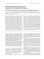
Different combinatorial strategies have been des-
cribed. Figure 1 illustrates the most basic scheme,
where the preparation of five samples leads to the
assignment of every [
15
N]-HSQC cross-peak to one of
K. Ozawa et al.
15
N-labelled proteins by cell-free synthesis
FEBS Journal 273 (2006) 4154–4159 ª 2006 The Authors Journal compilation ª 2006 FEBS 4155
19 amino acid residue types [10]. The five samples are
prepared with different combinations of [
15
N]-labelled
amino acids. The most abundant amino acids are
labelled in only one of the samples, while the least
abundant amino acids are labelled in up to three of the
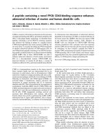
samples. The pattern of occurrence and nonoccurrence
of any particular cross-peak in the [
15
N]-HSQC spectra
recorded of these five samples identifies the amino acid
residue type associated with this cross-peak (Fig. 2).
Fig. 1. Combinatorial isotope labelling scheme. Oval symbols iden-
tify the
15
N-labelled amino acids used in the cell-free preparation of
the five different samples. The last column displays the average
amino acid abundance in proteins according to the NCBI database.
Fig. 2.
15
N-HSQC spectra of five combinatorially
15
N-labelled sam-
ples of the C-terminal 16 kDa domain of the E. coli DNA poly-
merase III subunit s. (A) Overview of the spectra. Numbers in the
top left corner refer to the five different labelling patterns of Fig. 1.
(B) Selected spectral region with all five spectra superimposed. The
pattern of peak occurrence in the different spectra identifies the
amino acid type.
15
N-labelled proteins by cell-free synthesis K. Ozawa et al.
4156 FEBS Journal 273 (2006) 4154–4159 ª 2006 The Authors Journal compilation ª 2006 FEBS
This analysis will be misleading only in situations
where there is complete overlap between two or more
cross-peaks so that they can no longer be distin-
guished from one another. Notably, cross-peak over-
lap is less likely to occur in these spectra, because
each contains only about one third of the cross-peaks
present in the [
15
N]-HSQC spectrum of the corres-
ponding uniformly labelled sample. Not a single case
of complete cross-peak overlap was encountered in the
case of the C-terminal domain of the s subunit of
DNA polymerase III from E. coli, a 16 kDa a-helical
protein [10].
Combinatorial [
15
N]-labelling depends on suppres-
sion of transamination reactions that would otherwise
obscure the labelling pattern. Thus, an early attempt
of combinatorial labelling in vivo had to exclude gluta-
mine, glutamate, asparagine and aspartate from the
labelling scheme because of excessive cross-labelling
[17]. In order to avoid the use of expensive [
15
N]-
amino acids, this particular in vivo labelling scheme
was designed for ‘[
15
N]-unlabelling’, where the protein
was produced on a medium containing inexpensive
15
NH
4
Cl and the [
15
N]-labelling of selected residues
was suppressed by the addition of amino acids at nat-
ural isotopic abundance [17]. In the case of cell-free
protein synthesis, however, the costs of the [
15
N]-
labelled amino acids are hardly limiting, considering
that adequate protein yields can be obtained from, at
most, a couple of milligrams of each amino acid [18].
A more sophisticated combinatorial labelling scheme
has been proposed by Parker et al. [25] based on dual
amino acid selective [
13
C ⁄
15
N]-labelling [12,26]. Five
protein samples were produced where each sample
contained a different combination of 16 [
15
N] or
[
15
N ⁄
13
C]-labelled amino acids. The [
15
N]-labelled
amino acids were used in 50% dilution with amino
acids at natural isotopic abundance, whereas the dou-
bly labelled amino acids were used undiluted. By
recording [
15
N]-HSQC and 2D HNCO spectra of each
sample, [
15
N]-HSQC cross-peaks could be assigned not
only by amino acid type, but also by the amino acid
type of the residue preceding it in the amino acid
sequence. Sequence specific resonance assignments of
the [
15
N]-HSQC peaks are obtained in this way so long
as the corresponding amino acid pairs are unique in
the amino acid sequence. The drawback of this
approach is the significantly larger cost of doubly
labelled amino acids, the requirement for more than
five samples if all 20 amino acids are to be included in
the labelling scheme, the spectral overlap in the [
15
N]-
HSQC spectrum which is the same as for a uniformly
[
15
N]-labelled sample, the need to quantify cross-peak
intensities, and the fact that the sequence specific
assignments will almost always be incomplete because
many amino acid pairs occur more than once in the
amino acid sequence.
The basic combinatorial [
15
N]-labelling scheme of
Fig. 1 provides the benefit of improved spectral resolu-
tion, cost-efficiency and sensitivity (as no dilute label-
ling is employed and no experiments other than
[
15
N]-HSQC spectra are required). It has been shown
that once the residue type assignment of the
[
15
N]-HSQC cross-peaks has been achieved by combi-
natorial [
15
N]-labelling, a single 3D HNCA spectrum
recorded of a uniformly [
15
N ⁄
13
C]-labelled sample can
be sufficient to complete the sequence specific reson-
ance assignment of the backbone amides [10].
Applications
The speed with which cell-free protein synthesis deliv-
ers [
15
N]-HSQC spectra of selectively [
15
N]-labelled
proteins makes it an attractive tool for preliminary
studies prior to the production of uniformly [
15
N ⁄
13
C]-
labelled samples for in-depth NMR analysis. Much
information can be gleaned already from a single selec-
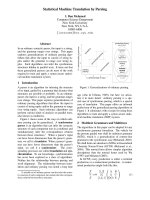
tively labelled sample. For example, binding interac-
tions with other (unlabelled) proteins can readily be
assessed (Fig. 3), as the increase in effective molecular
mass decreases the signal intensities in the [
15
N]-HSQC
spectrum [11].
Similarly, the presence of flexible polypeptide seg-
ments in the protein construct can be assessed by the
observation of intense and narrow [
15
N]-HSQC cross-
peaks. Often, these unstructured segments can be
localized in the amino acid sequence of the protein by
their amino acid composition, which can be derived
from all narrow [
15
N]-HSQC cross-peaks observed in
samples prepared with combinatorial [
15
N]-labelling,
without the need of sequence specific resonance assign-
ments [10].
One of the most attractive applications of combina-
torial [
15
N]-labelling, however, may be for the identifi-
cation of ligand binding sites on proteins with
established sequence specific resonance assignments of
the [
15
N]-HSQC spectrum, where it is often difficult to
assess the magnitude of chemical shift changes upon
ligand binding in [
15
N]-HSQC spectra of uniformly
labelled proteins due to severe spectral overlap [27]. In
this situation, combinatorial [
15
N]-labelling allows the
tracking of the cross-peaks at an effective spectral
resolution equivalent to that of samples prepared with
single [
15
N]-labelled amino acids [10]. Although combi-
natorial labelling requires at least five samples to
obtain complete residue type information, the protein–
ligand interaction can be probed by [
15
N]-HSQC
K. Ozawa et al.
15
N-labelled proteins by cell-free synthesis
FEBS Journal 273 (2006) 4154–4159 ª 2006 The Authors Journal compilation ª 2006 FEBS 4157
spectra of the reaction mixtures, which are quick to
prepare [21].
Conclusion
Over the past few years, cell-free protein synthesis has
been developed into a fast and inexpensive tool for
the production of stable isotope enriched proteins.
Increased amino acid incorporation yields, reduced iso-
tope scrambling and easier sample handling compared
to in vivo protein production render cell-free protein
synthesis particularly attractive for high-throughput
production of proteins and selective isotope labelling
starting from relatively expensive isotope labelled
amino acids. A straightforward combinatorial [
15
N]-
labelling scheme carries particular promise for acceler-
ated studies of proteins by NMR spectroscopy by
assigning residue type information to every amide
cross-peak observed in [
15
N]-HSQC spectra. We antici-
pate that high yield cell-free protein synthesis and
combinatorial isotope labelling will become routine
techniques in high-throughput NMR studies of pro-
teins.
Acknowledgements
GO and KO thank the Australian Research Council
(ARC) for a Federation Fellowship, and an Australian
Linkage (CSIRO) Postdoctoral Fellowship, respect-
ively. Financial support by the ARC for the 800 MHz
NMR facility at ANU is gratefully acknowledged.
References
1 Kigawa T, Muto Y & Yokoyama S (1995) Cell-free
synthesis and amino acid-selective stable isotope label-
ing of proteins for NMR analysis. J Biomol NMR 6,
129–134.
2 Kigawa T, Yabuki T, Yoshida Y, Tsutsui M, Ito Y,
Shibata T & Yokoyama S (1999) Cell-free production
and stable-isotope labeling of milligram quantities of
proteins. FEBS Lett 442 , 15–19.
3 Madin K, Sawasaki T, Ogasawara T & Endo Y (2000)
A highly efficient and robust cell-free protein synthesis
system prepared from wheat embryos: Plants apparently
contain a suicide system directed at ribosomes. Proc
Natl Acad Sci USA 97, 559–564.
4 Yokoyama S (2003) Protein expression systems for
structural genomics and proteomics. Curr Opin Chem
Biol 7, 39–43.
5 Vinarov DA, Lytle BL, Peterson FC, Tyler EM,
Volkman BF & Markley JL (2004) Cell-free protein
production and labeling protocol for NMR-based struc-
tural proteomics. Nat Methods 1, 149–153.
6 Kainosho M, Torizawa T, Iwashita Y, Terauchi T, Ono
AM & Gu
¨
ntert P (2006) Optimal isotope labelling for
NMR protein structure determinations. Nature 440,
52–57.
7 Ozawa K, Dixon NE & Otting G (2005) Cell-free synth-
esis of
15
N-labeled proteins for NMR studies. IUBMB
Life 57, 615–622.
8 Kigawa T, Yamaguchi-Nunokawa E, Kodama K,
Matsuda T, Yabuki T, Matsuda N, Ishitani R,
Fig. 3. Analysis of protein–protein interactions by NMR spectro-
scopy without sequence specific resonance assignment. This
example shows
15
N-HSQC spectra of selectively
15
N-Ala labelled w
in complex with v and c, where w, v and c are subunits of the
E. coli DNA polymerase III complex. (A) w was produced by cell-
free synthesis in the presence of separately purified, unlabelled v;
w produced in the absence of v is insoluble. A cross-peak is
observed for each of the 15 Ala residues of w. The most intense
cross-peaks are from residues with increased mobility. The wide
chemical shift distribution is indicative of a globular folded struc-
ture. The spectrum was recorded at pH 6.9 and 25 °Cona
600 MHz NMR spectrometer (Varian, Palo Alto, CA). The molecular
mass of the w–v complex is about 32 kDa. (B) Spectrum of the
w–v complex recorded in the presence of c. w was selectively
labelled with
15
N-Ala, whereas v and c were unlabelled. Due to the
high molecular mass of the complex (about 150 kDa), most cross-
peaks of w are broadened beyond detection, except for two cross-
peaks from flexible residues. Signals near 112 p.p.m. in the
15
N
dimension arise from highly mobile NH
2
groups of c at natural
isotopic abundance. The spectrum demonstrates that the w–v
complex binds to c. It was recorded at pH 6.9 and 25 °Cona
800 MHz NMR spectrometer (Bruker, Karlsruhe, Germany) [11].
15
N-labelled proteins by cell-free synthesis K. Ozawa et al.
4158 FEBS Journal 273 (2006) 4154–4159 ª 2006 The Authors Journal compilation ª 2006 FEBS
Nureki O & Yokoyama S (2001) Selenomethionine
incorporation into a protein by cell-free synthesis. J
Struct Funct Genomics 2, 29–35.
9 Ozawa K, Headlam MJ, Mouradov D, Watt SJ, Beck
JL, Rodgers KJ, Dean RT, Huber T, Otting G & Dixon
NE (2005) Translational incorporation of l-3,4-dihy-
droxyphenylalanine into proteins. FEBS J 272 , 3162–
3171.
10 Wu PSC, Ozawa K, Jergic S, Su XC, Dixon NE &
Otting G (2006) Amino-acid type identification
in
15
N-HSQC spectra by combinatorial selective
15
N-labelling. J Biomol NMR 34, 13–21.
11 Ozawa K, Jergic S, Crowther JA, Thompson PR,
Wijffels G, Otting G & Dixon NE (2005) Cell-free
in vitro protein synthesis in an autoinduction system for
NMR studies of protein–protein interactions. J Biomol
NMR 32, 235–241.
12 Kainosho M & Tsuji T (1982) Assignment of the
three methionyl carbon resonances in Streptomyces
subtilisin inhibitor by a carbon-13 and nitrogen-15
double labeling technique. A new strategy for struc-
tural studies of proteins in solution. Biochemistry 21,
6273–6279.
13 Le Master DM & Richards FM (1985)
1
H-
15
N hetero-
nuclear NMR studies of Escherichia coli thioredoxin in
samples isotopically labeled by residue type. Biochemis-
try 24, 7263–7268.
14 Griffey RH, Redfield AG, Loomis RE & Dahlquist FW
(1985) Nuclear magnetic resonance observation and
dynamics of specific amide protons in T4 lysozyme.
Biochemistry 24, 817–822.
15 McIntosh LP & Dahlquist FW (1990) Biosynthetic
incorporation of
15
N and
13
C for assignment and inter-
pretation of nuclear magnetic resonance spectra of pro-
teins. Q Rev Biophys 23, 1–38.
16 Yamazaki T, Yoshida M, Kanaya S, Nakamura H &
Nagayama K (1991) Assignments of backbone
1
H,
13
C,
and
15
N resonances and secondary structure of ribonu-
clease H from Escherichia coli by heteronuclear three-
dimensional NMR spectroscopy. Biochemistry 30,
6036–6047.
17 Shortle D (1994) Assignment of amino acid type in
1
H-
15
N correlation spectra by labeling with
14
N-amino
acids. J Magn Reson B 105, 88–90.
18 Ozawa K, Headlam MJ, Schaeffer PM, Henderson BR,
Dixon NE & Otting G (2004) Optimization of Escheri-
chia coli system for cell-free synthesis of selectively
15
N-labelled proteins for rapid analysis by NMR spec-
troscopy. Eur J Biochem 271, 4084–4093.
19 Klammt C, Lo
¨
hr F, Scha
¨
fer B, Haase W, Do
¨
tsch V,
Ru
¨
terjans H, Glaubitz C & Bernhard F (2004) High-
level cell-free expression and specific labeling of integral
membrane proteins. Eur J Biochem 271, 568–580.
20 Morita EH, Shimizu M, Ogasawara T, Endo Y, Tanaka
R & Kohno T (2004) A novel way of amino acid-speci-
fic assignment in
1
H-
15
N HSQC spectra with a wheat
germ cell-free protein synthesis system. J Biomol NMR
30, 37–45.
21 Guignard L, Ozawa K, Pursglove SE, Otting G &
Dixon NE (2002) NMR analysis of in vitro-synthesized
proteins without purification: a high-throughput
approach. FEBS Lett 524, 159–162.
22 Keppetipola S, Kudlicki W, Nguyen BD, Meng X,
Donovan KJ & Shaka AJ (2006) From gene to HSQC
in under five hours: high-throughput NMR proteomics.
J Am Chem Soc 128, 4508–4509.
23 Shi J, Pelton JG, Cho HS & Wemmer DE (2004) Pro-
tein signal assignments using specific labeling and cell-
free synthesis. J Biomol NMR 28, 235–247.
24 Trbovic N, Klammt C, Koglin A, Lo
¨
hr F & Bernhard
&Do
¨
tsch V (2005) Efficient strategy for the rapid back-
bone assignment of membrane proteins. J Am Chem
Soc 127, 13504–13505.
25 Parker MJ, Aulton-Jones M, Hounslow AM & Craven
CJ (2004) A combinatorial selective labeling method for
the assignment of backbone amide NMR resonances.
J Am Chem Soc 126, 5020–5021.
26 Yabuki T, Kigawa T, Dohmae N, Takio K, Terada T,
Ito Y, Laue ED, Cooper JA, Kainosho M & Yokoyama
S (1998) Dual amino acid-selective and site-directed
stable-isotope labeling of the human c-Ha-Ras protein
by cell-free synthesis. J Biomol NMR 11, 295–306.
27 Emerson SD, Palermo R, Liu CM, Tilley JW, Chen L,
Danho W, Madison VS, Greeley DN, Ju G & Fry DC
(2003) NMR characterization of interleukin-2 in com-
plexes with the IL-2Ra receptor component, and with
low molecular weight compounds that inhibit the
IL-2 ⁄ IL–Ra interaction. Protein Sci 12, 811–822.
K. Ozawa et al.
15
N-labelled proteins by cell-free synthesis
FEBS Journal 273 (2006) 4154–4159 ª 2006 The Authors Journal compilation ª 2006 FEBS 4159