Báo cáo khoa học: Interaction between catalytically inactive calpain and calpastatin Evidence for its occurrence in stimulated cells docx
Bạn đang xem bản rút gọn của tài liệu. Xem và tải ngay bản đầy đủ của tài liệu tại đây (348.16 KB, 9 trang )
Interaction between catalytically inactive calpain and
calpastatin
Evidence for its occurrence in stimulated cells
Monica Averna, Roberto Stifanese, Roberta De Tullio, Enrico Defranchi, Franca Salamino,
Edon Melloni and Sandro Pontremoli
Department of Experimental Medicine (DIMES), Section of Biochemistry and Centre of Excellence for Biomedical Research (CEBR),
University of Genova, Italy
In recent years, information has accumulated on the
3D structure of l-calpain and m-calpain [1–10], as well
as their isolated catalytic cores [11–16]. Much less is
known about the process by which calpain is activated
[3,4,6,17,18]. It is generally accepted that it is initiated
by the binding of calcium to several sites localized in
both calpain subunits and completed by a conforma-
tional change in domain II [19–29]. However, the role
of the two Ca
2+
-binding sites recently identified in this
catalytic domain is still to be defined [23]. More
intriguing is the possibility of detecting calpain activa-
tion in vivo, which has not been previously possible
because of the lack of reliable techniques for evaluat-
ing active calpain species and their intracellular local-
ization.
Identification of autolyzed calpain forms by means
of a specific monoclonal antibody does not seem to be
of a general use, as recent structural acquisitions have
suggested that calpain activation can also proceed
through a reversible process [1,2,4]. The proposed pro-
cedure involving the identification of calpain-degraded
target proteins appears not to be sufficiently specific
because of the very large number of calpain substrates
present in the cell (for reviews see [3,4,9,30]). The
recently devised fluorescence resonance energy transfer
technology has greatly improved the sensitivity, but
not the selectivity, required for the precise evaluation
of calpain activation and activity [31].
In this paper we report that, by means of a specific
monoclonal antibody that recognizes the calpain cata-
lytic domain [32], it is possible to detect conformation-
al changes in the calpain molecule that occur after it
binds to its natural effectors. Two conformational
states of calpain can be distinguished on the basis of
their affinity for this mAb: the native state shows low
affinity, whereas binding of specific ligands induces
Keywords
activation; calcium; calpain; calpastatin;
conformational states
Correspondence
S. Pontremoli, DIMES-Section of
Biochemistry, Viale Benedetto XV,
1–16132 Genova, Italy
Fax: +39 010518 343
Tel: +39 010353 8128
E-mail:
(Received 9 January 2006, accepted 15
February 2006)
doi:10.1111/j.1742-4658.2006.05180.x
Conformational changes in the calpain molecule following interaction with
natural ligands can be monitored by the binding of a specific monoclonal
antibody directed against the catalytic domain of the protease. None of
these conformational states showed catalytic activity and probably repre-
sent intermediate forms preceding the active enzyme state. In its native
inactive conformation, calpain shows very low affinity for this monoclonal
antibody, whereas, on binding to the ligands Ca
2+
, substrate or calpasta-
tin, the affinity increases up to 10-fold, with calpastatin being the most
effective. This methodology was also used to show that calpain undergoes
similar conformational changes in intact cells exposed to stimuli that
induce either a rise in intracellular [Ca
2+
] or extensive diffusion of calpast-
atin into the cytosol without affecting Ca
2+
homeostasis. The fact that the
changes in the calpain state are also observed under the latter conditions
indicates that calpastatin availability in the cytosol is the triggering event
for calpain–calpastatin interaction, which is presumably involved in the
control of the extent of calpain activation through translocation to specific
sites of action.
1660 FEBS Journal 273 (2006) 1660–1668 ª 2006 The Authors Journal compilation ª 2006 FEBS
transition to a conformation with significantly higher
affinity. The most extensive conformational change is
induced by calpastatin; the addition of substrate or
Ca
2+
proved to be less effective. Using this methodo-
logy, we have shown similar molecular transitions in
calpain in intact cells stimulated with agents known to
induce either a limited increase in intracellular [Ca
2+
]
or extensive redistribution and accumulation of cal-
pastatin in the cytosolic compartment. These data sug-
gest a new role for calpastatin in controlling the extent
of calpain translocation to and activation at specific
sites of action.
Results
To study the interaction of calpain with its natural
effectors, the purified protease isolated from human
erythrocytes was immobilized on a nitrocellulose
sheet and detected by a specific mAb that recognizes
the DI-DII polypeptide in both native calpain and the
fragment that accumulates during trypsin digestion
[14,24] (Fig. 1A). In Fig. 1B we provide evidence that
calpain bound to nitrocellulose reacts with mAb 56.3,
generating a light signal the intensity of which is a
function of the amount of mAb used, with a saturation
value at a concentration equal to 0.5–0.75 lg antibody.
After exposure of the immobilized calpain to a mixture
of Ca
2+
and a digestible substrate, catalytic activity
can be detected, demonstrating that immobilization on
the nitrocellulose sheet does not modify its catalytic
properties. This is demonstrated by the data in
Fig. 1C, which indicate that the catalytic activity of
immobilized calpain, as a function of Ca
2+
concentra-
tion, is 50% of the maximal at 25 lm Ca
2+
and
maximal at 100 lm Ca
2+
, as occurs when soluble
native enzyme is used [3,4,9,20,30]. Furthermore,
inhibition of the immobilized enzyme by E64 or
calpastatin was retained (Fig. 1D). The efficiency of
both inhibitors was identical with that observed in a
control assay using soluble enzyme (data not shown).
Together these results indicate that immobilized cal-
pain is an appropriate tool for the study of the effects
of natural ligands in changing its conformation, and
that these can be monitored by evaluating the intensity
of the light signal generated by the binding of mAb
56.3.
To perform these investigations, the two preferential
ligands of calpain, Ca
2+
ions and calpastatin, were tes-
ted. In the presence of Ca
2+
concentrations ranging
from zero to 5 lm (close to physiological values), a
twofold increase in the intensity of the signal was
detected, in the absence of any appreciable proteolytic
activity (Fig. 2). The concentrations of Ca
2+
used in
these experiments were selected to avoid undesired and
confusing changes in calpain structure such as mole-
cular aggregations or dissociation of the oligomers,
which have been shown to occur at higher concentra-
tions of the metal ion [45]. The addition of E64, a syn-
thetic inhibitor of calpain, did not modify the intensity
of the signal observed in the presence of Ca
2+
alone.
The increase in the mAb-binding capacity of calpain
observed in these conditions can be ascribed to
increased accessibility of the calpain epitope recognized
by this mAb. As this conformational transition does
not lead to the expression of catalytic activity, these
structural changes must precede the calpain active
state.
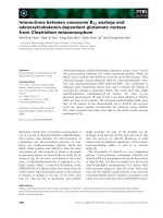
As shown in Fig. 3, when calpain was exposed to
increasing concentrations of recombinant calpastatin
RNCAST104, there was a much greater increase in the
A
B
CD
Fig. 1. Properties of nitrocellulose-immobilized native human eryth-
rocyte calpain. (A) Human erythrocyte calpain (10 lg) was incubat-
ed in the absence (lane 1) or presence of trypsin in a
calpain ⁄ trypsin ratio of 1000 : 1 (lane 2). Western blot analysis was
performed [41] using mAb 56.3. (B) Immobilized calpain was incu-
bated in 0.1 mL 50 m
M sodium borate buffer, pH 7.5, containing
10 m
M EDTA and the indicated amounts of mAb 56.3. The immu-
noreactive spots (see inset) were quantified with a Shimadzu
CS9000 densitometer and expressed as arbitrary units. (C) Immobil-
ized calpain (0.5 lg) was assayed as described in [33] using human
denatured globin as substrate for 60 min at 37 °C in the presence
of the indicated [Ca
2+
]. The nitrocellulose sheet was removed
before the addition of trichloroacetic acid (7% final concentration).
Calpain activity was quantified after the release of free NH
2
groups
as in [33]. (D) Activity of immobilized calpain was assayed as in (C)
in the presence of 1 m
M Ca
2+
and 10 lM E64 or 25 nmol rat brain
recombinant calpastatin RNCAST104.
M. Averna et al. Calpain–calpastatin interaction in stimulated cells
FEBS Journal 273 (2006) 1660–1668 ª 2006 The Authors Journal compilation ª 2006 FEBS 1661
light signal (7–8-fold) than when calpain was exposed
to Ca
2+
alone (Fig. 2). Moreover, the maximum effect
was reached with 10 nmol RNCAST104, which corres-
ponds approximately to a 1 : 1 protease ⁄ inhibitor
molar ratio. The addition of Ca
2+
even at a concen-
tration of 5 lm did not affect the RNCAST104-medi-
ated increase in light emission.
Thus in contrast with what occurs in the presence of
Ca
2+
, the addition of equimolar amounts of calpasta-
tin induces an increase of approximately one order of
magnitude in the affinity of calpain for mAb 56.3,
indicating that, in these conditions, the mAb epitope
on the protease has become more accessible.
These data not only indicate that calpastatin induces
a pronounced Ca
2+
-independent change in calpain
conformation, but also provide strong support for pre-
vious observations indicating that calpain and calpast-
atin can associate in a 1 : 1 molar ratio, regardless of
the presence of Ca
2+
(unpublished work).
In addition to Ca
2+
and calpastatin, a number of
digestible substrates can behave as calpain ligands and
may accordingly induce changes in the conformation
of the protease detectable by the mAb binding. To
investigate this, we exposed immobilized calpain to
digestible and nondigestible proteins and evaluated
their efficiency in promoting conformational change in
the protease. BSA, a protein not digested by calpain,
had no effect at any concentration tested. Casein, a cal-
pain substrate [4,9,30], induced a progressive increase
in the binding of mAb 56.3 to calpain, as revealed by
a 2.5–3-fold increase in the light signal at a concentra-
tion of 2 mgÆmL
)1
(Fig. 4).
The observations so far reported indicate that cal-
pain can exist in two freely convertible inactive confor-
mations. The former is mostly present in the absence
of any effector, and the other is induced, with different
degrees of efficiency, by interaction with micromolar
Fig. 2. Effect of Ca
2+
on binding of mAb 56.3 to nitrocellulose-
immobilized calpain. Immobilized calpain was incubated in 0.1 mL
50 m
M sodium borate buffer, pH 7.5, in the presence of 10 mM
EDTA or the indicated Ca
2+
concentration (d). After saturation,
mAb 56.3 was added and the binding of the mAb to calpain was
measured as described in Experimental procedures and the legend
to Fig. 1B and expressed as arbitrary units. Alternatively, immobi-
lized calpain was exposed to Ca
2+
in the presence of 10 lM E64
(s). Immobilized calpain was also incubated in 0.1 mL 50 m
M
sodium borate buffer, pH 7.5, containing the indicated Ca
2+
concen-
tration in the presence of human denatured globin as substrate,
and its activity was measured as described in the legend to Fig. 1C
(n). The results are expressed as the arithmetical mean ± SD from
four different experiments.
Fig. 3. Effect of calpastatin on the binding of mAb 56.3 to immobil-
ized calpain. Immobilized calpain was exposed to increasing con-
centrations of recombinant rat brain calpastatin RNCAST104 in the
presence of 10 m
M EDTA (h)or5lM Ca
2+
(d). The binding of
mAb 56.3 to immobilized calpain was measured as described in
Figs 1 and 2. The data obtained in these experiments were also
analyzed by Scatchard plot (inset), using the relationship [B] ⁄ [F] ¼
k(B
max
– B), where [B] is the bound ligand concentration, [F] is the
free ligand concentration, B
max
is the maximum ligand amount, and
k is the affinity constant.
+RNCAST104
Fig. 4. Effect of calpain digestible or nondigestible proteins on the
binding of mAb 56.3 to immobilized calpain. Immobilized calpain
was mixed with 10 m
M EDTA (control) in the presence of 10 pmol
rat brain recombinant calpastatin RNCAST104 (black bar) or the indic-
ated amounts of BSA (light grey bars) or casein (heavy grey bars).
The mAb bound to immobilized calpain was detected as described
in Experimental procedures and quantified as reported in the
legend to Fig. 2.
Calpain–calpastatin interaction in stimulated cells M. Averna et al.
1662 FEBS Journal 273 (2006) 1660–1668 ª 2006 The Authors Journal compilation ª 2006 FEBS
(physiological amounts) Ca
2+
concentrations, a digest-
ible protein substrate, and finally calpastatin.
The finding that calpastatin was the most efficient
ligand at promoting calpain transition, detected as an
increase in mAb that bound to calpain, is consistent
with the fact that the protease has an affinity for its
protein inhibitor that is more than 10 000-fold higher
than that for its substrates.
When native calpain was replaced with the autolyzed
75-kDa form, identical results were obtained in all the
experimental conditions tested (data not shown). This
indicates that the removal of part of the DI and DV
domain from the calpain molecule does not abolish the
conformational transition described above and may
explain the Ca
2+
dependence of the autolyzed enzyme
[34].
We then explored whether, in stimulated cells, cal-
pain undergoes conformational changes that could be
detected by mAb 56.3 binding. For this purpose,
human neutrophils were stimulated with the chemotac-
tic peptide f-Met-Leu-Phe, which is known to promote
intracellular mobilization of Ca
2+
[46–48]. As shown
in Fig. 5A,B and quantified in Fig. 5C, under these
conditions, an approximately 10-fold increase in fluor-
escence emission was detected, indicating that calpain
had undergone a transition from the low to the high
affinity mAb-binding form. Interestingly, these results
obtained in vivo were almost superimposable on those
obtained in vitro after exposure of native calpain to
calpastatin. This suggests that the same process is
operating in both experimental situations.
To establish if the data for human neutrophils could
be reproduced in a different cell line, we used murine
erythroleukemia (MEL) cells stimulated with the Ca
2+
ionophore A23187. In resting cells, calpain was poorly
stained by the mAb, indicating that it was mainly pre-
sent in the low-affinity form (Fig. 6A). Scanning the
fluorescence throughout the cell revealed that the pro-
tease is quite homogeneously diffuse throughout the
cytosol. After stimulation with the Ca
2+
ionophore
(Fig. 6B), the intensity of calpain staining increased 7–
8-fold, indicating that the protease was now mainly
present in the high-affinity form.
In these conditions, although the highest amount of
calpain is still in the cytosol, a small fraction is locali-
zed at the membrane, as indicated by the two small
fluorescent peaks detectable at both sides of the cell
scan. This further confirms that translocation to the
A
C
B
Fig. 5. Binding of mAb 56.3 to calpain in human neutrophils stimul-
ated with f-Met-Leu-Phe. Purified neutrophils (10
7
cells) were incub-
ated at 37 °C for 10 min in 10 m
M Hepes, pH 7.5, (10 mL),
containing 140 m
M NaCl, 5 mM MgCl
2
,5mM glucose and 50 lM
Ca
2+
in the absence (A) or presence (B) of 1 lM f-Met-Leu-Phe.
Cells were fixed and permeabilized as described in Experimental
procedures. They were then exposed to mAb 56.3, and its binding
to calpain was detected by confocal microscopy, after incubation
with fluorescein-labeled secondary antibody. (C) Fluorescence was
measured as described in Experimental procedures. The data repre-
sent the arithmetical mean ± SD of four different experiments.
A
B
Fig. 6. Binding of mAb 56.3 to calpain in MEL cells loaded with
Ca
2+
. MEL cells (10
7
cells) were incubated at 37 °C for 10 min in
10 m
M Hepes, pH 7.5 (10 mL), containing 140 mM NaCl, 5 mM
MgCl
2
,5mM glucose and 50 lM Ca
2+
in the absence (A) or pres-
ence (B) of 1 l
M A23187 Ca
2+
ionophore. Cells were fixed and per-
meabilized as described in Experimental procedures. They were
then exposed to mAb 56.3, and its binding to calpain was detected
by confocal microscopy, after incubation with fluorescein-labeled
secondary antibody. The fluorescence detected in each section
(0.5 lm) is shown at the right of each picture. The arrow points to
the calpain ring around the cell.
M. Averna et al. Calpain–calpastatin interaction in stimulated cells
FEBS Journal 273 (2006) 1660–1668 ª 2006 The Authors Journal compilation ª 2006 FEBS 1663
plasma membrane is an obligatory step in the directing
of the protease to its sites of action, the preferred cal-
pain substrates being transmembrane or membrane-
associated proteins [3,4,30].
We still required direct evidence of the nature of the
ligand responsible for the observed conformational
transition. Thus, to discriminate between the effect due
to the rise in free Ca
2+
from that induced by the inter-
action of calpain with calpastatin, we stimulated Jur-
kat cells with arachidonate, which is known to induce
apoptosis without producing, during the early phase
of stimulation, appreciable changes in intracellular
free [Ca
2+
] [49,50]. As previously observed (Fig. 7B)
in these cells, stimulation with ionophore A23187
promoted a calpain-mediated fluorescence increase of
7–8-fold. However, a sixfold increase in fluorescence
intensity was observed after brief stimulation with
arachidonate, indicating that a similar conformational
transition of calpain can be obtained in conditions in
which intracellular Ca
2+
homeostasis is almost unaf-
fected [49,50]. These findings excluded the involvement
of Ca
2+
in the conformational change in calpain,
strongly suggesting that it is the interaction with cal-
pastatin that is responsible for the observed effects.
Other ligands such as digestible substrates were exclu-
ded a priori because they would never be present in the
cytosol at suitable concentrations.
The different calpain fluorescence observed in control
(Fig. 7A) and arachidonate-stimulated (Fig. 7C) cells
after detection with the calpain mAb can be ascribed to
the presence of large amounts of calpastatin which,
after stimulation, becomes freely available in the cyto-
sol for interaction with calpain.
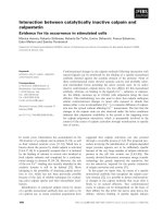
This hypothesis is confirmed by the effect of arachi-
donate treatment on the intracellular distribution of
calpastatin (Fig. 8). In untreated cells, cytosol contains
a very limited amount of calpastatin, the bulk of the
inhibitor being localized in perinuclear aggregates.
After stimulation with arachidonate, the cell image is
completely reversed, as calpastatin becomes freely
diffuse in the cytosol and only traces of aggregates
remain located in the perinuclear region. Thus, the
increased availability of calpastatin, occurring in con-
ditions of unmodified Ca
2+
homeostasis, clearly indi-
cates that the ligand responsible for the changes in
intracellular calpain conformation is its natural inhib-
itor, calpastatin.
The new conformational state acquired by calpain in
these experimental conditions may represent an interme-
diate, but still inactive, form which is stabilized by inter-
action with ligands, the most efficient being calpastatin.
Discussion
A major problem in understanding the physiological
role of calpain is the reliability of techniques capable
Fig. 7. Binding of mAb 56.3 to calpain in Jurkat cells loaded with
Ca
2+
or stimulated with arachidonate. Jurkat cells (10
7
cells) were
incubated at 37 °C for 10 min in 10 m
M Hepes, pH 7.5 (10 mL),
containing 140 m
M NaCl, 5 mM MgCl
2
,5mM glucose and 50 lM
Ca
2+
in the absence (A) or presence of (B) 1 lM A23187Ca
2+
-iono-
phore or (C) 100 l
M arachidonate. Cells were fixed and permeabil-
ized as described in Experimental procedures. They were then
exposed to mAb 56.3, and its binding to calpain was detected by
confocal microscopy, after incubation with fluorescein-labeled sec-
ondary antibody. The data represent the arithmetical mean ± SD of
four different experiments. The arrow points to the calpain ring
around the cell.
Fig. 8. Effect of arachidonate on the intracellular distribution of cal-
pastatin in Jurkat cells. Jurkat cells were incubated with arachido-
nate as described in the legend to Fig. 7C. After 30 min of
incubation, cells were fixed, and calpastatin was probed with
7 lgÆmL
)1
mAb 35.23 [35] followed by a fluorescein-labeled second-
ary antibody. Fluorescence was quantified using the software as
described in Experimental procedures. Cell nuclei were stained
with propidium iodide. The arrows indicate the perinuclear calpasta-
tin aggregates. C ¼ control; Ar. ¼ arachidonate.
Calpain–calpastatin interaction in stimulated cells M. Averna et al.
1664 FEBS Journal 273 (2006) 1660–1668 ª 2006 The Authors Journal compilation ª 2006 FEBS
of detecting the changes in its conformation that
accompany its activation and regulation in specific cell
compartments. In spite of several attempts to solve this
problem [19–29], no precise information is available,
because of the inadequacy of the methods so far pro-
posed for evaluating the interaction of calpain with
natural ligands, a process that must occur in defined
intracellular compartments and presumably precedes
activation of the protease. We explored the possibility
of approaching this problem by taking advantage of
the different accessibility of a calpain epitope to a spe-
cific mAb. We established that this short amino-acid
sequence is confined to the catalytic domain of the
protease, a region known to undergo profound con-
formational rearrangements [21–23], leading to expres-
sion of the catalytic activity.
In preliminary experiments we demonstrated that
the affinity of calpain for its mAb increases after expo-
sure to various ligands. These changes in the condi-
tions preserving the native conformation of the
protease were interpreted as the result of a molecular
transition converting the native form into a state in
which the mAb epitope sequence becomes more acces-
sible.
In this paper we report that the exposure of calpain
to micromolar physiological concentrations of Ca
2+
,
or a digestible protein substrate, or calpastatin is fol-
lowed by a change in its conformation, which can be
monitored by an increase in its affinity for its mAb.
Calpain does not show catalytic activity in any of these
conditions, indicating that this molecular transition
precedes the onset of the active enzyme form. Using a
Scatchard plot as a calibration curve, we established
that calpastatin promotes the conversion of almost all
the calpain molecules into the high-affinity conforma-
tion, whereas the other ligands promote the transition
of only 20–30% of the calpain molecules. Thus, this
procedure provides a tool for the identification of the
calpain states generated by its interaction with natural
ligands.
This methodology was successfully applied to intact
cells, and the results show that similar conformational
changes in calpain occur after stimulation with appro-
priate effectors.
In human neutrophils and MEL cells, even though
the limited increase in Ca
2+
could be regarded as the
event that promoted these changes, two relevant find-
ings suggest a different conclusion. The first concerns
the extent of the increase in calpain fluorescence, which
could not be induced by Ca
2+
alone as indicated in the
in vitro experiments (Figs 2 and 3). The second is rela-
ted to the availability of calpastatin in the cytosol, the
concentration of which increases when Ca
2+
homeosta-
sis is perturbed, as previously reported [43]. Thus, the
pronounced change in calpain conformation revealed
by the high fluorescence reached can only be attributed
to the interaction of calpain with calpastatin.
Stimulation of Jurkat cells with arachidonate provi-
ded experimental evidence in favor of this hypothesis
as it simultaneously promoted mobilization of calpast-
atin accompanied by a marked transition in the cal-
pain molecule, suggesting its interaction with the
inhibitor. Moreover, these events occur in the early
phase of Jurkat cell stimulation, in which no evidence
for alteration in Ca
2+
homeostasis has been obtained
[49,50]. Thus, the effect of arachidonate suggests that
conditions promoting calpain–calpastatin interaction in
the cytosol can be Ca
2+
independent and precede cal-
pain activation. Additional information was obtained
using this methodology in experiments with MEL cells
stimulated with a Ca
2+
ionophore. Analysis of the dis-
tribution of calpain in the cells revealed that, in addi-
tion to the massive conformational change in the
enzyme present in the cytosol, an increase in intracellu-
lar [Ca
2+
] also induces translocation of a small
amount of the protease to the plasma membrane.
These results are in agreement with the accepted evi-
dence that an increase in intracellular [Ca
2+
] induces
degradation of some proteins specifically localized at
the inner surface of the plasma membrane [3,4,30] and
with the observation that the extent of calpain activa-
tion at the plasma membrane is a function of the
amount of cytosolic calpastatin [43].
Taken together, these findings suggest that the for-
mation of a calpain–calpastatin complex before the
onset of calpain activation is functionally relevant, not
only for the modulation of calpain activation in the
cytosol, but also for controlling the amount of calpain
translocated to and activated at the plasma membrane.
Experimental procedures
Materials
Ca
2+
ionophore A23187, f-Met-Leu-Phe, arachidonic acid,
BSA, casein, nonfat skimmed milk powder, trypsin, and
E64 were purchased from Sigma Aldrich (Milan, Italy).
Purification of human erythrocyte calpain and
recombinant rat brain calpastatin
Human erythrocyte calpain was purified and assayed as
reported in [33]. Autoproteolyzed human erythrocyte cal-
pain (75 kDa) was prepared as described in [34]. Rat brain
recombinant calpastatin RNCAST104 (GenBank accession
number Y13588) was prepared as indicated in [35].
M. Averna et al. Calpain–calpastatin interaction in stimulated cells
FEBS Journal 273 (2006) 1660–1668 ª 2006 The Authors Journal compilation ª 2006 FEBS 1665
Monoclonal antibodies
The mAb against calpain (mAb 56.3) was obtained as
reported in [32]. It showed inhibitory properties when
added to a calpain activity assay mixture [32,36]. The
mAb against calpastatin (mAb 35.23) was produced as
described in [35].
Cell culture and human neutrophil isolation
MEL cells were obtained and cultured as specified in [37].
Jurkat cells (human T-lymphocyte line) were cultured at
37 °C (5% CO
2
) in RPMI 1640 (Sigma Aldrich) growth
medium containing 10% fetal bovine serum (Euroclone
Ltd, UK), 10 UÆmL
)1
penicillin (Sigma Aldrich),
100 lgÆmL
)1
streptomycin (Sigma Aldrich) and 4 mml-glu-
tamine. Human neutrophil isolation was based on a modifi-
cation [38] of the procedure described in [39].
Calpain digestion by trypsin
Human erythrocyte calpain (10 lg) was incubated in 50 lL
50 mm sodium borate buffer, pH 7.5, containing 0.5 mm 2-
mercaptoethanol and 1 mm EDTA for 60 min at room tem-
perature with or without trypsin in a calpain ⁄ trypsin ratio of
1000 : 1 [14]. After 60 min, 50 lL 120 mm Tris ⁄ HCl,
pH 6.8, containing 4% SDS, 4% 2-mercaptoethanol and
20% glycerol was added to the incubation mixture and then
heated for 3 min at 100 °C. Samples (50 lL) were submitted
to SDS ⁄ PAGE (10% gel) [40], and western blot was per-
formed as indicated in [41]. Proteins were probed with mAb
56.3. The immunoreactive material was revealed as reported
in [42].
Calpain immobilization
The procedure for immobilization of human erythrocyte
calpain is summarized in Scheme 1. The purified enzyme
(0.5 lgin5lL50mm sodium borate buffer, pH 7.5, con-
taining 0.1 mm EDTA) was spotted on a nitrocellulose
sheet (0.5 cm · 0.5 cm; Bio-Rad Laboratories, Bio-Rad Ita-
lia, Milan, Italy) and left for 15 min at 4 °C in a humidified
chamber. The sheet was then washed with 1 mm EDTA
and saturated with 5% nonfat skimmed milk powder. The
nitrocellulose sheet was then incubated in 0.1 mL sodium
borate buffer, pH 7.5, containing the calpain ligands in the
conditions specified elsewhere. The mixtures were incubated
at 4 °C for 30 min. mAb 56.3 (0.2 lg) was then added.
Calpain was detected using a peroxidase-conjugated secon-
dary antibody [42] developed with an ECL
Ò
detection sys-
tem (Amersham Pharmacia Biotech).
Immunoreactive material was detected by subjecting the
probed nitrocellulose sheets to autoradiography and quanti-
fied with a Shimadzu CS9000 densitometer using a fixed
wavelength of 590 nm.
Binding of mAb 56.3 to intracellular calpain
revealed by confocal microscopy and
fluorescence quantification
Control or stimulated MEL cells, Jurkat cells or human neu-
trophils were washed with NaCl ⁄ P
i
. Calpain and calpastatin
were detected by confocal analysis as described in [43], using
mAb 56.3 or mAb 35.23 respectively as primary antibody
and a fluorescein isothiocyanate-conjugated sheep anti-
mouse IgG as secondary antibody (Amersham Biosciences
Europe Gmbh, Milan, Italy). The excitation ⁄ emission wave-
lengths were 488 ⁄ 522 nm for fluorescein-labeled antibodies
and 488–568 ⁄ 605 nm for propidium iodide-stained chroma-
tin [44]. Fluorescence was quantified with Laser Pix Software
(Bio-Rad Bioscience).
Acknowledgements
This work was supported in part by grants from
MIUR, FIRB and PRIN projects, and from the Uni-
Scheme 1.
Calpain–calpastatin interaction in stimulated cells M. Averna et al.
1666 FEBS Journal 273 (2006) 1660–1668 ª 2006 The Authors Journal compilation ª 2006 FEBS
versity of Genova. We thank Mr Roberto Minafra for
his skilful technical assistance.
References
1 Moldoveanu T, Campbell RL, Cuerrier D & Davies PL
(2004) Crystal structures of calpain–E64 and –leupeptin
inhibitor complexes reveal mobile loops gating the act-
ive site. J Mol Biol 343, 1313–1326.
2 Hosfield CM, Elce JS & Jia Z (2004) Activation of cal-
pain by Ca
2+
: roles of the large subunit N-terminal and
domain III–IV linker peptides. J Mol Biol 343, 1049–
1053.
3 Pontremoli S, Melloni E & Salamino F (1999) Cal-
pain: from structure to biological function. In Calcium
as a Cellular Regulation (Carafoli E & Klee C, eds),
pp. 371–388. Oxford University Press, New York.
4 Goll DE, Thompson VF, LIH, Wei W & Cong J (2003)
The calpain system. Physiol Rev 83, 731–801.
5 Pal GP, De Veyra T, Elce JS & Jia Z (2003) Purificat-
ion, crystallization and preliminary X-ray analysis of a
mu-like calpain. Acta Crystallogr D Biol Crystallogr 59 ,
369–371.
6 Maki M, Kitaura Y, Satoh H, Ohkouchi S & Shibata H
(2002) Structures, functions and molecular evolution
of the penta-EF-hand Ca
2+
-binding proteins. Biochim
Biophys Acta 1600, 51–60.
7 Reverter D, Braun M, Fernandez-Catalan C, Strobl S,
Sorimachi H & Bode W (2002) Flexibility analysis
and structure comparison of two crystal forms of cal-
cium-free human m-calpain. Biol Chem 383, 1415–
1422.
8 Dainese E, Minafra R, Sabatucci A, Vachette P, Mello-
ni E & Cozzani I (2002) Conformational changes of cal-
pain from human erythrocytes in the presence of Ca
2+
.
J Biol Chem 277, 40296–40301.
9 Sorimachi H & Suzuki K (2001) The structure of cal-
pain. J Biochem (Tokyo) 129, 653–664.
10 Masumoto H, Nakagawa K, Iria S, Sorimachi H,
Suzuki K, Bourenkov GP, Bartunik H, Fernandez-Cata-
lan C, Bode W & Strobl S (2000) Crystallization and
preliminary X-ray analysis of recombinant full-length
human m-calpain. Acta Crystallogr D Biol Crystallogr
56, 73–75.
11 Pal G, De Veyra T, Elce JS & Jia Z (2003) Crystal
structure of a micro-like calpain reveals a partially acti-
vated conformation with low Ca
2+
requirement. Struc-
ture (Camb) 11, 1521–1526.
12 Leinala EK, Arthur JS, Grochulski P, Davies PL, Elce
JS & Jia Z (2003) A second binding site revealed by C-
terminal truncation of calpain small subunit, a penta-
EF-hand protein. Proteins 53, 649–655.
13 Wang C, Barry JK, Min Z, Tordsen G, Rao AG &
Olsen OA (2003) The calpain domain of the maize
DEK1 protein contains the conserved catalytic triad
and functions as a cysteine proteinase. J Biol Chem 278,
34467–34474.
14 Thompson VF, Lawson KR, Barlow J & Goll DE
(2003) Digestion of mu- and m-calpain by trypsin and
chymotrypsin. Biochim Biophys Acta 1648, 140–153.
15 Brandenburg K, Harris F, Dennison S, Seydel U &
Phoenix D (2002) Domain V of m-calpain shows the
potential to form an oblique-orientated alpha-helix,
which may modulate the enzyme’s activity via interac-
tions with anionic lipid. Eur J Biochem 269, 5414–
5422.
16 Cygler M, Grochulski P & Blauchard H (2002) Crystal-
lization and structural details of Ca
2+
-induced conform-
ational changes in the EF-hand domain VI of calpain.
Methods Mol Biol 172, 243–260.
17 Moldoveanu T, Hosfield CM, Lim D, Jia Z & Davies
PL (2003) Calpain silencing by a reversible intrinsic
mechanism. Nat Struct Biol 10, 371–378.
18 Nakagawa K, Masumoto H, Sorimachi H & Suzuki K
(2001) Dissociation of m-calpain subunits occurs after
autolysis of the N-terminus of the catalytic subunit, and
is not required for activation. J Biochem (Tokyo) 130,
605–611.
19 Moldoveanu T, Jia Z & Davies PL (2004) Calpain acti-
vation by cooperative Ca
2+
binding at two non-EF-
hand sites. J Biol Chem 279, 6106–6114.
20 Dutt P, Spriggs CN, Davies PL, Jia Z & Elce JS
(2002) Origins of the difference in Ca
2+
requirement
for activation of mu- and m-calpain. Biochem J 367,
263–269.
21 Moldoveanu T, Hosfield CM D.Lim Elce JS & Davies
PL (2002) A Ca
2+
switch aligns the active site of cal-
pain. Cell 108, 649–660.
22 Jia Z, Hosfield CM, Davies PL & Elce JS (2002) Crystal
structure of calpain and insights into Ca
2+
-dependent
activation. Methods Mol Biol 172, 51–67.
23 Hata S, Sorimachi H, Nakagawa K, Maeda T, Abe K
& Suzuki K (2001) Domain II of m-calpain is a
Ca
2+
-dependent cysteine protease. FEBS Lett 501,
111–114.
24 Michetti M, Salamino F, Minafra R, Melloni E & Pon-
tremoli S (1997) Calcium-binding properties of human
erythrocyte calpain. Biochem J 325, 721–726.
25 Tompa P, Emori Y, Sorimachi H, Suzuki K & Friedrich
P (2001) Domain III of calpain is a Ca
2+
-regulated
phospholipid-binding domain. Biochem Biophys Res
Commun 280, 1333–1339.
26 Hosfield CM, Moldoveanu T, Davies PL, Elce JS & Jia
Z (2001) Calpain mutants with increased Ca
2+
sensitiv-
ity and implications for the role of the C(2)-like domain.
J Biol Chem 276, 7404–7407.
27 Strobl S, Fernandez-Catalan C, Braun M, Huber R,
Masumoto H, Nakagawa K, Irie A, Sorimachi H,
Bourenkow G, Bartunik H, et al. (2000) The crystal
structure of calcium-free human m-calpain suggests an
M. Averna et al. Calpain–calpastatin interaction in stimulated cells
FEBS Journal 273 (2006) 1660–1668 ª 2006 The Authors Journal compilation ª 2006 FEBS 1667
electrostatic switch mechanism for activation by cal-
cium. Proc Natl Acad Sci USA 97, 588–592.
28 Hosfield CM, Elce JS, Davies PL & Jia Z (1999) Crystal
structure of calpain reveals the structural basis for
Ca
2+
-dependent protease activity and a novel mode of
enzyme activation. EMBO J 18, 6880–6889.
29 Blanchard H, Grochulski P, Li Y, Arthur JS, Davies
PL, Elce JS & Cygler M (1997) Structure of a calpain
Ca
2+
-binding domain reveals a novel EF-hand and
Ca
2+
-induced conformational changes. Nat Struct Biol
4, 532–538.
30 Croall DE & De Martino GN (1991) Calcium-activated
neutral protease (calpain) system: structure, function,
and regulation. Physiol Rev 71, 813–847.
31 Stockolm D, Bartoli M, Sillon G, Bourg N, Davoust J
& Richard I (2005) Imaging calpain protease activity by
multiphoton FRET in living mice. J Mol Biol 346, 215–
222.
32 Pontremoli S, Melloni E, Damiani G, Salamino F,
Sparatore B, Michetti M & Horecker BL (1988) Effects
of a monoclonal anti-calpain antibody on responses of
stimulated human neutrophils. Evidence for a role for
proteolytically modified protein kinase C. J Biol Chem
263, 1915–1919.
33 Melloni E, Sparatore B, Salamino F, Michetti M &
Pontremoli S (1982) Cytosolic calcium-dependent protei-
nase of human erythrocytes: formation of an enzyme-
natural inhibitor complex induced by Ca
2+
ions.
Biochem Biophys Res Commun 106, 731–740.
34 Michetti M, Salamino F, Tedesco I, Averna M, Minafra
R, Melloni E & Pontremoli S (1996) Autolysis of
human erythrocyte calpain produces two active enzyme
forms with different cell localization. FEBS Lett 392 ,
11–15.
35 Melloni E, De Tullio R, Averna M, Tedesco I, Salami-
no F, Sparatore B & Pontremoli S (1998) Properties of
calpastatin forms in rat brain. FEBS Lett 431, 55–58.
36 Pontremoli S, Sparatore B, Salamino F, De Tullio R,
Pontremoli R & Melloni E (1988) The role of calpain in
the selective increased phosphorylation of the anion-
transport protein in red cell of hypertensive subjects.
Biochem Biophys Res Commun 151, 590–597.
37 Melloni E, Pontremoli S, Damiani G, Viotti PL, Weich
N, Rifkind RA & Marks PA (1988) Vincristine-resistant
erythroleukemia cell line has marked increased sensitiv-
ity to hexamethylene bisacetamide-induced differentia-
tion. Proc Natl Acad Sci USA 85, 3835–3839.
38 De Tullio R, Stifanese R, Salamino F, Pontremoli S &
Melloni E (2003) Characterization of a new p94-like cal-
pain form in human lymphocytes. Biochem J 375, 689–
696.
39 Boyum A (1968) Isolation of mononuclear cells and gra-
nulocytes from human blood. Scan J Clin Lab Invest
21, 77–89.
40 Laemmli U.K. (1970) Cleavage of structural proteins
during the assembly of the head of bacteriophage T4.
Nature 227, 680–685.
41 Towbin J, Staehelin T & Gordon J (1979) Electrophore-
tic transfer of proteins from polyacrylamide gels to
nitrocellulose sheets: procedure and some applications.
Proc Natl Acad Sci USA 76, 4350–4354.
42 Palejwala S & Goldsmith LT (1992) Ovarian expression
of cellular Ki-ras p21 varies with physiological status.
Proc Natl Acad Sci USA 89, 4202–4206.
43 DeTullio R, Passalacqua M, Averna M, Salamino F,
Melloni E & Pontremoli S (1999) Changes in intracellu-
lar localization of calpastatin during calpain activation.
Biochem J 343, 467–472.
44 Coligan SE, Knisbeck AM, Margulies DH, Shevach
EM & Strober W (1991) Current Protocols in Immunol-
ogy. Wiley, New York.
45 Pal GP, Elce JS & Jia Z (2001) Dissociation and aggre-
gation of calpain in the presence of calcium. J Biol
Chem 276, 47233–47238.
46 Anderson R, Steel HS & Tintinger GR (2005) Inositol
1,4,5-triphosphate-mediated shuttling between intracellu-
lar stores and the cytosol contributes to the sustained
elevation in cytosolic calcium in FMLP-activated
human neutrophils. Biochem Pharmacol 69, 1567–1575.
47 Thompson NT, Bonser RW, Tateson JE, Spacey GD,
Randall RW, Hodson HF et al. (1991) A quantitative
investigation into the dependence of Ca
2+
mobilisation
on changes in inositol 1,4,5-triphosphate levels in the
stimulated neutrophil. Br J Pharmacol 103, 1592–1596.
48 Steel HC & Anderson R (2002) Dissociation of the
PAF-receptor from NADPH oxidase and adenylate
cyclase in human neutrophils results in accelerated
influx and delayed clearance of cytosolic calcium. Br J
Pharmacol 136, 81–89.
49 Doroshenko N & Doroshenko P (2004) Ca
2+
influx is
not involved in acute cytotoxicity of arachidonic acid.
Biochem Pharmacol 67, 903–909.
50 Farooqui AA, Ong WY & Horrocks LA (2004) Bio-
chemical aspects of neurodegeneration in human brain:
involvement of neural membrane phospholipids and
phospholipases A2. Neurochem Res 29, 1961–1977.
Calpain–calpastatin interaction in stimulated cells M. Averna et al.
1668 FEBS Journal 273 (2006) 1660–1668 ª 2006 The Authors Journal compilation ª 2006 FEBS