Báo cáo khoa học: SHIP2 interaction with the cytoskeletal protein Vinexin ppt
Bạn đang xem bản rút gọn của tài liệu. Xem và tải ngay bản đầy đủ của tài liệu tại đây (712.31 KB, 15 trang )
SHIP2 interaction with the cytoskeletal protein Vinexin
Nathalie Paternotte
1
, Jing Zhang
1
, Isabelle Vandenbroere
1
, Katrien Backers
1
, Daniel Blero
1
,
Noriyuki Kioka
2
, Jean-Marie Vanderwinden
3
, Isabelle Pirson
1
and Christophe Erneux
1
1 Interdisciplinary Research Institute (IRIBHM), Universite
´
Libre de Bruxelles, Brussels, Belgium
2 Division of Applied Life Sciences, Graduate School of Agriculture, Kyoto University, Japan
3 Laboratoire de Neurophysiologie, Universite
´
Libre de Bruxelles, Brussels, Belgium
The ubiquitous src homology 2 (SH2) domain
containing inositol 5-phosphatase 2 (SHIP2) dephos-
phorylates phosphatidylinositol 3,4,5-trisphosphate
[PtdIns(3,4,5)P
3
] in vitro [1,2]. The reaction product
catalysed by SHIP2 is phosphatidylinositol 3,4-bisphos-
phate [PtdIns(3,4)P
2
]. SHIP2 is a member of the
inositol 5-phosphatase family, and with the SH2
domain containing inositol 5-phosphatase 1 (SHIP1), is
Keywords
cellular adhesion; phosphatidylinositol
3,4,5-trisphosphate; SHIP2; signal
transduction; Vinexin
Correspondence
C. Erneux, Institute of Interdisciplinary
Research (IRIBHM), Campus Erasme
Building C, 808 Route de Lennik,
1070 Brussels, Belgium
Fax: + 32 2 555 4655
Tel: + 32 2 555 4162
E-mail:
(Received 25 August 2005, accepted
28 September 2005)
doi:10.1111/j.1742-4658.2005.04996.x
The src homology 2 (SH2) domain-containing inositol 5-phosphatase 2
(SHIP2) catalyses the dephosphorylation of phosphatidylinositol 3,4,5-tris-
phosphate [PtdIns(3,4,5)P
3
] to phosphatidylinositol 3,4-bisphosphate
[PtdIns(3,4)P
2
]. We report the identification of the cytoskeletal protein
Vinexin as a protein interacting with SHIP2. This was achieved by yeast
two-hybrid screening using the C-terminal region of SHIP2 as bait. Vinexin
has previously been identified as a vinculin-binding protein that plays a key
role in cell spreading and cytoskeletal organization. The interaction
between SHIP2 and Vinexin was confirmed in lysates of both COS-7 cells
and mouse embryonic fibroblasts (MEF). The C-terminus was involved in
the interaction, as shown by the transfection of a truncated C-terminus
mutant of SHIP2. In addition, we showed the colocalization between Vine-
xin a and SHIP2 at the periphery of transfected COS-7 cells. When added
in vitro to SHIP2, Vinexin did not affect the PtdIns(3,4,5)P
3
5-phosphatase
activity of SHIP2. Enhanced cell adhesion to collagen-I-coated dishes was
shown upon transfection of either SHIP2 or Vinexin to COS-7 cells. This
effect was no longer observed with either a catalytic mutant or the C-termi-
nus mutant of SHIP2. It also appears SHIP2 specific; this was not seen
with SHIP1. Adhesion to the same matrix was decreased in SHIP2– ⁄ –
MEF cells compared with MEF+ ⁄ + cells. Our data suggest that SHIP2
interaction with Vinexin promotes the localization of SHIP2 at the peri-
phery of the cells leaving its catalytic site intact. The complex formation
between Vinexin and SHIP2 may increase cellular adhesion. The data rein-
force the concept that SHIP2 is active both as a PtdIns(3,4,5)P
3
5-phospha-
tase and as a modulator of focal contact formation.
Abbreviations
AD, activation domain; BSA, bovine serum albumin; CAP, c-Cbl-associated protein; CHO-IR, Chinese hamster ovary cells overexpressing the
insulin receptor; DBD, DNA-binding domain; DMEM, Dulbecco’s modified Eagle’s medium; EGF, epidermal growth factor; FBS, foetal bovine
serum; GST, glutathione S-transferase; HGF, hepatocyte growth factor; IGF, insulin-like growth factor; Ins(1,4,5)P
3
, inositol 1,4,5-
trisphosphate; IP, immunoprecipitation; MAP, mitogen-activated protein; M-CSF, macrophage colony-stimulating factor; MEF, mouse
embryonic fibroblast; NRS, normal rabbit serum; PBS, phosphate buffer solution; PDGF, platelet-derived growth factor; PI3K,
phosphoinositide 3-kinase; PtdIns(3,4)P
2
, phosphatidylinositol 3,4-bisphosphate; PtdIns(3,4,5)P
3
, phosphatidylinositol 3,4,5-trisphosphate;
PtdIns(4,5)P
2
, phosphatidylinositol 4,5-bisphosphate; PKB, protein kinase B; PTB, phosphotyrosine binding; PTEN, phosphatase and tensin
homologue deleted on chromosome 10; SAM, sterile alpha motif; SH2, src homology 2; SH3, src homology 3; SHIP, SH2 domain containing
inositol 5-phosphatase.
6052 FEBS Journal 272 (2005) 6052–6066 ª 2005 The Authors Journal compilation ª 2005 FEBS
a type II mammalian inositol 5-phosphatase [3–5]. S HIP1
and SHIP2, a s well a s t he phosphatase an d t ensin homo-
logue deleted on chromosome 10 (PTEN), reduce the
signalling pathway(s) mediated by the phosphoinositide
3-kinase (PI 3K) product PtdIns(3,4,5)P
3
. Both SHIP1
and SHIP2 contain a series of protein-interacting
domains. Both proteins possess a SH2 domain at their
N-terminal end, a catalytic domain in the central part,
potential phosphotyrosine-binding (PTB) consensus
sequences (NPXY) and proline-rich sequences at the
C-terminal end. Although SHIP1 and SHIP2 are com-
parable in their N-terminal regions, i.e. the SH2
domain and catalytic region, they clearly differ (in their
proline-rich sequences) in the C-terminal region. It has
been reported that SHIP2 binds selectively to the SH3
domain of Abl, whereas SHIP1 binds to the SH3
domain of Src [6]. In addition, only SHIP2 contains a
sterile alpha motif (SAM) domain at the C-terminal
end of the protein. SHIP2 is ubiquitously expressed
both at the mRNA and protein levels [7–9].
SHIP2 appears to be tyrosine phosphorylated by a
very large number of extracellular ligands, for exam-
ple, epidermal growth factor (EGF), platelet-derived
growth factor (PDGF), insulin, macrophage colony-
stimulating factor (M-CSF) and hepatocyte growth
factor (HGF). Moreover, SHIP2 may be involved in
some of these agonist-induced signalling pathways
[10–13]. SHIP2 has also been found in a phosphotyro-
sine complex with several tyrosine kinase receptors
including the EGF receptor [12] and the c-Met recep-
tor [11]. In addition, SHIP2 has been reported to form
a complex with the low-affinity receptor for the Fc
portion of the IgG antibodies, FccRIIB [7,14]. In
HeLa cells, SHIP2 was found to interact with p130
CAS
via its SH2 domain. SHIP2 localizes at lamellipodia
and regulates cell adhesion and spreading [15,16].
Recently, the same authors reported an important role
for SHIP2 in endocytosis and downregulation of the
EGF receptor [17].
In resting human platelets, 20% of SHIP2 cosedi-
mented with the actin cytoskeleton [18]. SHIP2 local-
ization to membrane ruffles is mediated in COS-7 cells
via complex formation via its C-terminal end proline-
rich sequences with the actin-binding protein, filamin
[19]. In Chinese hamster ovary cells over-expressing
the insulin receptor (CHO-IR), SHIP2 has been repor-
ted to interact with c-Cbl and the c-Cbl-associated pro-
tein, CAP [20]. This interaction with the proline-rich
domain of SHIP2 was not established for SHIP1.
Therefore, the proline-rich sequences of SHIP2 may
interact with a complex set of proteins that do not
overlap with those of SHIP1. SHIP2 has also been
shown to interact with c-Cbl in HeLa cells [17].
Studies in knockout mice have provided evidence that
SHIP2 plays a role in enhancing insulin sensitivity and
regulating obesity in vivo [21,22]. This may involve a
change in protein kinase B (PKB) activity as shown in
liver and muscle by the injection of insulin in SHIP2– ⁄ –
and SHIP+ ⁄ + mice [21]. A decrease in PKB activity
has also been observed in SHIP2-transfected cells fol-
lowing stimulation by growth factors or insulin [12,23–
25]. This could in turn affect PKB-dependent events:
SHIP2 causes a potent cell cycle arrest in G
1
in gliobla-
stoma cells [26]. Mitogen-activated protein (MAP) kin-
ase activity has also been shown to be decreased when
SHIP2 was transfected in various cell models [23,27,28].
Biochemical studies of SHIP2 have provided at least
two clear-cut situations. SHIP2 is active only in stimu-
lated cells representing a mechanism of downregulation
of phosphoinositide 3-kinase (PI3K) activation [12,23].
SHIP2 also interacts with several cytoskeletal proteins
and may regulate localized changes in PtdIns(3,4,5)P
3
and the remodelling of submembranous actin [19]. The
interaction between SHIP2 and the cytoskeletal pro-
teins p130
CAS
, filamin and CAP occurs in the absence
of any stimulus [13,15,19,20,29].
This is the first report of an interaction between
SHIP2 and the cytoskeletal protein Vinexin. Vinexin
has previously been reported to be involved in signal
transduction, cellular contacts and adhesion events
[30]. The interaction between SHIP2 and Vinexin is
shown to occur in COS-7 cells and mouse embryonic
fibroblast (MEF) cells. Complex formation between
SHIP2 and Vinexin enables the correct localization of
SHIP2 at cell–matrix adhesion sites and may positively
control cellular adhesion.
Results
Identification of Vinexin as a novel SHIP2 binding
partner
In a previous report, we identified the c-Cbl-associated
protein (CAP) as a protein binding to the proline-rich
sequences of SHIP2. This was achieved by yeast two-
hybrid screening of a human brain cDNA library fused
to the GAL4 transcriptional activation domain using
the C-terminal proline-rich region of SHIP2 as bait [20].
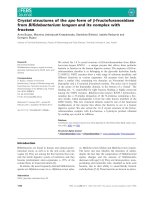
In this experiment, using the same procedure, Vinexin a
was identified as a SHIP2-binding protein: the recovered
library-derived plasmid which encoded a part of the
Vinexin sequence, induced reporter gene expression only
when coexpressed with the GAL4–SHIP2 C-terminal
fusion protein. This was not the case for the
unrelated GAL4-fusion protein, type I Ins(1,4,5)P
3
5-phosphatase protein (Fig. 1A).
N. Paternotte et al. SHIP2 interaction with Vinexin
FEBS Journal 272 (2005) 6052–6066 ª 2005 The Authors Journal compilation ª 2005 FEBS 6053
Vinexin is actually transcribed in two alternative
forms, referred to as a and b [30]. Sequence analysis
has shown that the 1028 bp fragment encoding amino
acids 328–671 of Vinexin a was in-frame with the
GAL4 activation binding domain. This isolated cDNA
encoded the C-terminal part of Vinexin a but lacked
the sorbin-like domain. This clone contained a large
fragment of Vinexin common to both a and b isoforms
(Fig. 1C). Using the same technique, we showed that
Vinexin interacted with the proline-rich domain of
SHIP2 but not with that of SHIP1 (Fig. 1B).
Association of Vinexin a with SHIP2 in transfected
COS-7 cells
The association of Vinexin a and SHIP2 was examined
in COS-7 cells. Vinexin a (the longest isoform) was
chosen rather than Vinexin b because the construct
identified by yeast two-hybrid screening appeared to be
common to Vinexin a and b (Fig. 1C). We compared
the transfection of His vector (pcDNA3His), Vinexin a
(HA-tagged) or SHIP2 (His-tagged) and Vinexin a
(HA-tagged) (Fig. 2A). The apparent molecular mass of
Vinexin a was shown to be 80 kDa, as previously repor-
ted in C2C12 cells [30]. The lysates were immunoprecipi-
tated with normal rabbit serum (NRS) or His antibody.
Vinexin a could be seen only in SHIP2 and Vinex-
in a-transfected immunoprecipitates. When probed with
antibodies to Vinexin, it could not be seen in pcDNA3-
His transfected immunoprecipitate, or when the immu-
noprecipitation was made with NRS (Fig. 2B).
We compared the transfection of either HA-tagged
Vinexin a ⁄ His vector, His-tagged SHIP2 ⁄ HA vector
or the cotransfection of His-tagged SHIP2 and HA-
tagged Vinexin a (Fig. 2C). When the three lysates
were immunoprecipitated with anti-His, Vinexin a was
A
B
C
Fig. 1. Isolation of Vinexin as SHIP2 (SH2
domain containing inositol 5-phosphatase 2)
partner using yeast two-hybrid screening.
(A) Yeast expressing GAL4-DBD fused to
the SHIP2 C-terminus, GAL4-DBD or
GAL4-DBD fused to type I
Ins(1,4,5)P
3
5-phosphatase baits were trans-
formed with GAL4-AD–Vinexin and plated
on medium lacking leucine, tryptophan,
histidine and adenine. (B) Yeast expressing
GAL4-DBD–SHIP2 C-terminus or GAL4-
DBD–SHIP1 C-terminus baits were trans-
formed with GAL4-AD–Vinexin and plated
on medium lacking leucine, tryptophan,
histidine and adenine. (C) Sequence of
human Vinexin a. The SH3 (Src homology 3)
domains are underlined and the SoHo
domain is boxed. Vinexin b starts at Met343
and the cDNA clone isolated from yeast
two-hybrid screening starts at His328. (DBD
DNA-binding domain; AD activation domain)
SHIP2 interaction with Vinexin N. Paternotte et al.
6054 FEBS Journal 272 (2005) 6052–6066 ª 2005 The Authors Journal compilation ª 2005 FEBS
detected only when both SHIP2 and Vinexin a were
transfected (Fig. 2D). In another series of experiments,
lysates were prepared from transfected COS-7 cells
stimulated or not with EGF (100 ngÆmL
)1
) for
3 min. The same amounts of SHIP2 were detected in
anti-His immunoprecipitates from unstimulated and
EGF-stimulated cells (data not shown).
Endogenous Vinexin a and b associate with
SHIP2 in MEF cells
MEF cells express both Vinexin a and b (Fig. 3A,C).
They were used to further confirm the interaction
between Vinexin and SHIP2. Lysates of MEF cells were
immunoprecipitated with Vinexin antibodies or NRS
(Fig. 3B,D). The resulting immunocomplexes were blot-
ted with SHIP2 antibodies. As shown in Fig. 3B,D,
SHIP2 coimmunoprecipitated with endogenous Vinexin
in SHIP2+ ⁄ + MEF cells. As expected, this was not
seen in the negative control SHIP2– ⁄ – MEF cells or
when the immunoprecipitation was carried out with
NRS (Fig. 3B,D, respectively). Equal amounts of these
proteins were immunoprecipitated from MEF cells
(Fig. 3B). The expression of Vinexin and SHIP2 was
shown in whole-cell extracts (Fig. 3A,C).
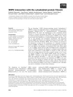
Intracellular localization of SHIP2 and Vinexin a
COS-7 cells were cotransfected with His–SHIP2 and
HA–Vinexin a and stimulated with EGF (100 ngÆmL
)1
)
for 3 min. The data of a representative cell are shown
for SHIP2 and Vinexin a (Fig. 4D–F). As seen previ-
ously [12,19], SHIP2 was shown to be localized at the
periphery of the COS-7 cells, particularly at membrane
ruffles (Fig. 4D). In the absence of EGF, we did not
detect SHIP2 at the periphery of the cells (Fig. 4A).
Vinexin has been shown to be localized at focal adhe-
sion sites as well as cell–cell contact sites in NIH 3T3
cells [30], and similar localization was seen in the
COS-7 cells (Fig. 4B,E). The colocalization of SHIP2
A
B
C
D
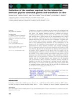
Fig. 2. Association of SHIP2 with Vinexin a in transfected COS-7 cells. (A) Whole-cell lysates (30 lg protein) immunoblotted with Vinexin or
His antibodies. (B) COS-7 cells (8 · 10
5
cells per condition) transfected with pcDNA3His, Vinexin a, SHIP2 and Vinexin a were lysed and
immunoprecipited with His antibodies or normal rabbit serum (NRS). The immunoprecipitates were immunoblotted with antibodies to Vinexin
or His. (C) Whole-cell lysates (30 lg protein) were immunoblotted with Vinexin or His antibodies. (D) COS-7 cells were transfected with
pcDNA3His, pcDNA3HA, SHIP2 His tagged or Vinexin a HA tagged as indicated. The lysates were immunoprecipitated with His antibodies
and immunoblotted with Vinexin or His antibodies. Equal amounts of SHIP2 were immunoprecipitated in the SHIP2-transfected cells.
(IP, immunoprecipitation)
N. Paternotte et al. SHIP2 interaction with Vinexin
FEBS Journal 272 (2005) 6052–6066 ª 2005 The Authors Journal compilation ª 2005 FEBS 6055
and Vinexin at the periphery of the cells (in yellow)
could be detected in EGF-stimulated cells (Fig. 4F)
and not in unstimulated cells (Fig. 4C).
Vinexin b does not affect PtdIns(3,4,5)P
3
5-phosphatase activity of SHIP2
A direct in vitro assay was used to determine whether
Vinexin b could modulate PtdIns(3,4,5)P
3
5-phospha-
tase activity of SHIP2. Purified His-tagged SHIP2 was
used as a source of activity in the presence of either
glutathione S-transferase (GST) or GST–Vinexin b.
PtdIns(3,4,5)P
3
5-phosphatase activity was measured
in the presence of His–SHIP2 and an excess of puri-
fied GST–Vinexin b or with GST alone at 5 lm.
The SHIP2 PtdIns(3,4,5)P
3
5-phosphatase activity
was comparable in the presence or absence of
GST–Vinexin b (data not shown).
C
A
B
D
Fig. 3. Endogenous Vinexin a and b associate with SHIP2 in MEF+ ⁄ + cells. (A, C) Whole-cell extracts (30 lg protein) were immunodetected
with antibodies to SHIP2 and Vinexin. (B, D) Lysates of MEF cells (1.5 mg protein) were immunoprecipitated with Vinexin antibodies or
NRS. The resulting immunoprecipitates were immunodetected with antibodies to SHIP2. The same membrane was also probed with anti-
bodies to Vinexin. (MEF, mouse embryonic fibroblast).
SHIP2 interaction with Vinexin N. Paternotte et al.
6056 FEBS Journal 272 (2005) 6052–6066 ª 2005 The Authors Journal compilation ª 2005 FEBS
Vinexin a did not influence PKB activity
in COS-7 cells
PKB activity has been reported to be inhibited in
SHIP2-transfected cells stimulated by EGF or insulin
[12,23] and this has been interpreted to be as a conse-
quence of a decrease in PtdIns(3,4,5)P
3
levels [12,23].
In this study, we used COS-7 cells stimulated or not
with EGF (100 ngÆmL
)1
). Overexpression of Vinexin a
did not affect basal or stimulated PKB activity (data
not shown).
Adhesion of MEF SHIP2 cells and COS-7 cells
upon SHIP2 and Vinexin a overexpression
Previous data have suggested that transfection of
SHIP2 in HeLa cells increased cell adhesion on colla-
gen I [15]. The mechanism involved requires the tyro-
sine phosphorylation of SHIP2 by a src kinase [16].
COS-7 cells transfected with SHIP2 and MEF cells
deficient for SHIP2 were used to carry out cell adhesion
experiments on culture dishes coated with collagen I.
SHIP2+ ⁄ + and SHIP2– ⁄ – MEF cells were detached
from plates and kept in suspension for 10 min. They
were then replated on collagen I. After 15 min of adhe-
sion, the cells were counted. These experiments showed
that the number of attached SHIP2+ ⁄ + MEF cells
was always higher than the number of SHIP2– ⁄ – fibro-
blast cells (Fig. 5B). Representative immunoblots
showed that there was no change in expression of
Vinexin a between SHIP2+ ⁄ + and – ⁄ – MEF cells
(Fig. 5A). As expected, SHIP2 was not expressed in
MEF– ⁄ – cells.
To determine whether overexpression of SHIP2 and
Vinexin had any influence on cell adhesion, COS-7
cells transfected with either SHIP2 or Vinexin a were
detached from their culture dishes and then replated
onto dishes coated with collagen I (Fig. 5D). The cells
that were attached to the culture dishes were counted
after 15 min adhesion. The number of attached cells
was 18 ± 2.1% (n ¼ 3) for the cells transfected with
the vector alone. This number was markedly increased
in COS-7 cells transfected with either SHIP2
(34 ± 6.1%, n ¼ 3) or Vinexin a (31 ± 4.7%, n ¼ 3).
The overexpression of either SHIP2 or Vinexin a was
verified by western blotting (Fig. 5C). The data suggest
that SHIP2 and Vinexin a increase cell adhesion on
collagen I in transfected COS-7 cells. We have tested
A
BC
FED
Fig. 4. Vinexin a and SHIP2 are colocalized at the periphery of transfected COS-7 cells. COS-7 cells cotransfected with Vinexin a and SHIP2
were stimulated or not with EGF 100 ngÆmL
)1
for 3 min. (A, D) Cells were stained with anti-His sera and fluorescein-labelled anti-mouse sec-
ondary sera. (B, E) Vinexin a was visualized by indirect immunofluorescence using anti-Vinexin sera and Texas-Red-labelled anti-rabbit secon-
dary sera. (C, F) Cells were double stained with anti-His and anti-Vinexin sera. The arrows indicate the colocalization of Vinexin and SHIP2 at
the periphery of the cells (in yellow).
N. Paternotte et al. SHIP2 interaction with Vinexin
FEBS Journal 272 (2005) 6052–6066 ª 2005 The Authors Journal compilation ª 2005 FEBS 6057
the coexpression of SHIP2 and Vinexin a on cell
adhesion but did not get higher adhesion than with
Vinexin a or SHIP2 alone. However, it is possible that
transfection of each condition alone reaches a maximal
adhesion value (data not shown).
In our adhesion assay, the mean value of adherent
cells was 20.33 ± 3.22 and 21.08 ± 5.11% for His
vector transfected or non transfected cells (n ¼ 3),
respectively. Transfection of vector alone does not
modify the percentage of attached cells. Therefore, the
control of SHIP2 (or his mutants) transfected cells
could be the result of either the suggested conditions
with no change in interpretation of the data.
Influence of SHIP2 proline-rich sequences and
catalytic mutant on cell adhesion
A truncated form of SHIP2 that lacked 366 amino
acids at the C-terminus [i.e. the proline-rich sequences
of the protein (DProline SHIP2)] has previously been
shown to be fully active as a PtdIns(3,4,5)P
3
5-phos-
phatase [31] and to be able to lower PtdIns(3,4,5)P
3
levels in EGF-stimulated cells [12]. It was therefore
interesting to compare the effect on cell adhesion of
SHIP2 with the DProline SHIP2; we also compared
the effect of SHIP2 with another construct that did
not have the SHIP2 SH2 domain (DSH2 SHIP2). Cells
expressing these constructs were detached from their
culture dishes and then replated onto dishes coated
with collagen I (Fig. 6). Cells attached to the culture
dishes were counted after 15 min incubation. The num-
ber of adherent cells was 19 ± 2.7% (n ¼ 3, non trans-
fected cells), 36 ± 0.5% (n ¼ 3, cells transfected with
SHIP2) or 29 ± 1.1% (n ¼ 3, cells transfected with
DSH2 SHIP2) (Fig. 6B). By contrast, 17.8 ± 0.1%
(n ¼ 3) cells were attached when the C-terminal-trun-
cated mutant was transfected compared with the value
obtained in SHIP2 transfected cells (50.5 ± 4.5%,
A
B
C
D
Fig. 5. Adhesion assay on MEF SHIP2 cells and on COS-7 cells overexpressing SHIP2 and Vinexin a. (A) Representative control immuno-
blots probed with SHIP2 and Vinexin antibodies. (B) Confluent monolayers of MEF SHIP2+ ⁄ + and – ⁄ – cells were incubated for 15 min at
37 °C in a serum-free medium. The numbers of attached and unattached cells on the dish were counted. The number of attached cells was
expressed as a percentage of the total number of adherent cells before the 15 min collagen I coating. Data are expressed as means ± SEM
(n ¼ 2; *P < 0.05). (C) Representative control immunoblots probed with SHIP2 and Vinexin antibodies. (D) Confluent monolayers of COS-7
transfected cells were incubated for the 15 min at 37 °C in a serum-free medium. The numbers of attached and unattached cells on the dish
were counted. The number of attached cells was expressed as a percentage of the total number of adherent cells before the 15 min colla-
gen I coating. Data are expressed as means ± SEM (n ¼ 3; *P < 0.05).
SHIP2 interaction with Vinexin N. Paternotte et al.
6058 FEBS Journal 272 (2005) 6052–6066 ª 2005 The Authors Journal compilation ª 2005 FEBS
n ¼ 3; Fig. 6D). The control of expression of the var-
ious constructs was confirmed by western blotting with
anti-His sera (Fig. 6A,C). The data suggest that the
C-terminal end of SHIP2 is involved in the increase in
cell adhesion observed in transfected cells. In order to
test the influence of SHIP2 catalytic activity on cell
adhesion, COS-7 cells were transfected with either
SHIP2 or SHIP2 D607A: the number of adherent
cells was 29 ± 3.6% (n ¼ 3, non transfected cells),
27.1 ± 7.5% (n ¼ 3) when the catalytic mutant was
transfected and 60.4 ± 1.6% (n ¼ 3) for the wild-type
SHIP2 (Fig. 7B). Therefore, the catalytic activity of
SHIP2 is taking part in the increase in cell adhesion
measured in SHIP2-transfected cells. Overexpression of
either SHIP2 or SHIP2 D607A was verified by western
blotting (Fig. 7A).
We used lysates cotransfected with both SHIP2
(and our two SHIP2 mutants, i.e. DProline SHIP2
and SHIP2 D607A) and Vinexin a. Using immuno-
precipitation with anti-His, Vinexin a was present in
the SHIP2 and SHIP2 D607A mutant, but was
markedly reduced in DProline SHIP2 immunoprecipi-
tations (Fig. 8B). The overexpression of SHIP2 and
mutants is shown in Fig. 8A. The data are consistent
with Vinexin interaction at the proline-rich sequences
of SHIP2.
To investigate the specificity of SHIP2 in mediating
the increase in cell adhesion, we tested the effect of
SHIP1. The number of adherent cells was 22 ± 6.1%
(n ¼ 3, in non transfected cells) and 39 ± 2.5% (n ¼
3, in cells transfected with SHIP2) (Fig. 7D). By con-
trast, 21 ± 2.1% (n ¼ 3) cells were attached in
SHIP1-transfected cells. The control of expression of
SHIP1 and SHIP2 was confirmed by western blotting
with anti-His and anti-SHIP1 sera (Fig. 7C). In con-
trast to SHIP2, SHIP1 does not increase cellular
adhesion in our model.
Discussion
It has been reported that SHIP2 displays inositol
5-phosphatase activity when PtdIns(3,4,5)P
3
and
A
B
C
D
Fig. 6. Effect of overexpression of SHIP2, DSH2 SHIP2 and DProline SHIP2 on adhesion. (A, C) Representative control immunoblots probed
with His antibodies. (B, D) Confluent monolayers of COS-7 transfected cells were incubated for the 15 min at 37 °C in a serum-free med-
ium. The numbers of attached and unattached cells on the dish were counted. The number of attached cells was expressed as a percentage
of the total number of adherent cells before the 15 min collagen I coating. Data are expressed as means ± SEM (n ¼ 3; *P < 0.05).
N. Paternotte et al. SHIP2 interaction with Vinexin
FEBS Journal 272 (2005) 6052–6066 ª 2005 The Authors Journal compilation ª 2005 FEBS 6059
PtdIns(4,5)P
2
were used as substrates in vitro
[12,18,23,26]. Moreover, the levels of PtdIns(3,4,5)P
3
were decreased in both EGF-stimulated COS-7 cells
and in insulin-stimulated CHO-IR cells transfected
with SHIP2. This has also been observed in rat vascu-
lar smooth muscle cells, where PtdIns(3,4,5)P
3
levels
were decreased in SHIP2-transfected cells stimulated
by PDGF or IGF-1 [28]. Both PKB and MAP kinase
activities have also been shown to decrease in SHIP2-
transfected cells, suggesting that SHIP2 is a downregu-
lator of both arms of receptor tyrosine kinase activa-
tion [12,23]. Consistent with this, is the upregulation of
PKB activity in cells deficient in SHIP2 in response to
M-CSF or serum [13,32]. These data have established
the role of SHIP2 in the acute control of PtdIns(3,4,5)P
3
and PKB activities in stimulated cells.
SHIP2 involvement in cytoskeleton organization was
initially seen in HeLa cells where SHIP2 associates
with p130
CAS
[15]. This association was observed in
cells attached to collagen I, which induces SHIP2 tyro-
sine phosphorylation that is secondary to activation of
src tyrosine kinases [16]. Immunofluorescence studies
have indicated that SHIP2 is localized to focal contacts
and to lamellipodia [15]. In resting human platelets,
20% of SHIP2 was recovered in the cytoskeleton
[18]. In this model, the affinity of SHIP2 for the cyto-
skeleton was always higher than that of SHIP1.
Because the major structural differences between
SHIP2 and SHIP1 are at the C-terminal end of the
proteins, the data suggest that their respective proline-
rich sequences may interact with different SH3-con-
taining protein partners. Indeed, in COS-7 cells, SHIP2
localization to membrane ruffles has been shown to
be mediated via complex formation through its
C-terminal proline-rich sequences with the actin-binding
protein filamin [19]. In another study in platelets,
SHIP2 formed a tetrameric complex with filamin, actin
and GPIb-IX-V [29]. The interaction of SHIP2 with
CAP and c-Cbl has been reported in CHO-IR regard-
less of cell stimulation by insulin [20]. Recently, it has
been reported that SHIP2 has a role in the internal-
ization and degradation of the EGF receptor in HeLa
cells [17]. It is not clear whether this involves SHIP2
phosphatase activity or a scaffolding protein type of
function to facilitate interaction with regulators of
the cytoskeleton. In HGF-stimulated MDCK cells,
cells overexpressing both SHIP2 and SHIP1 formed
lamellipodia at the membrane [11].
The important features of our results can be
summarized as follows. SHIP2 is associated with the
A
B
C
D
Fig. 7. Effect of a catalytic mutant SHIP2 and SHIP1 overexpression on adhesion. (A, C) Representative control immunoblots probed with
His and SHIP1 antibodies. (B, D) Confluent monolayers of COS-7 transfected cells were incubated for the 15 min at 37 °C in a serum-free
medium. The numbers of attached and unattached cells on the dish were counted. The number of attached cells was expressed as in
Fig. 5. Data are expressed as means ± SEM (n ¼ 3; *P < 0.05).
SHIP2 interaction with Vinexin N. Paternotte et al.
6060 FEBS Journal 272 (2005) 6052–6066 ª 2005 The Authors Journal compilation ª 2005 FEBS
cytoskeletal protein Vinexin, this was identified by
yeast two-hybrid screening and confirmed in two
cellular models by coimmunoprecipitation. SHIP2 and
Vinexin a were also shown to colocalize at the peri-
phery of the cells, at least in our model of COS-7 cells
stimulated by EGF. Transfection with either SHIP2
or Vinexin a increased COS-7 cell adhesion to colla-
gen I. This effect was not observed with SHIP1.
Higher cell adhesion was also measured in SHIP2+⁄ +
MEF cells compared with –⁄ – cells [3]. The addition of
Vinexin to purified SHIP2 did not affect SHIP2
PtdIns(3,4,5)P
3
5-phosphatase activity. As proposed
previously for filamin interaction with SHIP2, the
interaction did not appear to block the catalytic acti-
vity of SHIP2 [29].
Vinexin and CAP are members of the same adaptor
protein family and are shown to regulate cytoskeletal
organization and signal transduction cascades [33].
Vinexin, which was identified by yeast two-hybrid
screening as a vinculin-binding protein, also promoted
upregulation of actin stress fiber formation, suggesting
its implication in cytoskeletal organization. C2C12 cell
lines that express Vinexin b in a stable manner have
been shown to enhance cell spreading on fibronectin
[30]. In our study, SHIP2 was shown to be in a com-
plex with Vinexin both in COS-7 transfected cells and
MEF cells. Stimulation of the cells with EGF did not
modulate the association of the two proteins although
SHIP2 tyrosine phosphorylation was increased. This is
in agreement with previous results obtained with CAP
in stimulated COS-7 or CHO-IR cells [20]. Therefore,
the interaction of Vinexin with the proline-rich domain
of SHIP2 is not influenced by its tyrosine phosphoryla-
tion. In HeLa cells, Vinexin b is found in a complex of
proteins containing both SHIP2 and filamin (data not
shown) and the association of SHIP2 and p130
CAS
has
been reported by others [15]. A recent study of
ArgBP2, another adaptor protein of the same family
of Vinexin [33], shows that numerous interactors of
ArgBP2 are actin-regulatory proteins including dynam-
in or synapsin [34]. Therefore, rather than proposing
an interaction with a one protein partner Vinexin, we
prefer to suggest that SHIP2 interacts with several pro-
tein partners in the cytoskeleton network.
In our study, transfection of either SHIP2 or Vinex-
in a in COS-7 cells increased cellular adhesion to
collagen-I-coated dishes. Similar results for SHIP2
on adhesion have been obtained before in HeLa cells
[15]. In that model, p130
CAS
was identified as a protein
partner of SHIP2. This interaction with p130
CAS
involved the SHIP2 SH2 domain [15]. The catalytic
domain of SHIP2 appeared to be dispensable for
increased adhesion and a Dproline SHIP2 mutant was
not tested [15]. SHIP2+ ⁄ + and – ⁄ – MEF cells in our
study showed a difference in cell adhesion; adhesion
was always higher in SHIP2+ ⁄ + cells in which we
demonstrated complex formation between Vinexin and
SHIP2. This also suggests that the result is not an arte-
fact caused by overexpression and validates the data
we obtained in SHIP2-transfected COS-7 cells. Inter-
estingly, p130
CAS
, CAP, Vinexin and vinculin have
been found to be associated with the cell–matrix adhe-
sion sites in a network of protein-like integrins and
F-actin [35]. Our hypothesis is that the interaction of
SHIP2 with Vinexin (and perhaps other non enzymatic
components of the cell–matrix adhesion site) promotes
the localization of SHIP2 at cell–matrix adhesion sites
leaving its catalytic site intact. This complex formation
between Vinexin and SHIP2 may activate vinculin and
increase cell adhesion. A truncated form of SHIP2 that
lacked the C-terminal proline-rich sequences did not
increase cellular adhesion. This mutant did also not
interact with Vinexin in coimmunoprecipitation experi-
A
B
Fig. 8. Interaction between Vinexin a and DProline SHIP2 or SHIP2
D607A. (A) The whole cell lysates (30 lg proteins) were immuno-
blotted with either Vinexin a or His antibodies. (B) COS-7 cells
(8 · 10
5
cells ⁄ condition) were cotransfected with Vinexin a and
SHIP2 or mutants. Lysates were immunoprecipited with His anti-
bodies and immunoblotted with antibodies to either Vinexin or His.
N. Paternotte et al. SHIP2 interaction with Vinexin
FEBS Journal 272 (2005) 6052–6066 ª 2005 The Authors Journal compilation ª 2005 FEBS 6061
ments. This suggested that the C-terminal end of
SHIP2 is not correctly localized and is therefore no
longer able to increase cell adhesion. In contrast, the
SHIP2 SH2 domain deleted mutant behaved as a
wild-type, suggesting that the SH2 domain was not
involved, at least in our model of COS-7 cells. Interest-
ingly, transient reduction of PTEN expression by
RNAi has been shown to induce loss of adhesion in
HEK293 cells [36]. Therefore, it appears that both
SHIP2 and PTEN may increase cellular adhesion.
Whether the changes in cell adhesion that we
observed are linked to changes in the local cellular lev-
els of PtdIns(4,5)P
2
or PtdIns(3,4,5)P
3
is currently
unknown. In our adhesion assay, transfection of a cata-
lytic mutant of SHIP2 did not result in an increase in
cell adhesion compared with transfection of the wild-
type SHIP2. This suggests the involvement of SHIP2
catalytic activity in the mechanism of cell attachment
and therefore changes in PtdIns(3,4,5)P
3
levels. This
may occur in a second step after the correct localization
of a complex between Vinexin and SHIP2. Therefore,
the interaction of Vinexin and SHIP2 could be a link
between the role of Vinexin in cytoskeleton organiza-
tion and PtdIns(3,4,5)P
3
metabolism.
Experimental procedures
Materials
Monoclonal anti-His6 serum and the adult human brain
Matchmaker library were purchased from BD Biosciences
(Erembodegem, Belgium). Monoclonal anti-phosphotyro-
sine 4G10 and anti-PKB sera (directed against the PH
domain) were purchased from Upstate (Veenendaal, the
Netherlands). Collagen I was from Sigma (Bornem,
Belgium), Fugene, complete protease inhibitor cocktail
and Triton X-100 were from Roche (Vilvoorde, Belgium).
The rabbit polyclonal C-terminal SHIP2 antibodies have
been reported and characterized previously [7,31]. The
rabbit polyclonal antibody against Vinexin a and b has
been reported previously [30]. Protein G–Sepharose, horse-
radish peroxidase-linked secondary antibodies and ECL
kit were obtained from Amersham Pharmacia Biotech
(Roosendaal, the Netherlands). Easitides [
32
P]ATP[cP]
(3000 CiÆmmol
)1
) was from NEM (Perkin Elmer, Zaven-
tem, Belgium). DiC
8
-PtdIns(3,4,5)P
3
was from Echelon
Biosciences Incorporated (Salt Lake City, UT). The
cDNAs encoding SHIP1, type I inositol 5-phosphatase
and SHIP2 have been reported previously [12,31,37,38].
The constructions of the two mutants of SHIP2 in
pcDNA3His, lacking the SH2 domain (DSH2) and lack-
ing the proline-rich domain (DProline) have been repor-
ted previously [12]. The SHIP2 catalytic mutant
SHIP2 D607A was generated by PCR-based mutagenesis
using the QuickChange XL Site-Directed mutagenesis kit
from Stratagene (La Jolla, CA, USA). It was shown to
be inactive in transfected COS-7 cells in an assay of
PtdIns(3,4,5)P
3
5-phosphatase activity [31]. SHIP2+ ⁄ +
and – ⁄ – MEF cells were isolated as described previously
[32]. COS-7 cells were transfected with pcDNA3His–
SHIP2 to produce SHIP2. His-tagged SHIP2 was purified
using Talon metal affinity resin (BD Biosciences, Erem-
bodegem, Belgium) and eluted with 20 mm imidazole.
SHIP2 was identified by SDS ⁄ PAGE and Coomassie Blue
or western blotting. It was shown to be active with both
DiC
8
-PtdIns(3,4,5)P
3
and Ins(1,3,4,5)P
4
as substrate.
GST–Vinexin b [30] and GST alone were produced in
Escherichia coli, purified with glutathione agarose (Sigma)
and eluted with 10 mm gluthatione. PKB activity was
determined as described previously [23].
Yeast two-hybrid screening
The cDNA encoding the proline-rich domain (SHIP2
C-term, from Tyr956 to Pro1248) was cloned by PCR
downstream from the Gal4 DNA-binding domain in the
yeast two-hybrid vector pGBT9. The proline-rich domain
of SHIP1 (SHIP1 C-term, from Tyr864 to Val1077) and
the full-length type I Ins(1,4,5)P
3
5-phosphatase (type I,
from Met1 to Gln412) were cloned by PCR downstream
from the Gal4 DNA-binding domain in the yeast two-
hybrid vector pGBT9. The constructions were verified by
DNA sequencing. The yeast host strain used for the
screening and the reconstruction steps was the pJ69-4 A
strain (MAT a, ade 2 trp 1-D901 leu 2–3, 112 ura 3–52 his
3–200 Gal4D Gal80D LYS2::Gal1-HIS3 ADE2::Gal2-
ADE2 met1::Gal7-LACZ). To screen, the pJ69-4 A har-
boring pGBT9-SHIP2 C-terminal was transformed with an
adult human brain Matchmaker library in pACT2. The
transformants were first selected on aHIS medium and
then on aADE and finally reconstructed for specificity of
the bait [20].
Expression vector constructs
A HA epitope-tagged full-length Vinexin a in a eukaryotic
expression vector was constructed as follows. The cDNA of
mouse Vinexin a was amplified by PCR with Vinexin a
cDNA in p401FlagdeltaB as the template [30]. The 5¢-pri-
mer 5¢-CGC
GGATCCGGAATGGCCAGGATTCTTGG
AGTGGGA-3¢ was designed to have a BamHI restriction
site (underlined). The 3¢-primer 5¢-CCG
GAATTCTCA
CACTGGGGCTACATAATTTCC-3¢ contained an EcoRI
restriction site (underlined). The amplified DNA frag-
ment was digested with BamHI and EcoRI and ligated
in the pcDNA3-HA vector digested with BamHI and
EcoRI. The sequence of the HA-tagged Vinexin a cDNA
(HA–Vinexin a) in this vector was confirmed by DNA
sequencing.
SHIP2 interaction with Vinexin N. Paternotte et al.
6062 FEBS Journal 272 (2005) 6052–6066 ª 2005 The Authors Journal compilation ª 2005 FEBS
Cell lines
MEF fibroblasts were maintained in Dulbecco’s modified
Eagle’s medium (DMEM) supplemented with 10% fetal
bovine serum (FBS), 2% penicillin-streptomycin, 1% gluta-
mine, 1% sodium pyruvate and 0.1% b-mercaptoethanol.
COS-7 cells were cultured in DMEM supplemented with
10% FBS, 2% penicillin–streptomycin, 1% glutamine, 1%
sodium pyruvate and 1% fungizone.
Transfection of COS-7 cells
COS-7 cells (plated at 8 · 10
5
cells per dish one day before
the transfection) were transiently transfected in 10 cm
dishes using the Fugene method of transfection according
to the manufacturer’s instructions. Cells were stimulated or
not with 100 ngÆ mL
)1
EGF at 37 °C for 3 min. Cells were
washed with sterile NaCl ⁄ P
i
and lysed with a buffer con-
taining 80 mm Tris ⁄ HCl pH 7.5, 150 mm NaCl, 20 mm
EDTA, 1% Brij, 5 mm sodium pyrophosphate, 4 mm
sodium orthovanadate, 200 mm NaF, 10 nm okadaic acid
and complete protease inhibitor cocktail tablets. After sha-
king the lysates for 60 min at 4 °C and centrifugation at
10 000 g for 20 min at 4 °C, the supernatants were recov-
ered and immediately subjected to immunoprecipitation or
western blotting.
Immunoprecipitation and immunoblotting
After centrifugation at 10 000 g, the supernatants were pre-
cleared for 1 h at 4 °C with protein G–Sepharose. This was
centrifuged at 10 000 g for 5 min at 4 °C. The soluble frac-
tion was collected and incubated with the specified anti-
bodies for 3 h at 4 °C. After centrifugation, the immune
complexes were washed extensively with lysis buffer before
they were dissolved in Laemmli sample buffer. Bound pro-
teins were separated by SDS ⁄ PAGE and transferred to
nitrocellulose membranes. Individual proteins were detected
with the specified antibodies and visualized by blotting with
horseradish peroxidase-linked secondary antibodies and
developed using the ECL kit.
Malachite phosphatase assay
PtdIns(3,4,5)P
3
5-phosphatase activity was determined with
the phosphate release assay using an acidic malachite green
dye [39]. Di-C
8
PtdIns(3,4,5)P
3
was diluted in 30 lL assay
buffer [50 mm Hepes pH 7.4, 2 mm MgCl
2
,1mgÆmL
)1
bovine serum albumin (BSA)]. The phosphatase reaction
was initiated by adding SHIP2 diluted in assay buffer
(15 lL) to the substrate. Samples were incubated at 37 °C.
After 7 min, reactions were stopped by the addition of
15 lL 0.1 m EDTA. 75 lL of malachite green reagent
(0.5% (w ⁄ v) ammonium molybdate, 0.057% (v ⁄ v) Tween-20
and 0.34 mm malachite green oxalate) were added to 50 lL
of the reaction solution. Samples were left for 10 min for
colour development before measuring absorbance at
650 nm. Inorganic phosphate release was quantified by com-
parison with a standard curve of KH
2
PO
4
in H
2
O. Linearity
of the assay was checked with respect to time and protein
concentration. The enzymatic blank was prepared by adding
first the EDTA solution and then the enzyme to the sub-
strate. Each value resulted from duplicate determinations.
Cell adhesion assay
The efficiency of cell adhesion was determined by measu-
ring the number of cells that adhered to a matrix [40,41].
Cell culture dishes (6 cm dishes) were coated with colla-
gen I (50 lgÆmL
)1
) diluted in NaCl ⁄ P
i
overnight at 37 °C.
Nonspecific binding sites were blocked by incubation for
1 h at 37 °C with a solution of BSA (10 mgÆmL
)1
)in
NaCl ⁄ P
i
. Cells cultured in dishes in the presence of serum
until they had achieved a confluent monolayer were
detached by exposure to trypsin, collected by centrifuga-
tion, and washed once with medium containing 10% FBS
and once with NaCl ⁄ P
i
. They were then replated on culture
dishes in medium without serum, coated with collagen I,
and incubated for 15 min at 37 °C in serum-free medium
and in a humidified atmosphere containing 5% CO
2
. After
washing the cells once with NaCl ⁄ P
i
, the number of cells
that were attached or unattached to the dish was counted.
Confocal immunofluorescence microscopy
Immunofluorescence using anti-His and anti-Vinexin sera
was performed on transfected COS-7 cells. We grew
3.75 · 10
5
transfected COS-7 cells on uncoated glass cover-
slips in 10-cm diameter dishes. Cells were stimulated with
EGF, rinsed in NaCl ⁄ P
i
and then fixed in 100% methanol
for 10 min. The cells were washed in NaCl ⁄ P
i
three times
for 10 min each time, made permeable with 0.15% Tri-
ton X-100 in NaCl ⁄ P
i
for 10 min, and washed again with
NaCl ⁄ P
i
. The fixed cells were incubated for 1 h at room
temperature with 1% BSA in NaCl ⁄ P
i
. Incubation with
immune serum was performed overnight at room tempera-
ture in 1% BSA in NaCl ⁄ P
i
. The anti-His serum was used at
a 1 : 200 dilution, and the anti-Vinexin serum was used at a
1 : 500 dilution. After being rinsed with NaCl ⁄ P
i
, cells were
incubated for 60 min in the dark with a fluorescein-labelled
anti-mouse secondary serum (anti-His) and with a Texas
Red-labelled anti-rabbit secondary serum (anti-Vinexin) at a
1 : 200 dilution. The cells were then washed three times with
NaCl ⁄ P
i
for 10 min and mounted with the SlowFade light
antifade kit (Molecular Probes, Leiden, the Netherlands)
following the manufacturer’s instructions. Cells were
observed under a LSM510 NLO confocal microscope fitted
on an Axiovert M200 inverted microscope equipped with a
N. Paternotte et al. SHIP2 interaction with Vinexin
FEBS Journal 272 (2005) 6052–6066 ª 2005 The Authors Journal compilation ª 2005 FEBS 6063
C-Apochromat 40·⁄1.2 NA water immersion objective
(Zeiss, Iena, Germany). A 2.5–5· electronic zoom was used
across regions of interest. The 488 nm excitation wavelength
of the Argon ⁄ 2 laser, a main dichroic HFT 488 and a band-
pass emission filter (BP500–550 nm) were used for selective
detection of the green fluorochrome (fluorescein). The
543 nm excitation wavelength of the HeNe1 laser, a main
dichroic HFT488 ⁄ 543 ⁄ 633 and a long-pass emission filter
(LP560 nm) were used for selective detection of the red
fluorochrome (Texas Red). Optical sections, 2.5 lm thick,
were collected sequentially for each fluorochrome. The ima-
ges generated (512 · 512 pixels, pixel size: 0.14 lm) were
merged and displayed with the Zeiss lsm510 software and
exported in .jpg image format. All Figures showed a single
optical section across the regions of interest.
Acknowledgements
We would like to thank Mrs Colette Moreau, Drs
Xavier Pesesse, Gilles Doumont and Fabrice Vandeput
for many helpful discussions and experimental help.
This work was supported by grants from the Fonds de
la Recherche Scientifique Me
´
dicale (Belgium), Action
de Recherche Concerte
´
e of the Communaute
´
Franc¸ aise
de Belgique. J. M. Vanderwinden is Senior Research
Associate of the National Fund for Scientific Research
(Belgium) and Associated Partner in IUAP ⁄ PAI5⁄ 20
from the Belgian Federal Science Policy Office. Natha-
lie Paternotte was supported by a fellowship of
F.R.I.A. (Fonds pour la formation a
`
la recherche dans
l’industrie et dans l’agriculture), Te
´
le
´
vie, the Fondation
universitaire David et Alice Van Buuren (Belgium) and
the Fondation Rose et Jean Hoguet (Belgium).
References
1 Ishihara H, Sasaoka T, Hori H, Wada T, Hirai H,
Haruta T, Langlois WJ & Kobayashi M (1999) Mole-
cular cloning of rat SH2-containing inositol phosphatase
2 (SHIP2) and its role in the regulation of insulin signal-
ing. Biochem Biophys Res Commun 260, 265–272.
2 Pesesse X, Deleu S, De Smedt F, Drayer L & Erneux C
(1997) Identification of a second SH2-domain-contain-
ing protein closely related to the phosphatidylinositol
polyphosphate 5-phosphatase SHIP. Biochem Biophys
Res Commun 239, 697–700.
3 Erneux C, Govaerts C, Communi D & Pesesse X (1998)
The diversity and possible functions of the inositol poly-
phosphate 5-phosphatases. Biochim Biophys Acta 1436,
185–199.
4 Majerus PW (1996) Inositols do it all. Genes Dev 10,
1051–1053.
5 Mitchell CA, Brown S, Campbell JK, Munday AD &
Speed CJ (1996) Regulation of second messengers by
the inositol polyphosphate 5-phosphatases. Biochem Soc
Trans 24, 994–1000.
6 Wisniewski D, Strife A, Swendeman S, Erdjument-
Bromage H, Geromanos S, Kavanaugh WM, Tempst P
& Clarkson B (1999) A novel SH2-containing phospha-
tidylinositol 3,4,5-trisphosphate 5-phosphatase (SHIP2)
is constitutively tyrosine phosphorylated and associated
with src homologous and collagen gene (SHC)in
chronic myelogenous leukemia progenitor cells. Blood
93, 2707–2720.
7 Muraille E, Pesesse X, Kuntz C & Erneux C (1999) Dis-
tribution of the src-homology-2-domain-containing inosi-
tol 5-phosphatase SHIP-2 in both non-haemopoietic and
haemopoietic cells and possible involvement of SHIP-2 in
negative signalling of B-cells. Biochem J 342, 697–705.
8 Kudo M, Saito S, Owada Y, Suzaki H & Kondo H
(2000) Localization of mRNA for SHIP2, SH2 domain-
containing inositol polyphosphate 5-phosphatase, in the
brain of developing and mature rats. Brain Res Mol
Brain Res 75, 172–177.
9 Schurmans S, Carrio R, Behrends J, Pouillon V, Merino
L & Clement S (1999) The mouse SHIP2 (InppI1) gene:
complementary DNA, genomic structure, promoter ana-
lysis, and gene expression in the embryo and adult
mouse. Genomics 62, 260–271.
10 Habib T, Hejna JA, Moses RE & Decker SJ (1998)
Growth factors and insulin stimulate tyrosine phosphor-
ylation of the 51C ⁄ SHIP2 protein. J Biol Chem 273,
18605–18609.
11 Koch A, Mancini A, El Bounkari O & Tamura T
(2005) The SH2-domain-containing inositol 5-phospha-
tase (SHIP)-2 binds to c-Met directly via tyrosine resi-
due 1356 and involves hepatocyte growth factor
(HGF)-induced lamellipodium formation, cell scattering
and cell spreading. Oncogene 24, 3436–3447.
12 Pesesse X, Dewaste V, De Smedt F, Laffargue M, Giur-
iato S, Moreau C, Payrastre B & Erneux C (2001) The
Src homology 2 domain containing inositol 5-phospha-
tase SHIP2 is recruited to the epidermal growth factor
(EGF) receptor and dephosphorylates phosphatidylino-
sitol 3,4,5-trisphosphate in EGF-stimulated COS-7 cells.
J Biol Chem 276, 28348–28355.
13 Wang YJ, Keogh RJ, Hunter MG, Mitchell CA, Frey
RS, Javaid K, Malik AB, Schurmans S, Tridandapani S
& Marsh CB (2004) SHIP2 is recruited to the cell mem-
brane upon macrophage colony-stimulating factor
(M-CSF) stimulation and regulates M-CSF-induced
signaling. J Immunol 173, 6820–6830.
14 Bruhns P, Vely F, Malbec O, Fridman WH, Vivier E &
Daeron M (2000) Molecular basis of the recruitment of
the SH2 domain-containing inositol 5-phosphatases
SHIP1 and SHIP2 by fcgamma RIIB. J Biol Chem 275 ,
37357–37364.
15 Prasad N, Topping RS & Decker SJ (2001) SH2-con-
taining inositol 5¢-phosphatase SHIP2 associates with
SHIP2 interaction with Vinexin N. Paternotte et al.
6064 FEBS Journal 272 (2005) 6052–6066 ª 2005 The Authors Journal compilation ª 2005 FEBS
the p130 (Cas) adapter protein and regulates cellular
adhesion and spreading. Mol Cell Biol 21, 1416–1428.
16 Prasad N, Topping RS & Decker SJ (2002) Src family
tyrosine kinases regulate adhesion-dependent tyrosine
phosphorylation of 5¢-inositol phosphatase SHIP2 dur-
ing cell attachment and spreading on collagen I. J Cell
Sci 115, 3807–3815.
17 Prasad NK & Decker SJ (2005) SH2-containing 5¢-inosi-
tol phosphatase, SHIP2, regulates cytoskeleton organ-
ization and ligand-dependent down-regulation of the
epidermal growth factor receptor. J Biol Chem 280,
13129–13136.
18 Giuriato S, Pesesse X, Bodin S, Sasaki T, Viala C, Mar-
ion E, Penninger J, Schurmans S, Erneux C & Payrastre
B (2003) SH2-containing inositol 5-phosphatases 1 and
2 in blood platelets: their interactions and roles in the
control of phosphatidylinositol 3,4,5-trisphosphate
levels. Biochem J 376, 199–207.
19 Dyson JM, O’Malley CJ, Becanovic J, Munday AD,
Berndt MC, Coghill ID, Nandurkar HH, Ooms LM &
Mitchell CA (2001) The SH2-containing inositol poly-
phosphate 5-phosphatase, SHIP-2, binds filamin and reg-
ulates submembraneous actin. J Cell Biol 155, 1065–1079.
20 Vandenbroere I, Paternotte N, Dumont JE, Erneux C &
Pirson I (2003) The c-Cbl-associated protein and c-Cbl
are two new partners of the SH2-containing inositol
polyphosphate 5-phosphatase SHIP2. Biochem Biophys
Res Commun 300, 494–500.
21 Clement S, Krause U, De Smedt F, Tanti JF, Behrends
J, Pesesse X, Sasaki T, Penninger J, Doherty M, Mala-
isse W et al. (2001) The lipid phosphatase SHIP2 con-
trols insulin sensitivity. Nature 409, 92–97.
22 Sleeman MW, Wortley KE, Lai KM, Gowen LC, Kint-
ner J, Kline WO, Garcia K, Stitt TN, Yancopoulos
GD, Wiegand SJ et al. (2005) Absence of the lipid phos-
phatase SHIP2 confers resistance to dietary obesity. Nat
Med 11, 199–205.
23 Blero D, De Smedt F, Pesesse X, Paternotte N, Moreau
C, Payrastre B & Erneux C (2001) The SH2 domain
containing inositol 5-phosphatase SHIP2 controls phos-
phatidylinositol 3,4,5-trisphosphate levels in CHO-IR
cells stimulated by insulin. Biochem Biophys Res
Commun 282, 839–843.
24 Sasaoka T, Hori H, Wada T, Ishiki M, Haruta T, Ishi-
hara H & Kobayashi M (2001) SH2-containing inositol
phosphatase 2 negatively regulates insulin-induced gly-
cogen synthesis in L6 myotubes. Diabetologia 44, 1258–
1267.
25 Wada T, Sasaoka T, Funaki M, Hori H, Murakami S,
Ishiki M, Haruta T, Asano T, Ogawa W, Ishihara H
et al. (2001) Overexpression of SH2-containing inositol
phosphatase 2 results in negative regulation of insulin-
induced metabolic actions in 3T3-L1 adipocytes via its
5¢-phosphatase catalytic activity. Mol Cell Biol 21,
1633–1646.
26 Taylor V, Wong M, Brandts C, Reilly L, Dean NM,
Cowsert LM, Moodie S & Stokoe D (2000) 5¢ Phos-
pholipid phosphatase SHIP-2 causes protein kinase B
inactivation and cell cycle arrest in glioblastoma cells.
Mol Cell Biol 20, 6860–6871.
27 Choi Y, Zhang J, Murga CYuH, Koller E, Monia BP,
Gutkind JS & Li WQ (2002) PTEN, but not SHIP and
SHIP2, suppresses the PI3K ⁄ Akt pathway and induces
growth inhibition and apoptosis of myeloma cells.
Oncogene 21, 5289–5300.
28 Sasaoka T, Kikuchi K, Wada T, Sato A, Hori H,
Murakami S, Fukui K, Ishihara H, Aota R, Kimura I
et al. (2003) Dual role of Src homology domain 2-con-
taining inositol phosphatase 2 in the regulation of plate-
let-derived growth factor and insulin-like growth factor
I signaling in rat vascular smooth muscle cells. Endo-
crinology 144, 4204–4214.
29 Dyson JM, Munday AD, Kong AM, Huysmans RD,
Matzaris M, Layton MJ, Nandurkar HH, Berndt MC
& Mitchell CA (2003) SHIP-2 forms a tetrameric com-
plex with filamin, actin, and GPIb-IX-V: localization of
SHIP-2 to the activated platelet actin cytoskeleton.
Blood 102, 940–948.
30 Kioka N, Sakata S, Kawauchi T, Amachi T, Akiyama
SK, Okazaki K, Yaen C, Yamada KM & Aota S (1999)
Vinexin: a novel vinculin-binding protein with multiple
SH3 domains enhances actin cytoskeletal organization.
J Cell Biol 144, 59–69.
31 Pesesse X, Moreau C, Drayer AL, Woscholski R,
Parker P & Erneux C (1998) The SH2 domain con-
taining inositol 5-phosphatase SHIP2 displays phos-
phatidylinositol 3,4,5-trisphosphate and inositol
1,3,4,5-tetrakisphosphate 5-phosphatase activity. FEBS
Lett 437, 301–303.
32 Blero D, Zhang J, Pesesse X, Payrastre B, Dumont JE,
Schurmans S & Erneux C (2005) Phosphatidylinositol
3,4,5-trisphosphate modulation in SHIP2-deficient
mouse embryonic fibroblasts. FEBS J 272, 2512–2522.
33 Kioka N, Ueda K & Amachi T (2002) Vinexin, CAP ⁄
ponsin, ArgBP2: a novel adaptor protein family regula-
ting cytoskeletal organization and signal transduction.
Cell Struct Func 27 , 1–7.
34 Cestra G, Toomre D, Chang S & De Camilli P (2005)
The Abl ⁄ Arg substrate ArgBP2 ⁄ nArgBP2 coordinates
the function of multiple regulatory mechanism conver-
ging on the actin cytoskeleton. Proc Natl Acad Sci USA
102, 1731–1736.
35 Zamir E & Geiger B (2001) Molecular complexity and
dynamics of cell-matrix adhesions. J Cell Sci 114, 3583–
3590.
36 Mise-Omata S, Obata Y, Iwase S, Mise N & Doi TS
(2005) Transient strong reduction of PTEN expres-
sion by specific RNAi induces loss of adhesion of
the cells. Biochem Biophys Res Commun 328, 1034–
1042.
N. Paternotte et al. SHIP2 interaction with Vinexin
FEBS Journal 272 (2005) 6052–6066 ª 2005 The Authors Journal compilation ª 2005 FEBS 6065
37 De Smedt F, Boom A, Pesesse X, Schiffmann SN &
Erneux C (1996) Post-translational modification of
human brain type I inositol-1,4,5-trisphosphate 5-phos-
phatase by farnesylation. J Biol Chem 271, 10419–10424.
38 Drayer AL, Pesesse X, De Smedt F, Woscholski R,
Parker P & Erneux C (1996) Cloning and expression of
a human placenta inositol 1,3,4,5-tetrakisphosphate and
phosphatidylinositol 3,4,5-trisphosphate 5-phosphatase.
Biochem Biophys Res Commun 225, 243–249.
39 Baykov AA, Evtushenko OA & Avaeva SM (1988) A
malachite green procedure for orthophosphate determi-
nation and its use in alkaline phosphatase-based enzyme
immunoassay. Anal Biochem 171, 266–270.
40 Goel HL & Dey CS (2002) Insulin stimulates spreading
of skeletal muscle cells involving the activation of focal
adhesion kinase, phosphatidylinositol 3-kinase and
extracellular signal regulated kinases. J Cell Physiol 193,
187–198.
41 Inagaki K, Noguchi T, Matozaki T, Horikawa T, Fuku-
naga K, Tsuda M, Ichihashi M & Kasuga M (2000)
Roles for the protein tyrosine phosphatase SHP-2 in
cytoskeletal organization, cell adhesion and cell migra-
tion revealed by overexpression of a dominant negative
mutant. Oncogene 19, 75–84.
SHIP2 interaction with Vinexin N. Paternotte et al.
6066 FEBS Journal 272 (2005) 6052–6066 ª 2005 The Authors Journal compilation ª 2005 FEBS