Báo cáo khoa học: Induction of PPARb and prostacyclin (PGI2) synthesis by Raf signaling: failure of PGI2 to activate PPARb potx
Bạn đang xem bản rút gọn của tài liệu. Xem và tải ngay bản đầy đủ của tài liệu tại đây (387.65 KB, 10 trang )
Induction of PPARb and prostacyclin (PGI
2
) synthesis
by Raf signaling: failure of PGI
2
to activate PPARb
Tanja Fauti
1
, Sabine Mu
¨
ller-Bru
¨
sselbach
1
, Mihaela Kreutzer
1
, Markus Rieck
1
, Wolfgang Meissner
1
,
Ulf Rapp
2
, Horst Schweer
3
, Martin Ko
¨
mhoff
3
and Rolf Mu
¨
ller
1
1 Institute of Molecular Biology and Tumor Research (IMT), Philipps-University, Marburg, Germany
2 MSZ, University of Wu
¨
rzburg, Germany
3 Department of Pediatrics, Philipps-University, Marburg, Germany
All prostaglandins [PGD
2
, PGE
2
, PGF
2
, PGI
2
(prosta-
cyclin), 15-deoxy-D
12,14
-PGJ
2
] and thromboxane A
2
are
synthesized from the common precursor PGH
2
, which
is generated by cyclooxygenase (Cox)-1 and Cox-2
from arachidonic acid (AA) (see [1] and references
therein). Cyclooxygenase-2 is regulated by transcrip-
tional and post-translational mechanisms in response
to a plethora of stimuli, while Cox-1 expression is con-
stitutive. Prostaglandin D
2
, PGE
2
, PGF
2
and PGI
2
can
trigger signaling cascades by interacting with G-protein
coupled membrane receptors. Prostaglandin I
2
has also
been proposed as an agonist of the ‘peroxisome prolif-
erator activated receptor-b’ (PPARb; also known as
PPARoad) [2–5]. Prostanoids play essential roles in
many physiological processes, such as inflammation,
pain, fever and platelet aggregation, but some compo-
nents of the prostanoid signaling network also figure in
tumorigenesis, including PGE
2
and the PPARs. While
the former plays a predominant role in promoting
tumor angiogenesis through upregulation of proangio-
genic growth factors [6,7], PGI
2
and PPARb have been
suggested to play a role in cell proliferation, differenti-
ation and apoptosis [5,8–11].
A role for PPARb in tumorigenesis has been pro-
posed for human colon cancer cells where the APC
tumor suppressor gene product inhibits PPARb
Correspondence
R. Mu
¨
ller, Institute of Molecular Biology and
Tumor Research (IMT), Philipps-University,
Emil-Mannkopff-Strasse 2, 35033 Marburg,
Germany
E-mail:
(Received 25 August 2005, revised 24 Octo-
ber 2005, accepted 8 November 2005)
doi:10.1111/j.1742-4658.2005.05055.x
A role for the nuclear receptor peroxisome proliferator-activated recep-
tor-b (PPARb) in oncogenesis has been suggested by a number of obser-
vations but its precise role remains elusive. Prostaglandin I
2
(PGI
2
,
prostacyclin), a major arachidonic acid (AA) derived cyclooxygenase (Cox)
product, has been proposed as a PPARb agonist. Here, we show that the
4-hydroxytamoxifen (4-OHT) mediated activation of a C-Raf-estrogen
receptor fusion protein leads to the induction of both the PPARb and
Cox-2 genes, concomitant with a dramatic increase in PGI
2
synthesis. Sur-
prisingly, however, 4-OHT failed to activate PPARb transcriptional activ-
ity, indicating that PGI
2
is insufficient for PPARb activation. In agreement
with this conclusion, the overexpression of ectopic Cox-2 and PGI
2
syn-
thase (PGIS) resulted in massive PGI
2
synthesis but did not activate the
transcriptional activity of PPARb. Conversely, inhibition of PGIS blocked
PGI
2
synthesis but did not affect the AA mediated activation of PPARb.
Our data obtained with four different cell types and different experimental
strategies do not support the prevailing opinion that PGI
2
plays a signifi-
cant role in the regulation of PPARb.
Abbreviations
AA, arachidonic acid; ASA, acetylsalicylic acid; Cox, cyclooxygenase (EC 1.44.99.1); cPGI, carbaprostacyclin; cPLA
2
, cytosolic phospholipase
A
2
(EC 3.1.1.5); DBD, DNA-binding domain; EPA, eicosapentaenoic acid; ERK, extracellular signal-regulated kinase; 6-k-PGF
1a
, 6-keto-
prostaglandin F
1a
; LBD, ligand-binding domain; mPGES, microsomal prostaglandin E
2
synthase (EC 5.3.99.3); 4-OHT, 4-hydroxytamoxifen;
PGE
2
, prostaglandin E
2
; PGI
2
, prostaglandin I
2
(prostacyclin); PGIS, prostaglandin I
2
synthase (prostacyclin synthase; EC, 5.3.99.4); PPAR,
peroxisome proliferator activated receptor; qPCR, quantitative PCR (real-time PCR).
170 FEBS Journal 273 (2006) 170–179 ª 2005 The Authors Journal compilation ª 2005 FEBS
transcription by TCF-4 (Wnt pathway) [8,12]. In
mouse models of intestinal tumorigenesis (such as the
Apc
Min
mouse) PPARb has also been reported to
affect tumor growth, albeit with partly contradictory
conclusions: the homozygous deletion of PPARb resul-
ted in the formation of smaller tumors [13] but led to
enhanced tumor growth in a more recent study using a
different PPARb null mouse [14]. Furthermore, the
pharmacological activation of PPARb has been shown
to accelerate the growth of intestinal adenomas [15].
In line with a pro-oncogenic function of the pro-
posed PPARb agonist PGI
2
is the observation that in
human colon carcinoma PGI
2
released by stromal
fibroblasts promotes the survival of the tumor cells [5],
and that apoptosis in mesenchymal renal medullary
interstitial cells is reduced by overexpression of PPARb
and further decreased upon administration of cPGI
[16]. In apparent contrast to these observations is the
finding that the ectopic expression of prostaglandin I
2
synthase (prostacyclin synthase; EC 5.3.99.4) inhibits
mouse lung tumorigenesis [17] and promotes apoptosis
[3]. The interpretation of these studies is, however,
complicated because there is no definitive proof that
natural PGI
2
is a PPARb agonist and other potential
PPARb ligands may exist [18]. Moreover, other recent
studies support the hypothesis that PPARb inhibits cell
proliferation and promotes differentiation [11,19–22].
The Ras-Raf-ERK signaling pathway controls the
activity of numerous transcription factors that are
essential for the regulation of cell cycle progression
and cell survival [23,24]. Different Ras-triggered path-
ways have also been implicated in the regulation of
genes involved in prostanoid synthesis and signaling,
such as group IVA cytosolic, calcium-dependent phos-
pholipase A
2
(cPLA
2
), Cox-2 and PPARb, all of which
have been implicated in tumorigenesis (see [1] for
review). In the present study, we use a 4-hydroxy-
tamoxifen (4-OHT) inducible system (N-BxB-ER cells)
[25] to show that multiple components of the prosta-
noid signaling network are targets of C-Raf signaling
pathways. Triggering of C-Raf signaling resulted in a
dramatic Cox-2 and ERK-dependent increase in the
synthesis and release of PGE
2
and PGI
2
which was
mainly due to a strong transcriptional activation of the
Cox-2 gene (and to a lesser extent of PGIS and
mPGES-1). Under the same experimental conditions
expression of the PPARb gene was also augmented by
C-Raf signaling suggesting the presence of an auto-
crine or intracrine PGI
2
–PPARb signaling mechanism.
Surprisingly, however, the observed massive induction
of PGI
2
synthesis did not lead to the transcriptional
activation of PPARb. In agreement with this finding,
PPARb transcriptional activity was affected neither by
the pharmacological inhibition of PGI
2
synthesis nor
by the simultaneous overexpression of Cox-2 and
PGIS. Our data therefore provide no evidence for an
agonistic effect of PGI
2
on PPARb, indicating that
physiological highly potent PPARb agonists, if exist-
ent, remain to be identified.
Results
Induction of prostanoid synthesis by C-Raf
signaling
To investigate the effect of Raf signaling on prostanoid
synthesis we made use of the 3T3-derived N-BxB-ER
cells that express a 4-OHT inducible N-terminally
truncated oncogenic Raf protein fused to the estrogen
receptor [25]. Cells were treated with 4-OHT for differ-
ent times in the absence and presence of AA and the
concentrations of prostanoids was measured in the cell
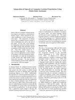
culture supernatants by GC-MS. Figure 1A shows a
dramatic induction of both PGE
2
and the stable PGI
2
metabolite 6-k-PGF
1a
. Induction of both prostanoids
was detectable within 2 h of 4-OHT treatment and
after 24 h reached values > 100-fold of the uninduced
basal levels. In the presence of AA (Fig. 1A, bottom
panel), synthesis of both prostanoids was greatly accel-
erated and reached higher maximum levels, indicating
that the level of endogenous AA generated by phos-
pholipase A
2
is rate-limiting even in the presence of
activated Raf. The induction of both prostanoids was
almost completely blocked by the Cox-1 ⁄ 2 inhibitor
acetylsalicylic acid (ASA) and the Cox-2 inhibitor
SC-58125 (Fig. 1B), pointing to a key role for Cox-2
in the induction of prostanoid synthesis by Raf. In
contrast to PGE
2
and 6-k-PGF
1a
, no significant
increase upon 4-OHT treatment was seen for throm-
boxane B2 (TxB2), PGD
2
and PGF
2a
(Fig. 1A).
Effects of c-Raf signaling on genes encoding
prostanoid-synthesizing enzymes
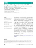
We next analyzed by quantitative real-time PCR
(qPCR) the effect of Raf activation on the expression
of genes that are relevant for the synthesis of PGE
2
and PGI
2
. Figure 2A shows a strong induction of
Cox-2 mRNA expression peaking at 240 min after
4-OHT addition, whereas no induction was seen for
cPLA
2
. This finding was confirmed by northern blot-
ting which showed a 10-fold induction of Cox-2
mRNA after 8 h (Fig. 2B and C). Induction was speci-
fic for Cox-2, since no significant change in expression
was seen with Cox-1 (Fig. 2B). These observations
explain the effects of exogenous AA and the Cox-2
T. Fauti et al. Raf induction of PGI
2
synthesis in the absence of PPARb activation
FEBS Journal 273 (2006) 170–179 ª 2005 The Authors Journal compilation ª 2005 FEBS 171
inhibitor on prostanoid synthesis in Fig. 1. We also
observed a 4-OHT triggered increase in the levels of
PGIS mRNA (Fig. 2A), but this was weak (1.3-fold)
and is therefore unlikely to contribute significantly
to the 4-OHT induced PGI
2
synthesis. Induction
mPGES-1 occurred relatively late after 4-OHT treat-
ment (4.7-fold 12 h post-treatment; Fig. 2B) suggestive
of a secondary event. Taken together, these results
indicate that Cox-2 is the key enzyme mediating the
dramatic induction of PGE
2
and PGI
2
synthesis after
Raf activation. The strong induction of Cox-2 expres-
sion was virtually abolished by both the ERK inhibitor
UO126 and the RNA polymerase inhibitor actinomy-
cin D (Fig. 2C) indicating the Raf-triggered increase in
Cox-2 mRNA expression is due to an ERK-mediated
induction of Cox-2 transcription.
Effects of C-Raf activation on PPARb expression
Activation of Raf not only led to a dramatic induction
of PGI
2
synthesis as described above, but in the same
experimental setting also induced the expression of
the PPAR b gene, which encodes the proposed nuclear
receptor for PGI
2
. As illustrated in Fig. 3A, an
approximately threefold increase in the level of PPARb
mRNA was seen within 8 h of 4-OHT treatment.
Induction was completely abolished by UO126 and
actinomycin D (Fig. 3B), suggesting an absolute
requirement for ERK function and unimpaired tran-
scription as already seen with Cox-2 above.
Effect of Raf activation on the transcriptional
activity of PPARb
The simultaneous upregulation of PGI
2
synthesis and
PPARb expression suggested the induction of an auto-
crine ⁄ intracrine signaling loop upon activation of Raf.
We therefore investigated whether 4-OHT treatment of
N-BxB-ER cells would lead to an activation of the
transcriptional activity of PPARb. To address this
question we constructed a luciferase reporter construct
consisting of seven LexA binding sites upstream of a
TATA-Initiator (TATA-Inr) module without any addi-
tional promoter elements. This reporter plasmid on its
own shows negligible luciferase activity and therefore
allows for a highly sensitive detection of the transcrip-
tional activity of a cotransfected transcriptional activa-
tor harboring a LexA DNA binding domain (DBD).
A
B
Fig. 1. Raf induces PGE
2
and PGI
2
synthesis. (A) Prostanoid levels in the culture medium of RafER3T3 cells after treatment with 4-OHT for the
indicated times in the absence ()AA; upper panel) or presence of 20 l
M arachidonic acid (+AA; bottom panel). 6-kPGF
1a
is a stable metabolite
of the unstable PGI
2
that is used as a direct measure of PGI
2
synthesis. (B) PGE
2
and 6-kPGF
1a
levels in the culture medium of RafER3T3 cells
after treatment with 4-OHT in the presence of 100 l
M ASA or 0.1 lM SC-58125. All data points represent the average of two measurements.
Raf induction of PGI
2
synthesis in the absence of PPARb activation T. Fauti et al.
172 FEBS Journal 273 (2006) 170–179 ª 2005 The Authors Journal compilation ª 2005 FEBS
In this system, the synthetic PPARb agonist
GW501516 gave a 30-fold induction with a fusion
protein consisting of the PPARb ligand binding
domain LBD and the LexA DBD (Fig. 4). In contrast,
no induction was seen after treatment with 4-OHT in
spite of the massive synthesis of the presumptive
PPARb agonist PGI
2
.
We also analyzed the effect of 4-OHT on a PPRE-
HSV-tk-pomoter-driven luciferase reporter construct
[26] in N-BxB-ER cells, but again were unable to
A
BC
Fig. 2. Raf induces genes encoding enzymes with key functions in prostaglandin synthesis expression. (A) RafER3T3 cells were treated with
4-OHT and mRNA levels of PLA
2
, Cox-2, mPGES-1 and PGIS were determined by qPCR. Values represent the average of triplicates; error
bars show the standard deviation. Significant differences from untreated cells are indicated by an asterisk (paired t-test: P < 0.05). (B) Analy-
sis of Cox-1 and Cox-2 expression in 4-OHT treated RafER3T3 cells by northern blotting. Quantitative evaluation by PhosphoImaging showed
that Cox-1 and PGIS mRNA levels did not fluctuate significantly during the time-course of the experiment. For a quantification of Cox-2
expression see (C). PGES mRNA was induced 4.7-fold at 16 h. (C) Analysis of Cox-2 induction in the presence of UO126 or actinomycin D.
Shown is the quantitative evaluation of a northern blot (PhosphorImager).
T. Fauti et al. Raf induction of PGI
2
synthesis in the absence of PPARb activation
FEBS Journal 273 (2006) 170–179 ª 2005 The Authors Journal compilation ª 2005 FEBS 173
detect any induction of transcriptional activity, both
in the presence and absence of a cotransfected
PPARb expression vector (data not shown). Likewise,
transcriptional activity was not increased by 4-OHT
in cells transfected with a RXRa expression vector
[26] and treated with the RXR agonist 9-cis retinoic
acid (data not shown). These findings strongly suggest
that the lack of PPARoad activation by Raf-induced
PGI
2
in the Lex system described above is not a pecu-
liarity of the experimental setup and is not due to a
rate-limiting level of the obligatory PPAR heterodime-
rization partner RXR. These observations are surpri-
sing and indicate that, at least in the experimental
systems used, PGI
2
may not act as agonist for
PPARb. We therefore addressed this issue in further
detail below.
Effect of PGI
2
synthesis on PPARb
Certain polyunsaturated fatty acids, such as AA and
eicosapentaenoic acid (EPA) have been described to
exert some agonistic effect on PPARb. This effect was
also observed in the LexA-DBD based luciferase assay
in the present study. An approximately 3-fold stimula-
tion of the transcriptional activity o PPARb was seen
with 10 lm AA, whereas EPA had a modest effect
only at a higher concentration of 30 lm (Fig. 5A).
Although the effect of AA was much weaker than that
of the synthetic PPARb agonists carbaprostacyclin
(cPGI) and GW501516, it was consistently and repro-
ducibly seen. Treatment with AA resulted in an
approximately sixfold increase in 6-k-PGF
1a
in the cul-
ture medium, and this increase could be completely
blocked by the PGIS inhibitor U51605 [27] (Fig. 5B).
U51605 also further reduced the low level of PGI
2
synthesis in the absence of AA by about threefold
(Fig. 5B). Thus, the extent of PGI
2
synthesis varied
over an overall range of nearly 15-fold, but no
correlation with PPARb transcriptional activity was
observed (Fig. 5B). Very similar results were obtained
with the Cox inhibitors ASA and SC-58125 (data not
shown).
Next, we overexpressed Cox-2 and ⁄ or PGIS in
HEK293 cells and monitored the effect on PGI
2
synthesis and PPARb transcriptional activity. As
depicted in Fig. 6, transfection of Cox-2 or PGIS
expression vectors alone only had a marginal effect
on 6-k-PGF
1a
levels in the culture medium, but
cotransfection of both vectors resulted in an almost
100-fold increased PGI
2
synthesis, both in the
A
B
Fig. 3. Raf induces PPARb gene expression. (A) RafER3T3 cells
were treated with 4-OHT and PPARb mRNA levels were deter-
mined by qPCR. Values represent the average of triplicates; error
bars show the standard deviation. Significant differences from
untreated cells are indicated by an asterisk (paired t-test:
P < 0.005). (B) Analysis by northern blotting of PPARb induction in
the presence of UO126 or actinomycin D. Shown is a quantitative
evaluation of a northern blot by PhosphorImaging.
Fig. 4. Induction of PPARb transcriptional activity by AA is not
dependent on PGI
2
synthesis. (A) Stimulation of PPARb-LBD medi-
ated transcriptional activity in NIH3T3 cells by polyunsaturated fatty
acids and the synthetic agonists carbaprostacyclin (cPGI) and
GW01516. For experimental details see legend to Fig. 6. Values
represent the average of triplicates; error bars show the standard
deviation. Significant differences from untreated cells are indicated
by an asterisk (paired t-test: P ¼ 0.01). (B) Effect of the PGIS inhib-
itor U51605 on 6-kPGF
1a
accumulation in the cell culture superna-
tant as a measure of PGI
2
synthesis (bar graph) and on PPARb-LBD
mediated transcriptional activity (bottom row). PPAR activities are
shown as the average of triplicates and standard deviation.
Raf induction of PGI
2
synthesis in the absence of PPARb activation T. Fauti et al.
174 FEBS Journal 273 (2006) 170–179 ª 2005 The Authors Journal compilation ª 2005 FEBS
absence and presence of exogenous AA. But again,
this dramatic increase in PGI
2
synthesis has no
inducing effect on the transcriptional activity of
PPARb.
Discussion
In the present study, we used a 4-OHT inducible sys-
tem (N-BxB-ER cells) [25] to investigate which com-
ponents of the prostanoid signaling network are
targets of Raf signaling. Our data show that C-Raf
activation leads to a dramatic ERK-dependent induc-
tion of Cox-2 transcription and to a modest increase
in mPGES-1 and PGIS mRNA expression (Fig. 2).
Induction of Cox-2 by Ras-dependent signaling, inclu-
ding the ERK pathway, has previously been reported
for several other experimental systems. Surprisingly,
we did not find any induction of cPLA2, even though
this gene has been described as a Ras target gene in
lung epithelial cells [28,29]. It is therefore likely that
Ras uses downstream effector pathways other than
Raf-MEK-ERK to regulate the cPLA2 gene. In
agreement with these observations, 4-OHT treatment
of N-BxB-ER cells led to a dramatic increase in
PGE
2
and PGI
2
synthesis, which, in keeping with a
lack of cPLA
2
induction, could be substantially
enhanced by adding AA to the growth medium
(Fig. 1A). These data suggest that Raf oncogenes can
contribute to tumorigenesis by augmenting the secre-
tion of tumor growth promoting prostaglandins, such
as PGE
2
.
In the same experimental system, we also observed a
clear induction of PPARb transcription upon Raf acti-
vation (Fig. 3). PPAR b has been shown to play a role
in diverse biological and biochemical processes, inclu-
ding lipid metabolism, wound healing, placenta
development and inflammation, but there is also con-
siderable evidence suggesting a function for PPARb in
oncogenesis [1,30]. This assumption is mainly based on
observations made with PPARb null mice where an
altered growth behavior of intestinal polyps was
observed [13–15]. In spite of this central biological role
for PPARb, the ligands that regulate its transcriptional
activity in vivo remain largely obscure [31]. Polyunsatu-
rated fatty acids, such as EPA, undoubtedly have an
agonistic effect, but this is weak and not isoform speci-
fic [32]. PGI
2
, an AA derivative formed by the succes-
sive action of Cox and PGIS, has been suggested as a
PPARb specific agonist [2,33,34]. Since 4-OHT induces
both PGI
2
synthesis and PPARb expression in N-BxB-
ER cells, we utilized this system to test whether Raf
activation establishes an autocrine ⁄ intracrine signaling
loop consistent with the notion of PGI
2
acting as
PPARb agonist.
Surprisingly, however, Raf activation did not lead
to any detectable increase in PPARb transcriptional
activity. This was seen with both a PPRE-tk reporter
construct measuring total PPAR activity (data not
shown) and with the b-isoform specific LexA-based
system established in this study (Fig. 4). The same
observation was made when an expression vector for
RxRa was cotransfected (data not shown), indicating
that the lack of activation by PGI
2
was not due to
rate-limiting levels of the obligatory PPAR hetero-
dimerization partner. These results clearly suggested
that PGI
2
is not a PPARb agonist in this experimen-
tal system (3T3 fibroblasts). We therefore performed
several additional experiments that all confirm the
A
B
Fig. 5. Overexpression of Cox-2 and PGIS does not induce PPARb
transcriptional activity. HEK293 cells were transiently transfected
with expression vectors for Cox-2, PGIS or both. Forty-eight hours
later, PPARb-LBD mediated transcriptional activity and 6-kPGF
1a
accumulation in the cell culture supernatant were determined. For
experimental details see legend to Fig. 6. Values represent the
average of triplicates; error bars show the standard deviation. Signi-
ficant differences from untreated cells are indicated by an asterisk
(paired t-test: P ¼ 0.01).
T. Fauti et al. Raf induction of PGI
2
synthesis in the absence of PPARb activation
FEBS Journal 273 (2006) 170–179 ª 2005 The Authors Journal compilation ª 2005 FEBS 175
conclusion that PGI
2
lacks agonistic activity for
PPARb in vivo.
The ectopic expression of Cox-2 and PGIS in
HEK293 cells resulted in a dramatic induction of
PGI
2
synthesis, but no increase in PPAR b transcrip-
tional activity was observed (Fig. 6). This is in con-
trast to a previously published observation made
with the human osteosarcoma cell line U2OS [2].
The reason for this discrepancy is not clear since we
were unable to reproduce the published results using
in the identical experimental set-up (U2OS cells and
Gal4-based reporter system; Tanja Fauti, unpublished
data). Prostacyclin-mediated regulation of PPARb
has also been claimed in another study using
HEK293 cells [3]. In this study, a PPRE-SV40-pro-
moter-luciferase construct was used as the reporter,
raising the possibility that the observed transcrip-
tional activation was mediated by a different PPAR
or even by a PPAR-unrelated event, e.g. through sti-
mulation of the SV40 promoter and ⁄ or via the PGI
2
membrane receptor IP. Unless supplemented by
appropriate controls, these data therefore do not
unequivocally show that PGI
2
can invoke a direct
transcriptional activation of PPARb.
The addition of pure PGI
2
(10 lm) to the culture
medium of Chinese hamster ovary cells did not alter
the transcriptional activity of PPARb to any significant
extent (unpublished data). This is in agreement with
two other previous studies. First, U2OS cells trans-
fected with a PPARb reporter did not show any
response to the addition of PGI
2
[35]. In a second
study, the same result was obtained with CV1 cells
[36]. Even though these results are in perfect agree-
ment, they have to be considered with some caution
since it is unclear how the biological instability of
PGI
2
might affect these kinds of experiments.
A weak agonistic effect was seen in 3T3 cells with
exogenously supplied AA, but this increase in PPARb
transcriptional activity was not influenced when PGI
2
synthesis was blocked by inhibitors of PGIS or Cox
(Fig. 5). Taken together, our observations made with
three different cell types and different experimental
approaches provide no evidence that PGI
2
acts as a
PPARb agonist.
Interestingly, in spite of the failure of PGI
2
to acti-
vate PPARb, the PGI
2
analog cPGI showed strong
agonistic properties in all four cell lines analyzed
(Fig. 5A; data not shown). It is possible that the subtle
differences in the chemical structures of PGI
2
and
cPGI have an unexpected effect on the ability to inter-
act with PPARb. Alternatively, the half-life of PGI
2
may be too short to allow for a sufficient concentra-
tion of intact molecules in transcription complexes in
the nucleus. While a very short interaction with the
PGI
2
membrane receptor (IP) may be sufficient for
triggering a signal, a much greater stability may be
required as a ligand of a nuclear receptor, where the
presence of ligand may be necessary for an extended
period of time.
As expected, AA was able to activate PPARb
activity, albeit at high concentrations (Fig. 5A). Even
though high local concentrations of specific lipids
can be achieved in vivo, so that there may be no
need for a high affinity ligand, it is unclear whether
AA itself can act as a PPARb agonist in vivo,or
whether AA is converted to PPARb stimulatory
metabolites by Cox-independent pathways. Further-
more, the existence of totally unrelated high affinity
PPARb agonists cannot be excluded at present. Fur-
ther studies systematically addressing this are neces-
sary to clarify this issue.
Fig. 6. Raf induction does not activate PPARb transcriptional activ-
ity. PPARb-LBD mediated transcriptional activity was determined in
untreated and 4-OHT-treated RafER3T3 cells in the presence of
20 m
M arachidonic acid. Cells were transiently transfected with an
expression vector encoding the LexA-PPARb fusion protein (Lex-
PPARb-LBD) or the empty vector (pcDNA3.1) together with a lexA-
luciferase reporter plasmid (7 L-TATAi). Luciferase activity was
determined 48 h after transfection; 4-OHT treatment was for 24 h.
As a positive control, cells were also treated with 1 m
M GW501516
for 24 h. Values represent the average of triplicates; error bars
show the standard deviation. Significant differences from untreated
cells are indicated by an asterisk (paired t-test: P < 0.003).
Raf induction of PGI
2
synthesis in the absence of PPARb activation T. Fauti et al.
176 FEBS Journal 273 (2006) 170–179 ª 2005 The Authors Journal compilation ª 2005 FEBS
Experimental procedures
Chemicals
Chemicals were purchased from the following companies:
acetylsalicylic acid (Cayman Chemical Company, Ann
Arbor, MI, USA), actinomycin D (Sigma-Aldrich, Tauf-
kirchen ⁄ Munich, Germany), carbaprostacyclin (Cayman
Chemical Company), GW501516 (Calbiochem ⁄ Merck Bio-
sciences, Bad Soden, Germany), 4-hydroxy-tamoxifen
(Sigma-Aldrich), PGI
2
(Alexis ⁄ AXXORA, Lausen, Switzer-
land), SC-58125 (Calbiochem ⁄ Merck Biosciences), U51605
(Cayman Chemical Company), UO126 (Promega, Man-
nheim, Germany).
Cell culture
NIH3T3, N-BxB-ER, HEK293 and CHO cells were cul-
tured in DMEM supplemented with 10% fetal bovine
serum, 100 UÆmL
)1
penicillin and 100 lgÆmL
)1
streptomy-
cin. Cells were maintained in culture at 37 °C with 5% CO
2
in a humidified incubator.
Plasmids
PGIS-pcDNA3.1 and COX2-pcDNA3.1 were obtained by
cloning the full-length human PGIS and Cox-2 cDNAs
into the expression vector pcDNA3.1(+) (Invitrogen,
Kahlsruhe, Germany). PPREx3-tk-pGL3 was constructed
by inserting the PPRE
3
-TK-fragment from PPRE
3
-TK-
LUC [36] (obtained from R.M. Evans, La Jolla, CA,
USA) into the pGL3 basic luciferase vector (Promega).
7 L-TATAi has been described previously [37]. pcDNA3.1-
LexA-PPARb-LBD was constructed as follows: the
PPARb-LBD fragment flanked by a 5¢-AseI- and a-3¢
BamHI-site was synthesized by PCR using pCMX-
mPPARb [36] as the template. The LexA-DBD fragment,
including a Kozak and a nuclear localization sequence,
was amplified from vWFnLexA by RT–PCR. The remain-
ing LexA-fragment was flanked with a 5¢ HindIII- and
a-3¢-NdeI-site. The fragments were cut with NdeI and
AseI, ligated with T4 DNA ligase (Roche diagnostics),
treated with Taq DNA polymerase to add 3¢ oligo(A)
overhangs and cloned into pCRIITOPO (Invitrogen).
Finally the LexA-PPARb-LBD fragment was cut with
BamHI and HindIII and subcloned into pcDNA3.1
zeo
(Invitrogen).
RNA isolation
RNA was isolated using the RNeasy
TM
kit from Qiagen
(Hilden, Germany) following the manufacturer’s protocol.
Briefly 30 lg of tissue were homogenized in 600 lL RLT
buffer and 6 lL b-mercaptoethanol with a warring blender
(Ultra-Thurrax; IKA, Staufen, Germany). Qia shredders
(Qiagen) were used to break down genomic DNA of lysed
tissue culture cells and homogenized tissue.
Northern blotting
RNA (5–20 lg) was mixed with sample buffer (0.5 mL 10
Mops buffer, 1.75 mL 37% formaldehyde, 5 mL forma-
mide) and loading buffer (50% glycerol, 1 mm EDTA,
0.25% Bromophenol blue, water) and separated on a 1%
agarose ⁄ formamide gels containing 2.2 m formaldehyde.
The RNA was blotted to Hybond-N (Amersham, Freiburg,
Germany) with 10 · NaCl ⁄ Cit and crosslinking under UV
light (Stratalinker 2400, 254 nm, 1200 J m
)2
; Stratagene,
La Jolla, CA, USA). Hybridization to P
32
-labeled probes
was performed as described [25]. Signal intensites on mem-
branes were quantitated by PhosphorImager (Fuji, Du
¨
ssel-
dorf, Germany).
Reverse transcriptase PCR
cDNA was synthesized using 1 lg of RNA, oligo dT primers
and reverse transcriptase according to the manufacturer’s
protocol (Roche Diagnostics, Mannheim, Germany). PCR
was performed for 25 cycles at an annealing temperature of
55 °C(PPARb) respective 58 °C(Cox-2) with Platinum Taq
polymerase (Invitrogen) using primers obtained from MWG
Biotech (Ebersberg, Germany) with the following sequences:
Cox-2 forward, 5¢—CCTTCTCCAACCTCTCCTAC—3¢;
Cox-2 reverse, 5¢—AGGGGGTGCCAGTGATAGAG—3 ¢;
PPARb forward, 5¢—AAGAGGAGAAAGAGGAAG
TGG—3¢; PPARb reverse, 5¢—ATTGAGGAAGAGGCTG
CTGA—3¢; actin forward, 5¢—GATGATGATATCGCCGC
GCTCGTCGTC—3¢; actin reverse, 5¢—GTGCCTCAGGG
CAGCGGACCGCTCA—3¢.
Quantitative PCR
Quantitative PCR was performed in a Mx3000P Real-Time
PCR system (Stratagene) for 45 cycles at an annealing tem-
perature of 57 °C. PCR reactions were carried out using
the Absolute QPCR SYBR Green Mix (Abgene, Hamburg,
Germany) and a primer concentration of 0.2 lm following
the manufacturer’s instructions. The following primers
MWG Biotech were used: actin forward, 5¢—AGAGGGA
AATCGTGCGTGAC—3¢; actin reverse, 5¢—CAATAGTG
ATGACCTGGCCGT—3¢; PPARb forward, 5¢—GTCGCA
CAACGCTATCC—3¢; PPARb reverse, 5¢—CTCCGGGCC
TTCTTTTTGGTCA—3¢; cPLA2 forward, 5¢—CATAAGT
TTACTGTTGTGGTTCTA—3¢; cPLA2 reverse, 5 ¢—AGT
GTCTCGTTCGCTTCC—3¢; COX-2 forward, 5¢—CCATG
GGTGTGAAGGGAAATAA—3¢; COX-2 reverse, 5¢—TTG
AAAAACTGATGGGTGAAG—3¢; mPGES-1 forward,
5¢—GGTGGCCCAGGAAGGAGACAGC—3¢; reverse
5¢—TGGCCTTCATGGGTGGGTAATA—3¢.
T. Fauti et al. Raf induction of PGI
2
synthesis in the absence of PPARb activation
FEBS Journal 273 (2006) 170–179 ª 2005 The Authors Journal compilation ª 2005 FEBS 177
Transient tansfections and luciferase assays
Transfections were performed with polyethylenimine (PEI,
average MW 25 000; Sigma-Aldrich). For each assay, 10
5
cells were transfected in DMEM plus 2% FCS with 5 lgof
plasmid DNA and 5 lLofa1⁄ 1000 PEI dilution (adjusted
to pH 7.0) preincubated for 15 min in 100 lL NaCl ⁄ P
i
for
complex formation. Four hours after transfection, the med-
ium was changed and cells were incubated in normal growth
medium for 24 h. Luciferase assays were performed as des-
cribed [38]. Values from three independent experiments were
combined to calculate averages and standard deviations.
Sample preparation for prostanoids by
GC ⁄ MS ⁄ MS-analysis
Samples were prepared as described [39] with minor modifi-
cations. Briefly, cell culture supernatants were spiked with
10 ng of deuterated internal standards, and solvent was
removed. The methoxime was obtained through reaction
with an O-methylhydroxylamine hydrochloride-acetate
buffer. After acidification to pH 3.5, prostanoid derivatives
were extracted, and the pentafluorobenzylesters were
formed. Samples were purified by TLC and two broad
zones with R
v
0.03–0.39 and 0.4–0.8 were eluted. After
withdrawal of the organic layers, trimethylsilyl ethers were
prepared by reaction with bis(trimethylsilyl)-trifluoroaceta-
mide and thereafter subjected to GC ⁄ MS ⁄ MS analysis.
GC ⁄ MS ⁄ MS analysis
A Finnigan (Thermo Electron Corp., Dreieich, Germany)
MAT TSQ700 GC ⁄ MS ⁄ MS equipped with a Varian (Palo
Alto, CA, USA) 3400 gas chromatograph and a CTC
A200S autosampler was used [39].
Acknowledgements
We are grateful to Margitta Alt and Bernhard Watzer
for excellent technical assistance. This work was sup-
ported by the Wihelm-Sander-Stiftung, the Dr Mildred
Scheel Stiftung and the Deutsche Forschungsgemeinsc-
haft (SFB-TR17).
References
1Mu
¨
ller R (2004) Crosstalk of oncogenic and prostanoid
signaling pathways. J Cancer Res Clin Oncol. 130, 429–
444.
2 Gupta RA, Tan J, Krause WF, Geraci MW, Willson
TM, Dey SK & DuBois RN (2000) Prostacyclin-
mediated activation of peroxisome proliferator-activated
receptor delta in colorectal cancer. Proc Natl Acad Sci
USA 97, 13275–13280.
3 Hatae T, Wada M, Yokoyama C, Shimonishi M &
Tanabe T (2001) Prostacyclin-dependent apoptosis
mediated by PPAR delta. J Biol Chem 276, 46260–46267.
4 Lim H & Dey SK (2002) A novel pathway of prostacy-
clin signaling-hanging out with nuclear receptors. Endo-
crinology 143, 3207–3210.
5 Cutler NS, Graves-Deal R, LaFleur BJ, Gao Z, Boman
BM, Whitehead RH, Terry E, Morrow JD & Coffey RJ
(2003) Stromal production of prostacyclin confers an
antiapoptotic effect to colonic epithelial cells. Cancer
Res 63, 1748–1751.
6 Amano H, Hayashi I, Endo H, Kitasato H, Yamashina
S, Maruyama T, Kobayashi M, Satoh K, Narita M,
Sugimoto Y, Murata T, Yoshimura H, Narumiya S &
Majima M (2003) Host prostaglandin E(2)-EP3 signal-
ing regulates tumor-associated angiogenesis and tumor
growth. J Exp Med 197, 221–232.
7 Sonoshita M, Takaku K, Sasaki N, Sugimoto Y, Ush-
ikubi F, Narumiya S, Oshima M & Taketo MM (2001)
Acceleration of intestinal polyposis through prostaglan-
din receptor EP2 in Apc (Delta 716) knockout mice.
Nat Med 7, 1048–1051.
8 He TC, Chan TA, Vogelstein B & Kinzler KW (1999)
PPARdelta is an APC-regulated target of nonsteroidal
anti-inflammatory drugs. Cell 99, 335–345.
9 Di-Poi N, Tan NS, Michalik L, Wahli W & Desvergne
B (2002) Antiapoptotic role of PPARbeta in keratino-
cytes via transcriptional control of the Akt1 signaling
pathway. Mol Cell 10, 721–733.
10 Mao-Qiang M, Fowler AJ, Schmuth M, Lau P,
Chang S, Brown BE, Moser AH, Michalik L, Des-
vergne B, Wahli W, Li M, Metzger D, Chambon PH,
Elias PM & Feingold KR (2004) Peroxisome-prolifera-
tor-activated receptor (PPAR)-gamma activation sti-
mulates keratinocyte differentiation. J Invest Dermatol
123, 305–312.
11 Kim DJ, Bility MT, Billin AN, Willson TM, Gonzalez
FJ & Peters JM (2005) PPARbeta/delta selectively
induces differentiation and inhibits cell proliferation.
Cell Death Differ doi:10.1038/sj.cdd.4401713.
12 Park BH, Vogelstein B & Kinzler KW (2001) Genetic
disruption of PPARdelta decreases the tumorigenicity of
human colon cancer cells. Proc Natl Acad Sci USA 98,
2598–2603.
13 Barak Y, Liao D, He W, Ong ES, Nelson MC, Olefsky
JM, Boland R & Evans RM (2002) Effects of peroxi-
some proliferator-activated receptor delta on placenta-
tion, adiposity, and colorectal cancer. Proc Natl Acad
Sci USA 99, 303–308.
14 Harman FS, Nicol CJ, Marin HE, Ward JM, Gonzalez
FJ & Peters JM (2004) Peroxisome proliferator-acti-
vated receptor-delta attenuates colon carcinogenesis.
Nat Med 10, 481–483.
15 Gupta RA, Wang D, Katkuri S, Wang H, Dey SK &
DuBois RN (2004) Activation of nuclear hormone
Raf induction of PGI
2
synthesis in the absence of PPARb activation T. Fauti et al.
178 FEBS Journal 273 (2006) 170–179 ª 2005 The Authors Journal compilation ª 2005 FEBS
receptor peroxisome proliferator-activated receptor-delta
accelerates intestinal adenoma growth. Nat Med 10,
245–247.
16 Hao CM, Redha R, Morrow J & Breyer MD (2002)
Peroxisome proliferator-activated receptor delta
activation promotes cell survival following hypertonic
stress. J Biol Chem 277, 21341–21345.
17 Keith RL, Miller YE, Hoshikawa Y, Moore MD,
Gesell TL, Gao B, Malkinson AM, Golpon HA,
Nemenoff RA & Geraci MW (2002) Manipulation of
pulmonary prostacyclin synthase expression prevents
murine lung cancer. Cancer Res 62, 734–740.
18 Shaw N, Elholm M & Noy N (2003) Retinoic acid is
a high affinity selective ligand for PPAR-beta ⁄ delta.
J Biol Chem 278, 41589–41592.
19 Westergaard M, Henningsen J, Svendsen ML, Johan-
sen C, Jensen UB, Schroder HD, Kratchmarova I,
Berge RK, Iversen L, Bolund L, Kragballe K &
Kristiansen K (2001) Modulation of keratinocyte gene
expression and differentiation by PPAR-selective
ligands and tetradecylthioacetic acid. J Invest Dermatol
116, 702–712.
20 Tan NS, Michalik L, Noy N, Yasmin R, Pacot C, Heim
M, Fluhmann B, Desvergne B & Wahli W (2001) Criti-
cal roles of PPAR beta ⁄ delta in keratinocyte response
to inflammation. Genes Dev 15, 3263–3277.
21 Schmuth M, Haqq CM, Cairns WJ, Holder JC, Dorsam
S, Chang S, Lau P, Fowler AJ, Chuang G, Moser AH,
Brown BE, Mao-Qiang M, Uchida Y, Schoonjans K,
Auwerx J, Chambon P, Willson TM, Elias PM & Fein-
gold KR (2004) Peroxisome proliferator-activated recep-
tor (PPAR)-beta ⁄ delta stimulates differentiation and
lipid accumulation in keratinocytes. J Invest Dermatol
122, 971–983.
22 Kim DJ, Murray IA, Burns AM, Gonzalez FJ, Perdew
GH & Peters JM (2005) Peroxisome proliferator-acti-
vated receptor-beta ⁄ delta inhibits epidermal cell prolifer-
ation by down-regulation of kinase activity. J Biol
Chem 280, 9519–9527.
23 Kerkhoff E & Rapp UR (1998) Cell cycle targets of
Ras ⁄ Raf signalling. Oncogene 17, 1457–1462.
24 Chang F, Steelman LS, Shelton JG, Lee JT, Navolanic
PM, Blalock WL, Franklin R & McCubrey JA (2003)
Regulation of cell cycle progression and apoptosis by
the Ras ⁄ Raf ⁄ MEK ⁄ ERK pathway (Review). Int J
Oncol 22, 469–480.
25 Kerkhoff E, Houben R, Loffler S, Troppmair J, Lee JE
& Rapp UR (1998) Regulation of c-myc expression by
Ras ⁄ Raf signalling. Oncogene 16, 211–216.
26 Shi Y, Hon M & Evans RM (2002) The peroxisome
proliferator-activated receptor delta, an integrator of
transcriptional repression and nuclear receptor signal-
ing. Proc Natl Acad Sci USA 99, 2613–2618.
27 Gorman RR, Hamilton RD & Hopkins NK (1979) Sti-
mulation of human foreskin fibroblast adenosine 3¢:
5¢-cyclic monophosphate levels by prostacyclin (prosta-
glandin I2). J Biol Chem 254, 1671–1676.
28 Heasley LE, Thaler S, Nicks M, Price B, Skorecki K &
Nemenoff RA (1997) Induction of cytosolic phospho-
lipase A2 by oncogenic Ras in human non-small cell
lung cancer. J Biol Chem 272, 14501–14504.
29 Blaine SA, Wick M, Dessev C & Nemenoff RA (2001)
Induction of cPLA2 in lung epithelial cells and non-
small cell lung cancer is mediated by Sp1 and c-Jun.
J Biol Chem 276, 42737–42743.
30 Michalik L, Desvergne B & Wahli W (2004) Peroxi-
some-proliferator-activated receptors and cancers: com-
plex stories. Nat Rev Cancer 4, 61–70.
31 Bishop-Bailey D & Wray J (2003) Peroxisome prolifera-
tor-activated receptors: a critical review on endogenous
pathways for ligand generation. Prostaglandins Other
Lipid Mediat 71, 1–22.
32 Forman BM, Tontonoz P, Chen J, Brun RP, Spiegel-
man BM & Evans RM (1995) 15-Deoxy-delta
12, 14-prostaglandin J2 is a ligand for the adipocyte
determination factor PPAR gamma. Cell 83, 803–812.
33 Lim H, Gupta RA, Ma WG, Paria BC, Moller DE,
Morrow JD, DuBois RN, Trzaskos JM & Dey SK
(1999) Cyclo-oxygenase-2-derived prostacyclin mediates
embryo implantation in the mouse via PPARdelta.
Genes Dev 13, 1561–1574.
34 Shao J, Sheng H & DuBois RN (2002) Peroxisome pro-
liferator-activated receptors modulate K-Ras-mediated
transformation of intestinal epithelial cells. Cancer Res
62, 3282–3288.
35 YuK, Bayona W, Kallen CB, Harding HP, Ravera CP,
McMahon G, Brown M & Lazar MA (1995) Differential
activation of peroxisome proliferator-activated receptors
by eicosanoids. J Biol Chem 270, 23975–23983.
36 Forman BM, Chen J & Evans RM (1997) Hypolipi-
demic drugs, polyunsaturated fatty acids, and eicosa-
noids are ligands for peroxisome proliferator-activated
receptors alpha and delta. Proc Natl Acad Sci USA 94,
4312–4317.
37 Nettelbeck DM, Jerome V & Muller R (1999) A dual
specificity promoter system combining cell cycle-regu-
lated and tissue-specific transcriptional control. Gene
Ther 6, 1276–1281.
38 Gehrke S, Jerome V & Muller R (2003) Chimeric tran-
scriptional control units for improved liver-specific
transgene expression. Gene 322, 137–143.
39 Schweer H, Watzer B & Seyberth HW (1994) Determi-
nation of seven prostanoids in 1 ml of urine by gas
chromatography-negative ion chemical ionization triple
stage quadrupole mass spectrometry. J Chromatogr 652,
221–227.
T. Fauti et al. Raf induction of PGI
2
synthesis in the absence of PPARb activation
FEBS Journal 273 (2006) 170–179 ª 2005 The Authors Journal compilation ª 2005 FEBS 179