Báo cáo khóa học: Some properties of human small heat shock protein Hsp20 (HspB6) potx
Bạn đang xem bản rút gọn của tài liệu. Xem và tải ngay bản đầy đủ của tài liệu tại đây (620.21 KB, 12 trang )
Some properties of human small heat shock protein Hsp20 (HspB6)
Olesya V. Bukach
1
, Alim S. Seit-Nebi
1
, Steven B. Marston
2
and Nikolai B. Gusev
1
1
Department of Biochemistry, School of Biology, Moscow State University, Moscow, Russia;
2
Imperial College School of Medicine
at National Heart and Lung Institute, London, UK
Human heat shock protein of apparent molecular mass
20 kDa (Hsp20) and its mutant, S16D, mimicking phos-
phorylation by cyclic nucleotide-dependent protein kinases,
were cloned and expressed in Escherichia coli. The proteins
were obtained in a homogeneous state without utilization
of urea or detergents. On size exclusion chromatography at
neutral pH, Hsp20 and its S16D mutant were eluted as
symmetrical peaks with an apparent molecular mass of
55–60 kDa. Chemical crosslinking resulted in the forma-
tion of dimers with an apparent molecular mass of 42 kDa.
At pH 6.0, Hsp20 and its S16D mutant dissociated, and
were eluted in the form of two peaks with apparent
molecular mass values of 45–50 and 28–30 kDa. At
pH 7.0–7.5, the chaperone activity of Hsp20 (measured by
its ability to prevent the reduction-induced aggregation of
insulin or heat-induced aggregation of yeast alcohol
dehydrogenase) was similar to or higher than that of
commercial a-crystallin. Under these conditions, the S16D
mutant of Hsp20 possessed lower chaperone activity than
the wild-type protein. At pH 6.0, both a-crystallin and
Hsp20 interacted with denatured alcohol dehydrogenase;
however, a-crystallin prevented, whereas Hsp20 either did
not affect or promoted, the heat-induced aggregation of
alcohol dehydrogenase. The mixing of wild-type human
Hsp27 and Hsp20 resulted in a slow, temperature-
dependent formation of hetero-oligomeric complexes, with
apparent molecular mass values of 100 and 300 kDa,
which contained approximately equal amounts of Hsp27
and Hsp20 subunits. Phosphorylation of Hsp27 by mito-
gen activated protein kinase-activated protein kinase 2 was
mimicked by replacing Ser15, 78 and 82 with Asp. A 3D
mutant of Hsp27 mixed with Hsp20 rapidly formed a
hetero-oligomeric complex with an apparent molecular
mass of 100 kDa, containing approximately equal quanti-
ties of two small heat shock proteins.
Keywords: small heat shock proteins; phosphorylation;
chaperone activity.
Human small heat shock proteins (sHsp) form a large
group of proteins, consisting of 10 members with a
molecular mass in the range of 17–23 kDa [1]. These
proteins are grouped together because all contain an
a-crystallin domain, of 80–100 amino acid residues, which
is located in the C-terminal part of the protein [2,3]. Some
sHsp, such as aB-crystallin and Hsp27, are ubiquitous
and expressed in practically all tissues [1,2,4,5], whereas
other sHsp (such as HspB7 and HspB9) are expressed
only in specific tissues [1,4,5]. sHsp tend to form large
oligomers that vary in structure and number of monomers
[6,7]. These complexes can be formed by identical or
nonidentical subunits. Subunits of a-crystallin, Hsp20,
Hsp22, and Hsp27 seem to be involved in the formation
of different heterooligomeric complexes [8–12]. Hsp27 and
aB-crystallin have been analyzed in detail [2–5,13,14],
whereas other members of the large superfamily of sHsp
are less well characterized.
Hsp20 was described by Kato et al. [8] as a byproduct of
purification of human aB-crystallin and Hsp27. Hsp20 is
expressed in practically all tissues, reaching a maximal level
of 1.3% of total proteins in skeletal, heart and smooth
muscles [2,9,15]. Since 1997, the laboratory of Colleen
Brophy has performed detailed investigations of the role of
Hsp20 in the regulation of smooth muscle contraction. It
has been shown that cAMP- and cGMP-dependent protein
kinases phosphorylate Ser16 of Hsp20 and that phosphory-
lation of Hsp20 is associated with smooth muscle relaxation
that is independent of the level of phosphorylation of the
myosin light chain [16–20]. These findings have been
confirmed and extended [21–23]. Insulin induces phos-
phorylation of rat Hsp20 at Ser157 [24] and Hsp20
phosphorylated at two different sites (Ser16 and Ser157)
differently affects glucose transport [25,26]. Recently, Hsp20
was detected in blood and it has been shown that Hsp20
binds to and inhibits platelet aggregation [27]. Thus,
significant progress has been achieved in revealing a possible
physiological role of Hsp20. However, investigation of the
biochemical properties of isolated Hsp20 lag behind.
Indeed, the biochemical properties of rat Hsp20 were only
briefly characterized in the reports of Kato et al. [8,9] and
van de Klundert et al. [15], whereas the corresponding
properties of human Hsp20 remain practically uncharac-
terized. Therefore, the present work was devoted to the
cloning and purification of wild-type human Hsp20 and its
Correspondence to N. B. Gusev, Department of Biochemistry,
School of Biology, Moscow State University, Moscow 119992, Russia.
Fax:/Tel.: + 7 095 9392747, E-mail:
Abbreviations: ADH, yeast alcohol dehydrogenase; DMS, dimethyl-
suberimidate; EDC, 1-(3-dimethylaminopropyl)-3-ethylcarbodiimide
hydrochloride; Hsp, heat shock protein; 3D mutant, human Hsp27
with replacement of Ser15, 78 and 82 by Asp; NHS, N-hydroxy-
succinimide; S16D, mutant of human Hsp20 with replacement of
Ser16 by Asp; sHsp, small heat shock proteins.
(Received 21 October 2003, accepted 17 November 2003)
Eur. J. Biochem. 271, 291–302 (2004) Ó FEBS 2003 doi:10.1046/j.1432-1033.2003.03928.x
mutant mimicking phosphorylation of Ser16, analysis of
their oligomeric state, chaperone activity and their ability to
interact with human Hsp27.
Materials and methods
Proteins
Cloning and mutagenesis of human Hsp20 and
Hsp27. The full-length cDNA encoding human Hsp20
(GenBank accession no.: AK056951) was amplified from
Marathon-Ready cDNA, Heart (Clontech) using the fol-
lowing forward 5¢-GAGATATA
CATATGGAGATCC
CTGTGC-3¢ (NdeI restriction site underlined) and reverse
5¢-GTG
CTCGAGTTACTTGGCTGCGGCTGGCGG-3¢
(XhoI restriction site underlined) primers, and Pwo DNA
polymerase (Roche). The 480 bp PCR product was purified
after electrophoresis in an agarose gel, then digested with the
restriction endonucleases NdeIandXhoI and inserted into
the plasmid vector pET23b (which had been predigested with
the same endonucleases). The resulting construct was verified
by DNA sequencing and used for expression and mutagen-
esis. A two step PCR-based ÔmegaprimerÕ method [28,29] was
used for the replacement of Ser16 of Hsp20 with Asp. In this
case, the primer S16D (5¢-GCCGCGCCGACGCCCCG
TTGC-3¢) was used for site-directed mutagenesis.
The human Hsp27 full-length cDNA (GenBank acces-
sion no.: NM001540) was amplified from Marathon-Ready
cDNA, Lung (Clontech) using the following forward
5¢-GAGATATA
CATATGGCCGAGCGC-3 and reverse
5¢-CC
GGATCCCTACTTCTTGGCTGG-3¢ primers con-
taining, respectively, NdeIandBamHI restriction sites
(underlined). The PCR product was purified and inserted
into the plasmid vector, pET11c (Novagen). The resulting
construct was verified by DNA sequencing and used for
expression and site-directed mutagenesis.
Three serine residues of Hsp27 (Ser15, Ser78 and
Ser82) were replaced with Asp. This was achieved by
using the following primers: 5¢-CGGGGCCCCGACTG
GGACCCC-3¢ for S15D and 5¢-GACCCCGCTGTC
GAGTTGCCGGTCGAGCGCGC-3¢ for the S78D and
S82D mutants. The two step PCR-based ÔmegaprimerÕ
method [28,29] permits creation of the so-called 3D
mutant of Hsp27 with replacements of Ser15, Ser78 and
Ser82 by Asp. This type of mutation mimics phosphory-
lation of Hsp27 by mitogen activated protein kinase-
activated protein kinase 2 [30,31].
Expression and purification of human Hsp20 and
Hsp27. Expression was performed in Escherichia coli
BL21(DE3) pUBS520. E. coli was cultured with aeration,
on Luria–Bertani (LB) media containing ampicillin
(150 lgÆmL
)1
) and kanamycin (40 lgÆmL
)1
), to an attenu-
ance (D
600
) of 0.5. Isopropyl thio-b-
D
-thiogalactoside
(IPTG) was added to a final concentration of 0.5 m
M
and
culture was continued for a further 4 h at 30 °C. The cells
were harvested, frozen and used for isolation of recombin-
ant human wild-type Hsp20, its S16D mutant, recombinant
human wild-type Hsp27 and its 3D mutant.
The initial stages of purification of Hsp20 and its S16D
mutant were performed as described previously [32]. Briefly,
the crude extract of Hsp20 in lysis buffer (50 m
M
Tris/HCl,
pH 8.0, 100 m
M
NaCl, 1 m
M
EDTA, 0.5 m
M
phenyl-
methanesulfonyl fluoride, 14 m
M
b-mercaptoethanol) was
fractionated with (NH
4
)
2
SO
4
(0–30% saturation) and
subjected to ion-exchange chromatography on a High-Trap
Q column (Amersham-Pharmacia) equilibrated with
buffer B (20 m
M
Tris/acetate, pH 7.6, 10 m
M
NaCl,
0.1 m
M
EDTA, 0.1 m
M
phenylmethanesulfonyl fluoride,
14 m
M
b-mercaptoethanol) and developed by a linear
(10–410 m
M
) gradient of NaCl. Further purification was
achieved by hydrophobic chromatography on a phenyl-
superose column (Amersham-Pharmacia) equilibrated with
20 m
M
phosphate buffer (pH 7.0), containing 0.3
M
(NH
4
)
2
SO
4
, and developed by a decreasing (0.3–0.005
M
)
gradient of (NH
4
)
2
SO
4
. The final preparations of Hsp20, or
its S16D mutant, were concentrated by ultrafiltration and
stored frozen in buffer B.
The initial steps of purification of Hsp27 or its 3D mutant
were similar to those described for Hsp20. Hsp27 and its 3D
mutant were fractionated by (NH
4
)
2
SO
4
(0–50% satura-
tion) and subjected to ion-exchange chromatography on a
High-Trap Q column (Amersham-Pharmacia), followed by
gel filtration on a Sephacryl S300 High-Prep 16/60 column
(Amersham-Pharmacia). If necessary, further purification
was achieved by hydrophobic chromatography on phenyl-
superose (Amersham-Pharmacia). Preparations of Hsp27
and its 3D mutant were concentrated by ultrafiltration and
stored frozen in buffer B containing 10% glycerol.
Denaturation and renaturation of sHsp. Denaturation
and renaturation of Hsp20 and commercial a-crystallin
(Sigma) was performed according to van de Klundert et al.
[15]. Recombinant wild-type Hsp20 in buffer B was freeze-
dried. The samples of freeze-dried Hsp20 or commercial
a-crystallin were dissolved in 50 m
M
phosphate (pH 7.5),
containing 100 m
M
Na
2
SO
4
,0.02%b-mercaptoethanol and
6
M
urea, up to a final protein concentration of 6 mgÆmL
)1
,
and then stored on ice for 2 h. After incubation, the samples
were diluted sixfold in the same buffer, minus urea and
b-mercaptoethanol, and dialyzed against two changes of the
same buffer overnight.
IEF and electrophoresis
Isoelectrofocusing (IEF) was performed, as described pre-
viously [29], in a 5.4% polyacrylamide gel containing 8.5
M
urea, 2% Triton-X-100, 0.4% ampholine (pH 3–10) and
1.6% ampholine (pH 5–7). Phosphoric acid (10 m
M
)and
sodium hydroxide (20 m
M
) were used as electrode buffers.
After fixation and removal of ampholine, the proteins were
stained with Coomassie R-250.
SDS gel electrophoresis was performed according to
Laemmli [33]. For quantitative measurements the gels were
stained with Coomassie R-250 and evaluated using the
program
ONEDSCAN
.
Size exclusion chromatography
The oligomeric state of sHsp was determined by size
exclusion chromatography on Superdex 200 HR 10/30
using the ACTA-FPLC system. The column was usually
equilibrated with buffer C (20 m
M
Tris/HCl, pH 7.5,
containing 150 m
M
NaCl and 15 m
M
b-mercaptoethanol).
292 O. V. Bukach et al. (Eur. J. Biochem. 271) Ó FEBS 2003
In the case of renaturation experiments, the same column
was equilibrated and developed with 50 m
M
phosphate
(pH 7.5) containing 100 m
M
Na
2
SO
4
.Thecolumnwas
calibrated using the following molecular mass markers:
thyroglobulin (669 kDa), ferritin (440 kDa), catalase
(240 kDa), aldolase (158 kDa), BSA (66 kDa) and chymo-
trypsinogen (25 kDa).
For investigating the exchange of subunits between
Hsp20 and Hsp27, equimolar quantities (0.4 mgÆmL
)1
Hsp20 and 0.54 mgÆmL
)1
Hsp27) of sHsp were mixed in
buffer B. The mixture obtained was either immediately
loaded onto the column or incubated for 3 h at 30 or 37 °C,
or for 15 h at 18 °C, before chromatography at room
temperature. In control experiments, isolated Hsp20 or
Hsp27 were incubated under exactly the same conditions
and subjected to size exclusion chromatography. The
protein composition of the fractions obtained in the course
of size exclusion chromatography was analyzed by means of
SDS gel electrophoresis [33].
The effect of pH on the oligomeric state of Hsp20 was
also analyzed by size exclusion chromatography. To achieve
this, the Superdex 200 HR 10/30 column was equilibrated
with buffer D (50 m
M
phosphate, 150 m
M
NaCl, 1 m
M
EDTA, 15 m
M
b-mercaptoethanol), pH-adjusted to 5.5,
6.0, 6.5, 7.0 or 7.5. The protein sample (150 lL,
0.6 mgÆmL
)1
) was mixed with an equal volume of 2· buffer
D at the test pH and incubated for 1 h at 20 °Cbefore
chromatography at room temperature.
Chemical crosslinking
Three different methods were used for crosslinking Hsp20.
In the first, Hsp20 (0.2 mgÆmL
)1
) was dialyzed overnight
against 50 m
M
phosphate buffer (pH 7.5), containing
100 m
M
Na
2
SO
4
. Before SDS gel electrophoresis, the
samples were either treated with an excess of b-mercapto-
ethanol or loaded onto the gel in the absence of
b-mercaptoethanol.
In the second method, Hsp20 (0.75 mgÆmL
)1
)in0.2
M
triethanolamine (pH 7.5) was incubated with dimethylsube-
rimidate (20 m
M
)for1hat20°C. The reaction was
stopped by the addition of SDS sample buffer. The protein
composition of the samples thus obtained was analyzed by
SDS gel electrophoresis.
In the third method, Hsp20 (1 mgÆmL
)1
), in 20 m
M
imidazole/HCl (pH 7.0) containing 150 m
M
NaCl, was
incubated for 1 h at 30 °C in the presence of 1-(3-
dimethylaminopropyl)-3-ethylcarbodiimide hydrochloride
(EDC) (5 m
M
)andN-hydroxysuccinimide (NHS) (5 m
M
).
The reaction was stopped by the addition of SDS sample
buffer and subjected to SDS/PAGE (15% gel) [33].
CD spectroscopy
sHsp ( 1mgÆmL
)1
) were dialyzed overnight, against
50 m
M
phosphate buffer containing 150 m
M
NaCl, at three
different pH values (6.0, 6.8 or 7.5). The samples thus
obtained were subjected to centrifugation (12 000 g,
20 min) and the pellet was discarded. Far UV CD spectra
were recorded in 0.05 cm cells at room temperature on a
Mark V. Jobin Yvon autodichrograph. All spectra presen-
ted represent the average of three accumulations.
Determination of chaperone activity
The chaperone activity of Hsp20 and of bovine lens
a-crystallin (Sigma) was determined by their ability to
retard or to decrease aggregation of the insulin B-chain
(Sigma) [15]. All experiments were performed in buffer E
(50 m
M
phosphate, pH 7.5, 100 m
M
Na
2
SO
4
). Insulin (6.5
mgÆmL
)1
), dissolved in 2.5% acetic acid, was added to the
incubation mixture (270 lL) to a final concentration of
0.25 mgÆmL
)1
. The mixture was incubated at 40 °Candthe
reaction started by addition of a water solution of dithio-
threitol up to a final concentration of 20 m
M
. Reduction of
the disulfide bonds of insulin was accompanied by aggre-
gation of the B-chain and an increase of turbidity that was
measured at 360 nm on an Ultraspec 3100 Pro spectro-
photometer.
The chaperone activity of Hsp20 and of commercial
a-crystallin was also determined by their ability to retard or
to prevent the heat-induced aggregation and precipitation of
yeast alcohol dehydrogenase (ADH) [29,34]. The incubation
mixture (280 lL) comprised equal volumes of buffer B and
buffer F (100 m
M
phosphate, 300 m
M
NaCl) at pH 6.0 or
7.0. Yeast ADH (Sigma) was added to the incubation
mixture to a final concentration of 0.15–0.26 mgÆmL
)1
and
thesamplewasincubatedat42 °C. The reaction was started
by the addition of dithiothreitol and EDTA up to final
concentrations of 30 m
M
and 2 m
M
, respectively. Heating
and removal of divalent cations induces the aggregation of
ADH; this process was followed at 360 nm on an Ultraspec
3100 Pro spectrophotometer. The optical measurement of
aggregation was complemented by a centrifugation assay
where the samples were withdrawn at different time-points
of incubation and subjected to centrifugation (12 000 g,
10 min). The protein composition of the pellet and super-
natant was determined by quantitative SDS gel electro-
phoresis [33].
Results
Isolation of human Hsp20 and its S16D mutant
As described in the Materials and methods, we developed
procedure for purification of recombinant wild-type human
Hsp20. All steps of extraction and purification were
performed in the absence of urea or detergents. The method
provided 5–7 mg of recombinant wild-type Hsp20 from 1 L
of the E. coli culture.
When the S16D mutant of Hsp20 was expressed in
E. coli, most of the protein was insoluble in the lysis buffer
and this buffer extracted less than 20% of the protein. The
S16D mutant that was extracted with lysis buffer was
subjected to the same steps of purification as the wild-type
protein and, according to the SDS gel electrophoresis, had
the same apparent molecular mass as the wild-type protein
(Fig. 1A). Most of the S16D mutant that was not soluble in
the lysis buffer could be dissolved in the same buffer
containing 6
M
urea and was subjected to ion-exchange
chromatography on a High-Trap Q column in buffer B,
containing 6
M
urea, at pH 8.5. According to SDS gel
electrophoresis, the apparent molecular mass of the protein
thus obtained was 2–3 kDa less than the corresponding
molecular mass of the wild-type Hsp20 or water-soluble
Ó FEBS 2003 Human 20 kDa small heat shock protein (Eur. J. Biochem. 271) 293
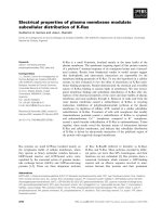
S16D mutant of Hsp20 (Fig. 1A). Tandem MS analysis
performed by Dr R. Wait (Kennedy Institute of Rheuma-
tology Division, Faculty of Medicine, Imperial College,
London) was unable to detect peptides beyond residue 102
in the urea-soluble S16D mutant (Fig. 1C), whereas both
N- and C-terminal peptides were clearly detected in the
wild-type Hsp20 and water soluble S16D preparations
(Fig. 1C). IEF, under denaturing conditions, indicated that
the pI value of the urea-soluble S16D mutant was higher
than that of the intact wild-type Hsp20 (Fig. 1B). By
analyzing the distribution of the charge residues in the
C-terminal region of Hsp20, and by calculating the theor-
etical pI values of differently truncated species of Hsp20, we
found that cleavage of the polypeptide chain only between
residues 122 and 127 resulted in the formation of a protein
species with a theoretical pI higher than that of intact
Hsp20. Thus, we propose that during expression or
purification, the S16D mutant tends to undergo proteolysis
of the C-terminal region. All experiments described in this
report were performed with an S16D mutant that was
soluble in the absence of urea. The pI value of this soluble
S16D mutant was 0.2 units lower than that of the wild-type
Hsp20. A similar shift of pI was observed previously for the
point mutants of Hsp25 with replacement of Ser with
Asp [29].
The method developed for purification of recombinant
human Hsp27 and its 3D mutant was similar to that
described previously [32,36] and yields 5–7 mg of homo-
geneous protein from 1 L of E. coli culture. As in the case
with Hsp20, all stages of Hsp27 purification were performed
in the absence of urea or detergents.
Oligomeric state of recombinant human Hsp20
Recombinant human wild-type Hsp20 was subjected to size
exclusion chromatography, at neutral pH, on a Superdex
200 column under three different experimental conditions.
In the first we loaded the column with 240 lLofa
2.6 mgÆmL
)1
concentration of protein (curve 1 on Fig. 2A).
In the second, the column was loaded with the same volume
of 0.3 mgÆmL
)1
protein (curve 2 on Fig. 2A). In the third,
the column was loaded with 30 lLofproteinata
concentration of 2.6 mg mL
)1
(curve 3 on Fig. 2A). Under
these experimental conditions, the apparent molecular mass
of recombinant human wild-type Hsp20 was 58, 54 and
56 kDa for the first, second and third experimental condi-
tions, respectively. We also analyzed the effect of urea
induced denaturation, followed by renaturation, on the
oligomeric state of Hsp20. As shown in Fig. 2B (curves 3
and 4) denaturation–renaturation showed practically no
effect on the oligomeric state of Hsp20, and both intact and
Fig. 1. Characterization of recombinant human wild-type Hsp20 and its
S16D mutant. SDS gel electrophoresis (A) and IEF (B) of wild-type
Hsp20 (1), and of its S16D mutant that is soluble in the absence (2) and
in the presence (3) of urea. Arrows indicate the position of molecular
mass markers (14 and 25 kDa) and direction of pH gradient. (C)
Primary structure of wild-type Hsp20, and of its S16D mutant soluble
in the absence and in the presence of urea, as determined by HPLC/
tandem MS. The experimentally determined sequence is shown in
bold; shadowed residues were not detected in the experiment.
Fig. 2. Size-exclusion chromatography of recombinant human wild-type
Hsp20. (A) Lack of effect of dilution or sample volume on the apparent
molecular mass of Hsp20. Equal volumes (240 lL) containing 624 lg
(1) or 72 lg (2) of Hsp20, or equal quantities (72 lg) of Hsp20
dissolved in 240 lL (2) or 30 lL (3) volumes, were subjected to
chromatography on a Superdex 200 HR 10/30 column. (B) Effect of
urea-induced denaturation followed by renaturation on the chroma-
tographic behavior of small heat shock proteins. a-Crystallin (1 and 2)
and wild-type Hsp20 (3 and 4) were subjected to size-exclusion
chromatography before (1 and 3) or after (2 and 4) urea-induced
denaturation, followed by renaturation.
294 O. V. Bukach et al. (Eur. J. Biochem. 271) Ó FEBS 2003
renatured proteins had an apparent molecular mass of
54–56 kDa. However, denaturation–renaturation of com-
mercial a-crystallin was accompanied by a significant
decrease of molecular mass. The molecular mass of
a-crystallin that was not subjected to urea treatment was
> 900 kDa, whereas after urea treatment and renaturation
itsmolecularmasswas570kDa(Fig.2B).
As size-exclusion chromatography was insufficient for the
exact estimation of oligomeric forms of Hsp20, and the
apparent molecular mass of 54–56 kDa determined by this
method may correspond to dimers or trimers of Hsp20, we
performed additional crosslinking experiments. The
removal of b-mercaptoethanol was accompanied by the
appearance of an additional band of molecular mass
40 kDa, as shown by SDS/PAGE (Fig. 3A). This band
disappeared if, prior to electrophoresis, the sample was
treatedwithanexcessofb-mercaptoethanol. Therefore, we
suggest that the 40 kDa band corresponds to Hsp20 dimer
crosslinked via single Cys46. Crosslinking of Hsp20 with
dimethylsuberimidate was also accompanied by the forma-
tion of an additional band with molecular mass 40 kDa
(Fig. 3B), that probably also corresponds to Hsp20 dimer.
Similar results were obtained if Hsp20 was subjected to
zero-length crosslinking by EDC and NHS (Fig. 3C). In
this case we observed two or three closely separated bands
with apparent molecular mass 38–40 kDa that probably
correspond to isomers of Hsp20 dimers. Thus, under the
experimental conditions used, Hsp20 predominantly forms
dimers of 40 kDa molecular mass, as judged by SDS gel
electrophoresis, and 54–58 kDa by size-exclusion chroma-
tography.
We considered that changes in pH might somehow affect
the quaternary structure of Hsp20. At pH 7.5–7.0 Hsp20
was eluted as a more or less symmetrical peak with apparent
molecular mass 54–58 kDa (Fig. 4). At pH 6.5, both wild-
type protein and its S16D mutant were eluted as broader
peaks with a slightly smaller apparent molecular mass
(46–47 kDa) (Fig. 4). When the pH was decreased to 6.0,
two peaks with apparent molecular masses of 47–50 and
28–30 kDa were observed on the chromatogram (Fig. 4).
At pH 5.5, the high molecular mass peak completely
disappeared and the small molecular mass peak became
broader and more asymmetric (Fig. 4). A decrease in pH
from 7.5 to 5.5 was accompanied not only by a decrease of
the apparent molecular mass of Hsp20, but also by a
decrease in the area under the protein peaks on the
chromatogram. Acidification probably results in the disso-
ciation of small oligomers of Hsp20 and its S16D mutant to
monomers that tend to unfold and aggregate. These
aggregates are retarded on the top of the column and
therefore not detected on the chromatogram.
The data presented indicates that acidic pH induced
unfolding of Hsp20. In order to confirm this, we analyzed
far UV CD spectra of Hsp20 and a-crystallin at different
pH values. At a high concentration of wild-type Hsp20
( 1.0 mgÆmL
)1
), dialysis against pH 6.0 buffer was accom-
panied by partial protein precipitation. The molar ellipticity
of Hsp20 remaining in the supernatant ( 0.5 mgÆmL
)1
)
had a negative maximum at 220 nm (Fig. 5A). After
dialysis at pH 6.8, wild-type Hsp20 ( 1.0 mgÆmL
)1
)was
predominant in the supernatant and the maximum peak of
molar ellipticity was shifted to 218 nm (Fig. 5A). Dialysis of
wild-type Hsp20 ( 1.0 mgÆmL
)1
)atpH7.5wasnot
accompanied by any precipitation and the molar ellipticity
at pH 7.5 was lower than that at acidic pH values with a
shift in the maximum to 216 nm. The data presented
confirm that acidification leads to partial unfolding and
precipitation of Hsp20 and indicate that the secondary (or
tertiary) structure of Hsp20 remaining in the supernatant at
acidic pH is different from that at neutral pH values. Similar
results were obtained with the S16D mutant of Hsp20 (data
Fig. 3. Crosslinking of Hsp20. (A) Formation of disulfide crosslinked
Hsp20 dimers. A sample of oxidized Hsp20 treated with an excess of
b-mercaptoethanol (2), or loaded onto the gel without the addition of
reducing agents (3). (B) Crosslinking of Hsp20 with dimethylsube-
rimidate. Hsp20 before (2) or after (3) incubation with 20 m
M
dimethylsuberimidate. (C) Zero-length crosslinking of Hsp20. Hsp20
before (2) and after (3) incubation with 1-(3-dimethylaminopropyl)-
3-ethylcarbodiimide hydrochloride (5 m
M
) and N-hydroxysuccinimide
(5 m
M
). In all cases the mixture of standards containing proteins with
molecular masses 94, 67, 43, 30, 20 and 14 kDa was loaded on the first
track.
Fig. 4. Effect of pH on the oligomeric state of recombinant human wild-
type Hsp20 and its S16D mutant. Three-hundred microliter samples
containing 90 lg of wild-type Hsp20 (solid lines) or its S16D mutant
(dotted lines) were loaded onto the column of Superdex 200 HR 10/30
equilibrated with buffer D (50 m
M
phosphate, 150 m
M
NaCl, 1 m
M
EDTA, 15 m
M
b-mercaptoethanol) with a pH of 7.5, 7.0, 6.5, 6.0 or
5.5. For clarity, the pairs of elution profiles obtained at different pH
values are shifted from each other by 10 mAu.
Ó FEBS 2003 Human 20 kDa small heat shock protein (Eur. J. Biochem. 271) 295
not shown). Analogous experiments were performed with
commercial a-crystallin. In this case, independently of pH,
a-crystallin was not precipitated and remained in the
supernatant. The changes of pH in the range of 6.0–7.5
weakly affect both amplitude and the position of maximum
on the far UV CD spectra of a-crystallin (Fig. 5B). Thus,
acidification induced small changes in the secondary (or
tertiary) structure of a-crystallin and these changes were not
accompanied by protein aggregation. As already men-
tioned, acidification induces substantial changes in the
secondary structure of Hsp20. These structural changes
probably result in the dissociation of Hsp20 dimers and
aggregation of partially unfolded monomers.
Chaperone activity of human Hsp20
The reduction of disulfide bonds induces dissociation and
aggregation of the insulin B-chain that is accompanied by a
substantial increase in the optical density (Fig. 6, curve 1).
At pH 7.5, the addition of increasing quantities of intact
wild-type Hsp20 results in an increase of the lag period and
a decrease in the amplitude of light scattering. Significant
retardation of the insulin B-chain aggregation was observed
at an insulin/Hsp20 ratio of 2 : 1. At a mass ratio of 1 : 1,
the sHsp almost completely prevented the aggregation of
reduced insulin (Fig. 6A). Denaturation by 6
M
urea
followed by renaturation had no effect on the chaperone
activity of the wild-type Hsp20, and complete prevention of
insulin aggregation was achieved at the same Hsp20/insulin
ratio as for intact protein (Fig. 6B). The S16D mutant of
Hsp20 also decreased the aggregation of insulin (Fig. 6C);
however, it was less effective than the wild-type protein.
Denaturation–renaturation of the S16D mutant only
weakly affected its chaperone properties (Fig. 6D). Com-
mercial a-crystallin that was not subjected to urea treatment
was very ineffective in preventing reduction-induced aggre-
gation of insulin. Even at a ratio of 1 : 1, a-crystallin only
slightly decreased the aggregation of insulin (Fig. 6E). Urea-
induced denaturation followed by renaturation significantly
improved the chaperone activity of a-crystallin (Fig. 6F).
This was probably caused by a change in the aggregation
state of a-crystallin that was induced by urea treatment and
identified by size-exclusion chromatography (see Fig. 2B).
However, even after treatment with urea, the chaperone
activity of a-crystallin was similar to that of the wild-type
Hsp20. Thus, at pH 7.5 and with reduced insulin as a model
substrate, the chaperone activity of the wild-type Hsp20
was comparable to or greater than that of commercial
a-crystallin.
At pH 7.0, the heating of isolated ADH in the absence of
divalent cations was accompanied by aggregation and a
large increase in the light scattering (Fig. 7, curve 1).
Addition of increasing quantities of the wild-type Hsp20
resulted in retardation of the onset of aggregation and a
decrease in the amplitude of light scattering (Fig. 7A, curves
2–5). At the ADH/Hsp20 ratio of 1 : 1 (wt/wt), aggregation
of ADH was completely prevented. Similar results were
obtained with the S16D mutant of Hsp20 (Fig. 7B) and
a-crystallin (Fig. 7C). However, at a lower concentration,
when the ratio of ADH/sHsp was 2 : 1 (wt/wt), the
efficiency of three sHsp decreased in the following order:
wild-type Hsp20 > S16D mutant > a-crystallin (Fig. 7D).
Thus, phosphorylation (or a mutation mimicking phos-
phorylation) decreased the chaperone activity of Hsp20
measured both with insulin and ADH (Figs 6 and 7). It is
worthwhile to note that at the same time, phosphorylation
of Ser16 of Hsp20 significantly enhanced the relaxation
effect of Hsp20 on the smooth muscle contraction [16–23].
The optical method used for measuring the chaperone
activity of Hsp20 was complemented by the centrifugation
assay. Upon heating, isolated ADH formed aggregates that
were easily precipitated and, after only 20 min of incubation,
more than 75% of the ADH was detected in the pellet
(Fig. 7E, curve 1). After 60 min of incubation, isolated ADH
was completely aggregated and precipitated. a-Crystallin
was rather ineffective in preventing the aggregation of ADH
(Fig. 7E, curve 2). This fact seems to contradict with the
results obtained by light scattering where a-crystallin at least
partially inhibited the aggregation of ADH (Fig. 7C).
However, this apparent contradiction can be explained by
the suggestion that small complexes are effectively precipi-
tated during centrifugation but contribute only slightly to
light scattering. These complexes can be formed either by
denatured ADH or by denatured ADH being bound to
sHsp. Indeed, we found that at the end of incubation more
than 30% of a-crystallin was coprecipitated with denatured
ADH (Fig. 7F). Hsp20 was more effective in preventing
Fig. 5. Far UV CD spectra of the wild-type Hsp20 (A) and commercial
a-crystallin (B). The spectra were recorded at pH 6.0 (1), 6.8 (2) or 7.5
(3).
296 O. V. Bukach et al. (Eur. J. Biochem. 271) Ó FEBS 2003
precipitation of denatured ADH (Fig. 7E, curve 3). Much
smaller quantities of Hsp20 were coprecipitated with dena-
tured ADH (Fig. 7F, curve 2). It is worthwhile mentioning
that isolated sHsp were not precipitated in the absence of
ADH, even after 60 min of incubation (Fig. 7F, curve 3).
Similar results were obtained with the S16D mutant of Hsp20
(data not shown). Thus, at pH 7.0, Hsp20 is a more potent
chaperone than a-crystallin, probably because complexes
formed by Hsp20 with denatured ADH are smaller or more
soluble than the corresponding complexes formed by dena-
tured ADH and a-crystallin.
As discussed above, a decrease in the pH to pH 6.0 may
induce partial unfolding and dissociation of small oligomers
formed by Hsp20 or its S16D mutant (Figs 4 and 5). As
unfolding and dissociation may affect the chaperone activity
of Hsp20, we analyzed the effect of different sHsps on the
aggregation of ADH at pH 6.0. Under these conditions,
heating also induced the aggregation of ADH (Fig. 8, curve
1). Addition of increasing quantities of wild-type Hsp20
increased the rate and amplitude of light scattering (Fig. 8A,
curves 2–5). Thus, the wild-type Hsp20, instead of prevent-
ing, promotes the aggregation of ADH. This is probably a
result of the formation of insoluble complexes of Hsp20 and
denatured ADH. Indeed, as shown in Figs 8E,F, incubation
of ADH with the wild-type Hsp20 resulted in the formation
of a pellet containing both proteins. At pH 6.0 and at the low
concentrations used in the experiment, isolated Hsp20 itself is
completely soluble and does not precipitate (Fig. 8A, curve
6, Fig. 8F, curve 3). However, complexes formed by partially
unfolded Hsp20 and denatured ADH tend to aggregate and
precipitate.
Qualitatively similar results were obtained with the S16D
mutant of Hsp20 (Fig. 8B). However, in this case addition
of increasing quantities of the S16D mutant resulted in a
small retardation of the onset of ADH aggregation and
either did not affect the amplitude of light scattering or
slightly increased it. Using the centrifugation assay we
found that after a short incubation, the S16D mutant
predominantly remained in the supernatant, whereas after a
long incubation a large proportion of the S16D mutant
coprecipitated with ADH (data not shown).
At pH 6.0, a low concentration of a-crystallin either did
not affect or slightly increased the thermal aggregation of
ADH (Fig. 8C). At an ADH/crystallin ratio of 1 : 1, a
significant decrease in the extent of ADH aggregation was
observed (Fig. 8C, curve 5). a-Crystallin was more effective
than Hsp20 in preventing the precipitation of ADH
(Fig. 8E) and smaller quantities of a-crystallin were copre-
cipitated with denatured protein (Fig. 8F). Therefore, at
pH 6.0, a-crystallin possessed higher chaperone activity
than Hsp20 or its S16D mutant.
Formation of mixed oligomer complexes between
recombinant human Hsp20 and Hsp27
In tissue extracts, Hsp20 forms high molecular weight
complexes [9,19] and is copurified with aB-crystallin and
Hsp27 [8,9]. Indirect data also indicate that Hsp20 may
interact with Hsp27 and aB-crystallin [10]. However, to our
knowledge, the hetero-oligomeric complexes formed by
Hsp20 with other sHsp have not been characterized and
reported in the literature. Therefore, we investigated the
Fig. 6. Influence of recombinant human Hsp20
(A and B), the S16D mutant of Hsp20 (C and
D) or commercial a-crystallin (E and F) before
(A, C and E) or after (B, D and F) urea treat-
ment followed by renaturation on the reduction
induced aggregation of insulin. The chaperone
activity was measured by the prevention of
dithiothreitol-induced aggregation of insulin
(0.25 mgÆmL
)1
)at40°C under conditions
described in the Materials and methods.
Insulin alone (1), or insulin in the presence of
0.06 mgÆmL
)1
(2), 0.12 mgÆmL
)1
(3) or
0.25 mgÆmL
)1
(4) small heat shock proteins.
Ó FEBS 2003 Human 20 kDa small heat shock protein (Eur. J. Biochem. 271) 297
interaction of Hsp20 and its mutant mimicking phosphory-
lation with Hsp27.
Interaction of the wild-type Hsp20 with the wild-type
Hsp27 was analyzed by means of size-exclusion chroma-
tography. Hsp20 and Hsp27 were eluted from the Superdex
200 column as single peaks with molecular masses of 56
and 560 kDa, respectively. The chromatographic behavior
of isolated Hsp20 and Hsp27 was not altered if, prior to
loading on the column, these proteins were preincubated for
3 h at 30 or 37 °Corfor15hat18°C. If equimolar
quantities of these two proteins were mixed and immediately
subjected to size-exclusion chromatography, two well sep-
arated peaks with apparent molecular masses 560 and
56 kDa, corresponding to isolated Hsp27 and Hsp20, were
detected on the chromatogram (Fig. 9A, curve 1). Accord-
ing to SDS/PAGE, the high molecular mass peak contained
exclusively Hsp27, whereas the small molecular mass peak
contained only Hsp20. The elution profile was not changed
upon preincubation of this mixture of proteins for 15 h at
18 °C (data not shown). If, prior to loading on the column,
the mixture of the wild-type Hsp27 and Hsp20 was
incubated for 3 h at 30 °C, the elution profile was signifi-
cantly changed. The amplitude of the high molecular mass
peak decreased and its apparent molecular mass was
470 kDa. This peak was asymmetric with a prominent
trailing edge. In addition, a new peak, with an apparent
molecular mass of 91 kDa, appeared on the chromatogram
and the peak corresponding to isolated Hsp20 (56 kDa)
decreased in size (Fig. 9A, curve 2). Even more prominent
changes were observed if the mixture of two wild-type
proteins was incubated for 3 h at 37 °C (Fig. 9A, curve 3).
In this case we observed two protein peaks with apparent
molecular masses of 300 and 100 kDa, and each of these
peaks, according to SDS/PAGE, contained almost identical
quantities of Hsp27 and Hsp20 (insert on Fig. 9A). Similar
results were obtained if the wild-type Hsp27 was mixed with
the S16D mutant of Hsp20. Thus, after mixing at 30–37 °C,
homo-oligomers of wild-type Hsp27 and Hsp20 (or the
S16D mutant of Hsp20) may rearrange, forming mixed
hetero-oligomers that contain similar quantities of these two
sHsp.
The isolated 3D mutant of Hsp27 produces a broad peak
with apparent molecular mass 96–106 kDa. A significant
decrease in molecular mass compared with the wild-type
Hsp27 is a result of the fact that mutations mimicking
phosphorylation induce dissociation of large oligomers of
Hsp27 [29–31]. As already mentioned, the wild-type Hsp20
and its S16D mutant are eluted as a single peak with an
apparent molecular mass of 56 kDa. A mathematical
summation of elution profiles obtained for the 3D mutant
of Hsp27 and the wild-type Hsp20 is presented on curve 1 of
Fig. 9B. Only one broad asymmetric peak, with an apparent
molecular mass of 100 kDa, was observed if, immediately
after mixing, the two proteins were loaded onto the column
(Fig. 9B, curve 2). The position and shape of this peak were
different from the sum of the two elution profiles obtained
for the isolated 3D mutant of Hsp27 and wild-type Hsp20
(compare curves 1 and 2 on Fig. 9B). If the mixture of the
Fig. 7. Effect of Hsp20, its S16D mutant and
a-crystallin on the heat-induced aggregation of
yeast alcohol dehydrogenase (ADH) at pH 7.0.
Aggregation of ADH (0.26 mgÆmL
)1
)was
induced by the addition of EDTA and
dithiothreitol and incubation at 42 °C, and
was measured either by light scattering (A–D)
or by centrifugation (E–F). Panels A–C, ADH
alone(1),orADHinthepresenceof
0.026 mgÆmL
)1
(2), 0.052 mgÆmL
)1
(3),
0.13 mgÆmL
)1
(4) or 0.26 mgÆmL
)1
(5)ofthe
wild-type Hsp20 (A), the S16D mutant of
Hsp20 (B), or a-crystallin (C). (D) Compar-
ison of the effect of different small heat shock
proteins (0.13 mgÆmL
)1
) on the aggregation of
ADH (0.26 mgÆmL
)1
). ADH alone (1), or
ADH in the presence of the wild-type Hsp20
(2), the S16D mutant of Hsp20 (3) or a-crys-
tallin (4). (E) Heat-induced precipitation of
isolated ADH (0.26 mgÆmL
)1
)(1),orADH
in the presence of either a-crystallin
(0.13 mgÆmL
)1
) (2) or wild-type Hsp20
(0.13 mgÆmL
)1
) (3). The percentage of ADH
in the pellet is plotted against the time of
incubation. (F) Co-precipitation of a-crystal-
lin (1) or wild-type Hsp20 (2) with heat
denatured ADH. The percentage of small heat
shock protein in the pellet is plotted against
the time of incubation. Lack of precipitation
of isolated small heat shock proteins is shown
on curve 3.
298 O. V. Bukach et al. (Eur. J. Biochem. 271) Ó FEBS 2003
3D mutant of Hsp27 and wild-type Hsp20 (or S16D mutant
of Hsp20) were incubated for 3 h at 30 °C, only one peak
with an apparent molecular mass 95 kDa was detected on
the chromatogram. Thus, homo-oligomers formed by the
3D mutant of Hsp27 and wild-type Hsp20 (or its S16D
mutant) rapidly rearrange, forming hetero-oligomeric
complexes.
Discussion
To our knowledge there are only two publications that
report a detailed investigation of the biochemical properties
of isolated Hsp20. Kato et al. [9] reported that Hsp20 is
presented in so-called aggregated and dissociated forms
with apparent molecular masses of 200–300 and 67 kDa,
respectively. Using size-exclusion chromatography, van de
Klundert et al. [15] also detected two forms of Hsp20, with
apparent molecular masses of 470 and 43 kDa, that,
depending on the protein concentration may convert to
each other. In our case, size-exclusion chromatography
revealed only an oligomer of Hsp20 with an apparent
molecularmassof 54–58 kDa (Fig. 2). According to our
crosslinking experiments, Hsp20 predominantly forms
dimers with an apparent molecular mass of 40 kDa, as
judged by SDS/PAGE (Fig. 3). Therefore, the question
arises as to why we did not observe the high molecular mass
oligomers of Hsp20 detected previously by Kato et al.[9]
and van de Klundert et al. [15].
We presumed that the exposure of Hsp20 to a high
concentration of urea [9,15], or to urea and detergents [35],
as used in the previously published reports, might affect the
quaternary structure of Hsp20. In order to verify this, we
denatured Hsp20 (purified by our method) by 6
M
urea and
renatured it under the conditions described by van de
Klundert et al. [15]. This treatment had no effect either on
the apparent molecular mass, as determined by size-
exclusion chromatography, or on the chaperone activity
measured by the prevention of insulin aggregation. Thus,
treatment with urea cannot explain the difference in
molecular mass identified in our experiments and in data
published previously [9,15]. Another explanation was based
on the fact that practically all previously published results
were obtained using rat Hsp20 [9,15,35], whereas in the
present study human Hsp20 was used. Although rat and
human Hsp20 are highly homologous ( 90% identity of
the primary structure), the rat Hsp20 consists of 162
residues, whereas the human protein consists of 160 residues
and the dipeptide deletion is located at the very C-terminal
end (residues 154–155 of rat Hsp20). It is known that the
C-terminal extension affects the oligomerization and chap-
erone action of Hsp27 [37]. Therefore, we propose that the
difference in the C-terminal extension of human and rat
Hsp20 results in a different oligomeric state of these two
proteins. However, this suggestion is speculative and needs
experimental verification. Finally, as previously mentioned,
when expressing the S16D mutant we found that truncation
of 30–50 C-terminal amino acid residues results in the
formation of protein aggregates that were soluble only in
the presence of a high concentration of urea. Previously it
has been shown that the truncation of a short C-terminal
Fig. 8. Influence of Hsp20, its S16D mutant
and a-crystallin on the heat-induced aggrega-
tion of yeast alcohol dehydrogenase (ADH) at
pH 6.0. Aggregation of ADH (0.15 mgÆmL
)1
)
was measured either by light scattering (A–D)
or by centrifugation (E–F). A–C, ADH alone
(1), or ADH in the presence of 0.015 mgÆmL
)1
(2), 0.03 mgÆmL
)1
(3), 0.075 mgÆmL
)1
(4) or
0.15 mgÆmL
)1
(5) of the wild-type Hsp20 (A),
the S16D mutant of Hsp20 (B) or a-crystallin
(C). Lack of aggregation of isolated small heat
shock proteins (0.15 mgÆmL
)1
)isshown
on curve 6. (D) Comparison of the effect
of different small heat shock proteins
(0.15 mgÆmL
)1
) on the aggregation of ADH
(0.15 mgÆmL
)1
). ADH alone (1), or ADH in
the presence of wild-type Hsp20 (2), the S16D
mutant of Hsp20 (3) or a-crystallin (4). (E)
Heat-induced precipitation of isolated ADH
(0.15 mgÆmL
)1
)(1),orADHinthepresenceof
either wild-type Hsp20 (0.15 mgÆmL
)1
)(2)or
a-crystallin (0.15 mgÆmL
)1
) (3). The percent-
age of ADH in the pellet is plotted against the
time of incubation. (F) Co-precipitation of
wild-type Hsp20 (1) or a-crystallin (2) with
heat denatured ADH. The percentage of small
heat shock proteins in the pellet is plotted
against the time of incubation. Lack of preci-
pitation of isolated small heat shock proteins
isshownoncurve3.
Ó FEBS 2003 Human 20 kDa small heat shock protein (Eur. J. Biochem. 271) 299
peptide increases the hydrophobicity of Hsp27 and decrea-
ses its chaperone effect [37]. There were no signs of
proteolytic degradation in the samples of Hsp20 purified
by Kato et al. [9] and van de Klundert et al. [15]; however,
truncation of a short (2–4 kDa) fragment can be easily
overlooked. Therefore, we suggest that during expression
and/or purification, Hsp20 can undergo limited proteolysis,
and deletion of a short C-terminal fragment may result in
the formation of a mixture of small and large aggregates
that were reported in the previous publications.
Van de Klundert et al. [15] claimed that Hsp20 is a poor
chaperone. In our investigation we found that at neutral or
slightly alkaline pH, Hsp20 has comparable or even higher
chaperone activity than commercial a-crystallin (Figs 6 and
7). sHsp protect the cell against unfavorable conditions,
among them acidosis. For instance, the data of Wang [38]
indicate that a-crystallin prevents acidification-induced
aggregation of creatine kinase and luciferase. In our study,
at pH 6.0, a-crystallin partially prevented the aggregation of
yeast ADH, whereas the wild-type Hsp20 retained its ability
to interact with denatured substrates, but, instead of
preventing, promoted the aggregation of denatured ADH
(Fig. 8). This was caused by the fact that at low pH Hsp20
tends to unfold, and dimers of Hsp20 dissociate to
monomers. Under these conditions, partially unfolded
monomers of Hsp20 interact with denatured ADH and
form poorly soluble complexes. Similar effects have been
observed for the truncated form of Hsp27 [37] and for the
alternative splicing product of aA-crystallin [39]. Thus,
although Hsp20 and a-crystallin are closely related, they
have different properties. Acidification induced a small
decrease of the chaperone activity of a-crystallin, but
significantly decreased the chaperone activity of Hsp20.
Similar conclusions were reached by van de Klundert et al.
[40], who postulated that Hsp20 and a-crystallin might be
involved in distinct protective activities in living cells. It is
worthwhile of note that the measurement of chaperone
activity and analysis of the quaternary structure of Hsp20
was performed in buffers with compositions that are not
completely physiological. This was implemented in order to
compare our results with data in the published literature.
However, limitations of biochemical experiments should be
taken into account when interpreting our results at a
physiological level.
The data obtained with the help of a yeast two-hybrid
system indicate that different sHsps may interact with each
other [10]. Moreover, Bova et al. [11], using the method of
fluorescence energy transfer, have directly shown that
Hsp27 and a-crystallin may form mixed oligomers. If crude
extracts of skeletal muscle or heart were subjected to size
exclusion chromatography, Hsp20 was eluted in one or two
high molecular mass peaks. Kato et al. [9] detected two
peaks with apparent molecular masses 200–300 and
68 kDa, whereas Pipkin et al. [19] detected only one peak
with an apparent molecular mass of 230 kDa. Brophy et al.
[17] postulated that cAMP-dependent phosphorylation
results in the change of macromolecular associations of
Hsp20. Finally, Hsp20 is usually copurified with Hsp27 and
a-crystallin [9,19]. Thus, all these data indirectly indicate the
formation of mixed oligomer complexes between Hsp20
and a-crystallin or Hsp27. To verify this, we analyzed the
chromatographic behavior of the mixture of Hsp20 and
Hsp27. In good agreement with Bova et al. [41], we found
that at low temperature the rate of subunit exchange
between the wild-type Hsp20 and Hsp27 was very slow.
However, at 30 or 37 °C the rate of exchange was
significantly increased and we detected two hetero-oligo-
meric complexes with apparent molecular masses 100 and
300 kDa that contained similar quantities of Hsp20 and
Hsp27 subunits (Fig. 9A). Mutation S16D, imitating phos-
phorylation of the Ser16 of Hsp20, had no significant effect
on the rate of subunits exchange or on the composition or
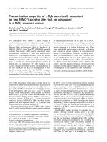
Fig. 9. Formation of hetero-oligomeric complexes between Hsp27 and
Hsp20. (A) Rearrangement of the complexes formed by the wild-type
Hsp27 and Hsp20. The wild-type Hsp27 and Hsp20 were loaded onto
the column immediately after mixing (1) or were incubated at 30 °C(2)
or at 37 °C (3) for 3 h. For clarity the profiles are shifted from each
other by 40 mAu. The protein composition of profile 3 fractions 25–33
is shown on the insert. The positions of Hsp27 and Hsp20 are marked
by arrows. (B) Rearrangement of the complexes formed by the 3D
mutant of Hsp27 and the wild-type Hsp20. A mathematical summa-
tion of the elution profiles of the isolated 3D mutant of Hsp27 and
isolated wild-type Hsp20 is presented on curve 1. Experimental elution
profiles of the mixture of the 3D mutant of Hsp27 and Hsp20 loaded
onto the column immediately after mixing (2), or after incubation for
3 h at 30 °C (3). For clarity the profiles are shifted from each other by
40 mAu.
300 O. V. Bukach et al. (Eur. J. Biochem. 271) Ó FEBS 2003
structure of the hetero-oligomeric complexes formed by
Hsp20 and Hsp27.
Mixing of the 3D mutant of Hsp27 with the wild-type
Hsp20 or its S16D mutant was accompanied by a very rapid
exchange of subunits and formation of a mixed oligomer
with an apparent molecular mass of 95 kDa (Fig. 9B).
Thus, Hsp20 and Hsp27 may form two types of hetero-
oligomeric complexes – one with an apparent molecular
mass of 100 kDa and another of 300 kDa. The low
molecular mass oligomer is formed by both the wild-type
and pseudophosphorylated sHsp, whereas only the wild-
type sHsp formed the large molecular mass complex. The
3D mutant of Hsp27 forms hetero-oligomeric complexes
more readily than the wild-type protein. Previously, a
similar conclusion was reached concerning the interaction of
Hsp22 with the wild-type Hsp27 and its 3D mutant [12].
Summing up, we conclude that Hsp20 and Hsp27 readily
form two types of hetero-oligomeric complexes with
molecular masses 100 and 300 kDa. The rate of exchange
of sHsp subunits depends on a mutation mimicking the
phosphorylation of Hsp27. The experimental conditions are
not directly comparable with those in living cells. However,
taking into account a high efficiency of complex formation
and a high concentration of Hsp20 and Hsp27 in certain
tissues [8,9], we may suppose that hetero-oligomeric com-
plexes are also formed in vivo. This will lead to a decrease in
the concentration of homo-oligomeric sHsp, affect oligomer
structure and result in the accumulation of hetero-oligo-
meric complexes with properties that might be different from
homo-oligomers. Isolation and detailed characterization of
hetero-oligomeric complexes will provide new, important
information on the functioning of sHsp in the cell.
Acknowledgements
The authors are grateful to Dr A. M. Arutunyan (Institute of Physico-
chemical Biology, Moscow State University) for his help in CD
measurements. This investigation was supported by the Russian
Foundation for Basic Research and by the Wellcome Trust.
References
1. Kappe, G., Franck, E., Verschuure, P., Boelens, W.C., Leunissen,
J.A. & de Jong, W.W. (2003) The human genome encodes 10
alpha-crystallin-related small heat shock proteins: HspB1–10. Cell
Stress Chaperones 8, 53–61.
2. Haslbeck, M. (2002) sHsps and their role in the chaperone net-
work. Cell. Mol. Life Sci. 59, 1649–1657.
3. Fontaine, J.M., Rest, J.S., Welsh, M.J. & Benndorf, R. (2003) The
sperm outer dense fiber protein is the 10th member of the super-
family of mammalian small stress proteins. Cell Stress Chaperones
8, 62–69.
4. Clark, J.I. & Muchowski, P.J. (2000) Small heat-shock proteins
and their potential role in human disease. Curr. Opin. Struct. Biol.
10, 52–59.
5. MacRae, T.H. (2000) Structure and function of small heat shock/
a-crystallin proteins: established concepts and emerging ideas.
Cell. Mol. Life Sci. 57, 899–913.
6. Kim, K.K., Kim, R. & Kim, S.H. (1998) Crystal structure of a
small heat-shock protein. Nature 394, 595–599.
7. van Monfort, R.L.M., Basha, E., Friedrich, K.L., Slingsby, C. &
Vierling, E. (2001) Crystal structure and assembly of a eukaryotic
small heat shock protein. Nat. Struct. Biol. 8, 1025–1030.
8. Kato, K., Shinohara, H., Goto, S., Inaguma, Y., Morishita, R. &
Asano, T. (1992) Copurification of small heat shock protein with
aB crystallin from human skeletal muscle. J. Biol. Chem. 267,
7718–7725.
9. Kato, K., Goto, S., Inaguma, Y., Hasegawa, K., Morishita, R. &
Asano, T. (1994) Purification and characterization of a 20-kDa
protein that is highly homologous to aB crystallin. J. Biol. Chem.
269, 15302–15309.
10. Sugiyama, Y., Suzuki, A., Kishikawa, M., Akutsu, R., Hirose, T.,
Waye, M.M., Tsui, S.K., Yoshida, S. & Ohno, S. (2000) Muscle
develops a specific form of small heat shock protein complex
composed of MKBP/HSPB2 and HSPB3 during myogenic dif-
ferentiation. J. Biol. Chem. 275, 1095–1104.
11. Bova,M.P.,McHaourab,H.S.,Han,Y.&Fung,B.K.(2000)
Subunit exchange of small heat shock proteins. Analysis of oligo-
mer formation of alphaA-crystallin and Hsp27 by fluorescence
resonance energy transfer and site-directed truncations. J. Biol.
Chem. 275, 1035–1042.
12. Benndorf, R., Sun, X., Gilmont, R.R., Biederman, K.J., Molloy,
M.P., Goodmurphy, C.W., Cheng, H., Andrews, P.C. & Welsh,
M.J. (2001) HSP22, a new member of the small heat shock protein
superfamily, interacts with mimic of phosphorylated HSP27
(
3D
HSP27). J. Biol. Chem. 276, 26753–26761.
13. Horwitz, J. (2003) Alpha-crystallin. Exp. Eye Res. 76, 145–153.
14. Ehrnsperger, M., Buchner, J. & Gaestel, M. (1998) Structure and
function of small heat-shock proteins. In Molecular Chaperones in
the Life Cycle of Proteins. Structure, Function and Mode of Action
(Fink, A.L. & Gioto, Y., eds). pp. 533–575. Marcel Dekker Inc.,
New York, USA.
15. van de Klundert, F.A.J.M., Smulders, R.H.P.H., Gijsen, M.L.J.,
Lindner, R.A., Jaenicke, R., Carver, J.A. & de Jong, W.W. (1998)
The mammalian small heat-shock protein Hsp20 forms dimers
and is a poor chaperone. Eur. J. Biochem. 258, 1014–1021.
16. Beall, A.C., Kato, K., Goldenring, J.R., Rasmussen, H. & Bro-
phy, C.M. (1997) Cyclic nucleotide-dependent vasorelaxation is
associated with the phosphorylation of a small heat shock-related
protein. J. Biol. Chem. 272, 11283–11287.
17. Brophy, C.M., Dickinson, M. & Woodrum, D. (1999) Phosphory-
lation of the small heat shock-related protein, HSP20, in vascular
smooth muscles is associated with changes in the macromolecular
associations of HSP20. J. Biol. Chem. 274, 6324–6329.
18. Beall, A., Bagwell, D., Woodrum, D., Stoming, T.A., Kato, K.,
Suzuki, A., Rasmussen, H. & Brophy, C.M. (1999) The small heat
shock protein, HSP20, is phosphorylated on serine 16 during cyclic
nucleotide-dependent relaxation. J. Biol. Chem. 274, 11344–11351.
19. Pipkin, W., Johnson, J.A., Creazzo, T.L., Bursh, J., Komalavilas,
P. & Brophy, C.M. (2003) Localization, macromolecular asso-
ciations, and functions of the small heat shock-related protein
HSP20 in rat heart. Circulation 107, 469–476.
20. Woodrum, D., Pipkin, W., Tessier, D., Komalavilas, P. & Brophy,
C.M. (2003) Phosphorylation of the heat shock-related protein,
HSP20, mediates cyclic nucleotide-dependent relaxation. J. Vasc.
Surg. 37, 874–881.
21. Rembold,C.M.,Foster,D.B.,Strauss,J.D.,Wingard,C.J.&Van
Eyk, J.E. (2000) cGMP-mediated phosphorylation of heat shock
protein 20 may cause smooth muscle relaxation without myosin
light chain dephosphorylation in swine carotid artery. J. Physiol.
524, 865–878.
22. Rembold, C., O’Connor, M., Clarkson, M., Wardle, R.L. &
Murphy, R.A. (2001) HSP20 phosphorylation in nitroglycerin-
and forskolin-induced sustained reductions in swine carotid media
tone. J. Appl. Physiol. 91, 1460–1466.
23. O’Connor, M. & Rembold, C. (2002) Heat-induced force sup-
pression and HSP20 phosphorylation in swine carotid media.
J. Appl. Physiol. 93, 484–488.
Ó FEBS 2003 Human 20 kDa small heat shock protein (Eur. J. Biochem. 271) 301
24. Wang, Y., Xu, A., Pearson, R.B. & Cooper, G.J.S. (1999) Insulin
and insulin antagonists evoke phosphorylation of P20 at serine
157 and serine 16 respectively in rat skeletal muscle. FEBS Lett.
462, 25–30.
25. Wang, Y., Xu, A. & Cooper, G.J.S. (1999) Phosphorylation of
P20 is associated with the actions of insulin in rat skeletal and
smooth muscle. Biochem. J. 344, 971–976.
26. Wang,Y.,Xu,A.,Ye,J.,Kraegen,E.W.,Tse,C.A.&Cooper,
G.J.S. (2001) Alteration in phosphorylation of P20 is associated
with insulin resistance. Diabetes 50, 1821–1827.
27. Kozawa, O., Matsuno, H., Niwa, M., Hatakeyama, D., Oiso, Y.,
Kato, K. & Uematsu, T. (2002) HSP20, low-molecular weight heat
shock-related protein, acts extracellularly as a regulator of platelet
functions: a novel defense mechanism. Life Sci. 72, 113–124.
28. Sarkar, G. & Sommer, S.S. (1990) The ÔmegaprimerÕ method of
site-directed mutagenesis. Biotechniques 8, 404–407.
29. Panasenko, O.O., Seit-Nebi, A., Bukach, O.V., Marston, S.B. &
Gusev, N.B. (2002) Structure and properties of avian small heat
shock protein with molecular weight 25 kDa. Biochim. Biophys.
Acta 1601, 64–74.
30. Ehrnsperger, M., Lilie, H., Gaestel, M. & Buchner, J. (1999) The
dynamics of Hsp25 quaternary structure. Structure and function
of different oligomeric species. J. Biol. Chem. 274, 14867–14874.
31. Rogalla, T., Ehrnsperger, M., Preville, X., Kotlyarov, A., Lutsch,
G.,Ducasse,C.,Paul,C.,Wieske,M.,Arrigo,A.P.,Buchner,J.&
Gaestel, M. (1999) Regulation of Hsp27 oligomerization, cha-
perone function, and protective activity against oxidative stress/
tumornecrosisfactora by phosphorylation. J. Biol. Chem. 274,
18947–18956.
32. Bukach,O.V.,Seit-Nebi,A.S.,Panasenko,O.O.,Kim,M.V.&
Gusev, N.B. (2002) Isolation of tissue and recombinant small heat
shock protein with molecular weight 25 kDa (HSP25) from avian
smooth muscles. Problems Biol. Med. Pharmaceut. Chem. 1,
50–57.
33. Laemmli, U.K. (1970) Cleavage of structural proteins during
the assembly of the head of bacteriophage T4. Nature 227,
680–685.
34.Sharma,K.K.,Kumar,R.S.,Kumar,G.S.&Quinn,P.T.
(2000) Synthesis and characterization of a peptide identified as
a functional element in alphaA-crystallin. J. Biol. Chem. 275,
3767–3771.
35. Brophy, C.M., Lamb, S. & Graham, A. (1999) The small heat
shock-related protein-20 is an actin-associated protein. J. Vasc.
Surg. 29, 326–333.
36. Buchner, J., Ehrnsperger, M., Gaestel, M. & Walke, S. (1998)
Purification and characterization of small heat shock proteins.
Methods Enzymol. 290, 339–349.
37. Lindner, R.A., Carver, J.A., Ehrnsperger, M., Buchner, J.,
Esposito,G.,Behlke,J.,Lutsch,G.,Kotlyarov,A.&Gaestel,M.
(2000) Mouse Hsp25, a small shock protein. The role of its
C-terminal extension in oligomerization and chaperone action.
Eur. J. Biochem. 267, 1923–1932.
38. Wang, K. (2001) aB- and aA-crystallin prevent irreversible aci-
dification-induced protein denaturation. Biochem. Biophys. Res.
Commun. 287, 642–647.
39. Smulders, R.H.P.H., van Geel, I.G., Gerards, W.L.H., Bloemen-
dal, H. & de Jong, W.W. (1995) Reduced chaperone-like activity
of aA
ins
-crystallin, an alternative splicing product containing a
large insert peptide. J. Biol. Chem. 270, 13916–13924.
40. van de Klundert, F.A.J.M., van den Ijssel, P.R.L.A., Stege, G.J. &
de Jong, W.W. (1999) Rat Hsp20 confers thermoresistance in a
clonal survival assay, but fails to protect coexpressed luciferase in
Chinese hamster ovary cells. Biochem. Biophys. Res. Commun.
254, 164–168.
41. Bova, M.P., Huang, Q., Ding, L. & Horwitz, J. (2002) Subunit
exchange, conformational stability, and chaperone-like function
of the small heat shock protein 16.5 from Methanococcus jan-
naschii. J. Biol. Chem. 277, 38468–38475.
302 O. V. Bukach et al. (Eur. J. Biochem. 271) Ó FEBS 2003