Báo cáo khóa học: A single mutation that causes phosphatidylglycerol deficiency impairs synthesis of photosystem II cores in Chlamydomonas reinhardtii pdf
Bạn đang xem bản rút gọn của tài liệu. Xem và tải ngay bản đầy đủ của tài liệu tại đây (264.56 KB, 10 trang )
A single mutation that causes phosphatidylglycerol deficiency impairs
synthesis of photosystem II cores in
Chlamydomonas reinhardtii
Bernard Pineau
1
, Jacqueline Girard-Bascou
2
, Stephan Eberhard
2
, Yves Choquet
2
, Antoine Tre
´
molie
`
res
1
,
Catherine Ge
´
rard-Hirne
1
, Annick Bennardo-Connan
1
, Paulette Decottignies
3
, Sylvie Gillet
3
and Francis-Andre
´
Wollman
2
1
Centre National de la Recherche Scientifique-Universite
´
Paris-Sud, UMR 8618, Institut de Biotechnologie des plantes, Orsay, France;
2
Centre National de la Recherche Scientifique, UPR 1261 ass. University Paris VI, Institut de Biologie Physico-Chimique, Paris,
France;
3
Centre National de la Recherche Scientifique-Universite
´
Paris XI, UMR 8619, Institut de Biochimie et Biophysique
Mole
´
culaire et Cellulaire, Orsay, France
Two mutants of Chlamydomonas reinhardtii, mf1 and mf2,
characterized by a marked reduction in their phosphatidyl-
glycerol content together with a complete loss in its D
3
-trans
hexadecenoic acid-containing form, also lost photosystem II
(PSII) activity. Genetic analysis of crosses between mf2 and
wild-type strains shows a strict cosegregation of the PSII and
lipid deficiencies, while phenotypic analysis of phototrophic
revertant strains suggests that one single nuclear mutation
is responsible for the pleiotropic phenotype of the mutants.
The nearly complete absence of PSII core is due to a severely
decreased synthesis of two subunits, D1 and apoCP47,
which is not due to a decrease in translation initiation. Trace
amounts of PSII cores that were detected in the mutants
did not associate with the light-harvesting chlorophyll a/b-
binding protein antenna (LHCII). We discuss the possible
role of phosphatidylglycerol in the coupled process of
cotranslational insertion and assembly of PSII core subunits.
Keywords: photosystem II; phosphatidylglycerol; D1 syn-
thesis; Chlamydomonas; thylakoid.
The utilization of light energy during photosynthesis to split
water molecules and generate reducing species and ATP
requires highly organized multimolecular complexes. These
complexes contain numerous proteins from chloroplast or
nucleo-cytosolic origin and various associated cofactors
including chlorophyll, carotenoid pigments and redox
components [1]. These large complexes are embedded in
a membrane containing a high proportion of glycolipids,
including monogalactosyldiacylglycerol (MGDG), digalac-
tosyldiacylglycerol (DGDG) and sulfoquinovosyldiacyl-
glycerol (SQDG) [2]. As reported by Joyard et al.[3],
thylakoid membranes do not contain phosphatidylcholine,
and phosphatidylglycerol (PtdGro or PG) is the only
phospholipid that is present in photosynthetic membranes
of cyanobacteria and eukaryotes. A particular fatty acid,
D
3
-trans hexadecenoic acid; C16:1(3t), esterified in the sn-2
position of glycerol, is present among PG found in
chloroplast membranes but it is absent from the other
eucaryotic cell membranes or from bacterial membranes.
Thylakoid membranes, similarly to mitochondrial inner
membranes, are very rich in proteins. The lipid to protein
mass ratio is low, especially in appressed regions where PSII
is located [4]. As a consequence, a large amount of lipid
molecules are directly exposed at the periphery of large
protein complexes and the proximal lipidic environment of
the protein surface is likely to be involved in the structural
and functional organization of integral membrane proteins
[5]. This view is consistent with the high degree of lateral and
transversal heterogeneity that characterizes the distribution
of lipids and their fatty acids along thylakoid membranes [2].
In the eukaryotic alga Chlamydomonas reinhardtii,
mutants mf1 and mf2 were described previously as being
deficient in PG with a total loss of its C16:1(3t)-containing
species [6]. They display alterations in the organization of
their light-harvesting antenna with a spectacular loss in the
oligomeric forms of light-harvesting chlorophyll a/b-binding
protein (LHCII), which is responsible for their low yield of
fluorescence [6,7]. They are also unable to grow photo-
autotrophically because they lack PSII activity [7,8]. Two
revertant strains, one from mf1 (pmf1) and the other from
mf2 (pmf2), selected for the restoration of phototrophic
growth, partially recovered a normal PG-C16:1(3t) content
[8]. These results suggested that PG-C16:1(3t) may have a
specific effect on the expression of PSII-related proteins.
However, while the two mf mutants grown in the presence
of exogenously added PG-C16:1(3t) recover oligomeric
LHCII, pointing to a specific role of this lipid in the
supramolecular organization of LHCII in vivo [9–11], they
did not recover any significant PSII activity [8], an
observation that questioned the relation between PG-
C16:1(3t) deficiency and PSII inactivation.
Correspondence to B. Pineau, IBP, Universite
´
Paris XI, baˆ t. 630,
F-91405 Orsay cedex, France. Fax: + 33 1 69 15 34 23,
E-mail:
Abbreviations: DGDG, digalactosyldiacylglycerol; MGDG, mono-
galactosyldiacylglycerol; SQDG, sulfoquinovosyldiacylglycerol; PG,
phosphatidylglycerol (PtdGro); C16:1(3t), D
3
-trans-hexadecenoic
acid; PSI, photosystem I; PSII, photosystem II; LHC, light-harvesting
chlorophyll protein complex; WT, wild-type strain; DCMU,
3-(3,4-dichlorophenyl)-1,1-dimethylurea.
(Received 16 September 2003, revised 10 November 2003,
accepted 18 November 2003)
Eur. J. Biochem. 271, 329–338 (2004) Ó FEBS 2003 doi:10.1046/j.1432-1033.2003.03931.x
Here, we developed a more thorough genetic approach
to demonstrate the correlation between recovery of PSII
activity and restoration of higher levels of C16:1(3t)-
containing PG. We show that impaired formation of the
PSII core complex in mf1 and mf2 results from a considerable
decrease in the rate of translation of the D1 and apoCP47
subunits of the PSII core. The marginal amounts of PSII
cores produced in these strains can not associate in LHCII–
PSII supercomplexes. These results point to a critical
function of PG at an early step in the biogenesis of PSII cores.
Materials and methods
Strains, cell growth and genetic analysis
C. reinhardtii mf1 and mf2 mutant strains were described
previously [6,7,10] as well as two phototrophic revertants
(pmf1 and pmf2) and were selected, respectively, from mf1
and mf2 strains [8]. Fl39 is a nuclear mutant completely
deficient in PSII activity [12]. The FUD7 strain bears a
deletion of the chloroplast psbA gene encoding the D1
subunit of the PSII reaction center [13]. The wild-type strain
(WT) used in this work was derived from the 137c strain
[14]. Cells were grown at 25 °C in tris/acetate/phosphate
medium at 7–10 lmol photonsÆm
)2
Æs
)1
cool fluorescent
light or in minimum medium at 40–60 lmol photonsÆ
m
)2
Æs
)1
cool fluorescent light [14]. Induction of gametes,
crosses and tetrad analysis were performed as described
previously [14]. Fluorescence induction kinetics of dark-
adapted cells were recorded on cells grown in solid or liquid
tris/acetate/phosphate medium as described previously [15].
Construction of the 5¢
psbA–petA
strain
A DNA fragment containing the promoter, 5¢ UTR and the
first 30 codons of the psbA gene was amplified by PCR using
oligonucleotide primers Aprom (forward): 5¢-CGC ATC
GAT GGA TCC TGC CAC TGA CGT CCT ATT TTA
ATA CTC C-3¢, and Acod (reverse): 5¢-CGC GGA TCC
ATG GAA TCG ATG TAT AAA CGG TTT TCA GTT
GAA GT-3¢,andtheEcoRI restriction fragment of the
chloroplast genome R14 [16] as a template. The resulting
DNA fragment was then digested by ClaIandNcoI(two
restriction sites generated by the oligonucleotides used), and
cloned into vector B
T
FFF [17] digested with the same
enzymes to form plasmid p(bAC)FFF. The aadA cassette
conferring spectinomycin resistance [18] excised with
EcoRV and SmaI was then cloned in reverse orientation
with respect to the petA coding region in p(bAC)FFF
linearized with HincIItoyieldplasmidpihK(bAC)FFF.
This plasmid was used for transformation of a WT, mt+
strain according to Boynton et al. [19]. Transformants in
which the endogenous petA gene was replaced by homo-
logous recombination by the 5¢psbA–petA chimera were
selected for spectinomycin resistance, brought to homo-
plasmy and assessed for the presence of the chimeric
5¢ psbA–petA gene in the chloroplast genome.
Pulse-labelling experiments
Pulse-labelling experiments were carried out according to
Kuras and Wollman [20]. In the experiments presented in
Fig. 5B, cells equivalent to 1 mg of chlorophyll were washed
in 50 m
M
Tris/HCl pH 7.8, 10 m
M
NaCl, 1 m
M
EDTA,
resuspended in the same medium containing 0.5 l
M
of the
protease inhibitor Pefabloc (Fluka, Switzerland) and briefly
disrupted by sonication. Cell extracts were centrifuged at
600 g for 2 min, the white-grey pellet discarded and the
supernatant centrifuged again at 36 000 g for 45 min to
sediment all the green material.
Cell fractionation
Cell membranes, highly enriched in thylakoids, were
prepared from French press-disrupted cells following the
method of Chua and Bennoun [21] including differential
centrifugation of cell extracts and flotation in sucrose layer
gradients. Minor changes were introduced in the molarity of
sucrose layers to take into account the lower density of the
thylakoid membranes of mf1 and mf2, and thus 1.15
M
instead of 1.3
M
sucrose was used in the layer above the
1.8
M
sucrose-containing initial membrane material. For
blue-native PAGE experiments, cell samples equivalent to
1 mg of chlorophyll were washed in 10 m
M
Hepes pH 7.6,
2m
M
EDTA, resuspended in 2 mL of 5 m
M
Hepes, 1 m
M
EDTA, 0.5 l
M
Pefabloc, and briefly disrupted by sonica-
tion. Cell extracts were centrifuged at 700 g for 2 min, the
white-grey pellet discarded and the supernatant centrifuged
again at 36 000 g for 25 min to sediment all the green
material. The pellet was further resuspended in 0.7 mL of
5m
M
Hepes, 1 m
M
EDTA, and loaded on two layers
consisting of 1.4 mL of 1.1
M
and 1.2 mL of 1.5
M
sucrose
in 5 m
M
Hepes, 1 m
M
EDTA, then centrifuged in a
Beckmann SW60 rotor at 270 000 g for 20 min. Thylakoid
membranes were harvested at the interface of the two
sucrose layers.
Blue-native PAGE
Purified thylakoid membranes were prepared from cells
disrupted with ultrasound and diluted in 50 m
M
Bistris,
0.75
M
amino-n-caproic acid, 0.5 m
M
Na
2
EDTA, 20%
(v/v) glycerol, pH 7, to a final chlorophyll concentration
of 0.3 mgÆmL
)1
. Membrane suspensions were solubilized
with dodecylmaltoside (1%, w/v) at 4 °C for 20 min and
centrifuged at 36 000 g for 4 min. The supernatants were
supplemented with Serva blue G-250 (final concentration
0.25%, w/v) prior to loading on the gel. Blue-native
electrophoreses were performed according to the general
method of Scha
¨
gger et al. [22] with minor modifications.
The separating gel consisted of a 4–13% (w/v) acrylamide
gradient whereas the stacking gel was 4% (w/v) acryl-
amide. Final concentrations of Bistris and amino-n-
caproic acid in gel buffer were 25 m
M
and 250 m
M
,
respectively. Cathodic buffer (50 m
M
tricine, 15 m
M
Bistris) was supplemented with 0.012% (w/v) Serva blue
G-250. Electrophoresis (using glass plates of 10 · 12 cm)
was run overnight (4 °C, 110 V) with replacement of the
blue cathodic buffer by a new colourless buffer for two
additional hours. Fragments of green bands or full-length
thin strips were excised from the gel, briefly rinsed with
water and frozen when not used immediately. Before the
second dimension denaturing electrophoresis, strips were
incubated for 30 min in 125 m
M
Tris pH 6.8, 50 m
M
330 B. Pineau et al.(Eur. J. Biochem. 271) Ó FEBS 2003
dithiothreitol, 20% (v/v) glycerol, 4% (w/v) SDS, heated
at 70 °C for 2 min and further analysed on 9–18% (w/v)
acrylamide gradient gels.
Denaturing gel electrophoresis
Electrophoresis in the presence of SDS was performed using
9–18% (w/v) acrylamide gradient gels as reported previ-
ously [23]. Polyacrylamide gels (12–18%, w/v) containing
8
M
urea were performed according to de Vitry et al. [24].
TMBZ staining of electrophoresis gels
After separation of whole cell proteins on denaturing
12–18% (w/v) polyacrylamide/urea gels, covalently bound
cytochromes were stained with 3,3¢,5,5¢-tetramethylbenzi-
dine (TMBZ) according to Thomas et al. [25].
Lipid analysis
Aliquots of cells (150 lg chlorophyll) were harvested by
centrifugation, fixed in boiling ethanol for 5 min and lipids
were extracted with chloroform according to Bligh and
Dyer [26]. Individual lipids were separated by thin layer
chromatography on silica gel 60 plates using the solvent
system chloroform/acetone/methanol/acetic acid/water
(50/20/10/10/5, v/v/v/v/v) and lipid spots detected with
iodine vapour [6]. Fatty acid methyl esters were prepared
by transesterification in methanol/BF
3
, recovered with
n-pentane, dissolved in methanol and analysed by capillary
gas-liquid chromatography using a 50 m long, 0.25 mm
diameter CP-wax 52 column. Heptanoic acid was used as
an internal standard.
Results
The absence of functional PSII and lack of PG-C16:1(3t)
result from a single nuclear mutation
To assess the relation between PG-C16:1(3t) deficiency
and PSII inactivation, we undertook a genetic analysis of
the mf1 and mf2 strains. First, we analysed segregation of
the two phenotypes on colonies arising from the four
products of meiosis (tetrads) from zygotes obtained in
mf2 · WT crosses. All 24 tetrads presented a 2 : 2
Mendelian segregation for PSII deficiency, as character-
ized by their fluorescent induction kinetics of dark-
adapted cells. Two colonies had a wild-type phenotype
[PSII
+
] with several phases of fluorescence rise and decay
that led to a steady-state level well below the F
max
level
reached upon inhibition of photosynthetic electron flow in
the presence of 3-(3,4-dichlorophenyl)-1,1-dimethylurea
(DCMU) (Fig. 1). The two others displayed a DCMU-
insensitive steady state fluorescence yield from the onset of
illumination typical of a PSII-deficient phenotype [PSII
–
].
This showed that the PSII deficiency in the mf2 strain
most probably resulted from a single nuclear mutation.
The progeny from five of these tetrads was used for lipid
analysis. The content in PG-C16:1(3t) together with the
content in total PG (relative to chlorophyll) displayed a
2 : 2 segregation in each tetrad, however the mean values
for total PG content indicated the contribution of a
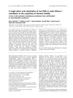
Fig. 1. Fluorescence induction kinetics of dark-adapted cells of the WT,
mf1 and revertant p3mf1 strains. Cells were grown on liquid tris/acet-
ate/phosphate medium. The F
o
level of the fluorescence response was
not calibrated. Kinetics were recorded with dark-adapted cells that
were either treated or untreated with 10 l
M
DCMU (that inhibits
electron flow from PSII) and thus in treated cells the F
max
level is
reached. Fluorescence induction kinetics from mf2 and [PSII
–
]mem-
bers of tetrads from the cross WT · mf2 wereidenticaltothatofthe
mf1 strain.
Table 1. PG and C16:1(3t) contents in tetrads from the cross mf2 xWT.
ContentofPGisexpressedinlg of PG total fatty acids per mg
chlorophyll and C16:1(3t) content is expressed in percent of PG total
fatty acids. Data of one tetrad and the mean values from the [PSII
–
]
and [PSII
+
] clones of the five tetrads analyzed are displayed with SD.
Clone from tetrad PG C16:1(3t)
One tetrad
1 [PSII
–]
51.4 0.6
2 [PSII
–
] 83.8 0.3
Mean of five tetrads 60 ± 16.2 <0.6
One tetrad
3 [PSII
+
] 130.3 11.6
4 [PSII
+
] 123.3 23.2
Mean of five tetrads 127.6 ± 51.6 14.8 ± 5
Ó FEBS 2003 Phosphatidylglycerol succeptibility of PSII core (Eur. J. Biochem. 271) 331
complex genetic context. Because, in each tetrad, all
[PSII
–
] progeny (and only these) were severely deficient in
PG-C16:1(3t) and partially deficient in PG (Table 1), this
demonstrated the cosegregation previously observed from
32 [PSII
–
] clones from a random progeny [8]. Thus, lipid
and PSII alterations result from a single or two tightly
linked mutation(s). The latter hypothesis is rather unlikely
as phototrophic revertant strain (pmf2) derived from mf2,
showed a joint reversion of PG-C16:1(3t) deficiency [8].
For strain mf1, we could not use a similar strategy based
on tetrad analysis, as this strain is completely sterile. This
prompted us to analyse more revertant strains selected
from mf1 (six revertants named p1mf1 to p6mf1)onthe
basis of photosynthetic growth (hence PSII activity, as
deduced from their fluorescence induction kinetics; Fig. 1).
They all showed partial restoration of their C16:1(3t) and
total PG content whether they were grown in mixotrophic
or phototrophic conditions (Table 2). This demonstrated
in a statistically valuable way the conclusion previously
drawn from only one pmf1 revertant strain [8]. Thus, a
single nuclear mutation is responsible for both the lipid
and PSII defects in the two strains mf1 and mf2.The
sterility of the mf1 strain prevented us from determining
whether these two mutations were allelic or not.
Loss of PSII core complexes in the
mf1
and
mf2
mutants
We then investigated the molecular basis for PSII deficiency,
by looking at the polypeptide pattern in thylakoid pre-
parations from the two mf mutants. The apoCP47 and
apoCP43 polypeptides that form the core antenna of PSII
were highly deficient as compared to the WT, whereas they
were partially restored in the pmf revertant strains (Fig. 2).
As observed in most PSII mutants identified so far in
C. reinhardtii [1], the strong decrease of apoCP47 and
apoCP43 was accompanied by a similar decrease in
polypeptides D1 and D2 (data not shown).
It should be noted that polypeptides from the LHCII
antenna (P11, P16 and P17) or from the minor antenna
complexes (CP26 and CP29 [27]) accumulated significantly
in the two mutants mf1 and mf2, although some limited
change in their amount was observed. With the use of a
polyclonal antiserum raised against LHCII from maize, we
detected the presence of P11, P16 and P17 in mf1 and mf2
thylakoids (data not shown), together with four to five
immunoreactive polypeptides of lower molecular mass that
were absent from WT membranes and presumably resulted
from the degradation of LHCII polypeptides [11]. The
position of CP29 and CP26 in our gel system was identified
by mass spectroscopy analysis of the individual bands
excised from the gels (data not shown).
These data unveil a marked deficiency in PSII core
complexes in mf mutants whereas the peripheral PSII
antenna was not altered to the same extent.
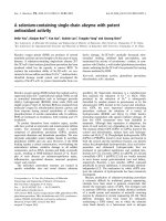
Fig. 2. Thylakoid membrane polypeptides from WT, mf1, mf2 and
revertant p3mf1 strains after electrophoresis on 9–18% (w/v) SDS/
PAGE with Coomassie blue staining. The three tracks on the left were
loaded with a thylakoid suspension equivalent to 7 lg chlorophyll,
andthetwoontherightwithathylakoidsuspensionequivalentto
10 lg chlorophyll. Other revertant pmf strains display similar patterns
to that of p3mf1.
Table 2. PG and C16:1(3t) contents in WT, mf mutants and pmf revertant strains. Content of PG is expressed in lg of PG total fatty acids per mg
chlorophyll and C16:1(3t) content is expressed in percent of PG total fatty acids. For WT, mf1, p3mf1, mf2 and pmf2 cells grown in TAP medium,
data are the mean of two to four independent cultures and SD are indicated; for the other strains only one determination was made. Strains mf1 and
mf2 are unable to grow in minimum medium. ND, not determined.
Lipid
Strain
WT mf1 p1mf1 p2mf1 p3mf1 p4mf1 p5mf1 p6mf1 mf2 pmf2
TAP medium
PG 103.4 ± 7.9 49 ± 18.5 55 64 64.6 ± 16.7 85.3 ND ND 34.8 ± 5.1 63.3 ± 3.1
C16:1(3t) 19.6 ± 3.2 < 0.66 7.3 6 8.6 ± 5.8 4.7 ND ND < 0.8 15.8 ± 3.1
Minimum medium
PG 154.7 – 80 98 80.7 94 108 64.7 – ND
C16:1(3t) 26.7 – 10 8.2 11.5 8.5 7.4 7.3 – ND
332 B. Pineau et al.(Eur. J. Biochem. 271) Ó FEBS 2003
Organization of the residual amount of PSII
in
mf
mutants
To test whether the minor amounts of PSII core subunits
still accumulating in mf mutants could be found in PSII
supramolecular assemblies, we used blue-native gel electro-
phoresis. Dodecylmaltoside-solubilized thylakoids of the
WT strain displayed several green bands (Fig. 3; A–E)
migrating above those containing free antennae. The free
antenna was recovered in three bands obviously reduced in
mf1 and mf2 but largely restored in all revertant pmf1 strains
(Fig. 3). From their polypeptide composition, showing the
presence of apoCPI and LHCI (Fig. 4), the three green
bands B, C and D can be assigned to PSI–LHCI complexes.
They were present in all strains (Fig. 3), although their
relative ratios were altered in strains mf1 and mf2, exhibiting
an increase in band D and splitting of band C. All of these
bands correspond to various forms of PSI supercomplexes
(Fig. 4). Thus, the lack of PG-C16:1(3t) modifies the state
of oligomerization of PSI–LHCI supercomplexes in
C. reinhardtii [7,9] as in Arabidopis thaliana [28]. Band A
corresponds to dimeric PSII core complexes with their
associated antenna [29] and contains the core complex
polypeptides apoCP47, apoCP43, the minor antenna com-
plex CP26, CP29 and the LHCII antenna polypeptides
(Fig. 4). Band A was completely missing in mf1 and mf2
membranes but partially restored in thylakoids from all
pmf1 strains tested (Fig. 3). In the WT strain, small amounts
of PSII core subunits were visible in band E migrating just
above the cytochrome b
6
–f complex, which represents
monomeric PSII without antenna polypeptide (Fig. 4).
Band E was clearly seen in the green pattern from pmf1
strains (Fig. 3). Inspection of two-dimensional gels reveals
that traces of apoCP47 and apoCP43 were also present but
restricted to band E in the two mutants mf1 (data not
shown) and mf2 (Fig. 4) and were not associated with any
LHCII antenna polypeptides.
We conclude that the small amount of PSII core
complexes that accumulate in these mutants cannot form
stable supercomplexes with the peripheral antenna or that
these supercomplexes did not resist electrophoretic separ-
ation. The absence of such associations is, however,
consistent with the previous data obtained from fluores-
cence spectra giving evidence for connection of LHCII to
PSI in mf1 and mf2 [6,7].
The lack of PSII cores in
mf1
and
mf2
is due to a
decreased synthesis of two PSII core subunits
The strongly reduced accumulation of PSII could result
from a defect in the synthesis of any of the major PSII core
subunits or from a post-translational degradation process.
To address that point, cells of two progeny from the
mf2 · WT cross (a [PSII
–
] and a [PSII
+
] member from the
same tetrad) were pulse labelled with [
14
C]acetate for 5 min
in the presence of cycloheximide. As controls, we also
labelled FUD7 cells, deleted for the psbA gene encoding D1
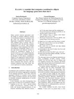
Fig. 3. Blue-native gel electrophoresis analysis of the pigment–protein
complexes in thylakoids from WT, mf1, mf2 and three revertant pmf1
strains. Samples equivalent to 16 lg chlorophyll were loaded in each
track. The upper part of the gel resolves PSII oligomers (band A), core
complex monomers (band E) and PSI oligomers (bands B–D). The
lower part of the gel resolves LHCII proteins.
Fig. 4. Two-dimensional separation of poly-
peptides from WT and mf2 chlorophyll-binding
complexes, resolved by blue-native gel electro-
phoresis in the first dimension. After electro-
phoresis, the gels were silver stained. A–E
represent the positions of green bands (as
shown in Fig. 3); stars designate the positions
of apoCP43 and apoCP47 belonging to the
core complex of PSII. Note that a PSII band
whose composition is close to that of band A
(PSII–LHCII) is visible just next to band C
(PSI–LHCI) in the WT profile. ., subunits of
the CF0–CF1 ATPsynthetase; r position of
cytochrome b
6
–f complexes.
Ó FEBS 2003 Phosphatidylglycerol succeptibility of PSII core (Eur. J. Biochem. 271) 333
(DpsbA), as well as the mf1, mf2 and WT cells. As observed
in Fig. 5A, the absence of D1 in the FUD7 mutant causes a
decreased synthesis of apoCP47 but not of D2 and apoCP43
as previously reported [24]. A similar situation was observed
in the mf mutants and [PSII
–
]progeny.SynthesisofD1and
apoCP47 were barely detectable (Fig. 5A), whereas synthe-
sis of D2 and apoCP43 remained similar to those in the WT
strain. We noted that labelling of D1 and apoCP47 were
clearly detectable in 40 min pulses in mf1 and mf2 strains
(Fig. 5B), suggesting that the low labelling observed in
5 min pulses is not due to rapid degradation of the
neosynthesized proteins. A similar result was also obtained
with the mf1 strain (data not shown). The tracks from the
[PSII
+
] and [PSII
–
] progeny were identical to those from
their WT and mf parents, respectively. These changes in
synthesis of PSII subunits followed the same segregation, as
that observed for PSII activity and PG content.
Translation initiation of the
psbA
mRNA is not affected
in the
mf2
strain
The dramatic decrease in D1 synthesis in mf1 and mf2 strains
did not correlate with any significant changes in the
accumulation of psbA mRNA, as revealed by RNA-filter
hybridization experiments (data not shown). This indicates
the likelihood of a translational defect of D1 synthesis
occuring in these strains. To determine if translation
initiation of the psbA mRNA was compromised, we inserted
a chimeric gene, containing the petA-coding region (enco-
ding cytochrome f) translated under the control of the
5¢ UTR of psbA in place of the endogenous petA gene. The
transformant, hereafter refered to as strain 5¢psbA–petA,
mt+ (Fig. 6A), was subsequently crossed with the mf2, mt–
strain, to compare the expression of the chimeric gene in
either the WT or mf2 nuclear background. The whole
progeny of that cross carries the chimeric gene because of the
uniparental inheritance of the chloroplast genome transmit-
ted from the mt+ parent [14]. Two members of each tetrad
also inherited the mf2 nuclear background and were
identified, using a fluorescence induction kinetics screen,
by their [PSII
–
] phenotype, whereas the other two members
displayed [PSII
+
] phenotype. Accumulation of cyto-
chrome f wasthenassayedbyTMBZstainingonwhole
cell extracts from 5¢psbA–petA and mf2 parental strains and
from two tetrads of that cross (Fig. 6B). We observed no
significant changes in the accumulation of cytochrome f
between the members of the two tetrads tested. We assessed
Fig. 5. Protein pulse labelling experiments in the mf1 and mf2 strains.
(A) Short pulse labelling (for 5 min with 5 lCiÆmL
)1
[
14
C]acetate) of
PSII core subunits in a half-tetrad (one [PSII
–
] and one [PSII
+
]pro-
geny) from the cross mf2 · WT as well as in mf1, mf2,WTandFUD7
cells. Labelled polypeptides were separated by electrophoresis on
9–18% (w/v) SDS/PAGE (for a better resolution of apoCP47 and
apoCP43; upper panel) or 12–18% (w/v) polyacrylamide/urea gels (for
a high resolution of D2 and also D1; lower panel). PS+, [PSII
+
]
strains (WT nuclear background); PS–, [PSII
–
]strains(mf2 nuclear
background). (B) Comparison of short and longer pulse-labelling
experiments performed on the WT and mf2 strains under
60 lmol photonsÆm
)2
Æs
)1
. Samples were harvested from a single cul-
ture, 8 and 40 min after addition of 1.2 lCiÆmL
)1
[
14
C]acetate and
analysed by electrophoresis on 9–18% (w/v) SDS/PAGE. Note the
clear identification of D1 labelling after 40 min pulse-labelling. Results
obtained with the mf1 strain were similar.
Fig. 6. Translation of the chimeric 5¢psbA–petA gene in WT and mf2
nuclear context. (A) Schematic maps of the petA gene in WT and
5¢psbA–petA strains. Relevant restriction sites (B, BglII; N*, an NcoI
site introduced by site directed mutagenesis around the petA initiation
codon for cloning purposes; H, HincII) are indicated. (B) Accumula-
tion of cytochrome f in the parents and offspring of two tetrads from
the cross 5¢psbA–petA · mf2.PS+,[PSII
+
]strains(WTnuclear
background); PS–, [PSII
–
]strains(mf2 nuclear background). Whole
cell proteins were separated on denaturing 12–18% (w/v) polyacryl-
amide/urea gels and stained with TMBZ; cyt f, cytochrome f;cytc
1
,
mitochondrial cytochrome c (loading control). (C) Short pulse label-
ling experiments (5 min with 5 lCiÆmL
)1
) for chloroplast-encoded
proteins in the parental strains and in two members of the first tetrad
presented above (B, *). Progeny number 11 has the mf2 nuclear
background while progeny number 14 has a WT nuclear background.
334 B. Pineau et al.(Eur. J. Biochem. 271) Ó FEBS 2003
directly the rate of synthesis of cytochrome f in the two
genetic backgrounds by 5 min pulse-labelling experiments
performed on the parental strains 5¢psbA–petA and mf2 and
on two members of tetrad 1. From fluorescence induction
kinetics, member 11 and member 14 have mf2 and WT nuc-
lear backgrounds, respectively. The expected near-absence
of synthesis of the D1 protein is clearly visible in the mf2 and
number 11 strains, whereas it is WT-like in the 5¢psbA–petA
and number 14 strains (Fig. 6C). In contrast, synthesis of
cytochrome f,drivenbythechimeric5¢psbA–petA tran-
script, was similar in all strains tested, after corrections for
14
C incorporation among the various strains. Thus, the
nuclear mf2 background had no effect on the translation rate
of this chimeric gene. We conclude that the mf2 mutation
does not act on the 5¢ UTR of the psbA transcript.
Discussion
A single nuclear mutation causes both the absence
of functional PSII and the lack of PG-C16:1(3t)
The mf1 and mf2 mutants were originally screened as
unusual PSII mutants because they lack variable fluores-
cence – a signature of the absence of PSII [15] – but have a
low (instead of a high) fluorescence yield. This unusual
feature was attributed to a major change in the supra-
molecular organization of the peripheral antenna. The
absence of LHCII oligomers in these strains leads to the
accumulation of LHCII monomers that presumably trans-
fer their excitation energy to PSI [7]. It was subsequently
proven that changes in supramolecular organization of the
peripheral antenna were due to the absence of one particular
fatty acid, C16:1(3t), which probably causes a decrease in
the overall content in PG [9]. Addition of PG-C16:1(3t) to
the growth medium allowed the recovery of oligomeric
LHCII [10,11]. However, in contrast to the changes in the
state of antenna oligomerization, the PSII defect was almost
insensitive to the addition of exogenous PG-C16:1(3t) [8],
raising the possibility that PG-C16:1(3t) deficiency was not
responsible for PSII inactivation. The extensive genetic
analysis performed in the present study nevertheless defines
a single nuclear mutational event that governs both
phenotypic characters, as suggested by preliminary data
from El Maanni et al. [8]. All phototrophic revertants
isolated from mf1 and mf2 also recovered some PG-
C16:1(3t), leading to an increase in total PG content. The
lack of PG-C16:1(3t) is not a mere consequence of PSII
deficiency, as PSII deficient mutants of C. reinhardtii such
as Fl39 (a nuclear mutant) or FUD7 (DpsbA) do not present
such lipid alterations (data not shown). Thus, the deficiency
in PG caused by the absence of the PG-C16:1(3t) form in
mf1 and mf2 thylakoid membranes is responsible for the
near-complete absence of PSII core complexes. The two
mutants hardly accumulated any PSII subunits. By blue-
native PAGE, we detected only trace amounts of PSII core
complexes, comprised of subunits apoCP47 and apoCP43.
None were associated with peripheral antenna, even though
our electrophoretic method preserved core–antenna com-
plex associations as shown by the presence of PSI–LHCI
and PSII–LHCII in the WT pattern. In contrast, PSII–
LHCII complexes were observed in revertant pmf1 cells.
Thus PG deficiency in mf1 and mf2 mostly targets PSII-
containing supercomplexes, although it also affects the
relative distribution of different types of PSI–antenna
supercomplexes [9]. Because PG was reported to be directly
involved in PSI functional organization, based on the study
of crystals of trimeric PSI from Synechococcus elongatus
[30], a detailed analysis of the early steps in PSI electron
transfer in the mf mutants would be required before one can
draw conclusions on the role of PG in PSI supramolecular
organization in C. reinhardtii.
Phosphatidylglycerol, photosynthesis and PSII biogenesis
The hf2 nuclear mutant of C. reinhardtii displays a severe
defect in SQDG, the other thylakoid-specific anionic lipid
[31]. Despite a partial alteration of PSII activity, it contained
the same amount of PSII core components as the WT strain
[32]. Thus, impaired PSII biogenesis in mf1 and mf2 mutants
does not simply result from a decrease in thylakoid anionic
lipids. Several studies emphasized the specific role of PG in
photosynthesis, in particular for PSII in cyanobacteria and
higher plants [33–36]. The requirement of PG in photo-
synthesis was recently established in vivo by the isolation of
two mutants of Synechocystis defective in the PG biosyn-
thesis pathway. These were inactivated in the genes respon-
sible for the last step of CDP-diacylglycerol synthesis [37]
or phosphatidyl-glycerol-3-phosphate synthesis [38]. They
both depended on PG supplementation for phototrophic
growth. The withdrawal of PG from the culture medium
correlated with alterations in PSII activity [37]. Recently,
PG was shown to be essential for the dimerization step of
PSII core monomers in the pgsA mutant of Synechocystis
bearing a disruption of the phosphatidyl-glycerol-3-phos-
phate synthase gene [39].
Photosynthetic mutants fully devoid of PG have not been
described up to now in eukaryotes. An A. thaliana mutant
deficient in phosphate accumulation was found to be
partially PG-deficient; its growth rate or photosynthetic
parameters were WT-like in two different light conditions
but its contents in SQDG and DGDG were increased [40].
In contrast, a pale green mutant of A. thaliana with
impaired photosynthesis was found to bear a mutation in
the gene encoding plastidic phosphatidylglycerolphosphate
synthase, leading to a reduced PG content [41]. Disruption
of the PGP1 gene by T-DNA insertion in A. thaliana
illustrated the importance of PG for the biogenesis of
thylakoid membranes [42,43]. Thus the essential function of
PG for photosynthetic viability, as demonstrated in cyano-
bacteria, can probably be extended to photosynthetic
eukaryotes even if the molecular mechanism(s) mediated
by PG remain(s) to be determined.
Here we show that the two PG-deficient mf1 and mf2
mutants of C. reinhardtii accumulate only trace amounts of
PSII core monomers that are unable to oligomerize in PSII–
LHCII supercomplexes. In this alga, LHCII mutants with
high PSII activity are easily recovered [44]. The loss in
LHCII–PSII core supercomplexes, therefore, should not be
responsible for the decreased content in PSII cores, pointing
to an effect of PG in PSII core biogenesis. The large but
partial PG deficiency occuring in mf1 and mf2 includes the
total loss of one PG form that contains the C16:1(3t) fatty
acid. Thus, it is reasonnable to assume that this fatty acid
plays a prominent role in the contribution of PG to PSII
Ó FEBS 2003 Phosphatidylglycerol succeptibility of PSII core (Eur. J. Biochem. 271) 335
biogenesis. Consistent with this view, the higher ratio of PG
to PSII in spinach preparations of dimeric PSII reaction
center than in monomeric PSII [45] can be interpreted in
light of the results from treatments with phospholipase A2,
that decrease the PG-C16:1(3t) content and lead to mono-
merization of dimeric PSII reaction centers. Conversely
dimerization of PSII reaction centers in vitro requires the
presence of PG-C16:1(3t). An A. thaliana mutant, totally
deficient in PG-C16:1(3t) did not display any significant
alteration of its photosynthetic properties but showed a
concomitant increase in PG-C16:0, which may be compen-
satory in this case [28]. As reported for the formation of the
trimeric LHCII antenna in C. reinhardtii,thefattyacid
C16:1(3t) could increase the efficiency of PG for PSII core
biogenesis in the situation of active synthesis determined by
the high growth rate of this alga [46]. Altogether, these
observations argue for a critical role of C16:1(3t)-containing
PG in the biogenesis of PSII core complexes and their
subsequent oligomerization in C. reinhardtii.
PG plays no part in translation initiation of D1
but could contribute to its cotranslational insertion
The drastic decrease in PSII core content in the mf1 and mf2
mutants could be attributed to a higher susceptibility of the
cores to proteolytic degradation or to their lower rate of
synthesis. When we studied the rates of synthesis of the
individual PSII subunits by 5 min pulse-labelling experi-
ments, we observed that synthesis of D1 and apoCP47 were
barely detectable in the mf1 and mf2 mutants, although the
mRNA levels for these two subunits were similar to those
observed in the WT strain. Due to the control by epistasy
of synthesis (CES) process [1], D1 and apoCP47 display
concerted rates of synthesis. In the absence of D1, the rate of
synthesis of apoCP47 is strongly reduced, while the rates of
synthesis of both D1 and apoCP47 drops when D1 cannot
assemble within PSII complexes, for example as a result of
the lack of D2 [24]. The decreased synthesis of D1 (and, as a
consequence, of apoCP47) in the mf mutants was thus
consistent with an impaired PSII assembly. We observed
recently that the CES behaviour of D1 (its much lower rate
of translation when it cannot assemble within PSII) resulted
from a translational regulation that depends on the 5¢ UTR
of psbA (L. Minai, F A. Wollman and Y. Choquet,
unpublished results). We thus tested whether the rate of
translation of a chimeric reporter gene harbouring the
coding region of petA translated under the control of the
5¢ UTR of psbA was decreased when it was expressed in mf2
strain. Much to our surprise, the level of synthesis of its
protein product (cytochrome f)inthemf2 nuclear back-
ground was identical to that observed in a WT nuclear
background. We are therefore bound to conclude that the
reduced synthesis of D1 is not due to a defect in translation
initiation but to a subsequent step that could be either
elongation, termination, membrane insertion or very rapid
cotranslational degradation of the D1 protein. However D1
synthesis in the mf strains, which is hardly detectable in
5 min pulse-labelling experiments, is still easily detectable in
longer pulses, arguing against a rapid degradation of the
polypeptide. It is difficult to discriminate further between
these alternatives at present, because chimeric genes made of
the coding sequence of psbA translated under the control of
unrelated 5¢ UTRs are only very poorly expressed
(L. Minai, unpublished results).
The inability of D1 to react with crosslinkers was
postulated to be due to a particular stability of its
conformation mediated by saturated fatty acids of
boundary lipids [47]. Later, PG was proposed to anchor
D1 into the thylakoid membranes of cyanobacteria by a
strong interaction with the hydrophobic part of the
molecule [35]. If this binding occurs at an early step in D1
synthesis, i.e. during the process of the cotranslational
insertion of the D1 protein into the thylakoid membrane,
then one could imagine that the absence of PG leads to a
drop in translational elongation of the D1 protein. Indeed
anionic phospholipids were shown to contribute to the
coupled translation–insertion of some protein subunits of
the electron transport chain from inner mitochondrial
membranes [48]. A null PGS1 mutant of S. cerevisiae,in
which the content of PG and cardiolipid could be
controlled by modulating the expression of a plasmid-
introduced PGS1 gene, was also used to demonstrate that
these anionic phospholipids have a mandatory function in
the translation of cytochrome b and the three largest
subunits of cytochrome oxidase [49]. On the other hand,
the activity of the SecYEG translocase in bacteria is
strictly dependent in vitro on the presence of PG in E. coli
and Bacillus subtilis [50]. Therefore, the requirement for
PG could arise at the level of the Sec translocon through
which D1 is inserted in the thylakoid membranes [51–53].
We note, however, that translocation of the other Sec
passenger proteins, such as cytochrome f, was not altered
in the mf mutants, an observation that does not support a
prominent role of PG-C16:1(3t) in Sec translocation
across thylakoid membranes. PG could still contribute
to cotranslational insertion of D1 in the thylakoid
membranes through a number of other steps that have
not been properly characterized yet.
Acknowledgements
This work was supported by CNRS UMR8618, UPR1261 and
UMR8619, Universite
´
s Paris Sud and Paris VI. S. Eberhard was
supported by a fellowship from the Ministe
`
re de l’Education et de la
Recherche. We wish to thank A. El Maanni for her participation in pmf
strains selection. We are grateful to Prof. R. Bassi for the gift of
antibodies to LHCII. We thank R. Boyer for the photographic pictures
and R. Kuras for critical reading of the manuscript.
References
1. Wollman, F.A., Minai, L. & Nechushtai, R. (1999) The biogenesis
and assembly of photosynthetic proteins in thylakoid membranes.
Biochim. Biophys. Acta 1411, 21–85.
2. Siegenthaler, P.A. (1998) Molecular organization of acyl lipids in
photosynthetic membranes of higher plants. In Lipids in Photo-
synthesis: Structure, Function and Genetics (Siegenthaler, P.A. &
Murata, N., eds), pp. 119–144. Kluwer Academic Publishers,
Dordrecht, the Netherlands.
3. Joyard, J., Mare
´
chal, E., Mie
`
ge, C., Block, M.A., Dorne, A. &
Douce, R. (1998) Structure, distribution and biosynthesis of gly-
cerolipids from higher plant chloroplasts. In Lipids in Photo-
synthesis: Structure, Function and Genetics (Siegenthaler, P.A. &
Murata, N., eds), pp. 21–52. Kluwer Academic Publishers,
Dordrecht, the Netherlands.
336 B. Pineau et al.(Eur. J. Biochem. 271) Ó FEBS 2003
4. Ivancich, A., Horvath, L.I., Droppa, M., Horvath, G. & Farkas,
T. (1994) Spin label EPR study of lipid solvation of supramole-
cular photosynthetic protein complexes in thylakoids. Biochim.
Biophys. Acta 1196, 51–56.
5. Fyfe, P.K., McAuley, K.E., Roszak, A.W., Isaacs, N.W., Cogdell,
R.J. & Jones, M.R. (2001) Probing the interface between mem-
brane proteins and membrane lipids by X-ray crystallography.
Trends Biochem. Sci. 26, 106–112.
6. Maroc, J., Tre
´
molie
`
res,A.,Garnier,J.&Guyon,D.(1987)
Oligomeric form of the light-harvesting chlorophyll a+b protein
complex CPII, phosphatidylglycerol, D
3
-trans-hexadecenoic acid
and energy transfer in Chlamydomonas reinhardtii, wild-type and
mutants. Biochim. Biophys. Acta 893, 91–99.
7. Garnier, J., Maroc, J. & Guyon, D. (1987) Characterization of
new strains of photosynthetic mutants of Chlamydomonas
reinhardtii. IV. Impaired excitation energy transfer in three
low fluorescent mutants. Plant Cell Physiol. 28, 1117–1131.
8. El Maanni, A., Dubertret, G., Delrieu, M.J., Roche, O. & Tre
´
-
molie
`
res, A. (1998) Mutants of Chlamydomonas reinhardtii
affected in phosphatidylglycerol metabolism and thylakoid
biogenesis. Plant Physiol Biochem. 36, 609–619.
9.Garnier,J.,Wu,B.,Maroc,J.,Guyon,D.&Tre
´
molie
`
res, A.
(1990) Restoration of both an oligomeric form of the light-har-
vesting antenna CPII and a fluorescence state II – state I transition
by D
3
-trans-hexadecenoic acid-containing phosphatidylglycerol, in
cells of a mutant of Chlamydomonas reinhardtii. Biochim. Biophys.
Acta 1020, 153–162.
10. Tre
´
molie
`
res, A., Roche, O., Dubertret, G., Guyon, D. &
Garnier, J. (1991) Restoration of thylakoid appression by
D
3
-trans-hexadecenoic acid-containing phosphatidylglycerol in a
mutant of Chlamydomonas reinhardtii. Relationships with the
regulation of excitation energy distribution. Biochim. Biophys.
Acta 1059, 286–292.
11. Dubertret, G., Mirshahi, A., Mirshahi, M., Gerard-Hirne, C. &
Tremolieres, A. (1994) Evidence from in vivo manipulations of
lipid composition in mutants that the D
3
-trans-hexadecenoic acid-
containing phosphatidylglycerol is involved in the biogenesis
of the light-harvesting chlorophyll a/b-protein complex of
Chlamydomonas reinhardtii. Eur. J. Biochem. 226, 473–482.
12. Maroc, J. & Garnier, J. (1981) Gel electrophoresis of chloroplast
membranes of mutants of Chlamydomonas reinhardtii which have
impaired photosystem II function and lack photosynthetic cyto-
chromes. Biochim. Biophys. Acta 637, 473–480.
13. Bennoun, P., Spierer-Herz, M., Erickson, J., Girard-Bascou, J.,
Pierre, Y., Delosme, M. & Rochaix, J.D. (1986) Characterization
of photosystem II mutants of Chlamydomonas reinhardtii lacking
the psbA gene. Plant Mol Biol. 6, 151–160.
14. Harris, E. (1989) The Chlamydomonas Sourcebook. A Compre-
hensive Guide to Biology and Laboratory Use. San Diego Academic
Press, San Diego, CA, USA.
15. Bennoun, P. & Delepelaire, P. (1982) Isolation of photosynthesis
mutants in Chlamydomonas.InMethods in Chloroplast Molecular
Biology (Edelman, M., Hallick, R.B. & Chua, N H., eds), pp. 25–
38. Elsevier Biomedical Press, Amsterdam, New York, Oxford.
16. Rochaix, J.D. (1978) Restriction endonuclease map of the
chloroplast DNA of Chlamydomonas reinhardtii. J. Mol. Biol. 126,
597–617.
17. Rimbault, B., Esposito, D., Drapier, D., Choquet, Y., Stern, D.B.
& Wollman, F.A. (2000) Identification of the initiation codon for
the atpB gene in Chlamydomonas chloroplasts excludes translation
of a precursor form of the b subunit of the ATP synthase. Mol.
Gen. Genet. 264, 486–491.
18. Goldschmidt-Clermont, M. (1991) Transgenic expression of ami-
noglycoside adenine transferase in the chloroplast: a selectable
marker of site-directed transformation of Chlamydomonas.
Nucleic Acids Res. 19, 4083–4089.
19. Boynton, J.E., Gillham, N.W., Harris, E.H., Hosler, J.P.,
Johnson, A.M., Jones, A.R., Randolph-Anderson, B.L., Robert-
son, D., Klein, T.M. & Shark, K.B. (1988) Chloroplast transfor-
mation in Chlamydomonas with high velocity microprojectiles.
Science 240, 1534–1538.
20. Kuras, R. & Wollman, F.A. (1994) The assembly of cytochrome
b6/f complexes: an approach using genetic transformation of the
green alga Chlamydomonas reinhardtii. EMBO J. 13, 1019–1027.
21. Chua, N.H. & Bennoun, P. (1975) Thylakoid membrane poly-
peptides of Chlamydomonas reinhardtii: wild-type and mutant
strains deficient in photosystem II reaction center. Proc. Natl
Acad. Sci. USA 72, 2175–2179.
22. Scha
¨
gger, H., Cramer, W.A. & von Jagow, G. (1994) Analysis of
molecular masses and oligomeric states of protein complexes by
blue native electrophoresis and isolation of membrane protein
complexes by two-dimensional native electrophoresis. Anal. Bio-
chem. 217, 220–230.
23. Pineau, B., Ge
´
rard-Hirne, C. & Selve, C. (2001) Carotenoid
binding to photosystem I and II of Chlamydomonas reinhardtii
cells grown under weak light or exposed to intense light. Plant
Physiol. Biochem. 39, 73–85.
24. deVitry,C.,Olive,J.,Drapier,D.,Recouvreur,M.&Wollman,
F.A. (1989) Posttranslational events leading to the assembly of
photosystem II protein complex: a study using photosynthesis
mutants from Chlamydomonas reinhardtii. J. Cell Biol. 109,
991–1006.
25. Thomas, P.E., Ryan, D. & Levin, W. (1976) An improved staining
procedure for the detection of the peroxidase activity of cyto-
chrome P-450 on sodium dodecyl sulfate polyacrylamide gels.
Anal Biochem. 75, 168–176.
26. Bligh, E.G. & Dyer, W.J. (1959) A rapid method of total lipid
extraction and purification. Can. J. Biochem. Physiol. 37, 911–917.
27. Bassi, R. & Wollman, F.A. (1991) The chlorophyll-a/b proteins of
photosystem II in Chlamydomonas reinhardtii. Isolation, charac-
terization and immunological cross-reactivity to higher plant
polypeptides. Planta 183, 423–433.
28. McCourt, P., Browse, J., Watson, J., Arntzen, C.J. & Sommer-
ville, C.R. (1985) Analysis of photosynthetic antenna function in
amutantofArabidopsis thaliana (L.) lacking trans-hexadecenoic
acid. Plant Physiol. 78, 853–858.
29. Thidolm, E., Lindstro
¨
m, V., Tissier, C., Robinson, C., Schro
¨
der,
W.P. & Funk, C. (2002) Novel approach reveals localization and
assembly pathway of the PsbS and PsbW proteins into the
photosystem II dimer. FEBS Lett. 513, 217–222.
30. Jordan,P.,Fromme,P.,Witt,H.T.,Klukas,O.,Saenger,W.&
Krauss, N. (2001) Three-dimensional structure of cyanobacterial
photosystem I at 2.5 A
˚
resolution. Nature 411, 909–917.
31. Sato,N.,Tsuzuki,M.,Matsuda,Y.,Ehara,T.,Osafune,T.&
Kawaguchi, A. (1995) Isolation and characterization of mutants
affected in lipid metabolism of Chlamydomonas reinhardtii. Eur. J.
Biochem. 230, 987–993.
32. Sato, N., Sonoike, K., Tsuzuki, M. & Kawaguchi, A. (1995)
Impaired photosystem II in a mutant of Chlamydomonas
reinhardtii defective in sulfoquinovosyl diacylglycerol. Eur. J.
Biochem. 234, 16–23.
33. Murata, N., Higashi, S.I. & Fujimura, Y. (1990) Glycerolipids in
various preparations of photosystem II from spinach chloroplasts.
Biochim. Biophys. Acta 1019, 261–268.
34. Kruse, O., Radunz, A. & Schmid, G.H. (1994) Phosphatidyl-
glycerol and b-carotene bound onto the D1 core peptide from
photosystem II in the filamentous cyanobacterium Oscillatoria
chalybea. Z. Naturforsch. 49c, 115–124.
35. Kruse, O. & Schmid, G.H. (1995) The role of phosphatidylglycerol
as a functional effector and membrane anchor of the D1 core
peptide from photosystem II particles of the cyanobacterium
Oscillatoria chalybea. Z. Naturforsch. 50c, 380–390.
Ó FEBS 2003 Phosphatidylglycerol succeptibility of PSII core (Eur. J. Biochem. 271) 337
36. Ducheˆ ne,S.,Smutny,J.&Siegenthaler,P.A.(2000)Thetopology
of phosphatidylglycerol populations is essential for sustaining
photosynthetic electron flow activities in thylakoid membranes.
Biochim. Biophys. Acta 1463, 115–120.
37. Sato, N., Hagio, M., Wada, H. & Tsuzuki, M. (2000)
Requirement of phosphatidylglycerol for photosynthetic function
in thylakoid membranes. Proc. Natl Acad. Sci. USA 97, 10655–
10660.
38. Hagio, M., Gombos, Z., Varkonyi, Z., Masamoto, K., Sato, N.,
Tsuzuki, M. & Wada, H. (2000) Direct evidence for requirement
of phosphatidylglycerol in photosystem II of photosynthesis. Plant
Physiol. 124, 795–804.
39. Sakurai, I., Hagio, M., Gombos, Z., Tyystjarvi, T., Paakkarinen,
V.,Aro,E.M.&Wada,H.(2003)Requirementofphosphati-
dylglycerol for maintenance of photosynthetic machinery. Plant
Physiol. 133, 1376–1384.
40. Ha
¨
rtel, H., Essigmann, B., Lokstein, H., Hoffmann-Benning, S.,
Peters-Kottig, M. & Benning, C. (1998) The phospholipid-defi-
cient pho1 mutant of Arabidopsis thaliana is affected in the orga-
nization, but not in the light acclimation, of the thylakoid
membrane. Biochim. Biophys. Acta 1415, 205–218.
41. Xu, C., Ha
¨
rtel,H.,Wada,H.,Hagio,M.,Yu,B.,Eakin,C.&
Benning, C. (2002) The pgp1 mutant locus of Arabidopsis encodes
a phosphatidylglycerolphosphate synthase with impaired activity.
Plant Physiol. 129, 594–604.
42. Hagio, M., Sakurai, I., Sato, S., Kato, T., Tabata, S. & Wada, H.
(2002) Phosphatidylglycerol is essential for the development of
thylakoid membranes in Arabidopsis thaliana. Plant Cell Physiol.
43, 1456–1464.
43. Babiychuk, E., Muller, F., Eubel, H., Braun, H.P., Frentzen, M. &
Kushnir, S. (2003) Arabidopsis phosphatidylglycerophosphate
synthase 1 is essential for chloroplast differentiation, but is dis-
pensable for mitochondrial function. Plant J. 33, 899–909.
44. Olive, J., Wollman, F.A., Bennoun, P. & Recouvreur, M. (1981)
Ultrastructure of thylakoid membranes in C. reinhardtii: evidence
for variations in the partition coefficient of the light-harvesting
complex-containing particles upon membrane fracture. Arch.
Biochem. Biophys. 208, 456–467.
45. Kruse, O., Hankamer, B., Konczak, C., Gerle, C., Morris, E.,
Radunz, A., Schmid, G.H. & Barber, J. (2000) Phosphatidylgly-
cerol is involved in the dimerization of photosystem II. J. Biol.
Chem. 275, 6509–6514.
46. Dubertret, G., Ge
´
rard-Hirne, C. & Tre
´
molie
`
res, A. (2002)
Importance of trans-D
3
-hexadecenoic acid-containing phos-
phatidylglycerol in the formation of the trimeric light-harvesting
complex in Chlamydomonas. Plant Physiol. Biochem. 40, 829–836.
47. Adir, N. & Ohad, I. (1988) Structural properties of the D1 and
surrounding photosystem II polypeptides as revealed by their
interaction with cross-linking reagents. J. Biol. Chem. 263,
283–289.
48. Chang, S.C., Heacock, P.N., Mileykovskaya, E., Voelker, D.R. &
Dowhan, W. (1998) Isolation and characterization of the gene
(CLS1) encoding cardiolipin synthase in Saccharomyces cerevisiae.
J. Biol. Chem. 273, 14933–14941.
49. Ostrander, D.B., Zhang, M., Mileykovskaya, E., Rho, M. &
Dowhan, W. (2001) Lack of mitochondrial anionic phospholipids
causes an inhibition of translation of protein components of the
electron transport chain. A yeast genetic model system for the
study of anionic phospholipid function in mitochondria. J. Biol.
Chem. 276, 25262–25272.
50. van der Does, C., Swaving, J., van Klompenburg, W. & Driessen,
A.J. (2000) Non-bilayer lipids stimulate the activity of the recon-
stituted bacterial protein translocase. J. Biol. Chem. 275, 2472–
2478.
51. Nilsson, R., Brunner, J., Hoffman, N.E. & van Wijk, K.J. (1999)
Interactions of ribosome nascent chain complexes of the chloro-
plast-encoded D1 thylakoid membrane protein with cpSRP54.
EMBO J. 18, 733–742.
52. Zhang, L., Paakkarinen, V., van Wijk, K.J. & Aro, E.M. (1999)
Co-translational assembly of the D1 protein into photosystem II.
J. Biol. Chem. 274, 16062–16067.
53. Zhang, L., Paakkarinen, V., Suorsa, M. & Aro, E.M. (2001) A
SecY homologue is involved in chloroplast-encoded D1 protein
biogenesis. J. Biol. Chem. 276, 37809–37814.
338 B. Pineau et al.(Eur. J. Biochem. 271) Ó FEBS 2003