Báo cáo khóa học: Expressed protein ligation Method and applications docx
Bạn đang xem bản rút gọn của tài liệu. Xem và tải ngay bản đầy đủ của tài liệu tại đây (368.79 KB, 15 trang )
REVIEW ARTICLE
Expressed protein ligation
Method and applications
Ralf David, Michael P.O. Richter and Annette G. Beck-Sickinger
Institute of Biochemistry, Faculty of Biosciences, Pharmacy and Psychology, University of Leipzig, Germany
The introduction of noncanonical amino acids and bio-
physical probes into peptides and proteins, and total or
segmental isotopic labelling has the potential to greatly aid
the determination of protein structure, function and protein–
protein interactions. To obtain a peptide as large as possible
by solid-phase peptide synthesis, native chemical ligation
was introduced to enable synthesis of proteins of up to 120
amino acids in length. After the discovery of inteins, with
their self-splicing properties and their application in protein
synthesis, the semisynthetic methodology, expressed protein
ligation, was developed to circumvent size limitation prob-
lems. Today, diverse expression vectors are available that
allow the production of N- and C-terminal fragments that
are needed for ligation to produce large amounts and high
purity protein(s) (protein a-thioesters and peptides or pro-
teins with N-terminal Cys). Unfortunately, expressed pro-
tein ligation is still limited mainly by the requirement of a Cys
residue. Of course, additional Cys residues can be introduced
into the sequence by site directed mutagenesis or synthesis,
however, those mutations may disturb protein structure
and function. Recently, alternative ligation approaches have
been developed that do not require Cys residues. Accord-
ingly, it is theoretically possible to obtain each modified
protein using ligation strategies.
Keywords: expressed protein ligation; IMPACT
TM
-system;
intein; native chemical ligation.
Introduction
Proteins and peptides that have been modified by intro-
ducing noncanonical amino acids, fluorescence tags, spin
resonance labels or cross-linking agents have great potential
for investigations into protein–protein interactions and can
help to elucidate protein structures. Furthermore, artificial
peptides and proteins with new properties and with a broad
range of applications can be obtained. Further interest lies
in fragmental or complete isotopic labelling for NMR
studies to determine protein structures.
Solid-phase peptide synthesis (SPPS) provides the pos-
sibility of introducing noncanonical amino acids into
peptides but is restricted to peptides of up to 60 amino
acids in length. By using expression systems in bacteria or
yeast, the recombinant generation of peptides and proteins
and their complete isotopic labelling has become possible
[1–3]. The size of the constructs is not restricted but the
insertion of noncanonical amino acids is difficult [4,5]. The
limitation of peptide size in SPPS was circumvented by
several approaches developed for the synthesis of proteins
by segment condensation [6]. Liu et al. used a glycolalde-
hyde peptide ester for the reaction of an unmasked aldehyde
with an amino-group of an N-terminal Cys or Ser to form
a thiazolidine- or oxazolidine-ring. Rearrangement of the
O-acyl-ester resulted in an amide bond with a pseudoproline
residue [7]. In the thiol capture approach, where only Cys
sidechains have to be protected, a 4-mercapto-dibenzofuran
ester forms an asymmetric disulfide bond with an
N-terminal Cys activated with an S-(methoxycarbonyl)sul-
fenyl (Scm) group of a second peptide. The free amino
function of this amino acid can attack the carbonyl group of
theesterandanOfiN-acyl transfer results in an amide-
bond. Reductive cleavage of the disulfide releases the free
Cys sidechain [8]. CNBr-cleavage fragments refold and
form noncovalent complexes and finally the missing peptide
bonds are reattached [9]. Cytochrome c CNBr fragments
1–65 and 66–104 were modified and religated by this
method [10], but this technique is limited by the occurrence
of Met at the cleavage site.
Dawson et al. introduced a simple and elegant method
called native chemical ligation (NCL) for the synthesis of
peptides by condensation of their unprotected segments.
The coupling of synthetic peptide-thioesters with peptides
carrying an N-terminal Cys leads to an amide-bond at the
ligation site. This approach has proven to be useful for
the synthesis of smaller proteins up to 120 amino acids in
Correspondence to A. G. Beck-Sickinger, Institute of Biochemistry,
University of Leipzig, Bru
¨
derstr. 34, D-04103 Leipzig, Germany.
Fax: + 49 341 97 36 909, Tel.: + 49 341 97 36 900,
E-mail:
Abbreviations: BAL, backbone amide linker; CBD, chitin binding
domain; eGFP, enhanced green fluorescent protein; EPL, expressed
protein ligation; FRET, fluorescence resonance energy transfer;
GFP, green fluorescent protein; HOBt, 1-hydroxybenzotriazole;
IMPACT
TM
, intein-mediated purification with an affinity chitin
binding tag; IPL, intein-mediated protein ligation; NCL, native
chemical ligation; PTPase, protein tyrosine phosphatase; SPPS,
solid-phase peptide synthesis; TROSY, transverse relaxation
optimized spectroscopy; TWIN, two intein system.
(Received 12 November 2003, revised 19 December 2003,
accepted 5 January 2004)
Eur. J. Biochem. 271, 663–677 (2004) Ó FEBS 2004 doi:10.1111/j.1432-1033.2004.03978.x
length; larger proteins cannot be obtained easily in one
ligation step. Multistep NCL of different peptide-segments,
however, can lead to larger proteins [11]. An extension of
this NCL strategy is the expressed protein ligation (EPL)
method [12] using recombinant thioesters and/or aCys-
peptides. This review gives an overview of this method and
its applications in the past few years.
Native chemical ligation
The method of native chemical ligation was introduced by
Dawson et al. [13,14] and is based on the reaction between a
thioester and the sidechain of a Cys residue – reported for
the first time by Wieland et al. [15]. Two fully unprotected
synthetic peptides react to form an amide bond, so they are
connected as in the native peptide backbone. The reaction
proceeds in aqueous conditions at neutral pH. The first step
of this process is the chemoselective transthioesterification
of an unprotected peptide Ca-thioester with an N-terminal
Cys of a second peptide. The so-formed thioester sponta-
neously undergoes an SfiN-acyl transfer to form a native
peptide bond and the resulting peptide product is obtained
in the final disposition. Internal Cys residues within both
peptide segments are permitted because the initial trans-
thioesterification step is reversible and no side products
are obtained, thus, no protecting groups are necessary. An
alternative method was introduced by Tam et al. [16,17],
where a C-terminal thiocarboxylic acid S-alkylates an
N-terminal a-bromoAla to form a covalent thioester. This
rearranges by SfiN-acyl shift and builds an -X-Cys- peptide
bond (Fig. 1).
To prevent the thiol of the N-terminal Cys from oxidation,
and thus forming an unreactive disulfide linked dimer, it is
necessary to add thiols or other reducing reagents like tris(2-
carboxyethyl)phosphine (TCEP) [18] to the reaction mix-
ture. Furthermore, the addition of an excess of thiols not
only keeps the thiol-functions reduced but also increases the
reactivity by forming new thioesters through transthioeste-
rification [19]. The addition of solubilizing agents such as
urea or guanidinium hydrochloride does not affect the
ligation reaction and can be used to increase the concentra-
tion of peptide segments and results in higher yields. The
compatibility and efficiency of all proteinogenic amino acids
at the C-terminus of the thioester peptide to react in NCL
was determined by Hackeng et al.[20].All20aminoacids
except Val, Ile and Pro can be placed in the -X-Cys- position
in NCL. Val, Ile and Pro are reported to react slowly. Also,
Asp and Glu as C-terminal residues are less favourable
because of the formation of side products [21].
A useful application of NCL is solid-phase chemical
ligation (SPCL) [22]. In this approach, one of the two
segments is bound to a polymer, while the other is applied in
aqueous solution and can be used inexcess. A simple washing
step completely removes the solubilized peptides and the
assembled full length protein can be cleaved from the resin.
In the tandem peptide ligation approach, the NCL is
applied to the synthesis of peptides and proteins requiring
two or more ligation steps. NCL is combined with a
pseudoproline ligation by imine capture [23], the third step
can be pseudoglycine ligation [24].
In addition to Cys, related amino acids, including
selenoCys [25] and selenohomoCys [26], have been reported
to work in a similar manner.
Thioester formation
The bottleneck in NCL is the generation of the thioester.
Several applications have been developed using solid-phase
peptide synthesis. Most of the strategies to obtain peptide
thioesters have used the Boc-strategy [13,17] because of the
base-lability of the thioester. However, different attempts
in the synthesis of thioesters were performed by using the
9-fluorenylmethoxycarbonyl (Fmoc) method. In general,
the Fmoc-strategy has several advantages over the Boc-
strategy, the first being the milder conditions used for
cleavage from the resin. To circumvent the susceptibility of
the thioester linkages to nucleophiles like piperidine, used
for the removal of the Fmoc-protecting group, several
cocktails for deprotection have been developed, e.g.,
1,8-diazabicyclo[5.4.0]undec-7-ene (DBU) with 1-hydroxy-
benzotriazole (HOBt) [27], 1-methylpyrrolidine with
hexamethyleneimine and HOBt [28] or DBU and HOBt
[29]. The final cleavage from the resin then results in the
peptide thioester.
Further methods were introduced that used different
resins. One is based on modifications of Kenner’s sulfon-
amide Ôsafety catchÕ linker [30]. The C-terminus of the
growingpeptidechainisattachedtotheresinwithanacid-
and base-stable N-acyl sulfonamide linker. The sulfonamide
is activated after peptide synthesis by N-alkylation using
diazomethane or iodoacetonitrile. The cleavage occurs with
nucleophile like thiols, which finally results in a peptide
thioester [31,32]. In the backbone amide linker (BAL)
strategy, the first carboxy terminally protected amino acid is
attached to the resin on the backbone nitrogen. The peptide
chain grows in the N-terminal direction. Deprotection,
activation and thioester formation at the carboxy terminus
occurs on the solid support. The peptide thioester can be
cleaved from the resin with trifluoroacetic acid [33].
Another approach uses standard resins like phenyl-
acetamidomethyl (PAM) or 4-hydroxymethyl benzoic acid
(HMBA), the Lewis acid, Al(CH
3
)
2
Cl and thiols in
Fig. 1. Ligation of unprotected peptide segments. In native chemical
ligation (A) the first step is a transthioesterification of a Ca-thioester by
the thiol function of an N-terminal Cys followed by a spontaneous
SfiN-acyl shift to obtain a native peptide bond. In an alternative
approach (B), a Ca-thiocarboxylic acid reacts with an a-bromo amino
acid by forming a thioester. This leads to the same product as in
method A.
664 R. David et al. (Eur. J. Biochem. 271) Ó FEBS 2004
methylenchloride [34]. Unfortunately, the alkylaluminium
thiolate method can lead to epimerization at the C-terminus
and reactions at the sidechains, e.g., sidechain thioesters and
aspartimide formation. This can be avoided by using a
weaker Lewis acid, e.g. Al(CH
3
)
3
[35]. A further possibility
is the synthesis of peptides on Cl-trityl-resin and the
cleavage of the fully protected peptide chain with acetic
acid and trifluoroethanol. The thioester can be obtained by
the treatment of the protected peptide with activating
reagents and thiols [36,37]. After deprotection of functional
sidechains with trifluoroacetic acid, the thioester can be
easily purified by HPLC (Fig. 2).
An alternative approach for the thioester synthesis of
larger peptides and proteins in high yields and purity uses
a bacterial expression system based on the intein mediated
self-splicing mechanism of precursor proteins as discussed
below.
Recombinant generation of proteins
with C-terminal thioester or N-terminal Cys
Inteins and their use in protein chemistry
Inteins are internal segments of precursor proteins that
catalyze their ipso excision, in an intramolecular process
called protein splicing, with the concurrent ligation of the
two flanking external regions (N- and C-exteins) through
a native peptide bond. This finally yields the host protein.
Thus, inteins are analogues of self-splicing RNA introns.
The first intein was discovered in 1987 and up to now over
100 inteins are listed [38–40]. The origin of inteins is not yet
clear. However, understanding of inteins, their evolution,
distributions and properties, will be easier if they are
considered as parasitic genetic elements. They will not
contribute to an organism’s fitness if they are propagated
into the next generation. The insertion of an intein gene into
a protein gene can be described through the so called
homing cycle. Homing is the transfer of a parasitic genetic
element to a cognate allele that lacks the element. This
process results in the duplication of the parasitic genetic
element and its rapid spread in a population [41–43]. Inteins
occur in organisms of all three domains of life as well as in
viral and phage proteins. There they are predominantly
found in enzymes involved in DNA replication and repair
[40,44]. Inteins can be divided into four classes: the maxi
inteins (with integrated endonuclease domain), mini inteins
(lacking the endonuclease domain), trans-splicing inteins
(where the splicing junctions are not covalently linked) and
Ala inteins (Ala as the N-terminal amino acid) [45]. The
sequences of inteins have some characteristics in common.
They appear in conserved regions of the host protein and all
intein sequences harbour different motifs termed A and B
(which contain a conserved Thr and His) at the N-terminal
splicing domain, F and G at the C-terminal splicing domain
(Fig. 3). Endonuclease containing inteins also bear the
blocks C, D, E and H [38,46]. The N-terminal amino acids
are typically Cys, Ser or Ala. The C-terminal block G
contains a conserved His/Asp pair and a downstream Cys,
Ser or Thr amino acid.
The nucleophilic thiol or hydroxyl sidechains of the
conserved amino acid residues led to the assumption that
(thio)esters that are formed by an NfiS- or an NfiO-shift
are intermediates of the internal rearrangement steps of the
splicing reaction. This was proven by various investigations.
Fig. 2. Formation of synthetic peptide a-thio-
esters. Peptide a-thioesters can be synthesized
by the Fmoc strategy by using backbone
amide linker resins (A), acidic cleavage from
mercaptoalkyl linker resins (B), Lewis acid
activated cleavage from common resins
(C), cleavage of fully protected peptides
(Boc, t-butyloxycarbonyl; tBu, t-Butyl) and
deprotection after thioester generation (D)
and by using of sulfonamide safety catch
linker resins (E).
Fig. 3. Characteristic positions of intein motifs and numbering. The
inserted intein carries the N-terminal extein (left shaded box) and the
C-terminal extein (right shaded box). The residues important for the
splicing process as well as the conserved segment blocks (A, B, C, D, E,
H, F, G) and some internal intein key amino acids are depicted in the
one letter code within the certain segments (bold black). Numbering of
the amino acids of a precursor protein is made in the following way:
the intein’s N-terminal amino acid (Cys, etc.) is numbered as 1
whereas the C-terminal amino acid of the N-terminal extein is num-
bered as )1 and the N-terminal residue of the C-terminal extein is
numbered beginning with +1.
Ó FEBS 2004 Expressed protein ligation (Eur. J. Biochem. 271) 665
Replacement of the amino acid residues at the N-terminus
containing a nucleophilic thiol or hydroxyl sidechain and
the Asp at the C-terminus, through site directed mutagen-
esis, ended up in a complete loss of splicing activity of the
intein [47,48].
Splicing mechanism
The first step of the well understood standard splicing
process of inteins (Fig. 4) is the transfer of the N-terminal
extein unit to the sidechain -SH or -OH group of a Cys/Ser
residue located at the immediate N-terminus of the intein
(NfiS-acyl shift). In some cases, inteins bear Ala at the
ultimate position at their N-terminus. In such cases, the first
step is circumvented [48,49] and the +1 nucleophile within
the C-extein attacks the carbon of the peptide’s N-terminal
splicing junction. This rearrangement seems to be thermo-
dynamically highly unfavourable but the molecular archi-
tecture of the intein forces the scissile peptide bond into a
twisted conformation of higher energy and thereby pushes
the equilibrium to the (thio)ester side. The following step is a
new transfer of the N-terminal extein to the Cys/Ser/Thr at
the +1 position of the C-extein, which leads to a branched
intermediate. In the last step, which might be a concerted
reaction, a conserved Asp residue at the C-terminus of the
intein cyclizes and a peptide bond is formed between the two
exteins through an SfiN-acyl shift [50].
This splicing mechanism implicates the importance of the
conserved amino acids flanking the splicing junctions such
as the block B Thr and His, and the block G His [45].
In the case of C-terminal splicing, the cumulative data
indicate that the present penultimate His appears to assist
the C-terminal Asp cyclization, although there are reported
mutants referring to this residue which did not prevent
splicing. The three dimensional structure of the splicing
domain at the N-terminal part of the intein forces the
peptide bond into a twisted conformation. This could also
be protonated through the penultimate His residue men-
tioned above. Mutation of this amino acid did not affect the
first steps of the splicing up to the branched intermediate but
abolished the final step. In the X-ray crystal structure of the
intein, Mycobacterium xenopi gyrase (Mxe GyrA) (Fig. 5),
the His197 is hydrogen bonded to Asn198 so that His197 is
oriented for the donation of a proton from Nd position to
the emerging alpha amino group of the C-extein, prior
to the SfiN-acyl shift [51,52]. Some putative inteins that
lack the penultimate His residue are either inactive or use
other amino acids. Accordingly, the penultimate His is not
absolutely required but increases the splicing rate. Block B
contains Thr and His that are separated through two amino
Fig. 4. Mechanism of intein-mediated protein
splicing. In the initial step a thioester
intermediate is formed by an NfiS-acyl shift
at the N-terminal Cys of the intein (Cys
1
).
Transthioesterification by a nucleophilic
attack of the sidechain of the N-terminal Cys
of the C-extein (Cys
+1
) on the thioester is
formed in the first step and results in a
branched intermediate. Peptide bond cleavage
coupled to succinimide formation of the
C-terminal intein–Asp releases the intein. The
knotted exteins undergo a spontaneous SfiN-
acyl shift and yield a peptide bond. Peptide
bond cleavage can occur independently at
both splicing sites. Mutation of Cys
1
to Ala
prevents splicing at the N-terminus and leads
to a C-terminal extein bonded with the intein.
C-terminal splicing cannot occur when the
C-terminal Asn is substituted by an Ala
residue and the N-terminal extein is cleaved
by nucleophilic attack.
666 R. David et al. (Eur. J. Biochem. 271) Ó FEBS 2004
acids. Both play a key role for the N-terminal splicing
process. Substitution of block B His to Leu in Sce VMA
abolished splicing [53,54] and only C-terminal cleavage
occurred. This implies that this His residue takes part in the
first NfiS rearrangement at the N-terminal splicing junc-
tion. X-ray crystal structures of Sce VMA1 [55–57] and
Mxe GyrA [51] with exteins showed a protonation of the
scissile peptide bond through the imidazole ring. This
interaction promotes the breakdown of the tetrahedral
intermediate formed by the +1 nucleophilic attack of the
N-terminal thioester bond. These findings were further
elucidated and confirmed through investigations of Ala
inteins. The exact role of Thr is not yet fully understood
because of the lack of available structural data. It has been
postulated that the Mxe GyrA intein stabilizes the tetra-
hedral intermediate at the N-terminal splicing junction by
the formation of an oxy anion hole through Nd of Asn74
and the block B Thr.
Both effects, the spatial constraints and the electronic
influence, lead to a reactive and accessible electrophilic
carbon of the scissile peptide bond as an acid/base catalysis
mechanism is suggested.
Furthermore, divalent transition metal cations influence
the protein splicing process. It was shown for the split
inteins Ssp DnaE and the Mtu RecA that micromolar
concentrations of Zn
2+
ions decreased the splicing rate and
Zn
2+
ion concentrations in the millimolar range stopped
completely the process through chelation of key amino
acids. A similar effect was obtained for Cd
2+
ions [58,59].
Classification of inteins
The elucidation of the splicing mechanism and the identi-
fication of the key amino acid residues involved in the
scission and ligation of the peptide bonds facilitated the
molecular engineering of artificial inteins as tools for
different applications in protein chemistry. Currently there
are five general methods of intein usage in this field so far:
(a) modified inteins with an inducible autocatalytic cleavage
activity are used for protein purification; (b) inteins are used
for trans-splicing. Here the inteins are split into two
fragments that can recombine and reconstitute their splicing
activity in vivo or in vitro. (c) Intein mediated protein ligation
(IPL) is used for the generation of specifically mono-
activated proteins, which can further be ligated with peptide
segments and provides access to artificially labelled proteins;
(d) inteins facilitate the synthesis of cyclic proteins and
(e) inteins are used for the detection of protein–protein
interactions [45,46].
Three dimensional structures of inteins
The structure of the intein Sce VMA1 that was determined
by X-ray crystallography clearly shows two domains
(Fig. 5) [55–57]. The structure of the splicing domain is
similar to that of the mini intein in the Mycobacterium
xenopi gyrase (Mxe GyrA) [51]. Residues from the endo-
nuclease domain of Sce VMA1 contribute to target
sequence-specific contacts as well as parts of the other
domain that are distant from the Sce VMA1 cleavage site.
Several studies have been made by photo-crosslinking to
identify these residues [60]. The splicing domains have
predominantly all b-structures and show high similarity to
the structure of the hedgehog proteins that are important in
the development of multicellular organisms [61].
Formation of C-terminal thioester-activated proteins
Protein engineering via NCL requires the specific generation
of C-terminal thioester-tagged proteins allowing ligation
with a second peptide or protein containing an N-terminal
Cys or Ser residue. The potent synthesis of Ca-thioesters of
bacterially expressed proteins was found through studies of
the N-terminal cleavage mechanism of inteins. In general,
the cleavage of the peptide bonds at either the N-terminus or
the C-terminus of the intein can occur independently.
Replacement of the C-terminal Asp by Ala blocked the
splicing process in the Pyrrococcus species GB-D intein.
However, the lack of the succinimide formation did not
affect the preceding NfiO-acyl rearrangement at the
N-terminal splicing junction. The same data were found
previously for the NfiS-acyl shift in the Sce VMA intein.
Incubation of this modified intein with thiols, like dithio-
threitol, releases the corresponding free C-terminal thioester-
tagged extein from the N-terminal splicing junction through
transthioesterification. This thiol-inducible cleavage activity
of an engineered intein was the beginning of the extensive
exploitation of other intein mutants as workhorses in the
area of biotechnology to obtain mono-thioester labelled
proteins and aCys-proteins [46,50].
Fig. 5. Comparison of Mxe GyrA (A) and Sce
VMA (B) intein structure. The structures of
both inteins have been determined by X-ray
crystallography [51,55,56] (PDP files 1AM2
and 1LWS, Blue
arrows indicate b-sheets whereas purple cyl-
inders symbolize a-helices. The N-termini are
coloured in green and C-terminal b-sheets in
red. The endonuclease domain of Sce VMA
(right part) is clearly separated from the self-
splicing domain (left part).
Ó FEBS 2004 Expressed protein ligation (Eur. J. Biochem. 271) 667
IMPACT
TM
-system
The IMPACT
TM
-system [62] [intein-mediated purification
with an affinity chitin binding tag (Fig. 6)] is commercially
available from New England Biolabs and allows the single
column isolation of protein thioesters by utilizing the thiol
induced self-cleavage activity of various inteins. In this
system, the target gene is cloned into an expression vector
right at the N-terminus of a modified intein. An additional
chitin binding domain (CBD) from Bacillus circulans is
fused to the C-terminal part of the intein and enables the
affinity purification of the further expressed three segmental
fusion proteins. All other cell proteins can be washed away
from the absorbed fusion protein, and after induction of the
cleavage with an excess of thiol and overnight incubation,
the protein of interest can be eluted as a C-terminal thioester
from the chitin resin. Several inteins are available (Table 1)
which differ with respect to the thiols used at 4 °C.
Additionally, there are recombinant inteins, which cleave
the C-terminal extein through the change of the pH or
temperature. This can be applied to protein purification or
EPL for the synthesis of the Cys segment. In the case of
C-terminal thioester synthesis, modified mini inteins are
commonlyusedwithaAsnfiAla mutation from the genes
of Mycobacterium xenopi (Mxe GyrA), Saccharomyces
cerevisiae (Sce VMA), Methanobacterium thermo-autotro-
phicum (Mth RIR1) and Synechocystis sp. PCC6803 (Ssp
DnaB). The cleavage takes place only at the N-terminus of
the intein because of the absence of the Asp cyclization.
These inteins can be cleaved through induction with various
thiols in great efficiency. This is an important chemical
aspect for ongoing protein ligation together with the
thioester stability.
Choice of thiols
For the thiolysis of the intein fusion proteins, a broad range
of thiols have been investigated. The choice of a certain thiol
depends on the accessibility of the catalytic pocket of the
intein/extein splicing domain and the properties of the target
protein of interest. In general, the thiols should be small,
nucleophilic molecules that can enter the catalytic pocket to
attack the thioester bond connecting the extein and the
intein. For further application of protein thioesters in EPL
two things have to be considered to be dependent on the
synthesis strategy. On one hand, the protein thioester should
be stable to hydrolysis in order to be isolated. On the other
hand, the thioester should also be reactive enough in EPL.
Simple alkyl thioesters are quite stable to hydrolysis but not
very reactive. Mixtures of alkylthiols and thiophenol [12,19]
or 2-mercaptoethansulfonic acid (MESNA) [63] improved
the reactivity. If there is no need for a thioester isolation,
MESNA or thiophenol could be used directly for the
induction of the cleavage and the subsequent reaction.
Instead of thiols, other nucleophiles like hydroxylamine [45]
can also be used to induce protein splicing and the isolation
of the target protein.
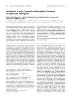
Fig. 6. Expressed protein ligation. The synthesis of proteins with
C-terminal thioester (left) and proteins with N-terminal Cys (right) can
be performed by using the IMPACT
TM
-system. Thioesters can be
obtained by fusing the protein of interest to the N-terminus of an
intein, proteins with N-terminal Cys by fusing to the C-terminus of a
mutated intein. Separation occurs by using the Chitin binding domain
(CBD). Both fragments can be synthesized by SPPS and specifically
labelled at the N- or C-terminus of the protein. Ligation of both
fragments proceeds under the conditions of NCL.
Table 1. Intein based vectors and their potential applications. Mxe GyrA, Mycobacterium xenopi gyrease A; Mth RIR1, Methanobacterium ther-
moautotrophicum; Ssp DnaB, Synechocystis sp. PCC6803; Sce VMA, Saccharomyces cerevisiae.
Vector Intein Splice junction Cleavage induction References Applications
pTXB1, 3 Mxe GyrA C-terminus Thiol
a
[64] Purification, generation of C-terminal thioesters
pTYB1, 2 Sce VMA C-terminus Thiol
a
[62] Purification, generation of C-terminal thioesters
pTWIN1 Ssp DnaB N-terminus pH and temperature [88] Purification C-terminal thioesters, aCys-proteins,
protein ligation, cyclization
Mxe GyrA C-terminus Thiol
a
[88]
pTWIN2 Ssp DnaB N-terminus pH and temperature [111] Purification, C-terminal thioesters, aCys-proteins,
protein ligation, cyclization
Mth RIR1 C-terminus Thiol
a
pTYB11, 12 Sce VMA N-terminus Thiol
a
[112] Purification
pTYB3, 4, pKYB1 Sce VMA C-terminus Thiol
a
[40] Purification, generation of C-terminal thioesters
a
Other nucleophiles might be used for the induction of the protein cleavage.
668 R. David et al. (Eur. J. Biochem. 271) Ó FEBS 2004
Generation of aCys proteins
The EPL requires a peptide or protein that contains an
amino terminal Cys residue (aCys) besides the a-thioester
moiety. To synthesize proteins possessing an aCys, the
protein cDNAs of interest can be cloned into various
commercially available vectors as mentioned above
(IMPACT
TM
-System). Thus, after the expression, the
intein/CBD fusion protein can be purified on a chitin
column and cleaved by changing the pH or temperature.
This will lead to the free aCys proteins. One drawback in
the intein-based synthesis of aCys proteins is the possible
spontaneous cleavage, which results in a loss of the
purification tag [45,64].
Expressed protein ligation (EPL)
Expressed protein ligation [12,50,65,66], also named intein-
mediated protein ligation [46], is an extension of the NCL
method. A recombinant Ca-thioester reacts with a chemi-
cally synthesized or expressed peptide/protein possessing an
N-terminal Cys under the conditions of NCL to form a
native peptide bond. This ligation method combines the
advantages of molecular engineering and chemical peptide
synthesis in many cases and allows site-specific introduction
of unnatural amino acids and chemical or biophysical tags
into large proteins. In former times, the difficulty of this
strategy was the chemical preparation of peptides or
proteins with a C-terminal thioester and the generation of
peptides and proteins with N-terminal Cys residues in large
quantities and high purity. Now, the expression of both
segments in high yields is possible by using the introduced
IMPACT
TM
-system. Thioesters can be obtained by fusing
the protein of interest with the N-terminus of an intein,
proteins with N-terminal Cys by fusing with the C-terminus
of a mutated intein [64]. Both fragments needed for ligation
can be synthesized alternately by SPPS as described already,
so it is possible to introduce specific labels either at the
N- or C-terminus of the protein. The chemically synthesized
section can be as small as possible whereas the expressed
part is not limited in size. This can lead to very large
specifically labelled proteins.
Expressed protein ligation can be performed directly on
chitin beads and thiolysis and ligation can occur simulta-
neously. It is disadvantageous if solubilizing agents are
needed for the ligation, because urea or guanidinium
hydrochloride for example denaturate the chitin binding
domain at concentrations higher than 2
M
. Alternatively,
the thioester may be eluted and the ligation reaction may
proceed in a second step. Detergents, urea or guanidinium
hydrochloride can be used in higher concentrations to
increase the solubility of peptides which may result in a
higher reaction yield.
If an amino acid within the protein sequence or several
amino acids on both ends was to be modified, the
protein would have to be split in three or more fragments
andtwoormoreligationstepswouldhavetobe
executed. The second peptide fragment carrying an
N-terminal Cys and an additional C-terminal thioester
has to be masked recombinantly at the N-terminus with
a protease cleavage site, e.g. factor Xa protease. After the
first ligation step, the N-terminal Cys is liberated by
protease treatment and the second ligation step can be
performed [50]. This protein can be synthesized from the
C- to N-direction.
Applications of expressed protein ligation
EPL chemistry applications are summarised in Table 2 and
described in more detail below.
Site specific protein modifications
The ability to change specific sidechains by the insertion of
noncanonical amino acids has great potential in protein
structure/function studies.
To determine the role of post-translational modifica-
tions it is necessary to insert phosphorylations or glyco-
sylations at defined positions. A phosphotyrosine peptide
is ligated to the C-terminus of the protein tyrosine kinase
C-terminal, Src kinase (Csk), which results in an intra-
molecular phosphotyrosine–Src homology 2 interaction
and increased catalytic phosphoryl transfer to a substrate
when compared with a nonphosphorylated control [12].
Table 2. Recent highlights show the scope of EPL chemistry. GFP, green fluorescent protein; CAR D1, immunoglobulin D1 domain of cox-
sackievirus-adenovirus receptor; MBP, maltose binding protein; proNPY, proneuropeptide Y; BBP, brain-binding peptide; RGD, (Arg-Gly-Asp)-
containing peptide.
Application Subject References
Investigation of protein–protein interactions Enhanced GFP [78,80,81]
Targeting CAR D1, calcitonin segment [86,113]
Internal isotopic labelling MBP [102]
Semisynthesis of prohormones proNPY [37,75,110]
Prenylation of proteins Rab7, YPT1 GTPase [114,115]
In vitro cyclization c-Crk-II [116]
Protein cyclization in vivo GFP [92]
Semisynthesis of cytotoxic proteins RNase A [63]
Incorporation of non natural amino acids Src [67]
Peptide and protein labelling with biophysical probes c-Crk-II, hIL-8 [73,76]
Conditional splicing in vivo MBP [83,84]
Cyclization using the TWIN system BBP, RGD [88]
In vitro screening for splicing inhibitors GFP [117]
Ó FEBS 2004 Expressed protein ligation (Eur. J. Biochem. 271) 669
The Csk–Src system was also investigated by Wang et al.
who displaced the Src–tyrosine by five unnatural Tyr
analogues to determine the role of the Tyr-sidechain for
Src affinity to Csk [67]. Lu et al. [68] observed the
influence of phosphorylation at two Tyr residues of
protein tyrosine phosphatase SHP-2 by introducing non-
hydrolyzable phospho-tyrosine analogues at the phos-
phorylation site of SHP-2 by expressed protein ligation.
Their results showed that phosphorylation at Tyr542 leads
to the basal inhibition of protein tyrosine phosphatase
(PTPase) activity by interacting with the N-terminal SH2
domain, whereas phosphorylated Tyr580 stimulates the
PTPase by interacting with the C-terminal SH2-domain.
The role of phosphorylation of the eukaryotic initiation
factor elF4E, which is implicated in the regulation of the
initiation step of translation, was observed by the
selectively phosphorylated version. Cap affinity of phos-
phorylated and unphosphorylated elF4E was determined
by fluorimetric time-synchronized titration [69].
The introduction of biophysical probes (spin labels or
fluorescence tags) allows the observation of protein–protein
interactions, membrane insertion or cellular uptake of
labelled peptides and proteins. Several fluorescence based
approaches [70–72] have been developed where the fluoro-
phore is attached to the sidechain of an amino acid (mainly
Lys) within the protein sequence.
Cotton et al. described the synthesis of a dual-labelled
version of the Crk-II adapter protein and its investigation
by fluorescence resonance energy transfer (FRET). A pair
of tetramethylrhodamine and fluoresceine was ligated to
the N- and C-terminus by solid-phase expressed protein
ligation. The construct reported the phosphorylation of
Crk-II by the nonreceptor tyrosine kinase by fluorescence
change that was affected by structural changes [73]. The
same FRET-pair was used to observe homo-oligomeriza-
tion of glutathione S-transferase, SH2 domain phospha-
tase-1 and serotonin N-acetyltransferase by measurement
of intermolecular FRET-effects [74]. We succeeded
recently in the semisynthesis of the 69 amino-acid
proNPY and its analogues to study prohormone proces-
sing. Five variants were synthesized containing either no
label or were labelled with carboxyfluorescein or biotin.
Western blot analysis was performed to determine the
binding site of anti-NPY and anti-proNPY antibodies
[75].
Furthermore we synthesized human interleukin-8, a
chemotactic cytokine, and its C-terminal carboxyfluo-
rescein-labelled analogue by expressed protein ligation.
Possessing four Cys residues, the formation of two disulfide-
bridges was necessary to obtain biological activity of hIL-8.
One of these Cys residues was chosen as a ligation site.
Internalization studies on HL60-cells expressing both
hIL-8-receptor subtypes and binding studies on HL60-
membranes provided an insight into the ligand receptor
interaction and the internalization of the interleukin-8-
receptor complex [76].
Also, single atoms like isotopes or atom homologues like
F instead of H, or Se instead of S can represent biophysical
probes. Wallace et al. introduced simultaneously (and site-
specific) selenium and bromine as reporter atoms into the
sequence of cytochrome c without significant changes of
structure and function [77].
Intermolecular protein splicing in
trans
to study
protein–protein interaction
Protein–protein interactions are essential for many biologi-
cal processes like receptor-ligand binding, protein polymer-
ization, gene expression, etc. To study these interactions
in vivo, several methods have been developed, one example
being the yeast two-hybrid system. The principle of these
methods is that potentially interacting proteins are tagged to
proteins with a particular function [78]. This function will be
recovered if an interaction of the tagged proteins is
accomplished. By using protein-splicing in trans [79] a split
intein is tagged to a split functional protein that is
reconstituted after interaction of the intein parts. Ozawa
et al. used halves of enhanced green fluorescent protein
(eGFP) as N- and C-terminal exteins and fused them to
N- and C-terminal fragments of a modified intein [80,81].
No fluorescence was observed from any construct expressed
in E.coli. In contrast, coexpression of calmodulin and its
target peptide M13 connected to the intein led to fluores-
cence of eGFP, suggesting that the interaction of calmo-
dulin and M13 triggers the refolding of the intein. A related
approach using firefly luciferase, was introduced by the
same group for mammalian cells [82].
The conditional protein splicing approach from Mootz
et al. [83,84] used the dimerization of the rapamycin
receptor FKBP and the rapamycin binding domain in the
presence of rapamycin to reconstitute a split intein in
mammalian cells. Maltose binding protein (MBP) and a
His-tag were used as exteins and the splicing product was
detected by Western blotting or by immunoprecipitation in
the cells. In a related approach by this group, GFP was
coupled to the N-terminus of an intein and expressed in
Chinese hamster ovary cells. The chemically synthesized
C-terminal part of the intein was coupled to a FLAG-
epitope and transported through the membrane by using a
protein transduction domain. The C-terminal intein can
associate with its N-terminal half within the cells and
ligation of GFP to the FLAG-epitope is performed [85].
By using the EPL-method, eGFP was ligated to an
amidated human calcitonin (hCT) derived carrier peptide.
Covalently bound calcitonin and its C-terminal fragments
were shown to permeate membranes of nasal epithelium,
but permeation was limited to peptides. Ligated eGFP-
hCT(8–32) shows specific mucosal internalization, whereas
enhanced GFP did not show internalization per se. The
shuttle-ability of hCT and its possible role in drug delivery
was demonstrated using eGFP [86].
Generation of cyclic peptides and proteins
Backbone cyclization can improve the stability and the
activity of peptides and proteins and reduce their conform-
ational flexibility. The production of circular proteins may
influence the rational design of enzymes and the develop-
ment of new agents by structure activity studies.
Cyclic structures can be obtained either by disulfide
formation or by formation of a peptide bond between
N- and C-termini or by sidechain cyclization. Several
methods have been developed by using modified inteins to
generate cyclic peptides and proteins. The aim is to create a
protein with both an N-terminal Cys and a C-terminal
670 R. David et al. (Eur. J. Biochem. 271) Ó FEBS 2004
thioester. Such a peptide can be generated by flanking the
protein of interest with two inteins (Fig. 7). The N-terminal
modified intein can be cleaved by a pH and temperature
shift, whereas the C-terminal intein is cleaved by the
addition of thiols. This ÔtwointeinsystemÕ (TWIN) also
allows the separation by chitin binding domains fused to the
inteins. The reaction of the two reactive groups leads to the
formation of cyclic peptides and proteins or multimers by
an amide bond [87,88].
Several approaches use intramolecular trans-splicing for
the generation of cyclic backbones in vivo and in vitro. In
these cases, the split intein is not coupled to a cleaved
protein or to two proteins which should be knotted, but the
intein parts flank one protein with an N-terminal Cys
residue. If the intein is reconstituted, a thioester intermediate
will be formed that undergoes transthioesterification. Cyc-
lization of Asp and SfiN-acyl transfer leads to a cyclic
product [89–92].
A simple approach for in vivo cyclization in Escherichia
coli cells was introduced by Camarero et al. [93]. An SH3
domain from murine c-Crk adapter protein with an
N-terminal Cys residue was N-terminally fused to an intein
with a chitin binding domain. After the expression of this
fusion protein, the N-terminal Met residue produced by the
start-codon is replaced by the Met-aminopeptidase, which
results in an active Cys residue. The amide-bond connecting
the protein to the intein can switch by NfiS-acyl shift to the
thioester bond. As this protein now possesses a reactive
N-terminal Cys residue and a C-terminal thioester it can
react to form an intramolecular bond by NCL.
Generation of cytotoxic proteins
In some cases, the expression of the desired proteins in
bacteria can cause cytotoxic side-effects because the target
protein competes with cellular components of the host.
Another problem is that overexpressed proteins may
aggregate as inclusion bodies in the cytosol. By using EPL
techniques this can be avoided through modular synthesis of
an artificial target protein as an intein fusion protein.
Subsequently, through ligation and refolding, the native
conformation and biological functionality of a cytotoxic
protein will be recovered. The potential cytotoxic RNase A
was expressed by this method [63]. One part of this protein
was produced as a segment carrying an intein at its
C-terminal site. After thiol-induced intein-mediated clea-
vage, the obtained thioester of the truncated RNase A was
joined with a fragment synthesized by SPPS that contained
a naturally occurring Cys residue at the N-terminus.
Ligation of both enzymatic inactive protein segments led
to the full length protein, which reconstituted its enzymatic
activity after several renaturation steps. Another intein-
based approach was used to purify the cytotoxic endonuc-
lease I-TevI by insertional inactivation followed by pH
controllable splicing [94]. In this case, a mini intein mutant
(DI-SIM) of the full length Mtu RecA intein was inserted
into the I-TevI sequence thereby inactivating the protein
in vivo. The intein triggered the splicing of the protein after
purification on a chitin column and the endonuclease could
be obtained in its native state. However, this method was
only successful when an appropriate Cys residue was in the
target protein allowing proper insertion of the intein.
Furthermore, the toxicity has to be low and the splicing
ratio in vitro/in vivo has to be as high as possible. Expression
of the whole protein is one of the big advantages in this
system as the folding of the endonuclease does not interfere
with the folding of the intein module. Intein-based trans-
splicing systems with either native or artificial split inteins
also seem to be adequate workhorses for the synthesis of
cytotoxic proteins [91,95].
Segmental isotopic labelling
Expressed protein ligation is of great use for the introduc-
tion of stable isotopes into protein segments (Fig. 8) [96,97].
This approach circumvents the practical size limitation
for structure determination by using NMR spectroscopy.
Generally, inadequate loss of structure resolution is based
on several effects that are proportional to the number of
amino acids. This includes line broadening, longer rota-
tional correlation times and an increased number of signals
of similar chemical shifts. Even though there are new NMR
techniques available, like transverse relaxation optimized
spectroscopy (TROSY) [98], the standard isotopic labelling
strategies through incorporation of uniformly labelled
15
N,
13
C and perdeuteration of amino acid sidechains bear the
Fig. 7. Generation of cyclic proteins. Intramolecular trans-splicing
(left). The two parts of a split intein flank one protein with N-terminal
Cys. If the intein is reconstituted, a thioester intermediate will be
produced that undergoes transthioesterification. After Asp cyclization
and SfiN-acyl transfer, a cyclic product is formed. Two intein (TWIN)
system (right). The protein of interest is cloned between two inteins.
The N-terminally modified intein can then be cleaved with a pH and
temperature shift, whereas the C-terminal intein is cleaved by addition
of thiols. The reaction of the two reactive groups leads to the formation
of cyclic peptides and proteins.
Ó FEBS 2004 Expressed protein ligation (Eur. J. Biochem. 271) 671
signal overlap of macromolecular systems. Yamazaki et al.
selectively labelled the C-terminal domain of the E. coli
RNA polymerase a-subunit [99] by using a trans-splicing
system based on a split PI-PfuI intein. Muir and coworkers
used the EPL strategy to introduce an
15
N-labelled domain
within the Src-homology domain 3 and 2 segment derived
from AbI protein tyrosine kinase [100]. In both cases the
part of the protein of interest was bacterially expressed in
15
N-isotope containing media. Fusion of this labelled
segment with the other recombinant protein part that was
unlabelled led to the specifically labelled protein. One of the
great advantages of these labelling strategies is the possibi-
lity to elucidate particular interactions of protein domains.
Such a phenomenon could be shown in bacterial sigma
factor [101]. In this case, the comparative NMR studies of
isotopic labelled model proteins of this protein obtained by
applying EPL revealed that the C-terminal DNA binding
domain does not interact directly with the N-terminal
autoregulatory domain. EPL and trans-splicing also have a
great impact in the preparation of labelled internal protein
segments. Yamazaki’s group presented a method for central
segmental isotopic labelling by using a tandem trans-splicing
approach [102,103]. To label an inner segment of the maltose
binding protein, the target protein was expressed as three
split intein fusion proteins. The central segment was thereby
expressed in isotope containing media as a fusion protein
with attached PI-PfuI and PI-PfuII inteins at its termini.
Consequently, the N-terminal parts of the desired protein
were expressed as fusion proteins carrying the other halves of
the split inteins. Simultaneous splicing yielded the target
protein including an inner isotopically labelled fragment.
Alternative ligation methods
The only disadvantage of NCL and EPL is the necessity of a
Cys residue or a homologue at the ligation site. The
occurrence of this amino acid in globular proteins is very
low and the insertion of additional Cys residues can alter the
protein structure and function by the formation of disulfide
bridges. Several approaches have been developed to
circumvent this limitation (Fig. 9).
NCL with Cys-mimetics
The NCL-methodology has been extended to -X-Gly- and
-Gly-X- ligation sites [104]. One peptide possessing a
C-terminal thioester reacts with a second one containing
either an Na(ethanethiol) peptide or a Na(oxyethanethiol)
peptide. The thioester intermediate forms a 5- or 6-member
ring and in a final SfiN-transfer an amide bond is formed.
In a subsequent step, the substitution at the amide bond can
beremovedbythetreatmentwithZnandH
+
to form a
native peptide bond.
NCL combined with desulfurization
In this application, NCL is extended to proteins without
Cys-residues [105]. Ala is a common amino acid in peptides
and proteins, thus, a specific Ala is replaced by a Cys residue
at the ligation site within the sequence of the protein of
interest. Then NCL is performed to ligate thioester and Cys-
peptide. In the following step the Cys is converted to an Ala
by desulfination using palladium or Raney-nickel and
hydrogen. This approach can be used for the synthesis of
linear and cyclic proteins and extends NCL-methodology
to -X-Ala As no selectivity of the desulfurization reaction
is possible, proteins that contain further Cys residues cannot
be made by this technique.
Staudinger ligation
This ligation method is inspired by the Staudinger reaction,
where a phosphine is used to reduce an azide to an amine.
An intermediate iminophosphoran possesses a nucleophilic
nitrogen which can react with an acyl donor to form an
amide. A peptide bearing a C-terminal phosphinothioester
is coupled to another peptide with an N-terminal a-azido
group to form a peptide bond. The final product has no
residual atoms [106,107]. This ligation method may also be
combined with NCL for tandem ligation applications. The
method however, has up to now only been used for small
peptides.
Expressed enzymatic ligation
This method combines the advantages of expressed protein
ligation with the substrate mimetic strategy of protease
mediated ligation. The reverse hydrolysis potential of a
protease, e.g. Glu/Asp-specific serine protease from
Staphylococcus aureus, is used to catalyze the peptide bond
formation [108]. The limiting enzyme substrate specificity
and possible proteolysis of peptides and ligated products
is eliminated by substrate mimetics carrying a site-specific
ester leaving group at the C-terminus of the former
Fig. 8. Segmental isotopic labelling. Protein
segments are expressed in unlabelled or iso-
topically enriched media as fusion proteins
with parts of split inteins. Reconstitution of
the inteins results in trans-splicing that leads
to terminally (A) or centrally (B) labelled
proteins.
672 R. David et al. (Eur. J. Biochem. 271) Ó FEBS 2004
unspecific peptide [109]. The IMPACT
TM
-system was used
for the synthesis of thioesters bearing V8-specific ester
leaving groups. These thioesters were used as acyl donors in
V8-mediated ligation of modified proNPY(1–69) [110].
Concluding remarks
Different ligation strategies for the fusion of proteins and
peptide segments have been established. The NCL has been
shown to be a significant benchmark for the latest protein
engineering tasks. Formation of a native peptide bond
between two or more different peptides can be performed
using several approaches that differ mostly in the way of
activating the N-terminal or C-terminal amino acid of the
reactions partners. All these clever strategies are, however,
restricted by the limited final size of the desired peptide or
protein. Therefore, one of the most important developments
within the last few years includes the approach based on
intein splicing. Although much has still to be learnt on the
mechanistic details and the defined roles of conserved amino
acids regarding the different inteins, these self-splicing
elements have become very powerful workhorses for the
multiple coupling of protein and peptide fragments in large
quantities and yields without the need for any protection
schemes. Native peptides and proteins and also synthetic or
semisynthetic proteins of all shapes and sizes can be
synthesized by this method. This technique has influences
particularly in the area of protein engineering. The further
development and understanding of intein-based splicing
systems, the discovery of inteins with novel properties and
the progress of other ligation methodologies will certainly
enlarge the repertoire of these interesting technologies.
Fig. 9. Alternate ligation methods. NCL with Cys-mimetics (A) results in Gly at the ligation site. NCL combined with desulfurization (B) leads to an
Ala residue. Staudinger ligation (C) is applicable to each amino acid at the ligation site. EEL uses the substrate mimetic approach and an inverse
working protease. The protein thioester used for ligation can be obtained by the IMPACT
TM
method (D).
Ó FEBS 2004 Expressed protein ligation (Eur. J. Biochem. 271) 673
Acknowledgements
Some of the work discussed in this article was supported by the DFG
grant 1264-5-1/2. Furthermore we kindly acknowledge the financial
support of the DFG for projects dealing with protein ligation (GK 378
and SFB 610).
References
1. Kohno, T., Kusunoki,H.,Sato,K.&Wakamatsu,K. (1998)A new
general method for the biosynthesis of stable isotope-enriched
peptides using a decahistidine-tagged ubiquitin fusion system: an
application to the production of mastoparan-X uniformly
enriched with 15N and 15N/13C. J. Biomol. NMR 12, 109–121.
2. Marley, J., Lu, M. & Bracken, C. (2001) A method for efficient
isotopic labeling of recombinant proteins. J. Biomol. NMR 20,
71–75.
3. Jansson, M., Li, Y.C., Jendeberg, L., Anderson, S., Montelione,
B.T. & Nilsson, B. (1996) High-level production of uniformly
15
N- and
13
C-enriched fusion proteins in Escherichia coli.
J. Biomol. NMR 7, 131–141.
4. Chin, J.W., Cropp, T.A., Anderson, J.C., Mukherji, M.,
Zhang, Z. & Schultz, P.G. (2003) An expanded eukaryotic genetic
code. Science 301, 964–967.
5. Wang, L. & Schultz, P.G. (2002) Expanding the genetic code.
Chem. Commun. 1–11.
6. Wallace, C.J. (1995) Peptide ligation and semisynthesis. Curr.
Opin. Biotechnol. 6, 403–410.
7. Liu, C F. & Tam, J.P. (1994) Peptide segment ligation strategy
without use of protecting groups. Proc. Natl Acad. Sci. USA 91,
6584–6588.
8. Kemp, D.S. & Carey, R.I. (1993) Synthesis of a 39-peptide and a
25-peptide by thiol capture ligations: observation of a 40-fold rate
acceleration of the intramolecular O,N-acyl-transfer reaction
between peptide fragments bearing only cysteine protective
groups. J. Org. Chem. 58, 2216–2222.
9. Dyckes, D.F., Creighton, T. & Sheppard, R.C. (1974)
Spontaneous re-formation of a broken peptide chain. Nature 247,
202–204.
10. Wallace, C.J. (1993) Understanding cytochrome c function:
engineering protein structure by semisynthesis. FASEB J. 7,
505–515.
11. Becker,C.F.,Hunter,C.L.,Seidel,R.,Kent,S.B.,Goody,R.S.&
Engelhard, M. (2003) Total chemical synthesis of a functional
interacting protein pair: the protooncogene H-Ras and the Ras-
binding domain of its effector c-Raf1. Proc.NatlAcad.Sci.USA
100, 5075–5080.
12. Muir, T.W., Sondhi, D. & Cole, P.A. (1998) Expressed protein
ligation: a general method for protein engineering. Proc. Natl
Acad. Sci. USA 95, 6705–6710.
13. Dawson, P.E., Muir, T.W., Clark-Lewis, I. & Kent, S.B. (1994)
Synthesis of proteins by native chemical ligation. Science 266,
776–779.
14. Dawson, P.E. & Kent, S.B. (2000) Synthesis of native proteins by
chemical ligation. Annu. Rev. Biochem. 69, 923–960.
15. Wieland, T., Bokelmann, E., Bauer, L., Lang, H.U. & Lau, H.
(1953) Bildung von S-haltigen Peptiden durch intramolekulare
Wanderung von Aminoacylresten. Annalen Chemie 583, 129–149.
16. Tam, J.P., Xu, J. & Eom, K.D. (2001) Methods and strategies of
peptide ligation. Biopolymers 60, 194–205.
17. Tam, J.P., Lu, Y.A., Liu, C.F. & Shao, J. (1995) Peptide synthesis
using unprotected peptides through orthogonal coupling meth-
ods. Proc. Natl Acad. Sci. USA 92, 12485–12489.
18. Burns, J.A., Butler, J.C., Moran, J. & Whitesides, G.M. (1991)
Selective reduction of disulfides by tris (2-carboxyethyl) phos-
phine. J. Org. Chem. 56, 2648–2650.
19. Dawson, P.E., Churchill, M., Ghadiri, M.R. & Kent, S.B.H.
(1997) Modulation of reactivity in native chemical ligation
through the use of thiol additives. J. Am. Chem. Soc. 119, 4325–
4329.
20. Hackeng, T.M., Griffin, J.H. & Dawson, P.E. (1999) Protein
synthesis by native chemical ligation: expanded scope by using
straightforward methodology. Proc. Natl Acad. Sci. USA 96,
10068–10073.
21. Villain, M., Gaertner, H. & Botti, P. (2003) Native chemical
ligation with aspartic and glutamic acids as C-terminal residues:
Scope and limitations. Eur. J. Org. Chem. 3267–3272.
22. Canne, L.E., Botti, P., Simon, R.J., Chen, Y., Dennis, E.A. &
Kent, S.B.H. (1999) Chemical protein synthesis by solid phase
ligation of unprotected peptide segments. J. Am. Chem. Soc. 121,
8720–8727.
23. Tam, J.P., Yu, Q. & Lu, Y.A. (2001) Tandem peptide ligation for
synthetic and natural biologicals. Biologicals 29, 189–196.
24. Eom, K.D., Miao, Z., Yang, J.L. & Tam, J.P. (2003) Tandem
ligation of multipartite peptides with cell-permeable activity.
J. Am. Chem. Soc. 125, 73–82.
25. Hondal, R.J., Nilsson, B.L. & Raines, R.T. (2001) Selenocysteine
in native chemical ligation and expressed protein ligation. J. Am.
Chem. Soc. 123, 5140–5141.
26. Roelfes, G. & Hilvert, D. (2003) Incorporation of seleno-
methionine into proteins through selenohomocysteine-mediated
ligation. Angew. Chem. Int. Ed. Engl. 42, 2275–2277.
27. Clippingdale, A.B., Barrow, C.J. & Wade, J.D. (2000) Peptide
thioester preparation by Fmoc solid phase peptide synthesis for
use in native chemical ligation. J. Pept. Sci. 6, 225–234.
28. Li, X., Kawakami, T. & Aimoto, S. (1998) Direct preparation of
peptide thioesters using an Fmoc solid-phase method. Tetra-
hedron Lett. 39, 8669–8672.
29.Bu,X.,Xie,G.,Law,C.W.&Guo,Z.(2002)Animproved
deblocking agent for direct Fmoc solid-phase synthesis of peptide
thioesters. Tetrahedron Lett. 43, 2419–2422.
30. Backes, B.J., Virgilio, A.A. & Ellman, J.A. (1996) Activation
method to prepare a highly reactive acylsulfonamide Ôsafety-
catchÕ linker for solid-phase synthesis. J. Am. Chem. Soc. 118,
3055–3056.
31. Shin,Y.,Winans,K.A.,Backes,B.J.,Kent,S.B.H.,Ellman,J.A.
& Bertozzi, C.R. (1999) Fmoc-based synthesis of peptide-alpha
thioesters: Application to the total chemical synthesis of a gly-
coprotein by native chemical ligation. J. Am. Chem. Soc. 121,
11684–11689.
32. Ingenito,R.,Bianchi,E.,Fattori,D.&Pessi,A.(1999)Solid
phase synthesis of peptide C-terminal thioesters by Fmoc/t-Bu
chemistry. J. Am. Chem. Soc. 121, 11369–11374.
33. Alsina, J., Yokum, T.S., Albericio, F. & Barany, G. (1999)
Backbone amide linker (BAL) strategy for N (alpha)-
fluorenylmethoxycarbonyl (Fmoc) solid-phase synthesis of
unprotected peptide p-nitroanilides and thioesters. J. Org. Chem.
64, 8761–8769.
34. Swinnen, D. & Hilvert, D. (2000) Facile, Fmoc-compatible solid-
phase synthesis of peptide C-terminal thioesters. Org. Lett. 2,
2439–2442.
35. Sewing, A. & Hilvert, D. (2001) Fmoc-compatible solid-phase
peptide synthesis of long C-terminal peptide thioesters. Angew.
Chem.Int.Ed.Engl.40, 3395–3396.
36. Futaki, S., Sogawa, K., Maruyama, J., Asahara, T. & Niwa, M.
(1997) Preparation of peptide thioesters using Fmoc-solid-phase
peptide synthesis and its application to the construction of a
template-assembled synthetic protein (TASP). Tetrahedron Lett.
38, 6237–6240.
37. von Eggelkraut-Gottanka, R., Klose, A., Beck-Sickinger, A.G. &
Beyermann, M. (2003) Peptide alpha-thioester formation using
standard Fmoc-chemistry. Tetrahedron Lett. 44, 3551–3554.
674 R. David et al. (Eur. J. Biochem. 271) Ó FEBS 2004
38. Gogarten, J.P., Senejani, A.G., Zhaxybayeva, O., Olendzenski, L.
& Hilario, E. (2002) Inteins: structure, function, and evolution.
Annu. Rev. Microbiol. 56, 263–287.
39.Shih,C.K.,Wagner,R.,Feinstein,S.,Kanik-Ennulat,C.&
Neff, N. (1988) A dominant trifluoperazine resistance gene from
Saccharomyces cerevisiae has homology with F0F1 ATP synthase
and confers calcium-sensitive growth. Mol. Cell. Biol. 8, 3094–
3103.
40. Perler, F.B. (2000) InBase, the intein database. Nucleic Acids Res.
28, 344–345.
41. Mira, A., Ochman, H. & Moran, N.A. (2001) Deletional bias and
the evolution of bacterial genomes. Trends Genet. 17, 589–596.
42. Neff, N.F. (1993) Protein splicing: Selfish genes invade cellular
proteins. Curr. Opin. Cell Biol. 5, 971–976.
43. Goddard, M.R. & Burt, A. (1999) Recurrent invasion and
extinction of a selfish gene. Proc.NatlAcad.Sci.USA96, 13880–
13885.
44. Pietrokovski, S. (2001) Intein spread and extinction in evolution.
Trends Genet. 17, 465–472.
45. Evans, T.C. Jr & Xu, M Q. (2002) Mechanistic and kinetic
considerations of protein splicing. Chem. Rev. 102, 4869–4883.
46. Evans, T.C. Jr & Xu, M Q. (2000) Intein-mediated protein
ligation: harnessing nature’s escape artists. Biopolymers 51,
333–342.
47. Xu, M.Q. & Perler, F.B. (1996) The mechanism of protein spli-
cing and its modulation by mutation. EMBO J. 15, 5146–5153.
48. Paulus, H. (2000) Protein splicing and related forms of protein
autoprocessing. Annu.Rev.Biochem.69, 447–496.
49. Noren, C.J., Wang, J. & Perler, F.B. (2000) Dissecting the
chemistry of protein splicing and its applications. Angew. Chem.
Int. Ed. Engl. 39, 450–466.
50. Muir, T.W. (2003) Semisynthesis of proteins by expressed protein
ligation. Annu. Rev. Biochem. 72, 249–289.
51. Klabunde, T., Sharma, S., Telenti, A., Jacobs, W.R. Jr &
Sacchettini, J.C. (1998) Crystal structure of GyrA intein from
Mycobacterium xenopi reveals structural basis of protein splicing.
Nat. Struct. Biol. 5, 31–36.
52. Chen, L., Benner, J. & Perler, F.B. (2000) Protein splicing in the
absence of an intein penultimate histidine. J. Biol. Chem. 275,
20431–20435.
53. Mizutani, R., Nogami, S., Kawasaki, M., Ohya, Y., Anraku, Y.
& Satow, Y. (2002) Protein-splicing reaction via a thiazolidine
intermediate: crystal structure of the VMA1-derived endo-
nuclease bearing the N and C-terminal propeptides. J. Mol. Biol.
316, 919–929.
54. Kawasaki, M., Nogami, S., Satow, Y., Ohya, Y. & Anraku, Y.
(1997) Identification of three core regions essential for protein
splicing of the yeast VMA1 protozyme. A random mutagenesis
study of the entire VMA1-derived endonuclease sequence. J. Biol.
Chem. 272, 15668–15674.
55. Duan, X., Gimble, F.S. & Quiocho, F.A. (1997) Crystal structure
of PI-SceI, a homing endonuclease with protein splicing activity.
Cell 89, 555–564.
56. Moure, C.M., Gimble, F.S. & Quiocho, F.A. (2002) Crystal
structure of the intein homing endonuclease PI-SceI bound to its
recognition sequence. Nat. Struct. Biol. 9, 764–770.
57. Poland, B.W., Xu, M.Q. & Quiocho, F.A. (2000) Structural
insights into the protein splicing mechanism of PI-SceI. J. Biol.
Chem. 275, 16408–16413.
58. Ghosh, I., Sun, L. & Xu, M.Q. (2001) Zinc inhibition of protein
trans-splicing and identification of regions essential for splicing
and association of a split intein*. J. Biol. Chem. 276, 24051–
24058.
59. Mills, K.V. & Paulus, H. (2001) Reversible inhibition of protein
splicing by zinc ion. J. Biol. Chem. 276, 10832–10838.
60.Hu,D.,Crist,M.,Duan,X.,Quiocho,F.A.&Gimble,F.S.
(2000) Probing the structure of the PI-SceI-DNA complex by
affinity cleavage and affinity photocross-linking. J. Biol. Chem.
275, 2705–2712.
61. Perler, F.B. (1998) Protein splicing of inteins and hedge-
hog autoproteolysis: structure, function, and evolution. Cell
92,1–4.
62. Chong, S., Mersha, F.B., Comb, D.G., Scott, M.E., Landry, D.,
Vence, L.M., Perler, F.B., Benner, J., Kucera, R.B., Hirvonen,
C.A., Pelletier, J.J., Paulus, H. & Xu, M.Q. (1997) Single-column
purification of free recombinant proteins using a self-cleavable
affinity tag derived from a protein splicing element. Gene 192,
271–281.
63. Evans, T.C. Jr, Benner, J. & Xu, M.Q. (1998) Semisynthesis of
cytotoxic proteins using a modified protein splicing element.
Protein. Sci. 7, 2256–2264.
64. Southworth, M.W., Amaya, K., Evans, T.C., Xu, M.Q. & Perler,
F.B. (1999) Purification of proteins fused to either the amino or
carboxy terminus of the Mycobacterium xenopi gyrase A intein.
Biotechniques 27, 110–114,116,118–20.
65. Severinov, K. & Muir, T.W. (1998) Expressed protein ligation, a
novel method for studying protein–protein interactions in tran-
scription. J. Biol. Chem. 273, 16205–16209.
66. Hofmann, R.M. & Muir, T.W. (2002) Recent advances in the
application of expressed protein ligation to protein engineering.
Curr. Opin. Biotechnol. 13, 297–303.
67. Wang, D. & Cole, P.A. (2001) Protein tyrosine kinase Csk-
catalyzed phosphorylation of Src containing unnatural tyrosine
analogues. J. Am. Chem. Soc. 123, 8883–8886.
68. Lu, W., Gong, D., Bar-Sagi, D. & Cole, P.A. (2001) Site-specific
incorporation of a phosphotyrosine mimetic reveals a role for
tyrosine phosphorylation of SHP-2 in cell signaling. Mol. Cell. 8,
759–769.
69. Zuberek,J.,Wyslouch-Cieszynska,A.,Niedzwiecka,A.,Dadlez,
M.,Stepinski,J.,Augustyniak,W.,Gingras,A.C.,Zhang,Z.,
Burley,S.K.,Sonenberg,N.,Stolarski,R.&Darzynkiewicz,E.
(2003) Phosphorylation of eIF4E attenuates its interaction with
mRNA 5¢-cap analogs by electrostatic repulsion: intein-mediated
proteinligationstrategytoobtainphosphorylatedprotein.RNA
9, 52–61.
70. Hofmann,R.M.,Cotton,G.J.,Chang,E.J.,Vidal,E.,Veach,D.,
Bornmann, W. & Muir, T.W. (2001) Fluorescent monitoring of
kinase activity in real time: development of a robust fluorescence-
based assay for Abl tyrosine kinase activity. Bioorg. Med. Chem.
Lett. 11, 3091–3094.
71. Ayers, B., Blaschke, U.K., Camarero, J.A., Cotton, G.J.,
Holford, M. & Muir, T.W. (1999) Introduction of unnatural
amino acids into proteins using expressed protein ligation.
Biopolymers 51, 343–354.
72. Cotton, G.J., Ayers, B., Xu, R. & Muir, T.W. (1999) Insertion of
a synthetic peptide into a recombinant protein framework: a
protein biosensor. J. Am. Chem. Soc. 121, 1100–1101.
73. Cotton, G.J. & Muir, T.W. (2000) Generation of a dual-labeled
fluorescence biosensor for Crk-II phosphorylation using solid-
phase expressed protein ligation. Chem. Biol. 7, 253–261.
74. Scheibner, K.A., Zhang, Z. & Cole, P.A. (2003) Merging fluor-
escence resonance energy transfer and expressed protein ligation
to analyze protein–protein interactions. Anal. Biochem. 317,
226–232.
75. von Eggelkraut-Gottanka, R., Machova, Z., Grouzmann, E. &
Beck-Sickinger, A.G. (2003) Semisynthesis and characterization
of the first analogues of pro-neuropeptide y. Chembiochemistry 4,
425–433.
76. David, R., Machova, Z. & Beck-Sickinger, A.G. (2003)
Semisynthesis and application of carboxyfluorescein-labelled
Ó FEBS 2004 Expressed protein ligation (Eur. J. Biochem. 271) 675
biologically active human interleukin-8. Biol. Chem. 384, 1619–
1630.
77. Wallace, C.J.A. & Clark-Lewis, I. (2000) Site-specific
independent double labeling of proteins with reporter atoms.
Bioch. Cell Biol. 78, 79–86.
78. Ozawa, T. & Umezawa, Y. (2001) Detection of protein–protein
interactions in vivo based on protein splicing. Curr. Opin. Chem.
Biol. 5, 578–583.
79. Lew, B.M., Mills, K.V. & Paulus, H. (1999) Characteristics of
protein splicing in trans mediated by a semisynthetic split intein.
Biopolymers 51, 355–362.
80. Ozawa, T., Takeuchi, T.M., Kaihara, A., Sato, M. &
Umezawa, Y. (2001) Protein splicing-based reconstitution of split
green fluorescent protein for monitoring protein–protein inter-
actions in bacteria: improved sensitivity and reduced screening
time. Anal. Chem. 73, 5866–5874.
81. Ozawa, T., Nogami, S., Sato, M., Ohya, Y. & Umezawa, Y.
(2000) A fluorescent indicator for detecting protein–protein
interactions in vivo based on protein splicing. Anal. Chem. 72,
5151–5157.
82. Ozawa, T., Kaihara, A., Sato, M., Tachihara, K. & Umezawa, Y.
(2001) Split luciferase as an optical probe for detecting protein–
protein interactions in mammalian cells based on protein splicing.
Anal. Chem. 73, 2516–2521.
83. Mootz, H.D., Blum, E.S., Tyszkiewicz, A.B. & Muir, T.W. (2003)
Conditional protein splicing: a new tool to control protein
structure and function in vitro and in vivo. J. Am. Chem. Soc. 125,
10561–10569.
84. Mootz, H.D. & Muir, T.W. (2002) Protein splicing triggered by a
small molecule. J. Am. Chem. Soc. 124, 9044–9045.
85. Giriat, I. & Muir, T.W. (2003) Protein semi-synthesis in living
cells. J. Am. Chem. Soc. 125, 7180–7181.
86. Machova, Z., Mu
¨
hle, C., Krauss, U., Trehin, R., Koch, A.,
Merkle, H.P. & Beck-Sickinger, A.G. (2002) Cellular inter-
nalization of enhanced green fluorescent protein ligated to a
human calcitonin-based carrier peptide. Chembiochemistry 3,
672–677.
87. Xu, M.Q. & Evans, T.C. Jr (2001) Intein-mediated ligation and
cyclization of expressed proteins. Methods 24, 257–277.
88. Evans, T.C. Jr, Benner, J. & Xu, M.Q. (1999) The cyclization and
polymerization of bacterially expressed proteins using modified
self-splicing inteins. J. Biol. Chem. 274, 18359–18363.
89. Scott, C.P., Abel-Santos, E., Wall, M., Wahnon, D.C. &
Benkovic, S.J. (1999) Production of cyclic peptides and proteins
in vivo. Proc. Natl Acad. Sci. USA 96, 13638–13643.
90. Scott, C.P., Abel-Santos, E., Jones, A.D. & Benkovic, S.J. (2001)
Structural requirements for the biosynthesis of backbone cyclic
peptide libraries. Chem. Biol. 8, 801–815.
91. Evans,T.C.Jr,Martin,D.,Kolly,R.,Panne,D.,Sun,L.,Ghosh,
I., Chen, L., Benner, J., Liu, X.Q. & Xu, M.Q. (2000) Protein
trans-splicing and cyclization by a naturally split intein from the
dnaE gene of Synechocystis species PCC6803. J. Biol. Chem. 275,
9091–9094.
92. Iwai, H., Lingel, A. & Pluckthun, A. (2001) Cyclic green fluor-
escent protein produced in vivo using an artificially split PI-PfuI
intein from Pyrococcus furiosus. J. Biol. Chem. 276, 16548–16554.
93.Camarero,J.A.,Fushman,D.,Cowburn,D.&Muir,T.W.
(2001) Peptide chemical ligation inside living cells: in vivo gen-
eration of a circular protein domain. Bioorgan. Med. Chem. 9,
2479–2484.
94. Wu,W.,Wood,D.W.,Belfort,G.,Derbyshire,V.&Belfort,M.
(2002) Intein-mediated purification of cytotoxic endonuclease I-
TevI by insertional inactivation and pH-controllable splicing.
Nucleic Acids Res. 30, 4864–4871.
95. Mills, K.V., Lew, B.M., Jiang, S. & Paulus, H. (1998) Protein
splicing in trans by purified N- and C-terminal fragments of the
Mycobacterium tuberculosis RecA intein. Proc. Natl Acad. Sci.
USA 95, 3543–3548.
96. Blaschke, U.K. Cotton, G.J. & Muir, T.W. (2000) Synthesis of
multi-domain proteins using expressed protein ligation: strategies
for segmental isotopic labeling of internal regions. Tetrahedron
56, 9461–9470.
97. Ottesen, J.J., Blaschke, U.K., Cowburn, D. & Muir, T.W. (2003)
Segmental isotopic labeling: prospects for a new tool to study the
structure-function relationships in multi-domain proteins. Biol.
Magnetic Resonance 20, 35–51.
98. Pervushin, K. (2001) The use of TROSY for detection and sup-
pression of conformational exchange NMR line broadening in
biological macromolecules. J. Biomol. NMR 20, 275–285.
99. Yamazaki,T.,Otomo,T.,Oda,N.,Kyogoku,Y.,Uegaki,K.,
Ito, N., Ishino, Y. & Nakamura, H. (1998) Segmental isotope
labeling for protein NMR using peptide splicing. J. Am. Chem.
Soc. 120, 5591–5592.
100. Xu, R., Ayers, B., Cowburn, D. & Muir, T.W. (1999) Chemical
ligation of folded recombinant proteins: segmental isotopic
labeling of domains for NMR studies. Proc. Natl. Acad. Sci. USA
96, 388–393.
101. Camarero, J.A., Shekhtman, A., Campbell, E.A., Chlenov, M.,
Gruber,T.M.,Bryant,D.A.,Darst,S.A.,Cowburn,D.&Muir,
T.W. (2002) Autoregulation of a bacterial sigma factor explored
by using segmental isotopic labeling and NMR. Proc. Natl Acad.
Sci. USA 99, 8536–8541.
102. Otomo, T., Ito, N., Kyogoku, Y. & Yamazaki, T. (1999) NMR
observation of selected segments in a larger protein: central-
segment isotope labeling through intein-mediated ligation.
Biochemistry 38, 16040–16044.
103. Otomo, T., Teruya, K., Uegaki, K., Yamazaki, T. & Kyogoku,
Y. (1999) Improved segmental isotope labeling of proteins and
application to a larger protein. J. Biomol. NMR 14, 105–114.
104. Canne, L.E., Bark, S.J. & Kent, S.B.H. (1996) Extending the
applicability of native chemical ligation. J. Am. Chem. Soc. 118,
5891–5896.
105. Yan, L.Z. & Dawson, P.E. (2001) Synthesis of peptides
and proteins without cysteine residues by native chemical
ligation combined with desulfurization. J. Am. Chem. Soc. 123,
526–533.
106. Nilsson, B.L., Kiessling, L.L. & Raines, R.T. (2000) Staudinger
ligation: a peptide from a thioester and azide. Org. Lett. 2, 1939–
1941.
107. Nilsson, B.L., Kiessling, L.L. & Raines, R.T. (2001) High-yield-
ing Staudinger ligation of a phosphinothioester and azide to form
apeptide.Org. Lett. 3, 9–12.
108. Bordusa, F. (2002) Proteases in organic synthesis. Chem. Rev.
102, 4817–4868.
109. Cerovsky, V. & Bordusa, F. (2000) Protease-catalyzed fragment
condensation via substrate mimetic strategy: a useful combina-
tion of solid-phase peptide synthesis with enzymatic methods.
J. Pept. Res. 55, 325–329.
110. Machova, Z., Von Eggelkraut-Gottanka, R., Wehofsky, N.,
Bordusa, F. & Beck-Sickinger, A.G. (2003) Expressed Enzymatic
Ligation for the Semisynthesis of Chemically Modified Proteins.
Angew. Chem. Int. Ed. Engl. 42, 4916–4918.
111. Mathys,S.,Evans,T.C.,Chute,I.C.,Wu,H.,Chong,S.,Benner,
J., Liu, X.Q. & Xu, M.Q. (1999) Characterization of a self-spli-
cing mini-intein and its conversion into autocatalytic N- and
C-terminal cleavage elements: facile production of protein
building blocks for protein ligation. Gene 231, 1–13.
112. Chong, S., Montello, G.E., Zhang, A., Cantor, E.J., Liao, W.,
Xu, M.Q. & Benner, J. (1998) Utilizing the C-terminal cleavage
activity of a protein splicing element to purify recombinant pro-
teins in a single chromatographic step. Nucleic Acids Res. 26,
5109–5115.
676 R. David et al. (Eur. J. Biochem. 271) Ó FEBS 2004
113. Nyanguile, O., Dancik, C., Blakemore, J., Mulgrew, K., Kaleko,
M. & Stevenson, S.C. (2003) Synthesis of adenoviral targeting
molecules by intein-mediated protein ligation. Gene Ther. 10,
1362–1369.
114.Alexandrov,K.,Heinemann,I.,Durek,T.,Sidorovitch,V.,
Goody Roger, S. & Waldmann, H. (2002) Intein-mediated
synthesis of geranylgeranylated Rab7 protein in vitro. J. Am.
Chem. Soc. 124, 5648–5649.
115.Rak,A.,Pylypenko,O.,Durek,T.,Watzke,A.,Kushnir,S.,
Brunsveld, L., Waldmann, H., Goody, R.S. & Alexandrov, K.
(2003) Structure of Rab GDP-dissociation inhibitor in complex
with prenylated YPT1 GTPase. Science 302, 646–650.
116. Camarero, J.A. & Muir, T.W. (1999) Biosynthesis of a head-to-
tail cyclized protein with improved biological activity. J. Am.
Chem. Soc. 121, 5597–5598.
117. Gangopadhyay, J.P., Jiang, S q. & Paulus, H. (2003) An
in vitro screening system for protein splicing inhibitors based on
green fluorescent protein as an indicator. Anal. Chem. 75, 2456–
2462.
Ó FEBS 2004 Expressed protein ligation (Eur. J. Biochem. 271) 677