Báo cáo khóa học: Chaperone activity of cytosolic small heat shock proteins from wheat pptx
Bạn đang xem bản rút gọn của tài liệu. Xem và tải ngay bản đầy đủ của tài liệu tại đây (412.85 KB, 11 trang )
Chaperone activity of cytosolic small heat shock proteins
from wheat
Eman Basha
1,
*, Garrett J. Lee
1,
†, Borries Demeler
2
and Elizabeth Vierling
1
1
Department of Biochemistry & Molecular Biophysics, University of Arizona, Tucson, AZ, USA;
2
Department of Biochemistry,
University of Texas, San Antonio, TX, USA
Small Hsps (sHsps) and the structurally related eye lens
a-crystallins are ubiquitous stress proteins that exhibit ATP-
independent molecular chaperone activity. We studied the
chaperone activity of dodecameric wheat TaHsp16.9C-I, a
class I cytosolic sHsp from plants and the only eukaryotic
sHsp for which a high resolution structure is available,
along with the related wheat protein TaHsp17.8C-II, which
represents the evolutionarily distinct class II plant cytosolic
sHsps. Despite the available structural information on
TaHsp16.9C-I, there is minimal data on its chaperone
activity, and likewise, data on activity of the class II pro-
teins is very limited. We prepared purified, recombinant
TaHsp16.9C-I and TaHsp17.8C-II and find that the class II
protein comprises a smaller oligomer than the dodecameric
TaHsp16.9C-I, suggesting class II proteins have a distinct
mode of oligomer assembly as compared to the class I
proteins. Using malate dehydrogenase as a substrate,
TaHsp16.9C-I was shown to be a more effective chaperone
than TaHsp17.8C-II in preventing heat-induced malate
dehydrogenase aggregation. As observed by EM, mor-
phology of sHsp/substrate complexes depended on the sHsp
used and on the ratio of sHsp to substrate. Surprisingly,
heat-denaturing firefly luciferase did not interact signifi-
cantly with TaHsp16.9C-I, although it was fully protected
by TaHsp17.8C-II. In total the data indicate sHsps show
substrate specificity and suggest that N-terminal residues
contribute to substrate interactions.
Keywords:sHsps;a-crystallins; protein folding; protein
aggregation; luciferase.
Small Hsps (sHsps) and the structurally related eye lens
a-crystallins are ubiquitous stress proteins that exhibit ATP-
independent molecular chaperone activity [1]. sHsps are
defined by a conserved C-terminal domain of 90 amino
acids, called the a-crystallin domain, which is flanked by a
short C-terminal extension and a variable length, noncon-
served N-terminal arm [2]. sHsps range in size between 15
and 40 kDa and form high molecular mass oligomers of
9–32 subunits, depending on the sHsp. sHsps are very
efficient at binding denatured proteins, and current models
propose that they function to prevent irreversible protein
aggregation and insolubilization, thereby increasing the
stress resistance of cells [1].
Plants are unusual among eukaryotes in that they express
multiple sHsp gene families that appear to have evolved
after the divergence of plants and animals [3–5]. While in
other organisms sHsps are found in the cytosol, plants
express both cytosolic sHsps and specific isoforms targeted
to intracellular organelles. There are at least two types of
sHsps in the cytosol, referred to as class I and class II
proteins, which share only 50% identity in the a-crystallin
domain and are estimated to have diverged over 400 million
years ago [6]. Five separate gene families encode mitochon-
drion, plastid, peroxisomal, nuclear and endoplasmic
reticulum-localized sHsps, each with appropriate organelle
targeting signals [3,4]. The evolutionary expansion of the
plant sHsp family may be the result of selection pressure
for tolerance to the many types of stresses encountered
by plants when they made the transition to growth on land.
In what way these sHsp families may serve specialized
functions is unknown.
High resolution structures of two sHsp oligomers are
now available: the class I plant sHsp, Triticum aestivum
(wheat) TaHsp16.9 C-I, and an sHsp from a prokaryotic
archeaon, Methanococcus jannaschii MjHsp16.5. Although
TaHsp16.9C-I is a dodecameric disk [7], and MjHsp16.5
forms a sphere composed of 24 subunits [8], both sHsp
oligomers are built from a conserved dimer structure, and
similar contacts between dimers stabilize the oligomer.
Although the oligomer is the dominant species at optimal
temperature for the organism, sHsp oligomers are in rapid
equilibrium with dissociated species as revealed by subunit
exchange [7,9–12], and some sHsps dissociate to a stable
suboligomeric species at the heat stress temperatures at
which they are predicted to be most active [7,13]. These
dynamic properties are likely to be important for sHsp
function.
The mechanism of sHsp chaperone action is an area of
active research. In vitro studies have shown that sHsps
Correspondence to E. Vierling, Department of Biochemistry,
University of Arizona, Tucson, AZ 85721–0106, USA.
Fax: +1 520 621 3709, Tel.: + 1 520 621 1601,
E-mail:
Abbreviations: Hsp(s), heat shock protein(s); sHsp(s), small heat shock
protein(s); MDH, porcine mitochondrial malate dehydrogenase;
Luc, firefly luciferase; SEC, size exclusion chromatography.
Present addresses: *Department of Botany, Tanta University, Tanta,
Egypt; Monsanto, Co., 800 N. Lindbergh Blvd., St. Louis, MO
63167, USA.
(Received 16 December 2003, revised 29 January 2004,
accepted 6 February 2004)
Eur. J. Biochem. 271, 1426–1436 (2004) Ó FEBS 2004 doi:10.1111/j.1432-1033.2004.04033.x
bind partially unfolded substrate proteins in an
ATP-independent fashion [1]. Current models suggest that
it is an sHsp dimer or other suboligomeric species that is
the active substrate-binding unit [7,14]. However, sHsp/
substrate complexes are typically significantly larger than
sHsp oligomers, consistent with some kind of reassocia-
tion of sHsp subunits after substrate binding. A few
studies have identified regions of sHsps that potentially
interact with substrate [15–17], and sHsp/substrate inter-
actions are proposed to involve hydrophobic contacts [1].
The inactive, non-native substrate that is associated with
the sHsp can be refolded by ATP-dependent chaperones,
primarily the Hsp70/DnaK system, although under some
conditions, Hsp100/ClpB or GroEL may also be required
[14,15,18–20].
To better define sHsp chaperone function and the
potential differences in function between the divergent
cytosolic class I and II plant sHsps, we initiated in vitro
studies of the chaperone activity of class I wheat
TaHsp16.9C-I in comparison to a wheat class II protein,
TaHsp17.8C-II. As mentioned above, TaHsp16.9C-I is the
only eukaryotic sHsp for which a high resolution structure is
available, but no significant characterization of its chaper-
one activity has been performed. Wheat TaHsp17.8C-II is
33% identical overall to TaHsp16.9C-I. Only two previ-
ous studies have examined the chaperone activity of this
classofsHsp,andnoplantclassIIsHsphasbeentested
for the ability to support substrate refolding [21,22]. Both
TaHsp16.9C-I and TaHsp17.8C-II are undetectable in
vegetative plant tissues, but accumulate dramatically during
heat stress and are also expressed during seed development
(E. Basha & E. Vierling, unpublished observation). In vivo
studies indicate that plant class I and II sHsps, although
both present in the cytosol, do not coassemble into mixed
oligomers, suggesting they have distinct functions in the
cell [23].
We prepared purified, recombinant TaHsp16.9C-I and
TaHsp17.8C-II and found that the class II protein compri-
ses a smaller oligomer than the dodecameric TaHsp16.9C-I,
suggesting class II proteins have a distinct mode of oligomer
assembly as compared to the class I proteins. Using malate
dehydrogenase (MDH) as a substrate, TaHsp16.9C-I was
shown to be a much more effective chaperone than
TaHsp17.8C-II in preventing heat-induced MDH aggrega-
tion. Surprisingly, heat-denaturing firefly luciferase (Luc), a
commonly used sHsp substrate, did not interact significantly
with TaHsp16.9C-I, although it was fully protected by
TaHsp17.8C-II. In total, the data indicate these sHsps show
substrate specificity and suggest that the divergent sHsp
N-terminal arm contributes significantly to substrate inter-
actions.
Materials and methods
Bacterial expression and purification of
Ta
Hsp16.9C-I
and
Ta
Hsp17.8C-II
Triticum aestivum TaHsp16.9C-I (AZ 369) and Ta-
Hsp17.8C-II (Accession number: AF350423) [24], were
produced as recombinant proteins in Escherichia coli BL21
cells using the pJC20 expression plasmid [25]. Cells were
grown in Luria–Bertani broth with 200 lgÆmL
)1
carbeni-
cillin at 37 °C (for TaHsp16.9C-I) or 32 °C(for
TaHsp17.8C-II), induced by the addition of isopropyl
thio-b-
D
-galactoside to 1 m
M
andthengrownforafurther
6 h. Purification of the recombinant sHsp from the soluble
cell fraction was performed essentially as described in
Lee and Vierling [25] with the following modifications.
TaHsp16.9C-I was enriched in the 55–90% (w/v) ammo-
nium sulfate fraction, while TaHsp17.8C-II was more
concentrated in the 40–70% (w/v) fraction. For
TaHsp17.8C-II, DEAE chromatography (diethylamino-
ethyl-Sepharose Fast Flow resin; Sigma) was performed in
3.2
M
urea (2.8
M
urea for TaHsp16.9C-I). After DEAE
chromatography, fractions containing sHsps were dialyzed
into 25 m
M
Tris/HCl, 1 m
M
EDTA, pH 7.5 (T25E1 buffer)
and applied to an hydroxyapatite column equilibrated in
10 m
M
Na/P
i
buffer, pH 7.5. The columns were eluted using
10–400 m
M
Na/P
i
buffer, pH 7.5. Fractions containing
sHsps were pooled and dialyzed against T25E1 and
concentrated, if necessary, to 1–2 mgÆmL
)1
with an Amicon
filter (YM10 membrane). Protein concentration was deter-
mined using the Bio-Rad protein assay with BSA as a
standard. Concentrations for the sHsp are expressed in
terms of subunit molecular mass, which is 16721.96 Da for
TaHsp16.9C-I and 17649.40 Da for TaHsp17.8C-II, as
calculated without the start Met residue, which is removed
in vivo in E. coli. Yields were 30 mgÆL
)1
bacterial culture.
Protein was stored at )80 °C.
Gel electrophoresis
SDS/PAGE was performed on 14.5% (w/v) acrylamide gels
using standard procedures. Non-denaturing pore exclusion
PAGE was performed on 4–18% (w/v) acrylamide gradient
gels as described by Helm et al. [23,26]. Gels were stained
with Coomassie Blue.
Electron microscopy
Proteins were applied to carbon-coated 200 mesh copper
grids (Ted Pella, Inc., Redding, CA, USA) in 50 m
M
phosphate buffer, pH 7.5 at 6.0 l
M
subunits and negat-
ively stained with 2% (w/v) uranyl acetate. The sHsp/
substrate complexes were obtained by incubating either
TaHsp16.9C-I or TaHsp17.8C-II with MDH under condi-
tions described for sHsp/substrate complex formation
assays (below). Grids were viewed in a Philips 420
transmission electron microscope (Philips Electronics, Ein-
dhoven, the Netherlands) and micrographs were taken at
82 000· magnification.
Sedimentation velocity experiments
Analytical ultracentrifugation was performed with a Beck-
man Optima XL-A ultracentrifuge. Samples (450 lL) were
centrifuged for 3.5 h at 4 °C and 40 000 r.p.m. in an AN 60
TI rotor using double sector epon centerpieces. Measure-
ments were taken at 230 and 280 nm using a 0.001 cm
radial step size in continuous measurement mode. Data
were analyzed with
ULTRA SCAN II
version 6.2 for Unix
( using the van Holde–
Weischet method [27] and finite element analysis as
described previously [28].
Ó FEBS 2004 Small Hsp chaperones (Eur. J. Biochem. 271) 1427
Thermal aggregation protection assays
Thermal aggregation protection assays were performed with
MDH essentially as described by Lee et al. [15] using 0.6 l
M
MDH and purified sHsps from 0.18 to 3.0 l
M
(monomer)
in 50 m
M
NA/P
i
buffer, pH 7.5. Samples were incubated in
1 mL quartz cuvettes in a thermostated water bath at 45 °C.
To quantify changes in light scattering, absorbance at
320 nm was taken before heating began and monitored
throughout heating every 5 min. Bovine IgG (reagent grade,
Sigma) at a final concentration equivalent to the weight
of 1.8 l
M
monomer TaHsp16.9C-I or 3.0 l
M
monomer in
thecaseofTaHsp17.8C-II was added to 0.3 l
M
MDH as a
negative control.
Aggregation protection of firefly luciferase (Luc) (Pro-
mega) was assessed as follows. Luc at 1 l
M
was heated with
12 l
M
TaHsp16.9C-I or TaHsp17.8C-II subunits in 50 m
M
Na phosphate, pH 7.5 (denaturation buffer) for 15 min at
42 °C in siliconized 0.65 mL microcentrifuge tubes. After
heating, samples were centrifuged for 15 min at 16 250 g
and the supernatant fractions removed. The supernatant
and pellet fractions were treated with SDS sample buffer
and the entire amount analyzed by SDS/PAGE and
Coomassie blue staining.
sHsp/substrate complex formation and size exclusion
chromatography
Purified TaHsp16.9C-I and TaHsp17.8C-II were analyzed
by size exclusion chromatography (SEC) on a Rainen
HPLC using a Toso-Haas TSK G4000 SWXL column, in a
mobile phase containing 250 m
M
Na/P
i
,pH7.3and
200 m
M
NaCl. For analysis of sHsp/MDH complexes,
6.0 l
M
of TaHsp16.9 C-I or TaHsp17.8C-II subunits were
incubated with different MDH concentrations in 50 m
M
Na/P
i
buffer, pH 7.5 for 30 or 90 min at 45 °C. After
incubation, samples were cooled on ice for 2 min and
centrifuged at 16 000 g for 15 min. NaCl was added to the
supernatant to a final concentration of 200 m
M
.Samples
were size-fractionated on the SEC column in a mobile phase
containing 250 m
M
Na/P
i
, pH 7.3 and 200 m
M
NaCl.
Analysis of sHsp/Luc complexes was performed similarly,
except the concentration of Luc was 1 l
M
and the
concentration of sHsps was 12 l
M
subunits. Samples were
heated for 15 min at 42 °C as described by Lee et al.[15].
SEC was performed as above except the mobile phase was
200 m
M
Na/P
i
, 100 m
M
NaCl, pH 7.3.
Firefly luciferase reactivation assays
Luc was heat-inactivated at 42 °C in the presence of sHsp
as described for formation of sHsp/Luc complexes above.
Luc reactivation in reticulocyte lysate was assayed as
described previously [18]. The sHsp/Luc mixture was
diluted to 25 n
M
Luc in 50% rabbit reticulocyte lysate
(Green Hectares, Oregon, WI, USA) in refolding buffer
and incubated at 30 °C and assayed as described previ-
ously. Luc activity was determined over time by adding
2.5 lL of the reticulocyte lysate reaction to 50 lLof
Luciferase Assay Mix (Promega) and monitoring light
emission in a Turner 20/20 luminominer. Activity is
plotted as a percentage relative to that of an equivalent
amount of native Luc measured prior to the heating step.
As a negative control, 0.11 lgÆlL
)1
bovine IgG was
substituted for sHsp (equivalent weight) in the initial heat-
inactivation step. Data points and error bars reflect the
mean and standard deviation of three replicates.
Results
Comparison of TaHsp16.9 C-I and TaHsp17.8 C-II
To produce recombinant wheat class I and II sHSPs for
these studies, we utilized the wheat class I cDNA,
TaHsp16.9C-I (Accession number, S21600), corresponding
to the sHsp for which the high resolution structure (2.65 A
˚
)
has been described [7], and a new wheat class II cDNA,
TaHsp17.8 C-II (Accession number, AAK51797) [24].
Amino acid sequence alignment illustrates the conserved
and divergent regions of these two sHsps (Fig. 1).
TaHsp16.9C-I and TaHsp17.8C-II have an overall identity
of only 33%, but regions corresponding to secondary
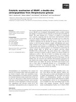
Fig. 1. Amino acid sequence alignment of TaHsp16.9C-I (Accession number S21600) and TaHsp17.8C-II (Accession number AAK51797). Identical
residues are indicated with * and highly conservative replacements indicated with colons or periods under the alignment. Regions of secondary
structure in TaHsp16.9C-I [7] are indicated above the alignment. The a-helices are displayed as open bars; b-strands as lines. The conserved
a-crystallin domain extends from b-strand 2 to b-strand 9. Regions in gray shaded boxes correspond to consensus regions within the a-crystallin
domain that show particularly high conservation between plant sHsps. Residues in the N-terminal region shown in bold correspond to sequences
conserved in all class I or class II proteins, respectively. Residues in the C-terminus in bold correspond to the conserved Basic-X-I/V-Q-I/V motif
identified by de Jong et al. [29]. Underlined residues in the C-terminus of TaHsp17.8C-II correspond to a conserved motif of class II proteins [6].
The alignment was performed using the
CLUSTAL
-
W
program (European Bioinformatics Institute; />1428 E. Basha et al. (Eur. J. Biochem. 271) Ó FEBS 2004
structure in the TaHsp16.9C-I a-crystallin domain along
with the conserved C-terminal Ôbasic-X-I/V-Q-I/VÕ motif
identified by de Jong et al. [29] show very high similarity. In
contrast, the N-terminal arms show very little similarity, as
is typical for sHsps [2,29], and each protein contains an
N-terminal consensus unique to the class I or class II plant
sHsps [6]. TaHsp17.8C-II also has a C-terminal motif
containing ProProPro that is typical of class II plant sHsps.
Thus, although these proteins would be predicted to have a
similar fold in the a-crystallin domain and to utilize the
hydrophobic residues of the basic-X-I/V-Q-I/V motif for
oligomer assembly, differences in the N-terminal arms and
flexibility of the C-terminal extension suggest that their
overall oligomeric structure may differ.
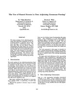
TaHsp16.9C-I, as reported previously [7], and
TaHsp17.8C-II were purified to greater than 98% homo-
geneity from E. coli cells, and the purified recombinant
proteins migrated as a single species at the expected monomer
mass on SDS/PAGE (Fig. 2A). Non-denaturing pore
exclusion PAGE and size exclusion chromatography (SEC)
were then utilized to compare the native structure of the two
sHsps. By both methods, although TaHsp16.9C-I has a
smaller monomeric size than TaHsp17.8C-II, TaHsp16.9 C-I
appears to exist as a larger oligomeric structure than the class
II sHsp. On nondenaturing PAGE TaHsp16.9C-I has an
estimated mass of 284 kDa, while TaHsp17.8C-II migrates
at 242 kDa. Similarly, on SEC the TaHsp16.9C-I peak
eluted at 10.32 min while TaHSP17.8C-II eluted later
at 10.65 min (Fig. 2C). Compared to TaHsp16.9C-I,
TaHsp17.8C-II always exhibited a fairly broad elution
profile, which could result from a variety of factors, including
oligomeric instability, nonuniformity of oligomer size or
interaction with the column matrix. As TaHsp16.9C-I
is a 12-subunit oligomer [7], these results suggest the
TaHsp17.8C-II oligomer is composed of fewer than 12
subunits.
Size of the recombinant sHsp oligomers
Although nondenaturing PAGE and SEC indicated the
class I and II sHsps have different oligomeric structures,
neither of these techniques are primary methods for size
determination. Therefore, to better understand the differ-
ence in subunit organization of these sHsps, we compared
them by EM using negative staining and by sedimentation
velocity centrifugation analysis.
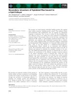
As shown in Fig. 3, purified preparations of either sHsp
appear as mostly uniform, roughly spherical particles. The
TaHsp16.9C-I particles have a diameter of approximately
11 nm, consistent with the crystal structure [7]. They are
clearly larger than the TaHsp17.8C-II particles, which
have an estimated diameter of only 9 nm. Therefore, the
relative sizes of the two oligomers are consistent with
their behavior on nondenaturing PAGE and SEC. Their
appearance is also similar to what has been observed for
class I and II sHsps from Pisum sativum (pea) [21],
suggesting conservation of the subunit stoichiometry of
class I and II oligomers.
In sedimentation velocity experiments, the sedimen-
tation distribution profile of both TaHsp16.9C-I and
TaHsp17.8C-II indicate that both proteins are associated
in a higher order structure (Fig. 4). The fact that these sHsps
exhibit nearly identical sedimentation coefficient distribu-
tions, but have different monomer molecular masses, is
consistent with the interpretation that TaHsp17.8C-II
contains either fewer subunits than TaHsp16.9C-I, or has
a more nonglobular shape. Finite element analysis of the
data [28] estimates a molecular mass of 201 kDa for
TaHsp16.9C-I and 173 kDa for TaHsp17.8C-II. These data
are consistent with a more extended shape for TaHsp16.9C-I
than for TaHsp17.8C-II and with a dodecameric organiza-
tion for TaHsp16.9C-I and an oligomer of TaHsp17.8C-II
containing 9–10 monomer units. These data are in good
agreement with the results from the other methods. In total,
the data indicate these two plant cytosolic sHsps have a
different oligomeric organization.
The wheat sHsps prevent heat-induced aggregation
of MDH
Although the TaHsp16.9C-I structure is known, there is
only one published experiment concerning its chaperone
Fig. 2. Purified TaHsp16.9C-I and TaHsp17.8C-II form high molecular
mass homo-oligomers. Purified TaHsp16.9C-I and TaHsp17.8C-II
(5 lg) were separated by SDS/PAGE (A), nondenaturing pore exclu-
sion PAGE (9 lg) (B), or size-exclusion HPLC (10 lg) (C). Gels were
stained with Coomassie blue. (C) Elution time in min is shown relative
to protein absorbance at 220 nm. Approximate elution positions of
molecular mass standards are indicated. Asterisk indicates a peak
arising from buffer absorbance.
Ó FEBS 2004 Small Hsp chaperones (Eur. J. Biochem. 271) 1429
activity, in which it was shown to form complexes with the
model substrate MDH when the two proteins are heated
together [7]. We undertook a more detailed examination of
the activity of TaHsp16.9C-I and, in comparison,
TaHsp17.8C-II. A well-established assay for sHsp chaper-
one activity is the ability to prevent heat-induced protein
aggregation as measured by light scattering [25]. In this
assay, the ratio of sHsp to substrate that is required to
suppress light scattering can be used as a measure of
effectiveness of the chaperone. Using this assay we tested the
relative activity of the two wheat proteins in suppressing
aggregation of MDH. As shown in Fig. 5, both wheat
sHsps were effective in preventing MDH aggregation as
assessed by suppression of an increase in light scattering
over time at 45 °C. TaHsp16.9C-I achieved maximum
protection of MDH at a ratio of two to three subunits of
Fig. 3. Visualization of negatively stained
TaHsp16.9C-I (left) and Ta Hsp17.8C-II (right)
by electron microscopy. Both proteins appear
as homogenous, roughly spherical particles,
but TaHsp17.8C-II is smaller. Bar indicates
50 nm for both.
Fig. 4. Sedimentation velocity analysis of TaHsp16.9C-I and
TaHsp17.8C-II. Shown is the integral distribution plot from the van
Holde–Weischet analysis of the data obtained for TaHsp16.9C-I (s)
and TaHsp17.8C-II (d). For both proteins, the majority of the sample
sedimented between 8.6 s and 9.2 s, with a small amount of smaller
association states (less than 6% of the total concentration) sedimenting
at S-values between 1 s and 8 s. Both samples also display some
amount of slightly higher order association states (< 10%) sedi-
mentingbetween9sand10s.
Fig. 5. Wheat sHsps suppress heat-induced aggregation of MDH.
Relative light scattering (320 nm) is plotted vs. time at 45 °Cfor
0.6 l
M
MDH (monomer) incubated with the indicated concentration
(in monomers) of either TaHsp16.9C-I (top) or TaHsp17.8C-II (bot-
tom). Numbers in parentheses indicate the ratio of sHsp monomer to
MDH monomer. For the IgG control, IgG was added at a weight
equivalent to 1.8 l
M
TaHsp16.9C-I or 3.0 l
M
TaHsp17.8C-II. Points
represent average and standard deviation of three replicates.
1430 E. Basha et al. (Eur. J. Biochem. 271) Ó FEBS 2004
sHsp to one subunit of MDH. In contrast, TaHsp17.8C-II
required four to five subunits of the sHsp per MDH subunit
to achieve the same level of protection. Note that no
aggregation protection is seen with the control protein IgG,
even when used at the same concentration as the highest
sHsp concentration (on a weight basis). Thus, while
both sHsps are effective chaperones, with this substrate
TaHsp16.9C-I is approximately twice as active on a subunit
(or weight) basis.
At the lowest concentrations of sHsp used (0.18 l
M
TaHsp16.9C-I and 0.60 l
M
TaHsp17.8C-II) the extent of
light scattering was actually higher than in the absence of
the sHsp. This may reflect the formation of very large
aggregates of MDH that also include the sHsp. At low sHsp
concentrations the sHsp could be bound to the MDH, but
not be abundant enough to prevent interaction of unfolded
MDH with itself.
Analysis of sHsp/MDH complexes
The differences in effectiveness of aggregation protection
between the two wheat sHsps suggests that the way in which
the sHsp and denatured substrate interact may be different.
To characterize sHsp/MDH complexes, SEC analysis
was performed after heating either TaHsp16.9C-I or
TaHsp17.8C-II (6.0 l
M
) with 1–4 l
M
MDH, yielding
sHsp/MDH ratios comparable to those used in the light-
scattering assays. MDH does not interact with either sHsp
when the proteins are incubated together at 22 °C (Fig. 6A);
the proteins elute at the predicted position based on their
individual native molecular masses. We have noted that
TaHsp17.8C-II consistently yields a lower absorbance
(A
220
)thanTaHsp16.9C-I on column chromatography.
We attribute this to either irreversible interaction of the
protein with the column, or presence of aggregates too large
to enter the column, but too small to be removed by brief
centrifugation prior to loading. MDH incubated alone at
45 °C becomes insoluble and does not enter the column
(Fig. 6B). However, a higher molecular mass species
becomes visible after heating sHsps and MDH together at
45 °C for 30 or 90 min (Fig. 6C).
At the ratio of sHsp:substrate of 6 : 1, TaHsp16.9C-I
gives full substrate protection; the size of the complex peak
increases between 30 and 90 min, and after 90 min the
MDH peak is completely depleted (Fig. 6C, upper panels;
compare to MDH peak in A). A majority of the MDH is in
complexes after the first 30 min, consistent with the rate of
aggregation protection measured by light scattering. The
same phenomenon occurs at a ratio of TaHsp16.9C-I to
MDH of 3 : 1, where full protection from aggregation is
also observed. However, as the ratio of TaHsp16.9C-I to
MDH is further decreased, the complex peak, while higher
at 30 min, actually decreases over time, and aggregated
MDH is now found in the sample pellet prior to column
loading (not shown). All of the 30 min samples show a
detectable complex peak that elutes earlier (7 min), which
may represent some kind of intermediate in complex
assembly. As the vast majority of complexes elute in the
column void volume, differences in complex size at the
different ratios cannot be estimated.
Complex formation with TaHsp17.8C-II reveals the
reduced capacity of this sHsp to protect MDH compared
to TaHsp16.9C-I (Fig. 6C, lower panels). Although com-
plete protection is observed at the sHsp:MDH ratio of
6 : 1, already at a 3 : 1 ratio the TaHsp17.8C-II/MDH
complex peak does not increase after 30 min. This result is
consistent with the light-scattering data, which showed the
sHsp was unable to fully protect MDH at this ratio. As
the amount of sHsp to substrate is further decreased, most
of the MDH is no longer found in complexes, but rather
is aggregated and removed by centrifugation prior to
sample loading on the column (not shown). Interestingly,
the sHsp itself does not appear to be complexed with the
insoluble MDH at the 2 : 1, sHsp/MDH ratio; after
90 min the free sHsp is still all accounted for in the peak
at 10.65 min. However, some sHsp is clearly lost to the
insoluble fraction, which is not loaded on the column,
when the ratio is only 1.5 : 1. This is consistent with
the maximum light-scattering values observed for
TaHsp17.8C-II/MDH at a 1 : 1 ratio, which were higher
than those for MDH alone. A potential intermediate-sized
species of complex is also evident at 6.5 min in most of
the samples. In total, as observed by light scattering,
substrate denaturation and aggregation are time-depend-
ent, the sHsps can be saturated with substrate, and
TaHsp17.8C-II is less effective in protecting MDH
compared to TaHsp16.9C-I.
To visualize the sHsp substrate complexes directly,
samples incubated as for the SEC analysis at 45 °Cfor
30 min were observed by EM and negative staining
(Fig. 7). Note that because samples were centrifuged prior
to application to the grid, only soluble material was
observed. Two consistent observations arose from this
analysis. First, complexes formed at an sHsp to substrate
ratio that was sufficient, or higher, than that required for
full protection (as determined in the light-scattering
experiments) were the most uniform. At the ratio
required for full protection, complexes formed with
TaHsp16.9C-I (3 : 1 ratio) had an average diameter of
54 nm, while complexes formed with TaHsp17.8C-II
(6 : 1 ratio) were somewhat larger (60 nm). Second,
complex regularity decreased as the amount of sHsp to
substrate decreased below the level of full protection for
either sHsp, with the irregular complexes looking more
like aggregates composed of smaller particles ( 40–
46 nm), as seen in the 2 : 1 and lower ratio mixtures.
MDH heated alone and applied to the grid before
centrifugation was a large amorphous mass, while after
centrifugation no proteinaceous material could be
observed (not shown).
Ta
Hsp17.8 C-II, but not
Ta
Hsp16.9 C-I, suppresses
aggregation of Luc
The above data indicate that TaHsp16.9C-I is more
effective in preventing aggregation of MDH than is
TaHsp17.8C-II. To test if this difference in chaperone
activity is the same with another heat sensitive substrate,
aggregation protection of firefly luciferase (Luc) was
examined. A simple differential centrifugation assay was
employed to determine if either wheat sHsp could prevent
insolubilization of Luc during heating. This assay was
employed in place of the spectrophotometric assay used
for MDH because of difficulties with adhesion of Luc to
Ó FEBS 2004 Small Hsp chaperones (Eur. J. Biochem. 271) 1431
the cuvette walls. As shown in Fig. 8A, Luc incubated
with TaHsp17.8C-II at 42 °C for 15 min was recovered
almost exclusively in the soluble fraction, indicating that
TaHsp17.8C-II was able to protect Luc from heat-induced
insolubilization. A similar weight of IgG gave no protec-
tion (not shown). Surprisingly, when Luc was incubated
with TaHsp16.9C-I, virtually all of the Luc was found in
the pellet fraction, while the sHsp remained soluble. Thus,
in contrast to results with MDH, TaHsp16.9C-I is a less
effective chaperone with Luc than is TaHsp17.8C-II. Note
that a higher ratio of sHsp to substrate is required to
protect Luc as compared to MDH. In parallel to these
observations, TaHsp17.8C-II formed a complex when
heated with Luc, which could be observed by SEC
(Fig. 8B). No such complex formed with TaHsp16.9C-I
(not shown). Thus, these two sHsps do not behave
equivalently with all substrates.
Denatured Luc bound to
Ta
Hsp17.8C-II can be
reactivated in a cell free lysate
The effectiveness of sHsp chaperone activity can also be
assessed by the ability of an sHsp to maintain substrate in
a state from which it can be refolded by ATP-dependent
Fig. 6. TaHsp16.9C-I is more effective in
forming complexes with MDH than is
TaHsp17.8C-II. All samples were separated by
SEC, and absorbance (220 nm) monitored
over elution time. Samples were centrifuged to
remove insoluble material prior to loading on
the column. (A) sHsps (6 l
M
)andMDH
(2 l
M
) incubated together at room tempera-
ture. (B) MDH heated alone. (C) High
molecular mass complexes formed between
sHsps and MDH after heating at 45 °Cfor
30 or 90 min. Concentrations were 6 l
M
sHsp
subunits for all samples, with from 1 to 4 l
M
MDH as indicated by the sHsp/MDH ratio.
Asterisk indicates a buffer peak.
1432 E. Basha et al. (Eur. J. Biochem. 271) Ó FEBS 2004
chaperones [18]. To test if the Luc protected by
TaHsp17.8C-II was in a conformation capable of reacti-
vation, the TaHsp17.8C-II/Luc complexes, or Luc heat-
denatured in the presence of TaHsp16.9C-I or an
equivalent weight of IgG, were incubated with reticulocyte
lysate plus or minus ATP (Fig. 8C). Reactivation of Luc
bound to TaHsp17.8C-II was highly efficient, achieving up
to 70% reactivation in less than 1 h in the presence of
ATP. As expected, TaHsp16.9C-I supported less than
15% reactivation of Luc because most of the Luc was
insoluble and not associated with the sHsp. Controls using
IgG, or in the absence of ATP, showed 5% or less
reactivation. Thus, formation of TaHsp17.8C-II/Luc com-
plexes is correlated with ability to support substrate
reactivation.
Discussion
Our data provide the first detailed analysis of the in vitro
chaperone activity of TaHsp16.9C-I, the only eukaryotic
sHsp for which a high resolution structure is available.
Surprisingly, although this sHsp effectively protects MDH
from insolubilization, it did not interact with a second
substrate, Luc, under the conditions tested. In parallel, we
analyzed a related wheat sHsp, TaHsp17.8C-II, which
proved to be less effective in protecting MDH, but
interacted well with Luc, both preventing aggregation and
supporting refolding. Thus, these results document the first
clear example of apparent substrate specificity for sHsps.
TaHsp16.9C-I and TaHsp17.8C-II represent two distinct
classesofcytosolicsHspsfromplants(classIandclassII),
Fig. 7. MDH/sHsp complexes visualized by
electron microscopy and negative staining.
TaHsp16.9C-I or TaHsp17.8C-II (6 l
M
sub-
units) heated with different concentrations of
MDH ranging from 6 to 1 subunit sHsp: 1
subunit substrate. Complexes were formed at
45 °C for 30 min then centrifuged and loaded
on the EM grids for visualization at magni-
fication of 820 000. Bar indicates 110 nm
for all.
Ó FEBS 2004 Small Hsp chaperones (Eur. J. Biochem. 271) 1433
estimated to have diverged at least 400 million years ago [6].
Comparing these two wheat proteins, amino acid sequence
identity is 33% overall, and 46% for the a-crystallin
domain. In addition to sequence differences, our analysis
of the purified recombinant proteins indicates that they
assemble into different quaternary structures. Solution
methods, from this work and previous studies, and a crystal
structure [7] demonstrate that native TaHsp16.9C-I is
dodecameric. In contrast, by EM, SEC and sedimentation
velocity experiments, the class II TaHsp17.8C-II was found
to form regular, but smaller oligomers, with an estimated
nine to ten subunits. Members of these same two sHsp
classes have also been characterized from Pisum sativum
(pea), PsHsp18.1C-I and PsHsp17.7C-II [21]. EM pictures
of the purified pea oligomers are remarkably similar to those
of the wheat proteins, with the class I sHsp having a
diameter of 10–11 nm and the class II protein a slightly
smaller diameter, despite the larger subunit size.
PsHsp18.1C-I was also found to be dodecameric by
sedimentation equilibrium analysis, like the homologous
wheat TaHsp16.9C-I (amino acid sequence identity/simi-
larity 68/75% throughout, and 80/86% in the a-crystallin
domain). However, sedimentation equilibrium analysis of
PsHsp17.7C-II estimated an oligomer of 11.3 ± 0.5 sub-
units [21], larger than our estimate for the wheat class II
protein. Therefore, it is unclear whether the stoichiometry of
oligomeric assembly is the same for all plant class II
proteins. However, we would predict that the assembly
should comprise an even number of subunits, based on the
dimeric building block of TaHsp16.9C-I, which involves
features conserved in the class II proteins as well [1,6].
Regardless of absolute subunit numbers, class I and II sHsp
oligomers clearly have distinct modes of assembly, as also
reflected in the fact that these two classes of sHsps do not
coassemble into mixed oligomers in vivo or in vitro, although
class I or II sHsps will coassemble into normal oligomers
when mixed with class I or II sHsps, respectively, from
different plant species ([7,23], and E. Basha & E. Vierling,
unpublished observation). Distinct assemblies of different
sHsps in the same cell have also been observed in humans
and bacteria [30,31], suggesting there are different,
conserved roles for specific sHsps.
Both TaHsp16.9C-I and TaHsp17.8C-II were able to
suppress the heat-dependent aggregation of MDH. How-
ever, TaHsp16.9C-I suppresses MDH aggregation com-
pletely at a stoichiometry of 2–3 subunits sHsp to 1
subunit MDH. In contrast, complete suppression of
MDH aggregation by TaHsp17.8C-II required the higher
ratio of 4–5 sHsp subunits:1 MDH subunit. From
previous work, PsHsp18.1C-I was found to be somewhat
more effective in the aggregation protection of MDH than
either of the wheat sHsps, suppressing MDH aggregation
at a ratio of 2 : 1, sHsp subunit:MDH [15]. The pea class
II protein was not tested with MDH, but when tested with
citrate synthase, it was more than sixfold less effective
than the PsHsp18.1C-I [21]. Thus, using these in vitro
assays with two different substrates, class II proteins have
proven to be less effective as chaperones than class I
proteins, consistent with some type of substrate specificity
for these two classes of proteins.
At the lowest concentrations of sHsp used (0.18 l
M
TaHsp16.9C-I and 0.6 l
M
TaHsp17.8C-II to 1 l
M
MDH)
the extent of light scattering was actually higher than in the
absence of the sHsp. This may reflect the formation of very
large aggregates of MDH that also include the sHsp, as
evidenced by the loss of sHsp from the SEC profile under
these conditions. At this low sHsp concentration, the sHsp
might be bound to the MDH but not be abundant enough
to prevent extensive interaction of unfolded MDH with
itself. Bova et al. [32] noticed such an effect using
aB-crystallin containing the R120G mutation linked to
desmin-related myopathy. One of the authors’ interpreta-
tions for the effect was a possible change in the availability
of substrate binding sites resulting in a less efficient
chaperone. When we used a low concentration of wheat
sHsps, therefore providing fewer binding sites, we may have
Fig. 8. TaHsp17.8C-II, but not TaHsp16.9C-I, maintains Luc in a soluble form during heating and facilitates Luc reactivation. (A) Coomassie blue
stained SDS/PAGE of soluble (S) and pellet (P) fractions prepared after heating 12 l
M
TaHsp16.9 or 17.8 with 1.0 l
M
Luc at 42 °C for 15 min.
(B) SEC analysis of 12 l
M
TaHsp17.8C-II plus 1.0 l
M
Luc either before (22 °C) or after heating at 42 °Cfor15min(42°C). Approximate elution
times of molecular mass markers are indicated. (C) Time course of Luc reactivation in reticulocyte lysate. (d) TaHsp17.8C-II + ATP; (j)
TaHsp17.8 C-II – ATP; (m) TaHsp16.9C-I + ATP; (h)Hsp16.9C-I–ATP;(s)IgG+ATP.
1434 E. Basha et al. (Eur. J. Biochem. 271) Ó FEBS 2004
imitated the same effect of the R120G mutation in
aB-crystallin.
SEC analysis showed that the complexes formed between
the two wheat sHsps and MDH are quite large. Working
with PsHsp18.1C-I, Lee et al. [15] found complexes with
MDH were much smaller than those formed with the wheat
sHsps, although the size observed by SEC was dependent
on the substrate concentration as well as the denaturation
temperature. The less efficient aggregation protection
obtained with the wheat sHsps (on a molar basis of sHsp:
substrate) compared to PsHsp18.1C-I suggests that the
MDH aggregates more rapidly than it can form stabilizing
interactions with the wheat sHsps. It is interesting that there
isalwaysafreepeakofsHsponSEC,evenwhensome
of the substrate has precipitated. The free sHsps could
still have a role in protection, by cycling on and off the
aggregates, as suggested by both Lindner et al.[33]and
Friedrich et al. [12].
The decrease in SEC complex peak height and the
eventual loss of sHsp at the highest substrate concentrations
is due to the insolubility of the sHsps bound to excess
substrate (as indicated by SDS PAGE; not shown).
Transition of sHsps to an insoluble fraction is observed
in vivo in many organisms [15,34,35], and may also result
from overloading of the sHsp with substrates. Experiments
in E. coli suggest that the chaperone ClpB is necessary
to resolubilize sHsp/substrate complexes in vivo [36], and
in vitro, protein aggregates containing sHsps are more
effective ClpB substrates than aggregates without sHsps
[19]. Therefore, even when complexed in an insoluble
fraction, the sHsps may confer an advantage for recovery of
protein activity in the cell.
We also observed by EM that at sHsp:substrate ratios
sufficient for complete substrate protection, sHsp/substrate
complexes had dimensions of 56 and 60 nm for
TaHsp16.9C-I and TaHsp17.8C-II, respectively. As repor-
ted previously, and consistent with the SEC comparisons,
PsHsp18.1C-I complexes with MDH were smaller on
average, being frequently 16 to 20 nm [15]. Complex
morphology also changed with decreasing sHsp to substrate
ratio, with much more heterogeneous particles and aggre-
gates of particles observed. These results are at odds with a
report by Stromer et al. [37] in which complex morphology
was reported to be dictated by substrate identity and
independent of the identity of the sHsp, although different
sHsp:substrate ratios were not observed by EM. It is
interesting that 40 nm particles, termed heat shock
granules, are found after heat stress in plants in vivo
[34,38]. To what extent the in vitro-formed complexes
resemble in vivo heat shock granules remains to be
determined.
Surprisingly, while TaHsp16.9C-I was more effective
than TaHsp17.8C-II in protecting MDH, TaHsp16.9C-I
showed no ability to protect Luc under the conditions
tested. In contrast, TaHsp17.8C-II protected Luc from
aggregation and formed high molecular mass complexes
with Luc. We also showed that TaHsp17.8C-II supported
Luc refolding using rabbit reticulocyte lysate as a source
of ATP-dependent eukaryotic chaperones. However, the
inability of TaHsp16.9C-I to protect Luc is not true for all
classIsHsps.PsHsp18.1C-I has been shown to protect
Luc with the same effectiveness as TaHsp17.8C-II and to
support Luc refolding [12,15,18]. This fact indicates that the
differences in sHsp–substrate interactions must be more
subtle than the differences between class I and II sHsps in
primary sequence or quaternary structure. TaHsp16.9C-I
and PsHsp18.1C-I show 80% identity and 86% similarity in
the conserved C-terminal a-crystallin domain. In contrast
they show only 41% identity and 50% similarity in the
N-terminal arm, suggesting substrate specificity is deter-
mined by the N-terminal arm. The N-terminus of
PsHsp18.1.C-I was also implicated in substrate interactions
in bis-ANS binding experiments [15]. As it is proposed that
substrate binding and protection involves oligomer dissoci-
ation and some type of reassociation to form the large sHsp/
substrate complexes [7,13], it must also be considered that
overall differences in oligomer stability and/or the kinetics
of oligomer dissociation, rather than specific sequence
differences, dramatically affect sHsp interactions with
different substrates.
Although to date sHsps have been ascribed little substrate
specificity, it is clear from this study and previous work
[21,37] that the effectiveness of substrate protection, on a
molar basis, by different sHsps can vary significantly under
the same conditions. A difference in effectiveness is obvious
to the extreme with TaHsp16.9C-I, which fails to interact
with Luc. It should be considered that minor differences
in the ratio of sHsp/substrate required for maximal
substrate protection are potentially functionally important
differences in the cellular environment and are essentially an
indication of substrate specificity. Full understanding of
sHsp substrate interactions will require not only considera-
tion of substrate binding sites and binding interactions, but
also the dynamics of the sHsp oligomer and the kinetics of
substrate aggregation.
Acknowledgements
This work was supported by National Institutes of Health grant RO1-
GM42762, USDA-NRICGP and University of Arizona Experiment
Station Funds, and American Cancer Society Faculty Research Award
#FRA-420 to E. V. G. J. L. was a recipient of a National Institutes of
Health Postdoctoral Fellowship. B. D. was supported by NSF BB1-
9974819. We thank Drs Kim Giese and Kenneth Friedrich for critical
reading of the manuscript.
References
1. van Montfort, R.L.M., Slingsby, C. & Vierling, E. (2002)
Structure and function of the small heat shock protein/a-crystallin
family of molecular chaperones. In Protein Folding in the
Cell (Horwich, A.L., ed.), pp. 105–156. Academic Press,
New York.
2. deJong, W.W., Leunissen, J.A. & Vooter, C.E. (1993) Evolution of
the a-crystallin/small heat-shock protein family. Mol. Biol. Evol.
10, 103–126.
3. Waters, E.R., Lee, G.J. & Vierling, E. (1996) Evolution, structure
and function of the small heat shock proteins in plants. J. Exp.
Bot. 47, 325–338.
4. Scharf, K D., Siddique, M. & Vierling, E. (2001) The expanding
family of small Hsps and other proteins containing an a-crystallin
domain. Cell Stress Chaperones 6, 225–237.
5. Waters, E.R. & Vierling, E. (1999) Chloroplast small heat shock
proteins: Evidence for atypical evolution of an organelle-localized
protein. Proc.NatlAcad.Sci.USA96, 14394–14399.
Ó FEBS 2004 Small Hsp chaperones (Eur. J. Biochem. 271) 1435
6. Waters, E.R. & Vierling, E. (1999) The diversification of plant
cytosolic small heat shock proteins preceded the divergence of
mosses. Mol. Biol. Evol. 16, 127–139.
7. van Montfort, R.L.M., Basha, E., Friedrich, K.L., Slingsby, C. &
Vierling, E. (2001) Crystal structure and assembly of a eukaryotic
small heat shock protein. Nat. Struct. Biol. 8, 1025–1030.
8. Kim, K.K., Kim, R. & Kim, S.H. (1998) Crystal structure of a
small heat-shock protein. Nature 394, 595–599.
9. Sobott,F.,Benesch,J.L.P.,Vierling,E.&Robinson,C.V.(2002)
Subunit exchange of multimeric protein complexes – Real-time
monitoring of subunit exchange between small heat shock proteins
by using electrospray mass spectrometry. J. Biol. Chem. 277,
38921–38929.
10. Bova,M.P.,Mchaourab,H.S.,Han,Y.&Fung,B.K.K.(2000)
Subunit exchange of small heat shock proteins – Analysis of
oligomer formation of aA-crystallin and Hsp27 by fluorescence
resonance energy transfer and site-directed truncations. J. Biol.
Chem. 275, 1035–1042.
11. Bova, M.P., Huang, Q.L., Ding, L.L. & Horwitz, J. (2002) Sub-
unit exchange, conformational stability, and chaperone-like
function of the small heat shock protein 16.5 from Methanococcus
jannaschii. J. Biol. Chem. 277, 38468–38475.
12. Friedrich, K.L., Giese, K., Buan, N.R. & Vierling, E. (2004)
Interactions between small heat shock protein subunits and sub-
strate in small heat shock protein/substrate complexes. J. Biol.
Chem. 279, 1080–1089.
13. Haslbeck, M., Walke, S., Stromer, T., Ehrnsperger, M., White,
H.E., Chen, S.X., Saibil, H.R. & Buchner, J. (1999) Hsp26: a
temperature-regulated chaperone. EMBO J. 18, 6744–6751.
14. Ehrnsperger, M., Gra
¨
ber,S.,Gaestel,M.&Buchner,J.(1997)
Binding of non-native protein to Hsp25 during heat shock creates
a reservoir of folding intermediates for reactivation. EMBO J. 16,
221–229.
15. Lee, G.J., Roseman, A.M., Saibil, H.R. & Vierling, E. (1997) A
small heat shock protein stably binds heat-denatured model sub-
strates and can maintain a substrate in a folding-competent state.
EMBO J. 16, 659–671.
16. Sharma, K.K., Kaur, H. & Kester, K. (1997) Functional elements
in molecular chaperone a-crystallin: Identification of binding sites
in aB-crystallin. Biochem. Biophys. Res. Commun. 239, 217–222.
17. Sharma, K.K., Kumar, G.S., Murphy, A.S. & Kester, K. (1998)
Identification of 1,1¢-Bi(4-anilino)naphthalene-5,5¢-disulfonic acid
binding sequences in a-crystallin. J. Biol. Chem. 273, 15474–15478.
18. Lee, G.J. & Vierling, E. (2000) A small heat shock protein
cooperates with heat shock protein 70 systems to reactivate a heat-
denatured protein. Plant Physiol. 122, 189–197.
19. Mogk, A., Schlieker, C., Friedrich, K.L., Scho
¨
feld, H.J., Vierling,
E. & Bukau, B. (2003) Refolding of substrates bound to small
Hsps relies on a disaggregation reaction mediated most efficiently
by ClpB/DnaK. J. Biol. Chem. 278, 31033–31042.
20. Veinger, L., Diamant, S., Buchner, J. & Goloubinoff, P. (1998)
The small heat-shock protein IbpB from Escherichia coli stabilizes
stress-denatured proteins for subsequent refolding by a multi-
chaperone network. J. Biol. Chem. 273, 11032–11037.
21. Lee, G.J., Pokala, N. & Vierling, E. (1995) Structure and in vitro
molecular chaperone activity of cytosolic small heat shock pro-
teins from pea. J. Biol. Chem. 270, 10432–10438.
22. Sun, W.N., Bernard, C., Van de Cotte, B., Van Montagu, M. &
Verbruggen, N. (2001) At-HSP17.6A, encoding a small
heat-shock protein in Arabidopsis, can enhance osmotolerance
upon overexpression. Plant J. 27, 407–415.
23. Helm, K.W., Lee, G.J. & Vierling, E. (1997) Expression and native
structure of cytosolic class II small heat shock proteins. Plant
Physiol. 114, 1477–1485.
24. Basha, E. & Vierling, E. (2003) Cloning and sequencing of a new
cytosolic class II small heat shock protein from wheat. Egypt. J.
Biotechn. 13, 103–113.
25. Lee, G.J. & Vierling, E. (1998) Expression, purification, and mo-
lecular chaperone activity of plant recombinant small heat shock
proteins. Methods Enzymol. 290, 350–365.
26. Helm, K.W., LaFayette, P.R., Nagao, R.T., Key, J.L. & Vierling,
E. (1993) Localization of small heat shock proteins to the higher
plant endomembrane system. Mol. Cell. Biol. 13, 238–247.
27. Van Holde, K. & Weischet, W.O. (1978) Boundary analysis of
sedimentation-velocity experiments with monodisperse and pau-
cidisperse solutes. Biopolymers 17, 1387–1403.
28. Demeler, B. & Saber, H. (1998) Determination of molecular
parameters by fitting sedimentation data to finite-element solu-
tions of the Lamm equation. Biophys. J. 74, 444–454.
29. De Jong, W.W., Caspers, G J. & Leunissen, J.A.M. (1998)
Genealogy of the a-crystallin-small heat-shock protein super-
family. Int. J. Biol. Macromol. 22, 151–162.
30. Studer, S. & Narberhaus, F. (2000) Chaperone activity and homo-
and hetero-oligomer formation of bacterial small heat shock
proteins. J. Biol. Chem. 275, 37212–37218.
31. Sugiyama, Y., Suzuki, A., Kishikawa, M., Akutsu, R., Hirose, T.,
Waye,M.M.,Tsui,S.K.,Yoshida,S.&Ohno,S.(2000)Muscle
develops a specific form of small heat shock protein complex
composed of MKBP/HSPB2 and HSPB3 during myogenic dif-
ferentiation. J. Biol. Chem. 275, 1095–1104.
32. Bova, M.P., Yaron, O., Huang, Q.L., Ding, L.L., Haley, D.A.,
Stewart, P.L. & Horwitz, J. (1999) Mutation R120G in aB-crys-
tallin, which is linked to a desmin-related myopathy, results in an
irregular structure and defective chaperone-like function. Proc.
NatlAcad.Sci.USA96, 6137–6142.
33.Lindner,R.A.,Treweek,T.M.&Carver,J.A.(2001)The
molecular chaperone a-crystallin is in kinetic competition with
aggregation to stabilize a monomeric molten-globule form of
a-lactalbumin. Biochem. J. 354, 79–87.
34. Nover, L., Scharf, K D. & Neumann, D. (1989) Cytoplasmic heat
shock granules are formed from precursor particles and are
associated with a specific set of mRNAs. Mol. Cell. Biol. 9,
1298–1308.
35. Arrigo, A P., Suhan, J.P. & Welch, W. (1988) Dynamic changes
in the structure and intracellular locale of the mammalian low-
molecular-weight heat shock protein. Mol. Cell. Biol. 8, 5059–
5071.
36. Mogk, A., Deuerling, E., Vorderwu
¨
lbecke, S., Vierling, E. &
Bukau, B. (2003) Small heat shock proteins, ClpB and the DnaK
system form a functional triade in reversing protein aggregation.
Mol. Microbiol. 50, 585–595.
37. Stromer, T., Ehrnsperger, M., Gaestel, M. & Buchner, J. (2003)
Analysis of the interaction of small heat shock proteins with
unfolding proteins. J. Biol. Chem. 278, 18015–18021.
38. Nover, L., Scharf, K D. & Neumann, D. (1983) Formation of
cytoplasmic heat shock granules in tomato cell cultures and leaves.
Mol. Cell. Biol. 3, 1648–1655.
1436 E. Basha et al. (Eur. J. Biochem. 271) Ó FEBS 2004