Báo cáo khoa học: to 3a-hydroxysteroid dehydrogenase from Pseudomonas sp. B-0831 using fluorescence stopped-flow procedures pptx
Bạn đang xem bản rút gọn của tài liệu. Xem và tải ngay bản đầy đủ của tài liệu tại đây (285.03 KB, 7 trang )
Transient-phase kinetic studies on the nucleotide binding
to 3a-hydroxysteroid dehydrogenase from
Pseudomonas
sp.
B-0831 using fluorescence stopped-flow procedures
Shigeru Ueda
1
, Masayuki Oda
2
, Shigeyuki Imamura
1
and Masatake Ohnishi
2
1
Department Diagnostics Research and Development, Division of Fine Chemicals and Diagnostics, Asahi Kasei Pharma Corporation,
Shizuoka, Japan;
2
Department of Cellular Macromolecule Chemistry, Graduate School of Agriculture, Kyoto Prefectural University,
Kyoto, Japan
The dual nucleotide cofactor-specific enzyme, 3a-hydroxy-
steroid dehydrogenase (3a-HSD) from Pseudomonas sp.
B-0831, is a member of the short-chain dehydrogenase/
reductase (SDR) superfamily. Transient-phase kinetic stud-
ies using the fluorescence stopped-flow method were con-
ducted with 3a-HSD to characterize the nucleotide binding
mechanism. The binding of oxidized nucleotides, NAD
+
,
NADP
+
and nicotinic acid adenine dinucleotide
(NAAD
+
), agreed well with a one-step mechanism, while
that of reduced nucleotide, NADH, showed a two-step
mechanism. This difference draws attention to previous
characteristic findings on rat liver 3a-HSD, which is a
member of the aldo-keto reductase (AKR) superfamily.
Although functionally similar, AKRs are structurally dif-
ferent from SDRs. The dissociation rate constants
associated with the enzyme–nucleotide complex formation
were larger than the k
cat
values for either oxidation or
reduction of substrates, indicating that the release of cofac-
tors is not rate-limiting overall. It should also be noted that
k
cat
for a substrate, cholic acid, with NADP
+
was only 6%
of that with NAD
+
, and no catalytic activity was detectable
with NAAD
+
, despite the similar binding affinities of
nucleotides. These results suggest that a certain type of
nucleotide can modulate nucleotide-binding mode and fur-
ther the catalytic function of the enzyme.
Keywords: aldo-keto reductase superfamily; nucleotide-
binding; fluorescence stopped-flow method; 3a-hydroxyster-
oid dehydrogenase; short-chain dehydrogenase/reductase
superfamily.
The NAD(P)
+
-dependent enzyme, 3a-hydroxysteroid
dehydrogenase (3a-HSD), catalyzes the reversible intercon-
version of hydroxy and oxo groups at position 3 of the
steroid nucleus, and has been found in many mammalian
cells and microorganisms [1,2]. Prokaryotic 3a-HSDs have
been described in Eubacterium lentum [3], Clostridium
perfringens [4], Pseudomonas putida [5], Comamonas (Pseu-
domonas) testosteroni ATCC11996 [6–9], and Pseudomonas
sp. B-0831 [10]. Despite similar substrate specificities,
eucaryotic and prokaryotic 3a-HSDs belong to two differ-
ent protein superfamilies: viz, the eucaryotic and prokary-
otic enzymes, respectively, belonging to the aldo-keto
reductase (AKR) (EC 1.1.1.213) and the short-chain dehy-
drogenase/reductase (SDR) superfamilies (EC 1.1.1.50)
[11–14]. Although structurally different, both HSDs are
functionally similar; the AKRs are monomeric and have an
a/b-barrel fold, whereas the SDRs are dimeric or tetrameric
and contain a Rossmann nucleotide-binding fold.
We have previously cloned 3a-HSD from Pseudomonas
sp. strain B-0831 and expressed it in Escherichia coli [10].
The SDS/PAGE and gel-filtration analyses revealed that
this enzyme forms a homodimer comprised of two 25 kDa
monomer proteins. The amino acid sequence shares about
50% identity with that of Comamonas (Pseudomonas)
testosteroni ATCC11996 [15], and contains two motifs
common to SDRs, the Gly-X-X-X-Gly-X-Gly cofactor-
binding and Tyr-X-X-X-Lys substrate-binding motifs.
Because 3a-HSD from C. testosteroni can catalyze not
only the oxidoreduction at position 3 of the steroid
nucleus but also carbonyl reduction of a variety of
nonsteroidal aldehydes and ketones, the enzyme was
named as 3a-hydroxysteroid dehydrogenase/carbonyl
reductase (3a-HSD/CR) [15,16]. In a similar manner,
P. sp. B-0831-derived 3a-HSD also shows carbonyl reduc-
tase activity [17]. Different from the NAD
+
-dependent
3a-HSD/CR, P. sp. B-0831-derived 3a-HSD has the
ability to use not only NAD
+
but also NADP
+
.In
clinical diagnostics, the enzyme has been used for the
measurement of total bile acids in serum [18]. Further-
more, a highly sensitive and unique enzyme cycling
method for total bile acids assay has been developed
Correspondence to M. Oda, Graduate School of Agriculture,
Kyoto Prefectural University, 1–5, Shimogamo Nakaragi-cho,
Sakyo-ku, Kyoto 606–8522, Japan.
Fax: + 81 75 7035673, Tel.: + 81 75 703 5673,
E-mail:
Abbreviations:3a-HSD, 3a-hydroxysteroid dehydrogenase; AKR,
aldo-keto reductase superfamily; CR, carbonyl reductase; NAAD
+
,
nicotinic acid adenine dinucleotide; SDR, short-chain dehydrogenase/
reductase superfamily.
Enzyme:3a-hydroxysteroid dehydrogenase (EC 1.1.1.213,
EC 1.1.1.50).
(Received 3 February 2004, revised 5 March 2004,
accepted 15 March 2004)
Eur. J. Biochem. 271, 1774–1780 (2004) Ó FEBS 2004 doi:10.1111/j.1432-1033.2004.04089.x
using this B-0831 enzyme in the presence of excessive
thio-NAD
+
(NAD
+
analogue) and NADH, which are
commercially available reagents [19].
Rat liver 3a-HSD, the first HSD to be assigned to the
AKRs, is involved in bile acid biosynthesis, and participates
in the inactivation of circulating steroid hormones such as
androgens, progestins and glucocorticoids [20]. Its nucleo-
tide cofactor-binding mechanisms have been extensively
studied [21–27]. This enzyme shows NADP
+
preference
and follows an ordered bi-bi reaction mechanism with
nucleotide binding first. The binding kinetics of NADP(H)
by the fluorescence stopped-flow method showed a two-step
mechanism which consists of fast formation of a loose
complex followed by slow formation of a tightly bound
complex [26], and the dissociation of NADP(H) is not rate-
limiting. The latter finding discriminates the rat enzyme
from other AKRs, such as human and pig muscle aldose
reductases [28,29].
As well as rat liver enzyme, 3a-HSD/CR from C. testo-
steroni ATCC11996 follows an ordered bi-bi mechanism
with pyridine nucleotide binding first and dissociation last [7].
In contrast to 3a-HSDs in AKRs, however, little is known
about the nucleotide-binding mechanism of 3a-HSD in
SDRs. In terms of nucleotide binding to the enzyme, the
nicotinamide ring adopts syn conformation in SDRs while it
adopts anti conformation in AKRs, exhibiting the opposite
stereospecificity of hydride transfer [27]. Therefore, it is of
great interest to elucidate the mechanism of the nucleotide
binding to 3a-HSD from P. sp. B-0831 as a member of SDRs.
In the present study, we analysed transient-phase kinetics
of nucleotide binding to bacteria-derived 3a-HSD by
fluorescence stopped-flow measurements for the first time.
Transient-phase kinetics can distinguish between the for-
mation and decay of individual complexes on either one- or
multiple-step reaction pathways. Application of this method
to 3a-HSD revealed that the reaction mechanism depends
on the type of nucleotides, and is different from that of
rat liver 3a-HSD. In addition to the unique features of
the kinetic mechanism, nucleotide preference of this enzyme
could be elucidated in comparison with the functional and
structural information of other SDRs and AKRs, such as
C. testosteroni 3a-HSD/CR and rat liver 3a-HSD.
Materials and methods
Materials
NAD
+
,NADP
+
, NAAD
+
, NADH, and NADPH (Ori-
ental Yeast Co., Ltd in Japan), steroids (Sigma Chem. Co.)
and other chemicals were of the highest commercial quality
available. Recombinant 3a-HSD from Pseudomonas sp.
strain B-0831 was expressed in E. coli and purified as
described previously [10]. The protein purity was deter-
mined to be over 95% by SDS/PAGE analysis, and the
protein concentration was determined by the method of
Lowry et al. [30].
Steady-state kinetic study
Assays were carried out at 37 °Cor15°Cin40m
M
Tris/
HCl (pH 8.5), in the presence of 1 m
M
NAD(P)
+
, 0.025%
nitrotetrazolium blue (NTB), 0.4% Triton X-100, and 2.5
units/mL diaphorase (Asahi Kasei) for oxidation reaction,
and in the presence of 0.3 m
M
NAD(P)H for reduction
reaction [10]. In the assay for substrate specificity, various
substrate concentrations were used for the determination of
K
m
and k
cat
. To determine K
m
for the NAD(P)
+
and
NAD(P)H binding, 1 m
M
cholic acid and 0.8 m
M
dehydro-
cholic acid were used, respectively. The reaction was
initiated by adding the enzyme, and terminated by adding
2 mL of 0.5% SDS after incubation for 5 min. The K
m
and
k
cat
values for oxidation reaction were determined by
measuring the formation of formazan dye at 550 nm, and
those for reduction reaction were determined by measuring
the decrease of NAD(P)H at 340 nm, using a Shimadzu
UV-2200 spectrophotometer. In the inhibition study by
NAAD
+
,1.0m
M
cholic acid (substrate) was used in the
presence of NAD
+
with concentration ranging from 5.0 l
M
to 200 l
M
,and2.0m
M
NAAD
+
as an inhibitor. The K
m
and K
i
values were calculated by the nonlinear least squares
method using the Taylor expansion [31].
Fluorescence titration measurements
The nucleotide binding to 3a-HSD was measured by
monitoring the quenching of intrinsic enzyme fluorescence
upon incremental addition of nucleotides. Emission spectra
(300–500 nm) were recorded on a JASCO Corporation
FP-777 fluorescence spectrophotometer at 280-nm excita-
tion. All titrations were carried out in 3-mL volumes with
0.49 l
M
of the enzyme in 50 m
M
Tris/HCl (pH 8.5) at
15 °C. The total volume change due to the addition of
nucleotide was less than 2%. The K
d
value was determined
by the nonlinear least squares method [31].
Stopped-flow kinetic study
The time course of fluorescence intensity caused by nucleo-
tide binding to 3a-HSD was monitored through cut-off filters
(Toshiba Kasei Kogyo), UV-31 with 50% transmittance
at 310 nm for oxidized nucleotides and UV-42 with 50%
transmittance at 420 nm for reduced nucleotides, equipped
with an Otsuka Electronics RA-401 stopped-flow apparatus
at 280-nm excitation by a 200 W D
2
lamp at 15 °C [32]. Using
a quartz cell (inner diameter: 2 mm), the dead time of the
apparatus was determined as 1.3 ms under the experimental
conditions described in a previous study [33]. In order to
determine the reliable kinetic range in the present experi-
ments, we measured the reduction rate of 2,6-dichloro-
phenol-indophenol by ascorbate, and confirmed the linearity
of the apparent first-order rate constant, k
app
, within the
range of 0–400 s
)1
in the Guggenheim plot [34].
Various concentrations of nucleotide solutions and
3a-HSD in 50 m
M
Tris/HCl (pH 8.5) were introduced into
the stopped-flow apparatus by means of a separate syringe.
The k
app
value for each nucleotide concentration was
determined in triplicate.
Data analysis of rapid reaction
The apparent first-order rate constant, k
app
,wasdetermined
from the Guggenheim plot. When the plots followed the
linear dependence of k
app
against initial concentration of
nucleotide, Eqn (1) was adopted:
Ó FEBS 2004 Transient kinetics of nucleotide binding to 3a-HSD (Eur. J. Biochem. 271) 1775
E þ Nuc
À!
k
þ1
À
k
À1
E-Nuc ð1Þ
where E is the enzyme, Nuc is the nucleotide, E-Nuc is the
enzyme–nucleotide complex, and k
+1
, k
)1
are the transient-
phase kinetic rate constants. On the condition that the initial
concentration of nucleotide, [Nuc], is greater than E, k
app
is thus expressed as Eqn (2) [35]:
k
app
¼ k
þ1
½Nucþk
À1
ð2Þ
The values of k
+1
and k
)1
were determined from the slope
and the intercept of the plot of [Nuc] vs. k
app
, respectively.
The dissociation constant of E-Nuc, K
)1
, can be calculated
from Eqn (3):
K
À1
¼ k
À1
=k
þ1
ð3Þ
In contrast, when the plots of [Nuc] vs. k
app
were nonlinear,
Eqn (4) was applied:
E þ Nuc
À!
k
þ1
À
k
À1
E-Nuc
À!
k
þ2
À
k
À2
E
Ã
-Nuc ð4Þ
where E-Nuc is an intermediate (loosely bound) form and
E*-Nuc is a more tightly bound isomerized form, and k
+1
,
k
)1
, k
+2
and k
)2
are the transient-phase kinetic rate
constants. In this mechanism, the reciprocal of the slower
relaxation time, k
app
, is expressed as Eqn (5) when
[Nuc] >>[E] [36].
k
app
¼
k
þ2
[NADH]
K
À1
þ [NADH]
þ k
À2
ð5Þ
where K
)1
(¼ k
)1
/k
+1
) is the dissociation constant for the
intermediate E-Nuc complex. The k
)2
value can be estima-
ted by linear extrapolation in the low [Nuc] region of k
app
vs.
[Nuc] plot. The k
+2
and K
)1
values were obtained with
the k
)2
value determined above, by the reciprocal of the
intercept and the slope/intercept of the secondary plot of
1/(k
app
) k
)2
) vs. 1/[Nuc], respectively [35]. In the two-step
mechanism, the overall dissociation constant, K
d
,forthe
E-NuccomplexcanbecalculatedfromEqn(6).
K
d
¼ K
À1
=½1 þðk
þ2
=k
À2
Þ ð6Þ
Results
Substrate specificity
The K
m
and k
cat
values of recombinant 3a-HSD from
P. sp. B-0831 toward typical substrates for both forward
and reverse reactions were determined (Table 1). There
was no difference between native and recombinant
enzymes with respect to these kinetic values (data not
shown). Compared with the enzyme from C. testosteroni
ATCC11996, the K
m
value for androsterone was 10 times
higher (210 to 31.1 l
M
), while that for 5a-androstan-3,17-
dione in the reverse reaction was of the same magnitude
(44 to 42.2 l
M
) [15]. For cholic acid as a substrate, the
k
cat
value with NADP
+
was only 6% of that with
NAD
+
, whereas both of the K
m
values were similar. In
the case of dehydrocholic acid as a substrate, the k
cat
value with NADPH was 4.2% of that with NADH
whereas the K
m
value with NADPH was of the same
order in magnitude as that with NADH. The k
cat
values
at 15 °C were also measured for comparison with the
dissociation constants of nucleotide binding obtained by
fluorescence stopped-flow procedure, as described below.
Nicotinic acid adenine dinucleotide (NAAD
+
), a precur-
sor of NAD
+
, does not work as cofactor in the reaction
[37]. However, the oxidation reaction of cholic acid in the
presence of NAD
+
was inhibited by NAAD
+
with 373 l
M
of the inhibitor constant (K
i
) in a competitive manner,
indicating that NAAD
+
also binds to the enzyme. The
NAAD
+
binding to the enzyme, together with the binding
of other nucleotides, was also investigated by both fluores-
cence titration and stopped-flow measurements, as des-
cribed below.
Fluorescence titration with nucleotides
P. sp. B-0831-derived 3a-HSD irradiated an intrinsic fluor-
escence emission spectrum of 336 nm k
max
at 280 nm
excitation. The incremental addition of NAD
+
and NADH
quenched the fluorescence emission signal (Fig. 1). Plots
of the degree of decrease in fluorescence intensity against
NAD(P)
+
,NAAD
+
, and NAD(P)H examined here were
fitted to a saturation absorption isotherm, yielding the K
d
value by the nonlinear least squares method. The K
d
values
for NAD
+
and NADP
+
approximated well, while the K
d
value for NADPH was 16-fold larger than that of NADH
(Table 2). As a reference, steady-state kinetic assays were
performed. The K
m
value of NADP
+
was slightly larger
than that of NAD
+
,andtheK
m
value of NADPH was
23-fold higher than that of NADH (Table 2).
Transient-phase kinetics on oxidized nucleotide binding
To determine whether nucleotide binding to 3a-HSD from
P. sp. B-0831 constituted a one- or two-step mechanism, we
used fluorescence stopped-flow measurement. The time
course for oxidized nucleotide binding demonstrated that
binding of not only NAD
+
but also NADP
+
is apparent by
the fluorescence kinetic transients. The k
app
values for
NAD
+
and NADP
+
were 84–225 s
)1
and 62–129 s
)1
,
respectively. From the relationship of k
app
against the
respective initial concentrations of NAD
+
and NADP
+
Table 1. Steady-state kinetic parameters for 3a-HSD from Pseudo-
monas sp. B-0831 with steroid substrates. n.d., Not determined.
Substrate K
m
a
(l
M
) k
cat
a
(s
)1
) k
cat
b
(s
)1
) Nucleotide
Androsterone 210 ± 8
c
134 ± 24
c
16.4 NAD
+
Cholic acid 31 ± 2
c
75 ± 1
c
n.d. NAD
+
Deoxycholic acid 54 ± 3 89 ± 6 n.d. NAD
+
Cholic acid 72 ± 2 4.5 ± 0.5 n.d. NADP
+
5a-Androstan-3,
17-dione
64 ± 3 98 ± 2 11.6 NADH
5a-Androstan-
17b-ol-3-one
44 ± 4 98 ± 3 11.6 NADH
Dehydrocholic acid 17 ± 1 78 ± 4 9.3 NADH
Dehydrocholic acid 85 ± 3 3.3 ± 0.3 n.d. NADPH
a
Determined at 37 °C.
b
Determined at 15 °C.
c
Data were taken
from Ueda et al. [37].
1776 S. Ueda et al. (Eur. J. Biochem. 271) Ó FEBS 2004
(Fig. 2), the linear dependence of k
app
against both [NAD
+
]
and [NADP
+
] indicated that the oxidized nucleotide
cofactor binding is in accordance with a simple one-step
mechanism. The NAAD
+
binding also showed a one-step
mechanism (data not shown). The kinetic rate constants,
k
+1
and k
)1
, and the dissociation constant, K
)1
,forthe
nucleotide binding to 3a-HSD (Table 3) are of the same
order to the corresponding K
m
value indicated in Table 2,
although the measurements were conducted at different
temperatures.
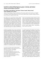
Fig. 1. Fluorescence emission spectra from binary complexes of
3a-HSD with NAD
+
or NADH, where an intrinsic fluorescence emis-
sion spectrum of 336 nm k
max
for 3a-HSD was portrayed at 280 nm
excitation. (A) Spectra of 6.43 l
M
3a-HSD in the absence (solid line) or
presence of 6.7 l
M
(dotted line), 23.3 l
M
(broken line), and 90 l
M
(dot-dashed line) NAD
+
.(B)Spectraof0.49l
M
3a-HSD in the
absence (solid line) or presence of 1.3 l
M
(dotted line), 2.6 l
M
(broken
line), and 6.7 l
M
(dot-dashed line) NADH.
Table 2. K
d
values obtained from fluorescence titration and K
m
values
obtained from steady-state kinetics for each nucleotide binding.
Nucleotide
Fluorescence titration
K
d
a
(l
M
)
Steady-state kinetics
K
m
b
(l
M
)
NAD
+
173 ± 24 29
NADP
+
192 ± 23 114
NAAD
+
258 ± 26 373
c
NADH 7.6 ± 0.9 11.4
NADPH 120 ± 4 268
a
Determined at 15 °C.
b
Determined at 37 °C.
c
K
i
value, deter-
mined in the inhibition assay.
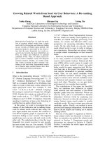
Fig. 2. Dependence of k
app
on the initial concentration of NAD
+
(A)
and NADP
+
(B). All reactions were carried out in 50 m
M
Tris/HCl
(pH 8.5) at 15 °C with 0.56 l
M
enzyme and various concentrations of
NAD
+
(from 2.5 to 75 l
M
) or NADP
+
(from 10 to 100 l
M
).Values
were expressed as the mean ± SD. The lines were drawn according to
Eqn (2), using k
+1
¼ 1.79, k
)1
¼ 84.8 for NAD
+
and k
+1
¼ 6.34,
k
)1
¼ 54.5 for NADP
+
.
Ó FEBS 2004 Transient kinetics of nucleotide binding to 3a-HSD (Eur. J. Biochem. 271) 1777
Transient-phase kinetics on reduced nucleotide binding
In the case of reduced nucleotide binding, the increase of
fluorescence intensity in NADH binding could be monit-
ored through a UV-42 cut-off filter. The k
app
value was
56–145 s
)1
, and the dependence of k
app
on the initial
concentration of NADH showed a hyperbolic curve
(Fig. 3). Different from oxidized nucleotides, the kinetic
feature of the NADH binding was consistent with a two-
step mechanism, which involves a fast bimolecular associ-
ation process followed by a slow unimolecular isomerization
process. The kinetic rate constants, k
+2
and k
)2
,andthe
dissociation constants, K
)1
and K
d
, are summarized in
Table 3.
The binding of NADPH to 3a-HSD was also investigated
by stopped-flow measurement. Although the quench in
intrinsic enzyme fluorescence emission spectrum following
NADPH binding was observed here as well, there were no
kinetic transients over a wide range of nucleotide concen-
trations. These results suggest different binding modes
toward NADPH and NADH.
Discussion
The transient-phase kinetics revealed that the binding of
oxidized nucleotide cofactors, NAD
+
and NADP
+
,to
3a-HSD from P. sp. B-0831 follows a one-step mechanism,
while the binding of reduced nucleotide cofactor, NADH,
follows a two-step mechanism. The binding of NAAD
+
,
not a cofactor but a competitive inhibitor, also follows a
one-step mechanism, as is the case of the oxidized cofactors.
The validity of the proposed mechanism was supported by
similar dissociation constants between the K
)1
or K
d
values
determined by stopped-flow measurements and the K
d
values determined by the fluorescence titration (Tables 2
and 3). For binding of oxidized nucleotides, the K
d
values
by fluorescence titration ranged from 2.2- to 3.6-fold the
corresponding K
)1
values. The K
d
value for the NADH
binding obtained by fluorescence titration was 2.5-fold that
obtained by stopped-flow analysis.
In HSD-related enzymes, whether or not the binding or
release of nucleotide cofactors is the rate-limiting step in the
reaction remains an issue [26,28,29]. The rate constant k
)1
for release of NAD
+
in a one-step mechanism (84.8 s
)1
)
was larger than the k
cat
value (11.6 s
)1
) for 5a-androstan-
3,17-dione reduction (Tables 1 and 3), indicating that the
dissociation of NAD
+
from the B-0831 3a-HSD is not
rate-limiting overall, a finding that coincides well with
dissociation reported on the enzyme from rat liver [26]. The
rate constant k
)2
for release of NADH from the B-0831
3a-HSD in a two-step reaction (24 s
)1
) was slightly larger
than the k
cat
value for androsterone oxidation (16.4 s
)1
).
While the dissociation of NADH in this reaction could
contribute to rate limiting, those in the oxidation of other
substrates examined in this study would not be rate limiting
as the k
cat
values are smaller than that for androsterone
oxidation (Table 1).
The rapid kinetic transients were not observed in the
NAD(H) binding to rat liver 3a-HSD, suggesting different
modes of binding toward NAD(H) and NADP(H) [26]. The
K
d
values of NADP(H) binding were much smaller (c., three
order in magnitude) than those of NAD(H) [27]. It was
concluded the interaction between the 2¢-phosphate of
NADP
+
and Arg276 was essential for the observation of
kinetic transients. In contrast, rapid kinetic transients were
observed in the binding of NAD
+
,NADP
+
and NADH to
P. sp. B-0831-derived 3a-HSD, albeit such was the case in
NADPH binding. The oxidized nucleotide cofactors
NAD
+
and NADP
+
bound to the enzyme with similar
affinity, following a one-step catalysis. In the reduced
nucleotide cofactors, the respective binding affinities were
different, and the relatively higher binding affinity nucleo-
tide, NADH, only showed a two-step catalysis. These
results indicate that the reaction mechanism depends on the
type of nucleotides, and is different from that of rat liver
3a-HSD.
In order to discuss the nucleotide cofactor preference, the
amino acid sequence in the nucleotide binding region was
compared with those of other SDRs. Similar to 3a-HSD
from P. sp. B-0831, 3a-HSD/CR from C. testosteroni and
Table 3. Transient-phase kinetic parameters for the formation and decay of enzyme-nucleotide complexes.
Nucleotide k
+1
(
M
)1
Æs
)1
) k
)1
(s
)1
) K
)1
(l
M
) k
+2
(s
)1
) k
)2
(s
)1
) K
)1
(l
M
) K
d
(l
M
)
NAD
+
1.79 ± 0.1 · 10
6
84.8 ± 0.8 47.6 ± 2.6
NADP
+
6.34 ± 1.0 · 10
5
54.5 ± 3.3 87.2 ± 8.0
NAAD
+
3.67 ± 0.3 · 10
5
41.5 ± 1.9 113.1 ± 4.7
NADH 278 ± 26 23.9 ± 3.1 39.2 ± 10.9 3.1 ± 0.9
Fig. 3. Dependence of k
app
on the initial concentration of NADH. All
reactions were carried out in 50 m
M
Tris/HCl (pH 8.5) at 15 °C, with
0.84 l
M
enzyme and various concentrations of NADH (from 5 to
37.5 l
M
). Values were expressed as the mean ± SD. The line was drawn
according to Eqn (5), using k
+2
¼ 278, k
)2
¼ 24, K
)1
¼ 39. Insert:
plot of 1/(k
app
– k
)2
) vs. 1/[NADH]. The k
)2
value was estimated by
linear extrapolation in the low concentration region of NADH.
1778 S. Ueda et al. (Eur. J. Biochem. 271) Ó FEBS 2004
7a-HSD from E. coli have common Asp residues at position
32 (numbering according to the B-0831 3a-HSD), which
is highly conserved among enzymes preferring NAD
+
[14,16,38,39]. It is noteworthy that the substitution of Asp
for Thr38 in mouse lung CR changed the nucleotide
preference from NADP
+
to NAD
+
[40]. The crystal
structure of C. testosteroni-derived 3a-HSD/CR complexed
with NAD
+
has shown that Ile33, located next to Asp32,
impedes NADP
+
with a 2¢-phosphate group [39]. The
corresponding residue in 3a-HSD from P. sp. B-0831 is
Arg33, which is conserved in the NADP
+
preferring
enzymes, such as mouse liver 11b-HSD and mouse lung
CR [16,41]. The crystal structure of mouse lung CR
complexed with NADPH displays a pair of basic residues,
Lys17 and Arg39, which correspond, respectively, to Ser11
and Arg33 in P. sp. B-0831-derived 3a-HSD, causing
electrostatic interaction with the 2¢-phosphate group of
NADP
+
[41]. These results indicate that the Asp32 and
Arg33 residues are, respectively, critical for NAD
+
and
NADP
+
preferences, resulting in the unique dual nucleotide
cofactor specificity of B-0831 3a-HSD, with the binding
affinity relatively lower than that of the NAD
+
-or
NADP
+
-preferring enzyme [7,26,42].
The ordered bi-bi reaction mechanism can be explained
by structural analyses: the substrate-binding loop of
3a-HSD is ordered when nucleotide cofactor binds to the
site next to the substrate-binding site. The conformational
change of C. testosteroni-derived 3a-HSD/CR induced by
nucleotide binding is more subtle than that of rat liver
3a-HSD [25,39]. The conformational change observed in
rat liver 3a-HSD is in good correlation with the slow
formation of a tightly bound complex via a two-step
reaction [26]. The NADH binding to P. sp. B-0831-derived
3a-HSD, which is a two-step reaction, may induce a
conformational change similar to that of rat liver 3a-HSD.
In contrast, the binding of NAD
+
or NADP
+
,whichisa
one-step reaction, may induce little conformational change
similar to that of C. testosteroni-derived 3a-HSD/CR. The
relatively lower binding affinity of these nucleotides
supports the notion that only a loose complex is formed
in the one-step reaction. It should also be noted that the k
cat
value for cholic acid as a substrate with NADP
+
is only 6%
of that for the same substrate with NAD
+
, although both
nucleotides bind to B-0831 3a-HSD with similar affinity.
Additionally, no catalytic activity was detectable with
NAAD
+
. In short, the different types of nucleotides can
modulate the dynamic conformation and the catalytic
function of the enzyme.
References
1. Cheng, K.C., White, P.C. & Qin, K.N. (1991) Molecular cloning
and expression of rat liver 3a-hydroxysteroid dehydrogenase. Mol.
Endocrinol. 5, 823–828.
2. Jez, J.M., Flynn, T.G. & Penning, T.M. (1997) A new nomen-
clature for the aldo-keto reductase superfamily. Biochem. Phar-
macol. 54, 639–647.
3. MacDonald, I.A., Jellet, J.F., Mahony, D.E. & Holdeman, L.V.
(1979) Bile salt 3a-and12a-hydroxysteroid dehydrogenases from
Eubacterium lentum and related organisms. Appl. Environ.
Microbiol. 37, 992–1000.
4. MacDonald, I.A., Meier, E.C., Mahony, D.E. &Costain,G.A.(1976)
3a-, 7a-and12a-hydroxysteroid dehydrogenase activities from
Clostridium perfringens. Biochim. Biophys. Acta 450, 142–153.
5. Uwajima, T., Takayama, K. & Terada, O. (1978) Production,
purification and crystallization of 3a-hydroxysterid dehydro-
genase from Pseudomonas putida. Agric. Biol. Chem. 42, 1577–
1583.
6. Boyer, J., Baron, D.N. & Talalay, P. (1965) Purification and
properties of 3a-hydroxysteroid dehydrogenase from Pseudo-
monas testosteroni. Biochemistry 4, 1825–1833.
7. Ska
˚
lhegg, B.A. (1975) 3a-hydroxysteroid dehydrogenase from
Pseudomonas testosteroni: kinetic properties with NAD and its
thionicotinamide analogue. Eur. J. Biochem. 50, 603–609.
8.Maser,E.,Oppermann,U.C.,Bannenberg,G.&Netter,K.J.
(1992) Functional and immunological relationships between
metyrapone reductase from mouse liver microsomes and 3a-hy-
droxysteroid dehydrogenase from Pseudomonas testosteroni.
FEBS Lett. 297, 196–200.
9. Oppermann, U.C. & Maser, E. (1996) Characterization of a
3a-hydroxysteroid dehydrogenase/carbonyl reductase from the
gram-negative bacterium Comamonas testosteroni. Eur. J. Bio-
chem. 241, 744–749.
10. Suzuki, K., Ueda, S., Sugiyama, M. & Imamura, S. (1993)
Cloning and expression of a Pseudomonas 3a-hydroxysteroid
dehydrogenase-encoding gene in Escherichia coli. Gene 130, 137–
140.
11. Persson, B., Krook, M. & Jo
¨
rnvall, H. (1991) Characteristics of
short-chain alcohol dehydrogenases and related enzymes. Eur. J.
Biochem. 200, 537–543.
12. Jo
¨
rnvall, H., Persson, B., Krook, M., Atrian, S., Gonzalez-
Duarte, R., Jeffery, J. & Ghosh, D. (1995) Short-chain dehy-
drogenases/reductases (SDR). Biochemistry 34, 6003–6013.
13. Jez, J.M., Bennett, M.J., Schlegel, B.P., Lewis, M. & Penning,
T.M. (1997) Comparative anatomy of the aldo-keto reductase
superfamily. Biochem. J. 326, 625–636.
14. Kallberg, Y., Oppermann, U., Jo
¨
rnvall, H. & Persson, B. (2002)
Short-chain dehydrogenases/reductases (SDRs). Eur. J. Biochem.
269, 4409–4417.
15. Maser, E., Mo
¨
bus, E. & Xiong, G. (2000) Functional expression,
purification, and characterization of 3a-hydroxysteroid dehydro-
genase/carbonyl reductase from Comamonas testosteroni.
Biochem. Biophys. Res. Commun. 272, 622–628.
16. Mo
¨
bus, M. & Maser, E. (1998) Molecular cloning, over-
expression, and characterization of steroid-inducible 3a-hydroxy-
steroid dehydrogenase/carbonyl reductase from Comamonas
testosteroni. A novel member of the short-chain dehydrogenase/
reductase superfamily. J. Biol. Chem. 273, 30888–30896.
17. Ueda, S., Oda, M., Imamura, S. & Ohnishi, M. (2004) Carbonyl
reductase activity of a pluripotent enzyme, 3a-hydroxysteroid
dehydrogenase from Pseudomonas sp. B-0831. J. Biol. Macromol.
4, 29–32.
18. Oda, K., Yoshida, S. & Takeda, T. (1989) Determination of total
3a-hydroxy bile acids in serum by a bioluminescent flow injection
system using a hollow-fiber reactor. Anal. Chim. Acta 225,
273–282.
19. Takahashi, M., Ueda, S., Misaki, H., Sugiyama, N., Mastumoto,
K., Matsuo, N. & Murao, S. (1994) Carnitine determination by an
enzymatic cycling method with carnitine dehydrogenase. Clin.
Chem. 40, 817–821.
20. Penning, T.M., Smithgall, T.E., Askonas, L.J. & Sharp, R.B.
(1986) Rat liver 3a-hydroxysteroid dehydrogenase. Steroids 47,
221–247.
21. Askonas, L.J., Ricigliano, J.W. & Penning, T.M. (1991) The
kinetic mechanism catalysed by homogeneous rat liver 3a-
hydroxysteroid dehydrogenase: evidence for binary and ternary
Ó FEBS 2004 Transient kinetics of nucleotide binding to 3a-HSD (Eur. J. Biochem. 271) 1779
dead-end complexes containing non-steroidal anti-inflammatory
drugs. Biochem. J. 278, 835–841.
22. Pawlowski, J.E. & Penning, T.M. (1994) Overexpression and
mutagenesis of the cDNA for rat liver 3a-hydroxysteroid/
dihydrodiol dehydrogenase. Role of cysteines and tyrosines in
catalysis. J. Biol. Chem. 269, 13502–13510.
23. Jez, J.M., Schlegel, B.P. & Penning, T.M. (1996) Characterization
of the substrate binding site in rat liver 3a-hydroxysteroid/
dihydrodiol dehydrogenase: the roles of tryptophans in ligand
binding and protein fluorescence. J. Biol. Chem. 271, 30190–
30198.
24. Bennett, M.J., Schlegel, B.P., Jez, J.M., Penning, T.M. & Lewis,
M. (1996) Structure of 3a-hydroxysteroid/dihydrodiol dehydro-
genase complexed with NADP
+
. Biochemistry 35, 10702–
10711.
25. Bennett, M.J., Albert, R.H., Jez, J.M., Ma, H., Penning, T.M. &
Lewis, M. (1997) Steroid recognition and regulation of hormone
action: crystal structure of testosterone and NADP
+
bound to
3a-hydroxysteroid/dihydrodiol dehydrogenase. Structure 5,
799–812.
26. Ratnam, K., Ma, H. & Penning, T.M. (1999) The arginine 276
anchor for NADP(H) dictates fluorescence kinetic transients in
3a-hydroxysteroid dehydrogenase, a representative aldo-keto
reductase. Biochemistry 38, 7856–7864.
27. Ma, H., Ratnam, K. & Penning, T.M. (2000) Mutation of
nicotinamide pocket residues in rat liver 3a-hydroxysteroid
dehydrogenase reveals different modes of cofactor binding.
Biochemistry 39, 102–109.
28. Kubiseski, T.J., Hyndman, D.J., Morjana, N.A. & Flynn, T.G.
(1992) Studies on pig muscle aldose reductase: kinetic mechanism
and evidence for a slow conformational change upon coenzyme
binding. J. Biol. Chem. 267, 6510–6517.
29. Grimshaw, C.E., Bohren, K.M., Lai, C.J. & Gabbay, K.H. (1995)
Human aldose reductase: rate constants for a mechanism includ-
ing interconversion of ternary complexes by recombinant wild-
type enzyme. Biochemistry 34, 14356–14365.
30. Lowry, O.H., Rosebrough, N.J., Farr, A.L. & Randall, R.J.
(1951) Protein measurement with the folin phenol reagent. J. Biol.
Chem. 193, 265–275.
31. Sakoda, M. & Hiromi, K. (1976) Determination of the best-fit
values of kinetic parameters of the Michaelis-Menten equation by
the method of least squares with the Taylor expansion. J. Biochem.
80, 547–555.
32. Tanaka, A., Ohnishi, M. & Hiromi, K. (1982) Stopped-flow
kinetic studies on the binding of gluconolactone and maltose to
glucoamylase. Biochemistry 21, 107–113.
33. Tonomura, B., Nakatani, H., Ohnishi, M., Yamaguchi-Ito, J. &
Hiromi, K. (1978) Test reactions for a stopped-flow apparatus:
reduction of 2,6-dichlorophenolindophenol and potassium ferri-
cyanide by 1-ascorbic acid. Anal. Biochem. 84, 370–383.
34. Guggenheim, E.A. (1926) On the determination of the velocity
constant of a unimolecular reaction. Phil. Mag. 2, 538–543.
35. Strickland, S., Palmer, G. & Massey, V. (1975) Determination of
dissociation constants and specific rate constants of enzyme-sub-
strate (or protein–ligand) interactions from rapid reaction kinetic
data. J. Biol. Chem. 250, 4048–4052.
36. Halford, S.E. (1975) Stopped-flow fluorescence studies on sac-
charide binding to lysozyme. Biochem. J. 149, 411–422.
37. Ueda,S.,Oda,M.,Imamura,S.&Ohnishi,M.(2004)Steady-state
kinetic properties of 3a-hydroxysteroid dehydrogenase from
Pseudomonas sp. B-0831: Steroid substrate specificity and nuc-
leotide cofactor dependency. J. Biol. Macromol. 4, 23–28.
38. Tanaka,N.,Nonaka,T.,Tanabe,T.,Yoshimoto,T.,Tsuru,D.&
Mitsui, Y. (1996) Crystal structures of the binary and ternary
complexes of 7a-hydroxysteroid dehydrogenase from Escherichia
coli. Biochemistry 35, 7715–7730.
39. Grimm, C., Maser, E., Mo
¨
bus,E.,Klebe,G.,Reuter,K.&Ficner,
R. (2000) The crystal structure of 3a-hydroxysteroid dehydro-
genase/carbonyl reductase from Comamonas testosteroni shows a
novel oligomerization pattern within the short chain dehydro-
genase/reductase family. J. Biol. Chem. 275, 41333–41339.
40. Nakanishi, M., Matsuura, K., Kaibe, H., Tanaka, N., Nonaka,
T., Mitsui, Y. & Hara, A. (1997) Switch of coenzyme specificity of
mouse lung carbonyl reductase by substitution of threonine 38
with aspartic acid. J. Biol. Chem. 272, 2218–2222.
41. Tanaka, N., Nonaka, T., Nakanishi, M., Deyashiki, Y., Hara, A.
& Mitsui, Y. (1996) Crystal structure of the ternary complex of
mouse lung carbonyl reductase at 1.8 A
˚
resolution: the structural
origin of coenzyme specificity in the short-chain dehydrogenase/
reductase family. Structure 4, 33–45.
42. Wilson, D.K., Nakano, T., Petrash, J.M. & Quiocho, F.A. (1995)
1.7 A
˚
structure of FR-1, a fibroblast growth factor-induced
member of the aldo-keto reductase family, complexed with coen-
zyme and inhibitor. Biochemistry 34, 14323–14330.
1780 S. Ueda et al. (Eur. J. Biochem. 271) Ó FEBS 2004