Báo cáo khoa học: Covalent activation of heart AMP-activated protein kinase in response to physiological concentrations of long-chain fatty acids docx
Bạn đang xem bản rút gọn của tài liệu. Xem và tải ngay bản đầy đủ của tài liệu tại đây (277.4 KB, 10 trang )
Covalent activation of heart AMP-activated protein kinase in response
to physiological concentrations of long-chain fatty acids
Hilary Clark
1
, David Carling
2
and David Saggerson
1
1
Department of Biochemistry and Molecular Biology, University College London, UK;
2
Cellular Stress Group, MRC Clinical Sciences
Centre, Imperial College School of Medicine, Hammersmith Hospital, London, UK
Rat hearts were perfused for 1 h with 5 m
M
glucose with or
without palmitate or oleate at concentrations characteristic
of the fasting state. The inclusion of fatty acids resulted in
increased activities of the a-1 or the a-2 isoforms of AMP-
activated protein kinase (AMPK), increased phosphoryla-
tion of acetyl-CoA carboxylase and a decrease in the tissue
content of malonyl-CoA. Activation of AMPK was not
accompanied by any changes in the tissue contents of
ATP, ADP, AMP, phosphocreatine or creatine. Palmitate
increased phosphorylation of Thr172 within AMPK
a-subunits and the activation by palmitate of both AMPK
isoforms was abolished by protein phosphatase 2C leading
to the conclusion that exposure to fatty acid caused activa-
tion of an AMPK kinase or inhibition of an AMPK phos-
phatase. Invivo, 24 h of starvation also increased heart
AMPK activity and Thr172 phosphorylation of AMPK
a-subunits. Perfusion with insulin decreased both a-1 and
a-2 AMPK activities and increased malonyl-CoA content.
Palmitate prevented both of these effects. Perfusion with
epinephrine decreased malonyl-CoA content without an
effect on AMPK activity but prevented the activation of
AMPK by palmitate. The concept is discussed that activa-
tion of AMPK by an unknown fatty acid-driven signalling
process provides a mechanism for a Ôfeed-forwardÕ activation
of fatty acid oxidation.
Keywords: AMP-activated protein kinase; fatty acids; heart;
insulin; protein phosphorylation.
The AMP-activated protein kinase (AMPK) is a heterotri-
meric enzyme complex with a key role in the regulation of
metabolism and other processes [1–4]. AMPK is activated
following an increase in the cellular AMP/ATP ratio.
Activation requires phosphorylation of Thr172 within the
a-subunit of AMPK, catalysed by an upstream AMPK
kinase. Dephosphorylation of Thr172 (in vivo by phospho-
protein phosphatase 2C [5]) leads to inactivation of AMPK.
Direct allosteric activation of AMPK also occurs following
an increase in the cellular AMP/ATP or creatine/phospho-
creatine ratios [6]. These processes constitute the ÔclassicalÕ
pathway allowing AMPK to be a sensor of the cellular
Ôenergy chargeÕ under conditions of increased ATP con-
sumption and/or impeded ATP production. Recently other
conditions have been described in which the AMPK is
covalently activated or inactivated without detectable
change in the cellular AMP/ATP ratio, e.g. changes due
to insulin [7], leptin [8], metformin [9,10], hyperosmotic
stress [9] and glucose deprivation [11] leading to proposals
[9,11] that covalent activation of the AMPK may also occur
through upstream processes independent of the ÔclassicalÕ
pathway, e.g. involving the LKB1 tumour-suppressor
kinase [12,13].
Malonyl-CoA has an important role in the regulation
of fuel selection by the heart [14,15] through its potent
inhibition [16] of carnitine palmitoyltransferase-1 (CPT1).
Malonyl-CoA is synthesized and disposed of by acetyl-CoA
carboxylase (ACC) and malonyl-CoA decarboxylase
(MCD), respectively. ACC is inactivated through phos-
phorylation by the AMPK [17–19]. By contrast, at present
there is conflicting evidence for or against the notion that
MCD can be activated following phosphorylation by the
AMPK [20–22]. Dyck and Lopaschuk [14] and Kudo et al.
[23] have shown during postischaemic reperfusion of the
rat heart that elevation of AMPK activity correlates with
decreased ACC activity, decreased malonyl-CoA content
andanincreasedrateofb-oxidation. Work from our
laboratory had shown that heart malonyl-CoA content was
increased by insulin [15,24] and insulin has been shown to
decrease AMPK activity in heart [7]. However, Awan and
Saggerson [15] and Hamilton and Saggerson [24] showed
that long-chain fatty acid (palmitate) both decreased
malonyl-CoA content and prevented the effect of insulin
to increase malonyl-CoA. Therefore we investigated the
effect of physiological concentrations of long-chain fatty
acids on AMPK activity in perfused rat hearts in the
expectation that AMPK activity would be increased. This
was found to occur through covalent modification of
AMPK a subunits driven by an unknown upstream protein
Correspondence to D. Saggerson, Department of Biochemistry and
Molecular Biology, University College London, Gower Street,
London, WC1E 6BT, UK.
Fax: + 44 20 7679 7193, Tel.: + 44 20 7679 7320,
E-mail:
Abbreviations: ACC, acetyl-CoA carboxylase; AMPK, AMP-activa-
ted protein kinase; CPT1, the overt form of mitochondrial carnitine
acyltransferase; KHB, Krebs–Henseleit bicarbonate; MCD, malonyl-
CoA decarboxylase; NEFA, non-esterified fatty acid; PKA, cyclic
AMP-dependent protein kinase; PP2C, phosphoprotein phosphatase
2C; PT-172, phosphorylation of Thr172 in AMPK a-subunits.
(Received 16 January 2004, revised 4 March 2004,
accepted 6 April 2004)
Eur. J. Biochem. 1–10 (2004) Ó FEBS 2004 doi:10.1111/j.1432-1033.2004.04151.x
phosphorylation mechanism that is not dependent upon
changes in the cellular AMP/ATP ratio. Similar changes in
AMPK phosphorylation and activation were seen when rats
were starved for 24 h. We also report on Ôcross-talkÕ between
this fatty acid-driven activation process and insulin and
adrenergic signalling pathways.
Experimental procedures
Chemicals
Antisera against AMPK a-subunits were raised in sheep
[25]. In one experiment (Fig. 6) a goat antiserum (Santa
Cruz) was used. These antisera or nonimmune serum were
prebound to protein G-Sepharose 4B. Antibody against a
peptide surrounding phospho-Thr172 on the a-subunits of
AMPK was from New England Biolabs. Antibody against
the phosphopeptide corresponding to amino acids 73–85 of
rat ACC-1 [HMRSSMS(PO
4
)GLHLVK] was from Upstate
Biotechnology. Recombinant phosphoprotein phosphatase
2C (PP2C; human a-isoform) was a generous gift from
R. Beri (AstraZeneca Pharmaceuticals). Sodium palmitate
or oleate were bound to fatty acid-poor BSA [26] and the
concentration of bound fatty acid was standardized with a
Wako NEFA test kit (Alpha Laboratories).
Animal procedures
1
Male Sprague–Dawley rats (300–350 g body weight) were
maintained at 20–22 °C on a 13 h light/11 h dark cycle with
light from 06:00 h to 19:00 h. Rats were anaesthetized with
sodium pentobarbitone (300 mgÆkg
)1
) injected intraperiton-
eally prior to removal of the heart. Hearts from fed animals
were perfused retrogradely via the aorta at 37 °Cwith
100 mL Krebs–Henseleit bicarbonate (KHB) medium
equilibrated with O
2
/CO
2
(19 : 1) containing 1.3 m
M
CaCl
2
,
5m
M
glucose and fatty acid-poor BSA (20 mgÆmL
)1
). The
medium was recirculated except in experiments with
epinephrine. The system [27] perfused the coronary circu-
lation and, because the ventricle was filled, required the
heart to work against a pressure of 80 ± 5 cm water. Any
hearts which did not have a sustained and steady beat
throughout the experiment or which showed discoloured
regions denoting inadequate perfusion were discarded.
After 20 or 60 min hearts were freeze-clamped (liquid
nitrogen). For in vivo measurements hearts from fed or
24 h-starved rats were directly freeze-clamped after removal
from the animal. Procedures conformed to the UK Animals
(Scientific Procedures) Act, 1986.
AMPK activity
Hearts were powdered under liquid nitrogen and homo-
genized (100 mgÆmL
)1
) in homogenization/immunopreci-
pitation buffer consisting of 50 m
M
Tris/HCl pH 7.8,
0.25 m
M
mannitol, 1 m
M
EDTA, 1 m
M
EGTA, 50 m
M
NaF, 5 m
M
Na
4
P
2
O
7
,1m
M
dithiothreitol, 1 m
M
phenyl-
methanesulfonyl fluoride, 1 m
M
benzamidine and soybean
trypsin inhibitor (4 lgÆmL
)1
). The homogenate was centri-
fuged at 4 °Cfor10minat13000g and 250 lLofthe
supernatant incubated for 2 h at 4 °C with anti-AMPK
serum (usually 15 lL) bound to Protein G-Sepharose.
Immunoprecipitates were collected by centrifugation (1 min
at 5200 g). Normally immunoprecipitates were washed/
recentrifuged once with 300 lL homogenization/immuno-
precipitation buffer and then twice (4 °C) with 300 lLof
AMPK assay buffer (40 m
M
Hepes pH 7.0 contain-
ing 80 m
M
NaCl, 0.8 m
M
EDTA, 8% v/v glycerol and
1m
M
dithiothreitol). Finally washed immunoprecipitates
were resuspended in 75 lL of AMPK assay buffer
which additionally contained 200 l
M
ÔSAMSÕ peptide
(HMRSAMSGLHLVKRR) [28], 5 m
M
MgCl
2
,withor
without 200 l
M
AMP. The AMPK assay was started by
addition of 200 l
M
[c-
33
P]ATP (250–500 d.p.m.Æpmol
)1
).
After 30 min at 37 °C the reaction was stopped by spotting
20 lL samples onto P81 Whatman phosphocellulose papers
which were washed twice for 10 min in a solution of
orthophosphoric acid (1%, v/v) and then twice for 10 min
in water before drying and scintillation counting. In
experiments with PP2C fresh immunoprecipitates (see
above) were washed with 300 lL homogenization/immu-
noprecipitation buffer and then twice (4 °C) with 300 lL
50 m
M
Tris/HCl pH 7.4 containing 1 m
M
dithiothreitol.
After recovery by centrifugation
2
(see above) these immu-
noprecipitates were resuspended in 25 lLof50m
M
Tris/
HClpH7.4,10m
M
MgCl
2
,1m
M
dithiothreitol, glycerol
(5%, v/v) and PP2C (160 lgÆmL
)1
). MgCl
2
was omitted
from control incubations. After 30 min at 30 °Cthe
immunoprecipitates were again recovered by centrifugation
and washed three times (4 °C) in 300 lL of AMPK assay
buffer before resuspension in 75 lL of the same buffer
together with the other components of the AMPK assay
(see above). AMPK activity is expressed as pmolÆmin
)1
per
mg 13 000 g supernatant protein (i.e. relative to the extract
immediately before immunoprecipitation). Preliminary
experiments established the optimum amounts of each
anti-AMPK serum. Blank activity with nonimmune sheep
serum was subtracted.
Western blotting
Hearts were powdered under liquid nitrogen and homo-
genized (200 mgÆmL
)1
) in homogenization/immunopreci-
pitation buffer followed by centrifugation (13 000 g)for
5 min. Supernatants (200 lg protein) were analysed by
SDS/PAGE, transferred to poly(vinylidene difluoride)
membranes and blotted with antiphospho-ACC Ig or
antiphospho-AMPK primary Ig. Following measurement
of phosphorylation of Thr172 within AMPK a-subunits
(P-T172) blots were stripped to measure total abundance of
AMPK a-subunits. The membranes were left at 50 °Cfor
30 min in 62.5 m
M
Tris/HCl pH 6.8 containing 100 m
M
2-mercaptoethanol and 2% SDS followed by three washes in
20 m
M
Tris/HCl pH 7.5 containing 0.14
M
NaCl and 0.1%
(v/v) Tween 20 (NaCl/Tris). After blocking with a solution
of milk powder (5% w/v) for 1 h the membranes were
washed again in NaCl/Tris and then re-blotted with AMPK
a-subunit primary antibody (Cell Signalling Technology).
Metabolites
ATP, ADP and AMP were measured in neutralized
trichloroacetic acid extracts of frozen heart after separation
by HPLC [29] and creatine and phosphocreatine as
2 H. Clark et al. (Eur. J. Biochem.) Ó FEBS 2004
described [30,31]. Malonyl-CoA was measured as described
by Awan & Saggerson [24]. Perfusion media and rat serum
were assayed for non-esterified fatty acid (NEFA; Wako
test kit) and glycerol [32]. Glucose was measured in
haemolysed blood samples [33].
Statistics
Values are given as means ± S.E.M. Statistical significance
was calculated using Student’s t-test for paired or unpaired
samples as indicated.
Results
Long-chain fatty acids cause phosphorylation of
a-subunits and activation of AMPK in perfused heart
Perfused hearts were fuelled by 5 m
M
glucose alone or by
glucose with 0.075 m
M
,0.25 m
M
or 0.5 m
M
long-chain fatty
acid. Palmitate (0.075 m
M
) was used because, with 2% BSA
present, this gave a NEFA/albumin molar ratio of
0.25 : 1, similar to that in fed plasma. Palmitate/oleate
at 0.25/0.5 m
M
gave NEFA/albumin ratios representative
of mild (1 day) and severe (> 1 day) starvation, respect-
ively. Over 60 min of perfusion we tested the effects of 0.25
and 0.5 m
M
fatty acids against two control conditions
(Table 1). Control 1 was where the initial perfusate fatty
acid concentration was zero. This is technically the correct
control but is unreal because a plasma NEFA concentration
of zero will not occur physiologically. In fact hearts started
with zero NEFA released a small but significant amount of
NEFA over 60 min (Table 2). Control 2 was where hearts
were started with 0.075 m
M
palmitate. Under this condition
the hearts were essentially in ÔNEFA balanceÕ whereas with
0.25 and 0.5 m
M
palmitate net removal of NEFA from the
perfusate occurred (Table 2).
Unexpectedly we found that a-1 and a-2 AMPK activities
tendedtobelowestinheartsstartedwith0.075m
M
palmitate and showed a significant decrease relative to
control 1 when assays contained the allosteric effector AMP
(Table 1). The adult heart normally supports much of its
ATP production from fatty acid oxidation [34]. Therefore
the control 1 condition may be one of metabolic stress,
reflected by a higher AMPK activity state than under
normal fed conditions.
Using control 1 as the baseline, 0.25 or 0.5 m
M
palmitate increased a-1 and a-2 AMPK activities by at
least2-foldwhenAMPwasomittedfromtheseassays.
Activation of a-2 AMPK by these fasting concentrations
of NEFA was also seen in assays with AMP. By contrast,
with AMP present, a-1 AMPK appeared to be insensitive
to palmitate. In essence, covalent activation following
exposure to fatty acid and allosteric activation by AMP
were mutually exclusive effects for a-1 AMPK whereas for
Table 1. The effect of perfusion with long-chain fatty acids on the activity state of heart AMPK. Hearts were perfused for 60 min with 5 m
M
glucose,
BSA (20 mgÆml
)1
) and sodium palmitate or oleate as indicated. The values are means ± S.E.M. of the numbers of independent measurements
shown in parentheses.
Initial NEFA concentration
in the perfusate
AMPK activity (pmolÆmin
)1
per mg of 13 000 g supernatant protein)
a-1 Complexes a-2 Complexes
Assayed without
200 lmAMP
Assayed
with AMP
Assayed without
200 lmAMP
Assayed
with AMP
Zero (control 1) 1.09 ± 0.27 (11) 2.70 ± 0.68
j
(6) 2.11 ± 0.49 (11) 4.21 ± 0.35
l
(11)
0.075 m
M
Palmitate (control 2) 0.70 ± 0.15 (5) 0.97 ± 0.30
a
(5) 1.49 ± 0.30 (6) 2.27 ± 0.63
b,I
(6)
0.25 m
M
Palmitate 2.46 ± 0.64
b,e
3.48 ± 0.92
e
(6) 4.14 ± 0.42
c,h
(6) 5.54 ± 0.50
a,f,I
(11)
0.5 m
M
Palmitate 2.79 ± 0.37
c,h
(11) 2.95 ± 0.53
g
(11) 5.16 ± 0.55
d,h
(11) 8.44 ± 0.90
d,h,l
(11)
0.5 mm Oleate 2.52 ± 0.27
c,h
(5) 2.67 ± 0.33
g
(5) 4.61 ± 0.63
c,g
(5) 8.15 ± 1.28
b,g,k
(5)
a,b,c,d
P < 0.05, < 0.02, < 0.01, < 0.001 respectively versus zero NEFA (unpaired test).
e,f,g,h
P < 0.05, < 0.02, < 0.01, < 0.001
respectively versus 0.075 m
M
palmitate (unpaired test).
i,j,k,l
P < 0.02, < 0.025, < 0.01, < 0.0005 respectively for the effect of AMP (paired
test).
Table 2. Net output or uptake of NEFA and glycerol by perfused hearts. Hearts were perfused for 60 min with 5 m
M
glucose, BSA (20 mgÆml
)1
)and
sodium palmitate as indicated. Values are means ± S.E.M. of between four and seven independent measurements.
Initial NEFA
concentration
in the perfusate
Final NEFA
concentration in
the perfusate (m
M
)
Change in perfusate NEFA
(lmolÆh
)1
Æg wet wtÆheart
)1
)
[A]
Glycerol release to perfusate
(lmol of fatty acid equivalentÆ
h
)1
Æg wet wtÆheart
)1
) [B]
Total fatty acid utilisation
(lmolÆh
)1
Æg wet wtÆheart
)1
)
[B–A]
Zero (control 1) 0.068 ± 0.003 +4.03 ± 0.60 7.03 ± 1.18 3.00
0.075 m
M
Palmitate
(control 2)
0.082 ± 0.005 +0.41 ± 0.38 6.73 ± 0.93 6.32
0.15 m
M
Palmitate 0.108 ± 0.005 )2.28 ± 0.34 5.22 ± 0.82 7.50
0.25 m
M
Palmitate 0.154 ± 0.005 )4.76 ± 0.28 5.88 ± 1.26 10.64
0.5 m
M
Palmitate 0.342 ± 0.016 )7.45 ± 1.22 5.45 ± 1.51 12.90
Ó FEBS 2004 Activation of AMPK by fatty acids (Eur. J. Biochem.)3
a-2 AMPK these appeared to be two independent effects
(Table 1).
Using control 2 as the baseline, perfusion with 0.25 or
0.5 m
M
palmitate increased a-1 AMPK activity by at least
3-fold and a-2 AMPK activity was increased by at least 2.5-
fold by 0.25 m
M
palmitate and by approximately 3.5-fold
by 0.5 m
M
palmitate. These effects of palmitate were seen
with or without AMP in the assays (Table 1). Perfusion
with 0.5 m
M
oleate caused activation of both AMPKs to
levels similar to those seen with 0.5 m
M
palmitate (Table 1).
Therefore the activation of AMPK was not peculiar to
palmitate but was a more generalized effect of long-chain
fatty acids.
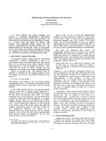
Downstream changes in ACC and malonyl-CoA follow-
ing activation of the AMPK were seen after perfusion with
0.5 m
M
palmitate for 60 min. Phosphorylation of ACC
under control 1 conditions was virtually undetectable using
the antibody which recognizes the AMPK phosphorylation
site sequence SMS(PO
4
)GLHLVK in ACC-1 (265 kDa)
and also recognizes the equivalent AMPK phosphorylation
site in ACC-2 (280 kDa). However after perfusion with
0.5 m
M
palmitate phosphorylation of both 265 and
280 kDa bands was clearly seen (Fig. 1). This was accom-
panied by a significant 51% decrease in malonyl-CoA
content (Table 3).
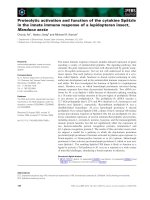
Figs 2 and 3 show experiments which support the
conclusion that activation of AMPK by fatty acid was
due to increased protein phosphorylation. Treatment of
immoprecipitates with PP2C abolished the activation due to
0.5 m
M
palmitate (Fig. 2). If Mg
2+
, which is required for
PP2C activity, was omitted the activation by palmitate was
not abolished (data not shown). The AMPK activities in
Fig. 2 are lower than those in Table 1 whilst the degree of
activation by palmitate was higher. The reason for this is
unclear but it is stressed that more extensive washing of
immunoprecipitates was necessary in order to remove
inhibitors of protein dephosphorylation before treatment
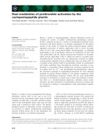
with PP2C. Fig. 3 shows that exposure of hearts to 0.5 m
M
palmitate significantly increased P-T172 abundance in the
combined AMPK a-1 and a-2 subunits by 2.5-fold without
causing any change in the abundance of AMPK a-subunit
protein.
Activation of AMPK by fatty acids does not require
changes in cellular adenine nucleotides
Perfusion with 0.5 m
M
palmitate for 60 min had no
significant effect on the contents of adenine nucleotides,
creatine and phosphocreatine or on the AMP/ATP ratio
and the Ôenergy chargeÕ [35] compared with either control 1
or control 2 conditions (Table 3). The only significant
change that was seen was a very small increase in the Ôenergy
Fig. 1. Effect of palmitate on the phosphorylation state of acetyl-CoA
carboxylase (Phospho-ACC). Hearts were perfused for 60 min with
5m
M
glucose and BSA (20 mgÆmL
)1
). C, Zero NEFA (control 1
conditions); P, 0.5 m
M
palmitate. Each of the measurements was
obtained from a separate heart.
Table 3. Measurements of adenine nucleotides, creatine, phosphocreatine and malonyl-CoA in perfused hearts. Heartswereperfusedfor20or60min
with 5 m
M
glucose, BSA (20 mgÆmL
)1
) and sodium palmitate as indicated. Values are means ± S.E.M. of the numbers of independent meas-
urements shown in parentheses and are expressed as lmolÆg wet weight heart
)1
except for malonyl-CoA (nmolÆgwetweight
)1
). The Ôenergy chargeÕ
was calculated from (ATP +
1
/
2
ADP)/(total adenine nucleotides) [35].
Perfusate fatty acid
initial concentration
and time of perfusion
Zero
(control 1)
0.075 m
M
palmitate
(control 2) 0.5 m
M
palmitate
20 min 60 min 60 min 20 min 60 min
AMP 0.131 ± 0.033 (5) 0.094 ± 0.011 (6) 0.092 ± 0.005 (7) 0.109 ± 0.013 (6) 0.095 ± 0.004 (7)
ADP 0.644 ± 0.048 (5) 0.478 ± 0.088 (6) 0.462 ± 0.021 (7) 0.577 ± 0.046 (6) 0.452 ± 0.014 (7)
ATP 2.19 ± 0.196 (5) 1.48 ± 0.294 (6) 2.09 ± 0.18 (7) 1.76 ± 0.233 (6) 1.84 ± 0.132 (7)
AMP/ATP ratio 0.063 ± 0.018 0.071 ± 0.011 0.047 ± 0.006 0.065 ± 0.009 0.054 ± 0.005
Energy charge 0.846 ± 0.018 0.844 ± 0.008 0.872 ± 0.009
a
0.833 ± 0.014 0.863 ± 0.010
Creatine ND 4.50 ± 0.66 (6) ND ND 3.70 ± 0.71 (6)
Phosphocreatine ND 6.54 ± 1.12 (6) ND ND 6.75 ± 1.08 (6)
Malonyl-CoA 3.35 ± 0.53 (6) 2.81 ± 0.33 (9) ND 2.77 ± 0.34 (6) 1.38 ± 0.13
b
(9)
a,b
P < 0.05, < 0.001, respectively, vs. zero NEFA (unpaired test). ND, Not determined.
Fig. 2. Effect of PP2C to abolish the activation of AMPK by palmitate.
Hearts were perfused for 60 min with 5 m
M
glucose and BSA
(20 mgÆmL
)1
) without (open bars) or with 0.5 m
M
palmitate (filled
bars). AMPK immunoprecipitates were incubated with 10 m
M
MgCl
2
and PP2C as indicated. Incubation without MgCl
2
abolished effects of
PP2C (data not shown). Values are means ± S.E.M. of four inde-
pendent measurements. AMPK activity was measured without AMP
and is expressed as pmolÆmin
)1
per mg 13 000 g supernatant protein.
4 H. Clark et al. (Eur. J. Biochem.) Ó FEBS 2004
chargeÕ in control 2 compared with that in control 1
(Table 3).
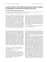
Time-dependence of the activation of AMPK
by fatty acids
No significant activation of AMPKs by 0.5 m
M
palmitate
was seen when the perfusion time was 20 min (Fig. 4).
From this finding it was correctly predicted that 0.5 m
M
palmitate would have no significant effect at 20 min on the
content of the downstream marker malonyl-CoA (Table 3).
The emergence of a significant effect of palmitate between
20 and 60 min was not accompanied by any significant
changes in the AMP/ATP ratio or in the Ôenergy chargeÕ
(Table 3), providing further evidence that covalent activa-
tion of AMPKs following exposure to fatty acid was not
driven by changes in adenine nucleotides.
Cross-talk between the activation of AMPK by fatty acids
and insulin and adrenergic signalling processes
Fig. 5 shows studies focused on the dominant a-2 AMPK
isoform. Insulin decreased a-2 AMPK activity by 55% in
the absence of palmitate. This effect was prevented by
0.5 m
M
palmitate (Fig. 5A). As a consequence the 4-fold
increase due to palmitate in this series of experiments
became 10-fold when insulin was also present. Insulin also
significantly decreased a-1 AMPK activity by 81%
(P<0.05) from 2.74 ± 0.68–0.53 ± 0.31 pmolÆmin
)1
per mg protein—an effect that also was prevented by
0.5 m
M
palmitate (data not shown). As expected from
previous studies [15,24] the content of the downstream
marker malonyl-CoA altered inversely with these changes in
AMPK activity (Fig. 5A).
Fig. 5B shows effects of epinephrine. Epinephrine
increased the rate of cardiac lipolysis measured as glycerol
accumulation in the perfusate from 0.13 ± 0.03 to
Fig. 3. Effect of palmitate on the phosphorylation state of Thr172 in
AMPK a-subunits. Hearts were perfused for 60 min with 5 m
M
glucose
and BSA (20 mgÆmL
)1
). C, Zero NEFA (control 1 conditions);
P, 0.5 m
M
palmitate. Each of the measurements was obtained from a
separate heart. Band intensities from immunoblots were determined by
phosphoimaging. These were expressed relative to the mean of the
valuesfromheartsexposedtopalmitatewhichwasgivenanarbitrary
value of 1.0. (A) Means ± S.E.M. for seven independent measure-
mentsinbothcases.a,indicatesP < 0.02 for the effect of palmitate.
(B) Representative images from immunoblots.
Fig. 4. Time-dependence of activation of AMPK by palmitate. Hearts
were perfused with 5 m
M
glucose and BSA (20 mgÆmL
)1
) without
(open symbols) or with 0.5 m
M
palmitate (filled symbols). AMPK
activity (expressed as pmolÆmin
)1
per mg 13 000 g supernatant pro-
tein) was measured without (squares) or with (circles) 200 l
M
AMP.
Values are means ± S.E.M. of 6–12 independent measurements. a,b,
indicate P < 0.01, < 0.001 for effects of palmitate vs. the control (at
60 min); c,d, indicate P < 0.05, P < 0.01 for comparison of 60 min
with 20 min values.
Fig. 5. Effects of palmitate, insulin and epinephrine on a-2 AMPK
activity and malonyl-CoA content. Hearts were perfused for 60 min
with 5 m
M
glucose and BSA (20 mgÆmL
)1
) and other additions as
indicated. C, No additions (control 1 conditions); I, 10 n
M
insulin,
E, 5 l
M
epinephrine; P, 0.5 m
M
palmitate; P + I, palmitate + insulin;
P + E, palmitate + epinephrine. The bars indicate ± S.E.M. Values
are means of between five and nine independent measurements. Open
bars: AMPK activity which was measured with 200 l
M
AMP present
and is expressed as pmolÆmin
)1
per mg 13 000 g supernatant protein.
Filled bars: malonyl-CoA content expressed as nmol per g wet weight
of heart. a,b,d, indicate P <0.05,<0.01,<0.001vs. thecontrol
(C);f,g,indicateP < 0.01vs. insulin or vs. epinephrine, respectively.
Ó FEBS 2004 Activation of AMPK by fatty acids (Eur. J. Biochem.)5
0.81 ± 0.16 lmolÆmin
)1
pergwetweightofheart
(P<0.01). For this reason the perfusate was not recircu-
lated in order to minimize any possible increase in AMPK
activity secondary to an increase in perfusate NEFA. As
expected from previous studies [24,36] epinephrine alone
significantly decreased malonyl-CoA content. However this
was not accompanied by any decrease in a-2 AMPK
activity. Epinephrine also had no effect on a-1 AMPK
activity (data not shown). With epinephrine in combination
with 0.5 m
M
palmitate, whilst the malonyl-coA content was
significantly lower than in the control condition, no
additivity in their effects on this parameter were seen.
Furthermore, with epinephrine in combination with palmi-
tate, a-2 AMPK activity was not different from that in the
control condition, i.e. epinephrine prevented the activating
effect of palmitate. With epinephrine in combination with
palmitate the rate of glycerol release into the perfusate was
0.93 ± 0.23 lmolÆmin
)1
pergwetweightofheartprovi-
ding reassurance that epinephrine was actually active under
these conditions.
Effect of fasting
in vivo
on AMPK activity
and the phosphorylation status of AMPK a-subunits
Fig. 6 shows that starvation for 24 h, which increased
serum NEFA concentration by almost 3-fold (and also
decreased blood glucose), significantly increased heart
P-T172 abundance by 2.2-fold and increased a-2 AMPK
activity to a similar extent. The a-2 AMPK activities in
Fig. 6 were appreciably lower than in Table 1 and in Figs 2,
4 and 5. In part this difference was due to the presence of
blood in these in vivo samples, i.e. the average protein in
13 000 g supernatantsfrom1gwetweightofperfused
heart was 58 mg whereas it was 130 mg for hearts sampled
in vivo (starvation had no effect on the protein content).
Also the goat antiserum used to immunoprecipitate the
AMPK for Fig. 6 yielded AMPK activities which were only
approximately half of those precipitated by the sheep
antiserum in all other experiments. Although these fed/
starved measurements were closely time-matched with each
other they were made some time after all of the perfusion
experiments. It is therefore possible that some degree of
animal variation could also have contributed to these
discrepancies.
Discussion
Our main conclusion was that an increase in NEFA
characteristic of the fed to fasted transition led to phos-
phorylation and activation of AMPK in perfused hearts
without changes in contents of AMP, ATP, phosphocrea-
tine and creatine. Therefore consideration must be given to
the likelihood of novel signalling processes that transmit,
through a protein phosphorylation mechanism, information
about the fat fuel availability or the relative fat/carbo-
hydrate availability. We are unaware of any previous report
of a such an effect of fatty acid in vitro except that
Kawaguchi et al. [37] showed that culture of hepatocytes
with palmitate for 12 h increased AMPK activity—but with
a 30-fold increase in the AMP/ATP ratio. We also showed
that an increase in serum NEFA after 24 h of starvation
was accompanied by increased AMPK activity and P-T172
abundance in vivo. This increase in AMPK activity is likely
to be a contributing factor to the 70% decrease in heart
malonyl-CoA after 24 h of starvation [38] although a
decrease in insulin would also have some effect.
We are satisfied that our heart perfusion conditions were
adequately physiological and closely matched those in the
literature as judged by three criteria. First, 31 individual
hearts gave values for the Ôenergy chargeÕ from 0.78 to 0.90
(Table 3). This range matches the highest values that we
could find in the literature for similarly made measurements
in rat hearts perfused in the Langendorff mode with glucose
in KHB-based medium [7,39,40] and also matches values
for hearts freeze-clamped in vivo [41,42]. Second, using
the estimate that the heart is 77% water (w/w) [43] to
interconvert values expressed per g wet weight and per g dry
weight our values for phosphocreatine (Table 3) were
comparable to the highest similarly made measurements
that we could find in the literature [39,42,44,45]. Third, a
plot of the reciprocal of the increase in fatty acid utilization
by the hearts (Table 2) vs. 1/[NEFA] was linear (r ¼ 0.977,
P < 0.05) with half-maximal increase in fatty acid utiliza-
tion at 0.26 m
M
palmitate (NEFA/albumin ratio ¼
0.85 : 1) and V
max
for total fatty acid utilization at
17.2 lmolÆh
)1
per g wet weight or 75 lmolÆh
)1
per g dry
weight. This value is close to those of Saddik and
Lopaschuk [34] who perfused working rat hearts at a
NEFA/albumin ratio of 2.7 : 1 and observed rates of fatty
acid utilization of between 63 and 59 lmolÆh
)1
per g dry
weight through an initial ÔpulseÕ and subsequent ÔchaseÕ
period.
TheextentofactivationoftheAMPKwith0.5m
M
palmitate depended to some extent on the chosen control
Fig. 6. Effect of starvation on a-2 AMPK activity and on the phos-
phorylation state of Thr172 in AMPK a-subunits. Hearts were obtained
from fed (F) or 24 h-starved rats (S). Each of the measurements was
obtained from a separate heart. Band intensities from immunoblots
were determined by phosphoimaging. These were expressed relative to
the mean of the values from hearts from starved rats which was given
an arbitrary value of 1.0. (A) Means ± S.E.M. for measurements of
blood glucose and serum NEFA (n ¼ 6 for fed and 10 for starved,
respectively), a-2 AMPK activity assayed with and without AMP
(n ¼ 5–6) and P-T172 abundance (n ¼ 8). Open bars, fed; filled bars,
starved. a,b,c,d, indicate P < 0.05, < 0.02, < 0.01, < 0.001,
respectively, for effects of starvation. (B) Representative images from
immunoblots.
6 H. Clark et al. (Eur. J. Biochem.) Ó FEBS 2004
conditions, the presence of AMP and whether or not insulin
was present. In general, whatever the assay conditions, the
degree of activation was not trivial and was comparable
in scale with the activation of cardiac AMPK following
ischaemia [23], ischaemia/reperfusion [46,47] or anoxia [7].
Cardiac lipolysis is increased during ishaemia [48,49]
suggesting that part of the ischaemic increase in AMPK
activity could be secondary to provision of NEFA. We also
suggest that increased AMPK activity in rat liver, adipose
tissue and skeletal muscle after treadmill running [22] may
be secondary to increased plasma NEFA.
At present the signalling process through which increased
NEFA causes covalent activation of AMPK is unclear but
fatty acids must now join the list of agents which do this
without any of the relatively large changes in cellular
adenine nucleotides that are typical of the ÔclassicalÕ pathway
for AMPK activation. However we cannot discount the
possibility that a subpopulation of AMPK and its upstream
kinase(s) are activated by a very localized change in the
AMP/ATP ratio undetectable by present methods and it is
of note that some a-2 AMPK activity in heart is tightly
associated with ACC [19]. If we had found that no fatty
acids other than palmitate caused activation of the AMPK
it would have been reasonable to propose that sphingolipid
signalling processes might be involved because palmitate is a
metabolic precursor for sphingolipid signalling molecules
which in turn produce effects that are not seen with other
long-chain fatty acids [50–52]. However oleate was as
effective as palmitate in causing activation of AMPK
making an involvement of sphingolipid signalling unlikely.
Phosphorylation of Thr172 within AMPK a-subunits was a
significant feature of the activation of AMPK by fatty acids
andonethatiscommontotheÔclassicalÕ activation
pathway. However at this time we cannot discount the
possibility that exposure to fatty acids may promote other
phosphorylation events (e.g. within AMPK b-subunits)
which modify activity, subcellular localization or substrate
recognition [53–57]. It was noted that a-1, but not a-2
AMPK complexes lost sensitivity to AMP after exposure of
hearts to fatty acid (Table 1). The AMP binding site on
AMPK appears to be a higher order structure contributed
by two or more of the a, b and c subunits of the AMPK
heterotrimer [58,59]. It is possible that protein phosphory-
lation driven by NEFA selectively modifies this AMP
binding site in a-1 AMPK complexes.
Alone, epinephrine and palmitate each decreased malo-
nyl-CoA content (Fig. 5). Cyclic AMP-dependent protein
kinase
3
(PKA) phosphorylates and inactivates heart ACC-2
[18,19,60] and isoprenaline increases phosphorylation of
ACC in cardiac myocytes [18]. As epinephrine had no effect
on AMPK activity in the absence of palmitate it is likely
that it decreased malonyl-CoA through this PKA-depend-
ent mechanism. The finding that epinephrine totally blocked
the activation of AMPK by palmitate is of note and requires
further investigation of the adrenergic signalling mechanism
that is involved. Although effects of epinephrine and
palmitate to decrease malonyl-CoA were not additive, the
content of malonyl-CoA was still low when epinephrine and
palmitate were both present (Fig. 5) suggesting that, whilst
phosphorylation/inactivation of ACC by AMPK is blocked
under these conditions, phosphorylation/inactivation of
ACC by PKA is still possible (summarized in Fig. 7).
Phosphorylation of the rat ACC1 isoform on Ser77 by PKA
blocks phosphorylation of Ser79 by AMPK and vice-versa
[61], i.e. phosphorylations of these two adjacent sites (which
are also found in ACC2 [62]) are mutually exclusive. Our
findings now extend this notion of mutual exclusivity. In the
physiological context it is of note that neither epinephrine
[63] nor cyclic AMP [64] enhanced cardiac oxidation of
readily available fatty acid when carbohydrate was also
available; rather, enhancement of carbohydrate usage was
favoured.
Insulin decreases heart AMPK activity under normal and
anoxic conditions [7,65] and decreases the phosphorylation
state of Thr172 within AMPK a-subunits [7]. Apart from
being blocked by the phosphatidylinositol 3-kinase inhibitor
wortmannin, no other details of this insulin process are
known. A novel and physiologically interesting observation
of the present study was that palmitate totally blocked
inactivation of the AMPK by insulin (Fig. 7), suggesting a
dominance of the fatty acid-driven pathway for activation
of AMPK over at least some aspects of insulin signalling.
This dominance of the fatty acid effect on AMPK activity
provides an explanation for the previous finding that
palmitate overrode the effect of insulin to increase malo-
nyl-CoA content in the heart [15,24]. It could also explain
why Sakamoto et al. [66] observed no effect of insulin on
heart AMPK activity since these authors perfused hearts
with 3% BSA and 0.4 m
M
or 1.2 m
M
palmitate. The study
of Gamble and Lopaschuk [65] though is at variance with
that of Sakamoto et al. [66]: Gamble and Lopaschuk [65]
used an identical perfusion system to Sakamoto et al. [66]
but reported that insulin caused a 40% decrease in AMPK
activity in hearts perfused with 3% BSA and 0.4 m
M
palmitate. However we have calculated that utilization of
fatty acid in Gamble and Lopaschuk’s experiments [65] was
approximately twice that reported by Sakamoto et al. [66].
The volume of the recirculated perfusate was not stated by
the former [65] and it is possible that their perfusate fatty
Fig. 7. Summary of the interplay between the effects of long-chain fatty
acid, insulin and epinephrine on AMPK activity and subsequent down-
stream changes to ACC, CPT1 and b-oxidation.
Ó FEBS 2004 Activation of AMPK by fatty acids (Eur. J. Biochem.)7
acid had been depleted to such an extent that complete
blockade of insulin’s action was no longer seen.
A covalent activation leading to phosphorylation/inac-
tivation of ACC, decreased malonyl-CoA and activation
of CPT1 provides a novel insight into ways in which a
ÔfeedforwardÕ activation of b-oxidation could occur in the
heart, and possibly in other cells/tissues. Previously it has
been suggested for adipose tissue [67], skeletal muscle [68]
and hepatocytes [37] that AMPK could be activated
following increased AMP generation by increased flux of
fatty acid through fatty acyl-CoA synthetase. However
our measurements of AMP and AMP/ATP do not
support this notion, at least in the heart. From the data
given by Saddik and Lopaschuk [34] we have calculated
that activation of long-chain fatty acids to their CoA
thioesters by aerobic working rat heart accounted for only
0.6% of total ATP utilization when exogenous fatty acid
was absent. This increased to only 1.9% of total ATP
utilization when exogenous fatty acid was high (1.2 m
M
palmitate with 3% BSA). Therefore activation of long-
chain fatty acid is not an appreciable fraction of overall
cardiac ATP expenditure and is not likely to cause
appreciable perturbation of the AMP/ATP ratio. Despite
our ignorance of the upstream mechanisms, a process for
ÔfeedforwardÕ activation of b-oxidation is of physiological
interest. For example, it could provide a mechanism for
ÔkickstartingÕ Randle’s Ôglucose fatty acid cycleÕ which has
proposed an explanation for the acute decrease in
utilization of carbohydrate fuels when provision of NEFA
is increased [69]. A key feature of the Randle model is the
necessity for b-oxidation to increase prior to suppression
of carbohydrate utilization [70]. It is difficult to see how
this could be achieved without a preceding decrease in
malonyl-CoA sufficient to allow activation of CPT1, in
which case an early decrease in ACC activity (and/or an
increase in MCD activity) is also required. We have now
shown that this can be driven by an unidentified signalling
pathway though which increased NEFA activates the
AMPK. Carling et al. [71] reported that long-chain fatty
acyl-CoA can stimulate the phosphorylation and activa-
tion of AMPK in a semipurified system. However we have
no evidence for such a role of fatty acyl-CoA because in
cardiac myocytes 0.5 m
M
palmitate causes activation and
phosphorylation of AMPK to the same extent as in
perfused heart (Y. Tsuchiya and D. Saggerson, unpub-
lished data) without any change in the myocyte content of
fatty acyl-CoA [15].
Our finding that AMPK and malonyl-CoA are not
significantly changed after 20 min of perfusion (Fig. 4) is
potentially problematic. It could mean that these changes
are quite slow in onset, in which case they would not be
relevant to an acute ÔkickstartingÕ of the Randle cycle.
However removal from the anaesthetized animal followed
by cooling, cannulation and then the initiation of perfusion
will cause considerable metabolic stress to the heart which
could mask other underlying metabolic changes. The period
of time that is necessary for the AMPK system to settle
down after this trauma is not known and requires clarifi-
cation by further experimental work. Studies with rat
cardiac myocytes (Y. Tsuchiya and D. Saggerson, unpub-
lished data) have also shown that activation of AMPK by
palmitate is relatively slow.
The list of the AMPK’s other immediate phosphorylation
targets or downstream processes that are affected in various
cells/tissues following activation of the AMPK is now very
extensive and includes HMG-CoA reductase, mitochond-
rial glycerolphosphate acyltransferase, nitric oxide synthase,
hormone-sensitive lipase, creatine kinase, glycogen syn-
thase, phosphofructokinase 2, ceramide synthesis, glucose
uptake, apoptosis, insulin receptor substrate 1, mammalian
target of rapamycin kinase (mTOR), mitogen-activated
protein kinase kinase 3, c-Jun N-terminal kinase, translation
elongation factor eEF2 and various transcriptional events
(see Introduction for reviews). It is questionable whether it is
desirable that all of these processes should be modified
together following an elevation in plasma NEFA. Therefore
further work is needed to investigate the extent and time-
scale over which NEFA-driven AMPK-mediated responses
in the heart extend beyond the ACC/MCD/malonyl-CoA/
CPT1 axis.
Acknowledgements
This work was supported by the British Heart Foundation (H.C and
D.S) and by the Medical Research Council (D.C).
References
1. Leclerc, I., Viollet, B., da Silva Xavier, G., Kahn, A. & Rutter,
G.A. (2002) Role of AMP-activated protein kinase in the
regulation of gene transcription. Biochem. Soc. Trans. 30, 307–
311.
2. Kemp, B.E., Stapleton, D., Campbell, D.J., Chen, Z.P., Murthy,
S., Walter, M., Gupta, A., Adams, J.J., Katsis, F., van Denderen,
B.,Jennings,I.G.,Iseli,T.,Michell,B.J.&Witters,L.A.(2003)
AMP-activated protein kinase, super metabolic regulator.
Biochem. Soc. Trans. 31, 162–168.
3. Hue, L., Beauloye, C., Bertrand, L., Horman, S., Krause, U.,
Marsin, A.S., Meisse, D., Vertommen, D. & Rider, M.H. (2003)
New targets of AMP-activated protein kinase. Biochem. Soc.
Trans. 31, 213–215.
4. Hardie, D.G. (2003) Minireview: the AMP-activated protein
kinase cascade: the key sensor of cellular energy status. Endo-
crinology 144, 5179–5183.
5. Moore, F., Weekes, J. & Hardie, D.G. (1991) Evidence that AMP
triggers phosphorylation as well as direct allosteric activation of
rat liver AMP-activated protein kinase. A sensitive mechanism
to protect the cell against ATP depletion. Eur. J. Biochem. 199,
691–697.
6. Ponticos, M., Lu, Q.L., Morgan, J.E., Hardie, D.G., Partridge,
T.A. & Carling, D. (1998) Dual regulation of the AMP-activated
protein kinase provides a novel mechanism for the control of
creatine kinase in skeletal muscle. EMBO J. 17, 1688–1699.
7. Beauloye, C., Marsin, A.S., Bertrand, L., Krause, U., Hardie,
D.G., Vanoverschelde, J.L. & Hue, L. (2001) Insulin antagonizes
AMP-activated protein kinase activation by ischemia or anoxia in
rat hearts, without affecting total adenine nucleotides. FEBS Lett.
505, 348–352.
8. Minokoshi, Y., Kim, Y B., Peroni, O.D., Fryer, L.G.D., Muller,
C., Carling, D. & Kahn, B.B. (2002) Leptin stimulates fatty acid
oxidation by activating AMP-activated protein kinase. Nature
(London) 415, 339–343.
9. Fryer, L.G., Parbu-Patel, A. & Carling, D. (2002) The Anti-dia-
betic drugs rosiglitazone and metformin stimulate AMP-activated
protein kinase through distinct signaling pathways. J. Biol. Chem.
277, 25226–25232.
8 H. Clark et al. (Eur. J. Biochem.) Ó FEBS 2004
10. Hawley, S.A., Gadalla, A.E., Olsen, G.S. & Hardie, D.G. (2002)
The antidiabetic drug metformin activates the AMP-activated
protein kinase cascade via an adenine nucleotide-independent
mechanism. Diabetes 51, 2420–2425.
11. Itani, S.I., Saha, A.K., Kurowski, T.G., Coffin, H.R., Tornheim,
K. & Ruderman, N.B. (2003) Glucose Autoregulates Its Uptake in
Skeletal Muscle: Involvement of AMP-Activated Protein Kinase.
Diabetes 52, 1635–1640.
12. Hong, S.P., Leiper, F.C., Woods, A., Carling, D. & Carlson, M.
(2003) Activation of yeast Snf1 and mammalian AMP-activated
protein kinase by upstream kinases. Proc. Natl Acad. Sci. USA
100, 8839–8843.
13. Hawley, S.A., Boudeau, J., Reid, J.L., Mustard, K.J., Udd, L.,
Makela, T.P., Alessi, D.R. & Hardie, D.G. (2003) Complexes
between the LKB1 tumor suppressor, STRADalpha/beta and
MO25alpha/beta are upstream kinases in the AMP-activated
protein kinase cascade. J. Biol. 2, 28.
14. Dyck, J.R. & Lopaschuk, G.D. (2002) Malonyl CoA control of
fatty acid oxidation in the ischemic heart. J. Mol. Cell. Cardiol. 34,
1099–1109.
15. Hamilton, C. & Saggerson, E.D. (2000) Malonyl-CoA metabolism
in cardiac myocytes. Biochem. J. 350, 61–67.
16. Saggerson, E.D. & Carpenter, C.A. (1981) Carnitine palmitoyl-
transferase and carnitine octanoyltransferase activities in liver,
kidney cortex, adipocyte, lactating mammary gland, skeletal
muscle and heart. FEBS Lett. 129, 229–232.
17. Brownsey, R.W., Zhande, R. & Boone, A.N. (1997) Isoforms of
acetyl-CoA carboxylase: structures, regulatory properties and
metabolic functions. Biochem. Soc. Trans. 25, 1232–1238.
18. Boone, A.N., Rodrigues, B. & Brownsey, R.W. (1999) Multiple-
site phosphorylation of the 280 kDa isoform of acetyl-CoA car-
boxylase in rat cardiac myocytes: evidence that cAMP-dependent
protein kinase mediates effects of beta-adrenergic stimulation.
Biochem. J. 341, 347–354.
19. Dyck, J.R., Kudo, N., Barr, A.J., Davies, S.P., Hardie, D.G. &
Lopaschuk, G.D. (1999) Phosphorylation control of cardiac
acetyl-CoA carboxylase by cAMP- dependent protein kinase and
5¢-AMP activated protein kinase. Eur. J. Biochem. 262, 184–190.
20. Saha, A.K., Schwarsin, A.J., Roduit, R., Masse, F., Kaushik, V.,
Tornheim, K., Prentki, M. & Ruderman, N.B. (2000) Activation
of malonyl-CoA decarboxylase in rat skeletal muscle by contrac-
tion and the AMP-activated protein kinase activator 5- amino-
imidazole-4-carboxamide-1-beta-D-ribofuranoside. J. Biol. Chem.
275, 24279–24283.
21. Habinowski, S.A., Hirshman, M., Sakamoto, K., Kemp, B.E.,
Gould, S.J., Goodyear, L.J. & Witters, L.A. (2001) Malonyl-CoA
decarboxylase is not a substrate of AMP-activated protein kinase
in rat fast-twitch skeletal muscle or an islet cell line. Arch. Biochem.
Biophys. 396, 71–79.
22. Park, H., Kaushik, V.K., Constant, S., Prentki, M., Przybyt-
kowski,E.,Ruderman,N.B.&Saha,A.K.(2002)Coordinate
regulation of malonyl-CoA decarboxylase, sn-glycerol-3-phos-
phate acyltransferase, and acetyl-CoA carboxylase by AMP-acti-
vated protein kinase in rat tissues in response to exercise. J. Biol.
Chem. 277, 32571–32577.
23. Kudo,N.,Barr,A.J.,Barr,R.L.,Desai,S.&Lopaschuk,G.D.
(1995) High rates of fatty acid oxidation during reperfusion of
ischemic hearts are associated with a decrease in malonyl-CoA
levels due to an increase in 5¢-AMP-activated protein kinase
inhibition of acetyl-CoA carboxylase. J. Biol. Chem. 270, 17513–
17520.
24. Awan, M.M. & Saggerson, E.D. (1993) Malonyl-CoA metabolism
in cardiac myocytes and its relevance to the control of fatty acid
oxidation. Biochem. J. 295, 61–66.
25. Woods, A., Salt, I., Scott, J., Hardie, D.G. & Carling, D. (1996)
The alpha1 and alpha2 isoforms of the AMP-activated protein
kinase have similar activities in rat liver but exhibit differences in
substrate specificity in vitro. FEBS Lett. 397, 347–351.
26. Evans, W.H. & Mueller, P.S. (1963) Effects of palmitate on the
metabolism of leukocytes from guinea pig exudate. J. Lipid. Res. 4,
39–45.
27. Mowbray, J. & Ottaway, J.H. (1973) The flux of pyruvate in
perfused rat heart. Eur. J. Biochem. 36, 362–368.
28. Davies, S.P., Carling, D. & Hardie, D.G. (1989) Tissue distribu-
tion of the AMP-activated protein kinase, and lack of activation
by cyclic-AMP-dependent protein kinase, studied using a specific
and sensitive peptide assay. Eur. J. Biochem. 186, 123–128.
29. Lawson, R. & Mowbray, J. (1986) Purine nucleotide metabolism:
the discovery of a major new oligomeric adenosine tetraphosphate
derivative in rat heart. Int.J.Biochem.18, 407–413.
30. Wahlefeld, A W. & Siedel, J. (1985) Creatine and Creatinine. In
Methods of Enzymatic Analysis (Bergmeyer, H.U., Bergmeyer, J.
& Grassl, M., eds), pp. 500–507. VCH Publishers, Deerfield
Beach.
31. Heinz, F. & Weisser, H. (1985) Creatine Phosphate. In Methods of
Enzymatic Analysis (Bergmeyer,H.U.,Bergmeyer,J.&Grassl,
M., eds), pp. 507–514. VCH Publishers, Deerfield Beach.
32. Garland, P.B. & Randle, P.J. (1962) A rapid enzymic assay for
glycerol. Nature (London) 196, 987–988.
33. Kunst, A., Draeger, B. & Ziegenhorn, J. (1985) D-Glucose. In
Methods of Enzymatic Analysis (Bergmeyer, H.U., Bergmeyer, J.
& Grassl, M., eds), pp. 163–172. VCH Publishers, Deerfield
Beach.
34. Saddik, M. & Lopaschuk, G.D. (1991) Myocardial triglyceride
turnover and contribution to energy substrate utilization in iso-
lated working rat hearts. J. Biol. Chem. 266, 8162–8170.
35. Atkinson, D.E. (1977) Cellular Energy Metabolism and its Regu-
lation. Academic Press, New York.
36. Goodwin, G.W., Taylor, C.S. & Taegtmeyer, H. (1998) Regula-
tion of energy metabolism of the heart during acute increase in
heart work. J. Biol. Chem. 273, 29530–29539.
37. Kawaguchi, T., Osatomi, K., Yamashita, H., Kabashima, T. &
Uyeda, K. (2002) Mechanism for fatty acid ÔsparingÕ effect on
glucose-induced transcription: regulation of carbohydrate-
responsive element-binding protein by AMP-activated protein
kinase. J. Biol. Chem. 277, 3829–3835.
38. McGarry, J.D., Mills, S.E., Long, C.S. & Foster, D.W. (1983)
Observations on the affinity for carnitine, and malonyl-CoA sen-
sitivity, of carnitine palmitoyltransferase I in animal and human
tissues. Demonstration of the presence of malonyl-CoA in non-
hepatic tissues of the rat. Biochem. J. 214, 21–28.
39. Raatikainen, M.J., Peuhkurinen, K.J. & Hassinen, I.E. (1994)
Contribution of endothelium and cardiomyocytes to
hypoxia-induced adenosine release. J. Mol. Cell Cardiol. 26, 1069–
1080.
40. Khatib, S.Y., Farah, H. & El-Migdadi, F. (2001) Allopurinol
enhances adenine nucleotide levels and improves myocardial
function in isolated hypoxic rat heart. Biochemistry (Moscow) 66,
328–333.
41. Casey, T.M., Dufall, K.G. & Arthur, P.G. (1999) An improved
capillary electrophoresis method for measuring tissue metabolites
associated with cellular energy state. Eur. J. Biochem. 261, 740–
745.
42. Tveita, T., Skandfer, M., Refsum, H. & Ytrehus, K. (1996)
Experimental hypothermia and rewarming: changes in mechanical
function and metabolism of rat hearts. J. Appl. Physiol. 80,
291–297.
43. Diem, K. (1962) Composition of the body, body fluids and
secretions. In: Documenta Geigy, Scientific Tables. (Diem, K., ed.),
pp. 516–598. Geigy Pharmaceutical Co., Ltd., Manchester.
44. Hearse, D.J., Ferrari, R. & Sutherland, F.J. (1999) Cardiopro-
tection: intermittent ventricular fibrillation and rapid pacing can
Ó FEBS 2004 Activation of AMPK by fatty acids (Eur. J. Biochem.)9
induce preconditioning in the blood-perfused rat heart. J. Mol.
Cell. Cardiol. 31, 1961–1973.
45. Smolenski, R.T., Amrani, M., Jayakumar, J., Jagodzinski, P.,
Gray, C.C., Goodwin, A.T., Sammut, I.A. & Yacoub, M.H.
(2001) Pyruvate/dichoroacetate supply during reperfusion accel-
erates recovery of cardiac energetics and improves mechanical
function following cardioplegic arrest. Eur. J. Cardio-Thorac Surg.
19, 865–872.
46. Kudo, N., Gillespie, J.G., Kung, L., Witters, L.A., Schulz, R.,
Clanachan, A.S. & Lopaschuk, G.D. (1996) Characterization of
5¢AMP-activated protein kinase activity in the heart and its role in
inhibiting acetyl-CoA carboxylase during reperfusion following
ischemia. Biochim. Biophys. Acta. 1301, 67–75.
47. Lopaschuk, G.D. (1997) Alterations in fatty acid oxidation during
reperfusion of the heart after myocardial ischemia. Am. J Cardiol.
80, 11A–16A.
48. Trach, V., Buschmans-Denkel, E. & Schaper, W. (1986) Relation
between lipolysis and glycolysis during ischemia in the isolated rat
heart. Basic Res. Cardiol. 81, 454–464.
49. van Bilsen, M., van der Vusse, G.J., Willemsen, P.H., Coumans,
W.A., Roemen, T.H. & Reneman, R.S. (1989) Lipid alterations in
isolated, working rat hearts during ischemia and reperfusion: its
relation to myocardial damage. Circ. Res. 64, 304–314.
50. de Vries, J.E., Vork, M.M., Roemen, T.H., de Jong, Y.F.,
Cleutjens, J.P., van der Vusse, G.J. & van Bilsen, M. (1997)
Saturated but not mono-unsaturated fatty acids induce apoptotic
cell death in neonatal rat ventricular myocytes. J. Lipid Res. 38,
1384–1394.
51. Hickson-Bick, D.L., Buja, M.L. & McMillin, J.B. (2000) Palmi-
tate-mediated alterations in the fatty acid metabolism of rat neo-
natal cardiac myocytes. J. Mol. Cell Cardiol. 32, 511–519.
52. Sparagna, G.C., Hickson-Bick, D.L., Buja, L.M. & McMillin, J.B.
(2000) A metabolic role for mitochondria in palmitate-induced
cardiac myocyte apoptosis. Am.J.Physiol.HeartCirc.Physiol.
279, H2124–H2132.
53. Hawley, S.A., Davison, M., Woods, A., Davies, S.P., Beri, R.K.,
Carling, D. & Hardie, D.G. (1996) Characterization of the AMP-
activated protein kinase kinase from rat liver and identification of
threonine 172 as the major site at which it phosphorylates AMP-
activated protein kinase. J. Biol. Chem. 271, 27879–27887.
54. Stein,S.C.,Woods,A.,Jones,N.A.,Davison,M.D.&Carling,D.
(2000) The regulation of AMP-activated protein kinase by phos-
phorylation. Biochem. J. 345, 437–443.
55. Mitchelhill, K.I., Michell, B.J., House, C.M., Stapleton, D., Dyck,
J.,Gamble,J.,Ullrich,C.,Witters,L.A.&Kemp,B.E.(1997)
Posttranslational modifications of the 5¢-AMP-activated protein
kinase beta1 subunit. J. Biol. Chem. 272, 24475–24479.
56. Warden, S.M., Richardson, C., O’Donnell, J. Jr, Stapleton, D.,
Kemp, B.E. & Witters, L.A. (2001) Post-translational modif-
ications of the beta-1 subunit of AMP-activated protein kinase
affect enzyme activity and cellular localization. Biochem. J. 354,
275–283.
57. Woods,A.,Vertommen,D.,Neumann,D.,Turk,R.,Bayliss,J.,
Schlattner,U.,Wallimann,T.,Carling,D.&Rider,M.H.(2003)
Identification of phosphorylation sites in AMP-activated protein
kinase (AMPK) for upstream AMPK kinases and study of their
roles by site-directed mutagenesis. J. Biol. Chem. 278, 28434–
28442.
58. Kemp, B.E., Mitchelhill, K.I., Stapleton, D., Michell, B.J., Chen,
Z.P. & Witters, L.A. (1999) Dealing with energy demand: the
AMP-activated protein kinase. Trends Biochem. Sci. 24, 22–25.
59. Cheung, P.C., Salt, I.P., Davies, S.P., Hardie, D.G. & Carling, D.
(2000) Characterization of AMP-activated protein kinase gamma-
subunit isoforms and their role in AMP binding. Biochem. J. 346,
659–669.
60.Winz,R.,Hess,D.,Aebersold,R.&Brownsey,R.W.(1994)
Unique structural features and differential phosphorylation of the
280- kDa component (isozyme) of rat liver acetyl-CoA carbox-
ylase. J. Biol. Chem. 269, 14438–14445.
61. Munday, M.R., Carling, D. & Hardie, D.G. (1988) Negative in-
teractions between phosphorylation of acetyl-CoA carboxylase by
the cyclic AMP-dependent and AMP-activated protein kinases.
FEBS Lett. 235, 144–148.
62. Abu-Elheiga, L., Almarza-Ortega, D.B., Baldini, A. & Wakil, S.J.
(1997) Human acetyl-CoA carboxylase 2. Molecular cloning,
characterization, chromosomal mapping, and evidence for two
isoforms. J. Biol. Chem. 272, 10669–10677.
63. Goodwin,G.W.,Ahmad,F.,Doenst,T.&Taegtmeyer,H.(1998)
Energy provision from glycogen, glucose, and fatty acids on
adrenergic stimulation of isolated working rat hearts. Am. J.
Physiol. 274, H1239–H1247.
64. Depre,C.,Ponchaut,S.,Deprez,J.,Maisin,L.&Hue,L.(1998)
Cyclic AMP suppresses the inhibition of glycolysis by alternative
oxidizable substrates in the heart. J. Clin. Invest. 101, 390–397.
65. Gamble, J. & Lopaschuk, G.D. (1997) Insulin inhibition of
5¢-adenosine monophosphate-activated protein kinase in the heart
results in activation of acetyl coenzyme A carboxylase and inhi-
bition of fatty acid oxidation. Metabolism 46, 1270–1274.
66. Sakamoto, J., Barr, R.L., Kavanagh, K.M. & Lopaschuk, G.D.
(2000) Contribution of malonyl-CoA decarboxylase to the high
fatty acid oxidation rates seen in the diabetic heart. Am. J. Physiol.
Heart Circ. Physiol. 278, H1196–H1204.
67. Hardie, D.G. & Carling, D. (1997) The AMP-activated protein
kinase – fuel gauge of the mammalian cell? Eur. J. Biochem. 246,
259–273.
68. Alam, N. & Saggerson, E.D. (1998) Malonyl-CoA and the reg-
ulation of fatty acid oxidation in soleus muscle. Biochem. J. 334,
233–241.
69. Randle, P.J., Garland, P.B., Hales, C.N. & Newsholme, E.A.
(1963) The glucose fatty acid cycle. Its role in insulin sensitivity
and the metabolic disturbances of diabetes mellitus. Lancet i,
785–789.
70. Caterson, I.D., Fuller, S.J. & Randle, P.J. (1982) Effect of the fatty
acid oxidation inhibitor 2-tetradecylglycidic acid on pyruvate
dehydrogenase complex activity in starved and alloxan- diabetic
rats. Biochem. J. 208, 53–60.
71. Carling, D., Zammit, V.A. & Hardie, D.G. (1987) A common
bicyclic protein kinase cascade inactivates the regulatory enzymes
of fatty acid and cholesterol biosynthesis. FEBS Lett. 223,
217–222.
10 H. Clark et al. (Eur. J. Biochem.) Ó FEBS 2004