Báo cáo khoa học: Copper-containing nitrite reductase fromPseudomonas chlororaphis DSM 50135 Evidence for modulation of the rate of intramolecular electron transfer through nitrite binding to the type 2 copper center pot
Bạn đang xem bản rút gọn của tài liệu. Xem và tải ngay bản đầy đủ của tài liệu tại đây (306 KB, 9 trang )
Copper-containing nitrite reductase from
Pseudomonas chlororaphis
DSM 50135
Evidence for modulation of the rate of intramolecular electron transfer through nitrite
binding to the type 2 copper center
Dora Pinho
1,2,
*, Ste
´
phane Besson
2
, Carlos D. Brondino
2,3
, Baltazar de Castro
1
and Isabel Moura
2
1
REQUIMTE, Departamento de Quı
´
mica, Faculdade de Cie
ˆncias,
Universidade do Porto, Portugal;
2
REQUIMTE/CQFB,
Departamento de Quı
´
mica, Faculdade de Cie
ˆncias
e Tecnologia, Universidade Nova de Lisboa, Caparica, Portugal;
3
Facultad de Bioquı
´
mica y Ciencias Biolo
´
gicas, Universidad Nacional del Litoral, Santa Fe, Argentina
The nitrite reductase (Nir) isolated from Pseudomonas
chlororaphis DSM 50135 is a blue enzyme, with type 1 and
type 2 copper centers, as in all copper-containing Nirs des-
cribed so far. For the first time, a direct determination of
the reduction potentials of both copper centers in a Cu-Nir
was performed: type 2 copper (T2Cu), 172 mV and type 1
copper (T1Cu), 298 mV at pH 7.6. Although the obtained
values seem to be inconsistent with the established electron-
transfer mechanism, EPR data indicate that the binding of
nitrite to the T2Cu center increases its potential, favoring the
electron-transfer process. Analysis of the EPR spectrum of
the turnover form of the enzyme also suggests that the
electron-transfer process between T1Cu and T2Cu is the
fastest of the three redox processes involved in the catalysis:
(a) reduction of T1Cu; (b) oxidation of T1Cu by T2Cu; and
(c) reoxidation of T2Cu by NO
2
–
. Electrochemical experi-
ments show that azurin from the same organism can donate
electrons to this enzyme.
Keywords: copper nitrite reductase; EPR; redox-titration;
type 1 copper; type 2 copper.
Several microorganisms reduce nitrate in a stepwise manner
via nitrite to form sequentially NO, N
2
O, and eventually
dinitrogen as part of their energy-generating metabolism, in
a process known as denitrification. Nitrite reductase (Nir)
plays a key role among the four dissimilatory reductases of
the denitrifying pathway, as this is the step where losses
of ÔfixedÕ nitrogen from soil into the atmosphere become
irreversible. In denitrifying bacteria two rather different
types of Nir have been found, one of which is a cytochrome
cd
1
, while the other contains copper, and no iron [1].
Copper-containing nitrite reductases present a trimeric
structure [2–5] and contain both type 1 (T1Cu) and type 2
(T2Cu) copper centers. Each subunit has a molecular
mass of % 40 kDa and, although the total number of
copper atoms found in enzymes from different organisms
varies considerably depending on the purification and
storage process, six copper atoms have been found in all
the crystal structures determined so far. Cu-Nirs have
been classified in two groups, according to the spectro-
scopic properties of their T1Cu centers. Blue reductases
(e.g. from Pseudomonas aureofaciens or Alcaligenes
xylosoxidans) exhibit a very intense absorption band at
% 590 nm and axial EPR signals. Green reductases (e.g.
from Achromobacter cycloclastes or Alcaligenes faecalis)
present two intense absorption bands (% 460 and
600 nm) and rhombic EPR signals. The T1Cu site is
bound by four ligands (His95, His145, Cys136 and
Met150 in the Ac. cycloclastes numbering) and the
geometry is an axially flattened tetrahedron in green
Nir or an axially distorted tetrahedron in blue Nir [4,5].
The T2Cu site is coordinated by a water molecule and
three His residues, two from one monomer (His100 and
His135) and another from the adjacent monomer
(HisB306), and shows a distorted tetrahedral geometry.
T1Cu center is involved in the intramolecular electron
transfer [6], while the T2Cu is the catalytic center [7]. The
study of this protein therefore entails the investigation of
three redox processes: the reduction of T1Cu by an
external electron donor, the intramolecular electron
transfer from T1Cu to T2Cu, and the reduction of
nitrite at the T2Cu center.
In Ac. cycloclastes, electron transfer between pseudo-
azurin and nitrite reductase has been investigated by
cyclic voltammetry [8]. Pseudoazurin accepts the electrons
Correspondence to I. Moura, REQUIMTE/CQFB, Departamento de
Quı
´
mica, Faculdade de Cieˆ ncias e Tecnologia, Universidade Nova de
Lisboa, 2829–516 Caparica, Portugal. Fax: + 351 212948385,
Tel.: + 351 212948300, E-mail:
Abbreviations: Az-iso2, azurin iso-2 from Methylomonas sp.; cd
1
-Nir,
cytochrome cd
1
nitrite reductase; Cu-Nir, copper-containing nitrite
reductase isolated from Pseudomonas chlororaphis DSM 50135;
cyt., cytochrome; DDC, diethyldithiocarbamate; k
app
,apparent
rate constant; MADH, methylamine dehydrogenase; NHE,
normal hydrogen electrode; pAz, pseudoazurin; T1Cu, type 1
copper; T2Cu, type 2 copper.
Enzyme: nitrite reductase (EC 1.7.2.1).
*Present address: Institute of Pharmacology and Therapeutics, Faculty
of Medicine of Porto, and Institute for Molecular and Cell Biology,
University of Porto, Alameda Prof. Hernaˆ ni Monteiro,
4200–319 Porto, Portugal.
(Received 3 March 2004, revised 23 March 2004,
accepted 7 April 2004)
Eur. J. Biochem. 271, 2361–2369 (2004) Ó FEBS 2004 doi:10.1111/j.1432-1033.2004.04155.x
from the electrode and donates them to the Nir, in the
presence of nitrite. Under these conditions, the shape of
the voltammogram becomes sigmoidal, with an increase
of the cathodic current (catalytic current) due to the
regeneration of oxidized pseudoazurin in the diffusion
layer near the electrode. In Al. xylosoxidans GIFU 1015,
however, the voltammetric response is unaffected in the
presence of Nir and nitrite, which indicates a slower
electron transfer process [9]. Unlike earlier reports [10],
recent studies seem to indicate that cytochrome c
551
could
be the physiological electron donor to the aforementioned
Nir [11].
The electrons donated by the donor to the T1Cu center
are transferred to the catalytic T2Cu center through a
chemical path involving the residues Asp98 and HisB255
(Al. xylosoxidans GIFU1051 numbering). Studies of site-
directed mutagenesis showed that both amino acids
control the intramolecular electron transfer process
through the formation of a hydrogen bond network,
which is involved in the proton supply for substrate
reduction [12,13]. The intramolecular electron transfer rate
changes smoothly with pH in the absence of nitrite, but
decreases very sharply with increasing pH when nitrite is
present [14], suggesting that nitrite binding to the enzyme
breaks the hydrogen bond network surrounding the T2Cu
center. The shape of this dependence is identical to the pH
dependence of the enzyme activity, which suggests that the
catalytic process and the intramolecular electron transfer
are closely linked.
The most recent model for the catalytic mechanism of
Cu-Nir supposes that nitrite binds to the oxidized form of
the T2Cu center, displacing a solvent molecule and forming
a hydrogen bond between one of its oxygen atoms and the
Asp98 residue. After reduction of the T2Cu center with an
electron from the T1Cu center, the proton of this hydrogen
bond is transferred from the Asp98 residue to the oxygen
atom of the substrate, yielding an O¼N–O–H intermediate.
The N–O bond in this oxygen atom is then broken, yielding
the product NO, whose release re-establishes the original
coordination of the active center. HisB255 could be involved
in the formation of additional hydrogen bonding, stabilizing
the deprotonated form of the Asp98 residue [15] or the
O¼N–O–H intermediate itself [16].
In order to study the electron flow in the denitrifying
pathway of the Gram-negative bacterium Pseudomonas
chlororaphis DSM 50135, we have purified and character-
ized its nitrite reductase (Cu-Nir). This blue copper-
containing enzyme is able to accept electrons from the
azurin isolated from the same strain at moderate rates.
Spectroscopic characterization allowed, for the first time,
the determination of the redox potentials of both copper
centers. Our studies suggest that the presence of substrate
plays an important role in the modulation of the redox
potential of the T2Cu center influencing the intramolecular
electron-transfer rate between both copper centers.
Materials and methods
Organisms and growth
Ps. chlororaphis DSM 50135 was grown in microaerobic
conditions at 28 °C and pH 7.0 in a well-defined medium,
continuously stirred during growth. The composition of the
growth medium was (gÆL
)1
): KH
2
PO
4
, 2; di-hydrated tri-
sodium citrate, 5; MgSO
4
Æ7H
2
O, 1; CaCl
2
Æ2H
2
O, 0.05;
NaCl, 1; NaNO
3
,4.5;NH
4
Cl, 3; KCl, 0.75. The medium
was also supplemented with oligoelements (l
M
): FeCl
3
,74;
CuCl
2
,1;ZnSO
4
, 1; MnSO
4
,1;(NH
4
)
6
Mo
7
O
24
,0.2;
Ni(NO
3
)
2
,0.03;Na
2
SeO
3
, 0.03; CoCl
2
,0.6andNa
2
B
2
O
7
,
0.1. The optical density, the concentrations of nitrate [17]
and nitrite [18], the pO
2
and the pH of the medium were
monitored, and the composition of the gaseous phase in the
fermentor was analyzed by mass spectrometry. At the end
of the exponential growth phase, after the depletion of both
nitrate and nitrite in the medium, the cells were harvested at
4 °C, using a Sharples centrifuge, at 9900 g,witha60LÆh
)1
flow. Cell yield was 1.7 g wet weight per L. Cell paste was
stored at )20 °C.
Protein purification
Cu-Nir was isolated by chromatographic procedures from
Ps. chlororaphis strain DSM 50135. All steps were per-
formed at 4 °C. In every step, all fractions were dialyzed and
concentrated by ultrafiltration on Diaflo cells (Amicon
Corp., Danvers, MA, USA), using YM30 membranes, and
analyzed by electronic spectroscopy (spectral ratio: A
280
/
A
600
). Activity staining of the enzyme in native electrophor-
esis gels was also used to follow the protein during
purification. Five hundred and seventeen grams of cells
(wet weight) were suspended in 10 m
M
Tris/HCl pH 7.6,
supplemented with 10 l
M
CuSO
4
(standard buffer) and
lysed with a Manton Gaulin press at 9000 MPa. Cell debris
and intact cells were removed by centrifugation (20 000 g
for 30 min, at 4 °C) and the membrane fraction was
separated by ultracentrifugation (180 000 g for 90 min, at
4 °C). The soluble fraction was then applied to a DEAE-
cellulose 52 column (5 · 40 cm) equilibrated with standard
buffer. A linear gradient was then applied onto the column,
from standard buffer to 400 m
M
Tris/HCl pH 7.6. Several
fractions with nitrite reductase (Nir) activity eluted from the
column,uptoanionicstrengthof% 200 m
M
.Allthese
fractions were sequentially applied onto an ionic exchange
column, Source 15Q (1.6 cm · 30 cm), equilibrated in
standard buffer, and eluted at ionic strengths between 20
and 50 m
M
Tris/HCl pH 7.6. Finally, the protein was
submitted to a gel filtration in a Sephadex 75 column
(2.6 · 60 cm) equilibrated with 0.3
M
Tris/HCl pH 7.6
buffer. An electrophoretically pure sample (SDS/PAGE)
with a spectral ratio, A
280
/A
600
, of 18.8 was concentrated,
frozen in liquid nitrogen and stored at )70 °C until use.
Azurin from the same strain was purified as described
before [19].
Protein and copper determination
Protein was assayed with the microbiuret method [20].
Bovine serum albumin was used as standard. The copper
content was determined by atomic absorption spectroscopy
on a PerkinElmer spectrophotometer, Model 5000,
equipped with a copper hollow cathode lamp. The standard
solutions in the concentrations 0, 0.5, 1.0, 1.5 and 2.0 p.p.m.
were prepared in water by appropriate dilution from a 1000
mgÆL
)1
Cu stock solution (Titrisol, Merck).
2362 D. Pinho et al.(Eur. J. Biochem. 271) Ó FEBS 2004
Electrophoresis, activity stain, molecular mass
and isoelectric point determination
Purity of the proteins was established by polyacrylamide gel
electrophoresis. Molecular mass was determined by SDS/
PAGE, according to the method of Laemmli [21], using the
Pharmacia low molecular mass kit as standards for
calibration (values in kDa): phosphorylase b (94.0), albumin
(67.0), ovalbumin (43.0), carbonic anhydrase (30.0), trypsin
inhibitor (20.1) and a-lactalbumin (14.4).
Nitrite reductase activity was detected directly in the
native gel as a clear band of oxidized methylviologen after
addition of nitrite [22]. After electrophoresis, the gel was
placed for 10 min in a degassed solution containing 100 m
M
potassium phosphate buffer pH 7, 4 m
M
methylviologen
and 10 m
M
sodium nitrite. Then 3 mL of sodium dithionite
70 mgÆmL
)1
in 0.1
M
NaHCO
3
were added to the reaction
mixture. The gel was shaken slowly until clear bands
appeared against the dark blue gel background, and placed
in 2.5% triphenyltetrazolium chloride (TTC) to fix the
bands. Afterwards, the gel was washed in water and kept in
50% ethanol before drying.
The molecular mass of the purified proteins was also
estimated by gel filtration using a Superdex 75 HR 10/30
column (Pharmacia) equilibrated with 50 m
M
Tris/HCl
buffer pH 7.6, 100 m
M
KCl, with a flow rate of 0.5
mLÆmin
)1
. Albumin (66.0 kDa), carbonic anhydrase
(29.0 kDa), chemotrypsinogen A (25.0 kDa), ribonuclease
A (13.7 kDa), cytochrome c (12.4 kDa), and aprotinin
(6.5 kDa) were used as calibration markers. The void
volume was determined with Dextran blue.
The isoelectric point (pI) of Cu-Nir was determined by
isoelectric focusing with a Pharmacia AmpholineÒ PAG-
plate gel, with polyacrylamide matrix total monomer
concentration (T) 5% and cross-linking factor (C) 3%
and pH values between 3.5 and 9.5. The focusing conditions
were: constant power, P ¼ 10 W; focusing time, 1 h
30 min, until equilibrium (V % 1500 V and I % 0); tem-
perature, 10 °C; anodic and cathodic solutions, 1
M
H
3
PO
4
and 1
M
NaOH, respectively. A 20 lLsamplewasused,
with % 5 lg protein. Low-pI Pharmacia standards were
used to calibrate the gel: amyloglucosidase (3.50), methyl
red (3.75), glucose oxidase (4.15), soybean trypsin inhibitor
(4.55), b-lactoglobulin (5.20), bovine carbonic anhydrase
(5.85) and human carbonic anhydrase (6.55).
Activity assays and protein handling
Nitrite reductase activity was measured using dithionite-
reduced benzylviologen as the electron donor. The assays
were performed at room temperature, in a degassed rubber-
sealed UV-visible cell with 100 m
M
phosphate buffer
pH 7.0, 0.5 m
M
benzylviologen and a sample aliquot, in a
2 mL total volume. Dithionite (10–20 lL, 50 m
M
)was
added to reduce the benzylviologen until A
540
% 1.2. The
reaction was initiated with the addition of nitrite in a final
concentration of 50 m
M
. The time-course assay monitored
the oxidation of benzylviologen at 540 nm. The specific
activity was calculated using the value 13.1 m
M
)1
Æcm
)1
for
the reduced benzylviologen molar absorptivity (e
540
)[23].
The activity values were expressed in UÆmg
)1
total protein
(1 U ¼ 1 lmol NO
2
–
min
)1
), after correction for the slow
nonenzymatic oxidation of benzylviologen. Dithionite-
reduced azurin from the same organism was also used as
electron donor, in a final concentration of 40 l
M
,in0.1
M
phosphate buffer pH 7.0 with 50 m
M
nitrite. Azurin
oxidation was followed at 625 nm (e ¼ 3.86 m
M
)1
Æcm
)1
[19]). Inhibition assays were performed as described, with
addition of the inhibitors (azide, cyanide and the copper
chelator diethyldithiocarbamate, DDC) to a final concen-
tration of 500 l
M
.
Ascorbate and dithionite reduction of the enzyme and
addition of nitrite to reduced samples were performed under
an inert atmosphere. Buffer exchange was accomplished
by simultaneous dilution and concentration in centricon
systems (Amicon).
Spectroscopy
UV-visible optical spectra were recorded on a Shimadzu
UV-2101PC split-beam spectrophotometer using 1-cm
quartz cuvettes. Time-course activity assays were performed
on an HP 8452 A Diode-Array spectrophotometer.
Variable-temperature EPR measurements at X-band
were performed on a Bruker EMX spectrometer equipped
with a rectangular cavity (Model ER 4102ST) and an
Oxford InstrumentsÕ continuous flow cryostat. EPR spectra
were simulated using the program
WIN
-
EPR SIMFONIA
1.2
(Bruker Instruments). Spin quantifications were performed
under nonsaturating conditions by double integration of the
spectra and comparison to a copper-EDTA standard.
Anaerobic redox titration of nitrite reductase was carried
out as follows. An 81 l
M
enzyme solution in 75 m
M
Tris/
HCl buffer, pH 7.6, was poised at different redox potentials
in the presence of redox mediators (2 l
M
). The mediators
and their respective potentials were: potassium ferricyanide
(430 mV), tetramethylphenylene diamine (260 mV), 2,6-
dichloro-4-[4-(hydroxyphenyl)imino]-2,5-cyclohexadien-one
(217 mV), 1,2-naphthoquinone (118 mV), 1,4-naphtho-
quinone (60 mV), 5-hydroxy-1,4-naphthoquinone
(30 mV), duroquinone (5 mV), indigo tetrasulfonate
()46 mV), indigo carmine ()111 mV), phenazine
()125 mV), 2-hydroxy-1,4-naphthoquinone ()145 mV),
antraquinone-2-sulfonate ()225 mV), phenosafranin
()275 mV), safranine O ()280 mV), neutral red
()325 mV), methylviologen ()436 mV) and triquat
()550 mV). Ascorbate/dithionite-reduced enzyme was oxid-
atively titrated with ferricyanide. After a suitable equilibra-
tion time, samples were frozen and kept in liquid N
2
.
Electrochemistry
The electron transfer process between the azurin and the
nitrite reductase was studied by cyclic voltammetry in the
presence of substrate. The electrochemical experiments were
performed with a modified gold electrode (1.6 mm diam-
eter, Bioanalytical Systems) arranged in a two-compartment
nylon cell designed for small volumes of material. The side
arm, containing the reference electrode [Ag/AgCl (3
M
KCl), Bioanalytical Systems], was connected to the working
compartment by a Luggin capillary. A platinum wire served
as counter electrode. Voltammetry was performed with an
Autolab 10 electrochemical analyzer (Eco Chemie, Utrecht,
the Netherlands) controlled by
GPES
4.0 software. The
Ó FEBS 2004 Cu-containing nitrite reductase from Ps. chlororaphis (Eur. J. Biochem. 271) 2363
potentials are referred to the normal hydrogen electrode
(NHE). The electrode surface was polished for 15 min with
0.3 lm alumina (Buehler) on a polishing cloth and then
cleaned for about 5 min in Millipore water using an
ultrasonic pool. Electrode modification was performed by
dipping the freshly polished electrode surface into 1 m
M
4,4¢-dithiodipyridine solution for 4 min. Excess modifier
was then removed by rinsing thoroughly with Millipore
water. The working electrode shows, after modification, an
effective surface area of 0.018 cm
2
. Before each measure-
ment, both the cell and the sample (70–100 lL) were flushed
with argon for 15 min. During the measurements the
solution was kept under a flow of argon. The voltammetric
experiments were performed at room temperature, in the
presence of 0.1 m
M
4,4¢-dithiodipyridine and an excess of
nitrite. The electrochemical response of the azurin was
measured in the absence and presence of nitrite reductase.
Data were analyzed according to Nicholson & Shain [24], as
described by Hoogvliet et al.[25].
Results and discussion
Isolation of nitrite reductase from the cells
A blue protein with nitrite reductase activity was isolated
from the soluble extract of Ps. chlororaphis DSM 50135
cells, grown under denitrifying conditions. The purification
process yielded %40 mg of active and electrophoretically
pure enzyme (Fig. 1).
Biochemical characterization of the protein
The molecular mass of the protein was determined by gel
filtration (107 kDa) and by SDS/PAGE (37.7 kDa). These
results are consistent with a homotrimeric structure for the
protein, as found in other copper-containing nitrite reduc-
tases reported in the literature [1].
When submitted to isoelectric focusing, the protein
migrated to form a smear, at pH values located between
5.4 and 6.2. As the protein is highly pure (cf. SDS/PAGE
results), this smear probably does not result from the
existence of any contaminants; rather, it may be due to some
heterogeneity of the oxidation states of the metal centers in
the protein. The pI of other copper nitrite reductases
described in the literature are also acidic, except for the
Al. xylosoxidans NCIB 11015 protein [1]. The pI deter-
mined for Ps. aureofaciens Cu-Nir is 6.05, which is clearly
inside the interval obtained for the Cu-Nir in study.
Copper quantification yielded 3.2 Cu per trimer. Accord-
ing to the crystallographic structure of several copper nitrite
reductases [2,26], two Cu per monomer, i.e. six copper
centers for each protein molecule are to be expected, three
T1Cu and three T2Cu. However, the values reported in the
literature seldom exceed 4.6 Cu per holoenzyme [27], which
reflects losses during the purification process, especially of
the more labile T2Cu [7,28]. Regeneration of the demeta-
lated centers was attempted by incubating a protein aliquot
with CuSO
4
, followed by extensive dialysis against Tris/HCl
100 m
M
pH 7.6 buffer. EPR spectroscopy confirmed partial
regeneration of T2Cu centers under these conditions: the
T1Cu/T2Cu ratio increased from 1 : 0.37 (as isolated) to
1 : 0.50 (regenerated sample). All electrochemical and
kinetic studies were performed with the regenerated aliquot,
while spectroscopic studies were performed with as-isolated
protein.
Enzymatic assays
Preliminary assays for the determination of kinetic param-
eters of the enzyme were performed with dithionite-reduced
benzylviologen as electron donor. A specific activity of
130 UÆmg
)1
protein was determined in presence of a large
excess of substrate (50 m
M
nitrite, pH 7.0), i.e. a turnover
number of 243 (reduced NO
2
–
)s
)1
Æ(Cu-Nir)
)1
. The purified
enzyme represents only % 2% of the total enzyme activity in
the cell extract. This low yield cannot be explained by the
usual protein losses during a purification process only, but is
probably also due to the high lability of the T2Cu center,
whose content (< 50% of the stoichiometric value
as observed by EPR) greatly influences the enzyme activity
[7]. As expected, the protein is inhibited by DDC and
cyanide [1].
Electronic absorption spectroscopy
The UV-vis spectrum of the native form of the Cu-Nir
(Fig. 2) exhibits absorption maxima at 280, 411, 460 and
598 nm and also a broad band at %780 nm, with molar
absorptivity values of e
460
¼ 4.89 m
M
)1
Æcm
)1
, e
598
¼
9.87 m
M
)1
Æcm
)1
and e
780
¼ 4.63 m
M
)1
Æcm
)1
, assuming a
molecular mass of 113 kDa. The 598 nm band is a S
Cys
p fi Cu d
x2-y2
charge-transfer band, typical of type 1
Fig. 1. SDS/PAGE of Cu-Nir. Ps. chlororaphis DSM 50135 nitrite
reductase (left lane); molecular mass (in Da) markers (right lane).
2364 D. Pinho et al.(Eur. J. Biochem. 271) Ó FEBS 2004
copper centers, while the 460 nm band originates in a
second S
cys
fi Cu transition [29]. In the literature, an
increase in the intensity of this second band has been
correlated with a higher rhombic distortion of the T1Cu
EPR signal, and with the presence of a green, rather than
blue, color [30]. This is observed for Ps. chlororaphis DSM
50135 Nir, which exhibits a blue color and an A
460
/A
598
ratio of 0.496, while the green nitrite reductases present
A
460
/A
598
ratio values above unity [31]. These different
spectroscopic characteristics reflect different orientations of
the axial methionine side chain in the blue and green
reductases [4,5]. According to Dodd et al.[4],structure
comparison of the blue Nir from Al. xylosoxidans NCIMB
11015 with the green Nir from Al. faecalis S-6, reveals that
the deviation of the S
d
(Met150) atom from the axial
position of the NNS plane formed by two N
d
(His95 and
His145) atoms and one S
b
(Cys136) atom causes the different
colors in the enzymes.
The spectrum of Cu-Nir also exhibits a small peak at
411 nm, probably due to a minor cytochrome contamin-
ation, evaluated in less than 2% (mass/mass) of the total
protein (based on known cytochrome c extinction coeffi-
cients).
Azurin-Cu-Nir electron transfer
It has been suggested that blue Cu-Nirs receive the electrons
needed for nitrite reduction from cognate blue copper-
containing proteins [4]. We have therefore studied the
electron transfer between these two proteins by spectro-
photometric and electrochemical methods.
In the spectrophotometric assay the reoxidation of the
dithionite-reduced azurin was followed at 625 nm, under an
argon atmosphere. Under these conditions, no reoxidation
was observed unless both nitrite and nitrite reductase were
present in the assay vial. The oxidation curve observed for
the azurin was biphasic, with an initial linear region,
followed by an extense nonlinear phase (not shown). Using
25 l
M
azurin, at pH 7.0, in the presence of 50 m
M
nitrite, a
specific activity of 0.33 UÆmg
)1
Cu-Nir was determined,
which is equivalent to a turnover number of 0.62 (reduced
NO
2
–
)s
)1
(Cu-Nir)
)1
, considerably higher than the values
reported for Al. xylosoxidans NCIB 11015 azurins I and II,
0.07 and 0.06 (reduced NO
2
–
)s
)1
(Cu-Nir)
)1
, respectively
(values calculated from data presented in reference [10]).
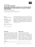
The electron transfer between azurin and nitrite reductase
from Ps. chlororaphis was also studied by cyclic voltamme-
try (Fig. 3). In the presence of Cu-Nir and nitrite, the cyclic
voltammograms of azurin exhibit a sigmoidal shape, with
enhanced cathodic currents and decreased anodic currents,
particularly at low scan rates. This behavior is consistent
with a reaction mechanism involving an initial heterogene-
ous electron transfer reaction at the electrode, followed by
an irreversible homogeneous chemical reaction in solution.
The measured catalytic current is independent of the scan
rate and proportional to the square root of the enzyme
concentration (not shown), which indicates that the
enhanced cathodic current is due to the catalytic regener-
ation of the azurin reoxidized by Nir [24]. The theory
describing this kind of mechanism has been developed by
Nicholson and Shain [24] and by Save
´
ant and Vianello [32]
and is frequently applied to kinetic studies of reactions
between redox enzymes and mediators. Second order rate
constants (k) were calculated as described in [25]: the kinetic
parameter k was calculated from the catalytic efficiencies
(the ratio of cathodic current in the presence and absence of
substrate) using the values computed by Nicholson and
Shain, and plotted vs. the inverse of the scan rate. The plots
yielded straight lines, confirming the applicability of the
Nicholson and Shain theory to the present system, and the
variation of the pseudo-first order rate constant with CuNir
concentrations (between 1 and 4 l
M
) yielded a value of
k ¼ (2.9 ± 0.9) · 10
4
M
)1
Æs
)1
for the rate constant between
reduced azurin and nitrite reductase.
The treatment described above is valid when the rate of
recycling of Cu-Nir
ox
[expressed by Eqn (1)] is not a limiting
factor for the catalytic current, i.e. when the limiting step of
the mechanism is the electron transfer between azurin and
Cu-Nir, whose rate is expressed by Eqn (2).
Fig. 3. Effect of the addition of Ps. chlororaphis DSM 50135 Cu-Nir on
the electrochemical response of azurin from the same organism, in the
presence of nitrite. Upper trace: 400 l
M
azurin in 50 m
M
Mes buffer
pH 6.2 and 50 m
M
KCl. Lower trace: addition of 6.5 l
M
Nir.
[nitrite] ¼ 50 m
M
.Scanrate,2mVÆs
)1
.
Fig. 2. Electronic absorption spectrum of the as-purified (blue) form of
the Ps. chlororaphis DSM 50135 nitrite reductase. [Cu-Nir] ¼ 23 l
M
in
20 m
M
Tris/HCl buffer pH 7.6.
Ó FEBS 2004 Cu-containing nitrite reductase from Ps. chlororaphis (Eur. J. Biochem. 271) 2365
V
1
¼ k
app
½CuÀNirðconstant pH, large excess of NO
À
2
Þ
ð1Þ
V
2
¼ k½azurin½CuÀNirð2Þ
The applicability of the Nicholson and Shain treatment
implies that V
2
< V
1
, which allows the determination of a
minimal value of 13 s
)1
for k
app
. This value is clearly smaller
than the observed turnover number of 243 s
)1
,which
confirms the applicability of the referred treatment to this
system.
Table 1 gathers several electron transfer constants
obtained by cyclic voltammetry in other physiologically
relevant systems. These data suggest that the electron
transfer between azurin and the Cu-Nir in Ps. chlororaphis
occurs at lower rates than those observed for other systems,
which raises the question whether the azurin is the
physiological reductant of Cu-Nir in this organism or not.
Ps. chlororaphis azurin reacts with its cognate Cu-Nir at
much higher rates than observed in similar experiments with
Al. xylosoxidans NCIB 11015 Cu-Nir and cognate azurins
[10], which have been reported as its electron donors.
However, the relevance of this comparison is hard to
ascertain, as recent results [9,11] suggest that, in the related
strain Al. xylosoxidans GIFU 1051, Cu-Nir may accept
electrons from cytochrome c
551
rather than from azurins I
and II.
EPR spectroscopy
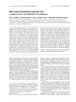
EPR spectra of Ps. chlororaphis nitrite reductase in the
as-purified, ascorbate-reduced, dithionite-reduced and turn-
over forms are presented in Fig. 4. The turnover form was
obtained by brief incubation of the dithionite reduced-form
with substrate under anaerobic conditions, and probably
corresponds to an enzyme form involved in the catalytic
cycle.
The as-purified form spectrum of Cu-Nir exhibits two
magnetically isolated components in a 3 : 1 ratio (Fig. 4A).
Addition of ascorbate reduces one of the components,
which becomes EPR silent, while the other component
remains in the oxidized form (Fig. 4B). The latter shows
a rhombic spectrum (g
z
¼ 2.350, g
y
¼ 2.110, g
x
¼ 2.040)
with hyperfine structure with the copper nucleus (I ¼ 3/2) at
the g
z
region (
Cu
A
z
¼ 10.7 mT). These properties identify
the system as a T2Cu center. Subtraction of this semi-
reduced spectrum from the native spectrum yielded an
almost axial spectrum (g
z
¼ 2.220, g
y
¼ 2.052, g
x
¼ 2.036),
showing also hyperfine structure at g
z
(
Cu
A
z
of 5.8 mT),
which identifies it clearly as originating from a T1Cu center
(Fig. 4A–B). EPR parameters obtained by simulation are
given in Table 2. EPR spectra of both T1Cu and T2Cu
centers are consistent with a d
x
2
-y
2
ground state, with
g
||
> g
^
> 2 [33].
Table 1. Electron transfer second order rate constants determined by
cyclic voltammetry in physiologically relevant systems.
Organism Electron transfer k (10
5
M
)1
Æs
)1
)
Al. faecalis
a
pAz fi Cu-Nir 18
Ac. cycloclastes
b
pAz fi Cu-Nir 7.3
Al. xylosoxidans
c
Cyt. c
551
fi Cu-Nir 4.0
Ps. nautica
d
Cyt. c
552
fi cd
1
-Nir 4.7
Methylomonas sp.
e
Az-iso2 fi MADH 1.0
Ps. chlororaphis
f
Az fi Cu-Nir 0.29
a
[40];
b
[8];
c
[11];
d
[41];
e
[42];
f
this work.
Fig. 4. Electron paramagnetic resonance spectra of Ps. chlororaphis
DSM 50135 nitrite reductase. (A) Native, (B) ascorbate-reduced, (C)
dithionite-reduced (the observed spectrum is a weak background from
the cavity), and (D) ÔturnoverÕ forms (see text for details); (A–B), dif-
ference spectrum, native minus ascorbate-reduced. Cu-Nir was 340 l
M
in 300 m
M
Tris/HCl pH 7.6. Instrument conditions: microwave fre-
quency, 9.49 GHz; microwave power, 2 mW; modulation amplitude,
0.4 mT
pp
; modulation frequency, 100 kHz and temperature, 40 K.
Table 2. EPR parameters for the type 1 and type 2 Cu centers in the
nitrite reductase from Ps. chlororaphis DSM 50135. ND, not deter-
mined. g-values ± 0.001, A-values ± 0.1.
Cu center g
z
g
y
g
x
Cu
A
z
(mT)
T1Cu 2.220 2.052 2.036 5.8
T2Cu
As-purified 2.350 2.110 2.040 10.7
With NO
2
–
2.34 ND ND 12.0
2366 D. Pinho et al.(Eur. J. Biochem. 271) Ó FEBS 2004
Upon reduction with excess of dithionite, T2Cu is also
completely reduced to an EPR-silent form (Fig. 4C).
Reoxidation of the protein with nitrite under anaerobic
conditions yielded a partially oxidized (ÔturnoverÕ)formof
the enzyme (Fig. 4D). The EPR spectrum shows that in this
form the reoxidation of T1Cu center is much more complete
than that of T2Cu, which suggests the T1Cu fi T2Cu
electron-transfer is faster than both the reoxidation of T2Cu
by nitrite and the reduction of T1Cu by dithionite, i.e. is the
fastest of the three redox processes involved in the catalysis
(cf [14]). The g
z
value and hyperfine coupling constant of the
spectrum of the turnover form of the T2Cu center are
slightly different from that of the as-purified form (cf.
Table 2) which suggests the existence of modifications in the
catalytic center of the enzyme. These modifications may be
attributed to either the presence of the substrate and/or a
product in the vicinity of the T2Cu active center rather than
to disruption of this center, as reoxidation by ferricyanide is
complete and originates a spectrum virtually identical to
that observed in the native form (data obtained during the
redox titration). Changes in the EPR spectrum of the
Al. xylosoxidans NCIB 11015 Nir upon addition of nitrite
have also been reported [34].
Redox titration
The development of appropriate models for the Cu-Nir
mechanism requires the determination of the potentials
of both copper centers, especially in order to understand
the relationship between redox catalysis and intramole-
cular electron transfer. However, the redox potentials of
copper nitrite reductases are not fully characterized.
Information available has been provided by electrochem-
ical titrations monitored by UV-vis spectroscopy [35],
that only follow the oxidation state of the T1Cu center
[36]. T2Cu center potentials have only been indirectly
estimated from pulse-radiolysis kinetic studies, and not
by electrochemical studies under equilibrium conditions
(cf. Table 3) [6,14,36,37].
Unlike UV-vis spectroscopy, EPR enables the simul-
taneous monitoring of both type 1 and T2Cu centers.
Therefore, in order to obtain reliable equilibrium redox
potentials, we performed for the first time an EPR
monitored redox-titration for a copper-containing Nir.
The protein was fully reduced with dithionite and
reoxidized stepwise with ferricyanide. The spectra of the
initial as-purified and the final reoxidized forms are
virtually identical, which demonstrates that the whole
process does not affect the integrity of the protein
sample. The results of the titration are presented in
Figs 5 and 6. As inferred in other Cu-Nirs (cf. Table 3),
the active center redox-potential in Ps. chlororaphis DSM
50135 Cu-Nir (E
m
T2Cu ¼ 172±5mV) is lower than
the electron-transfer center redox-potential (E
m
T1Cu ¼
298 ± 7 mV). Therefore, a slow electron transfer rate
between T1Cu and T2Cu centers should be expected
under these conditions, as it occurs against the electric
Table 3. Redox potentials for the copper centers in some nitrite reduc-
tases.
Organism E
m
T1Cu (mV) E
m
T2Cu (mV)
Ac. cycloclastes IAM 1013
b,d
240 250
a
Al. xylosoxidans GIFU 1051
b,d
280 280
a
Al. xylosoxidans NCIB 11015
b,e
260 240
a
Rhodobacter sphaeroides 2.4.3
b,f
247 <200
Ps. chlororaphis DSM 50135
c,g
298 172
a
Indirectly estimated value;
b
pH 7.0;
c
pH 7.6;
d
[14];
e
[36];
f
[35];
g
this work.
Fig. 5. EPR spectra observed during the redox titration of the
Ps. chlororaphis DSM 50135 nitrite reductase. Instrument conditions:
temperature, 20 K; microwave frequency, 9.49 GHz; microwave
power, 0.63 mW; modulation frequency, 100 kHz and modulation
amplitude, 1.0 mT
pp
.
Fig. 6. Redox titration of the Ps. chlororaphis DSM 50135 nitrite
reductase, at pH 7.6, monitored by EPR spectroscopy. m,oxidized
T1Cu signal intensity; s, oxidized T2Cu signal intensity. Experimental
data were normalized to the respective maximum intensities obtained
from Nernst equation fits (solid lines). EPR conditions given in Fig. 5.
Ó FEBS 2004 Cu-containing nitrite reductase from Ps. chlororaphis (Eur. J. Biochem. 271) 2367
potential. However, addition of nitrite to the reduced
form of the enzyme causes more complete reoxidation of
the T1Cu than of the T2Cu center (cf. Fig. 4), indicating
that the electron transfer process is kinetically and
thermodynamically favored in the presence of NO
2
–
.
Several explanations for these apparent contradictions
can be found in the literature. The binding of nitrite to
the oxidized form of T2Cu may increase its redox-
potential, thus making the electron transfer more spon-
taneous, as suggested by ENDOR experiments performed
in Rh. sphaeroides Cu-Nir [38]. Based on observations of
Al. xylosoxidans NCIB 11015 Cu-Nir, Prudeˆ ncio et al.
[39] have suggested that in vivo the T2Cu redox potential
may be increased considerably (even in the absence of
nitrite) by conformational changes induced by the
interaction of Cu-Nir with its natural electron donor.
In Al. xylosoxidans GIFU 1051 Nir, the T2Cu potential
depends on pH, and it is about 100 mV higher in the
protonated than in the deprotonated form of the enzyme
[14], while the T1Cu potential is pH-independent. Sup-
posing a similar dependence of the E
m
T2Cu with pH in
the Ps. chlororaphis Nir, it would be expected that at
lower pH values (the titration was performed at pH 7.6)
the electron transfer in the presence of nitrite should be
even more favored.
Conclusions
The nitrite reductase isolated from Ps. chlororaphis DSM
50135 is a blue protein with two types of copper-containing
centers, T1Cu and T2Cu, like other described Cu-Nirs.
Enzyme assays and electrocatalysis studies have shown that
the Cu-Nir from Ps. chlororaphis DSM 50135 accepts
electrons carried by the azurin purified from the same
organism. The direct determination of the redox potentials
of both copper centers yielded values (E
m
T1Cu ¼ 298 mV
and E
m
T2Cu ¼ 175 mV vs. NHE) which seem not to be
consistent with the proposed electron transfer pathway
(from electron donor to T1Cu to T2Cu to nitrite). However,
the EPR data indicate that nitrite binding to the T2Cu
increases the redox potential of this center, thereby making
the intramolecular electron-transfer more favorable, as
proposed by Veselov et al. [38]. EPR studies with the
ÔturnoverÕ form of the enzyme also suggest that, in the
presence of nitrite, the electron transfer between T1Cu and
T2Cu is the fastest of the three redox processes involved in
the catalysis: (a) reduction of T1Cu; (b) oxidation of T1Cu
by T2Cu; and (c) reoxidation of T2Cu by NO
2
–
(cf [14]).
Moreover, as it has been recently observed in Al. xylosoxi-
dans GIFU 1051 Nir [14] that the T2Cu potential increases
at low pH, it is likely that at that pH the intramolecular
electron transfer in the presence of nitrite will be even more
favorable. Further studies on the changes in enzyme activity
and both copper centers’ redox potentials with pH will
probably shed further light on the mechanisms underlying
the relieving of the apparent thermodynamic impediments
to the electron transfer to the substrate.
Acknowledgements
DP thanks the Fundac¸ a
˜
oparaaCieˆ ncia e Tecnologia for a PRAXIS
XXI PhD grant (BD/5041/95).
References
1. Zumft, W.G. (1997) Cell biology and molecular basis of denitri-
fication. Microbiol. Mol. Biol. Rev. 61, 533–616.
2. Godden, J.W., Turley, S., Teller, D.C., Adman, E.T., Liu, M.Y.,
Payne, W.J. & LeGall, J. (1991) The 2.3 angstrom X-ray structure
of nitrite reductase from Achromobacter cycloclastes. Science 253,
438–442.
3. Kukimoto, M., Nishiyama, M., Murphy, M.E.P., Turley, S.,
Adman, E.T., Horinouchi, S. & Beppu, T. (1994) X-ray structure
and site-directed mutagenesis of a nitrite reductase from Alcali-
genes faecalis S-6: roles of two copper atoms in nitrite reduction.
Biochemistry 33, 5246–5252.
4. Dodd, F.E., van Beeumen, J., Eady, R.R. & Hasnain, S.S. (1998)
X-ray structure of a blue-copper nitrite reductase in two crystal
forms. The nature of the copper sites, mode of substrate binding
and recognition by redox partner. J. Mol. Biol. 282, 369–382.
5. Inoue, T., Gotowda, M., Deligeer, Kataoka, K., Yamaguchi, K.,
Suzuki, S., Watanabe, H., Gohow, M. & Kai, Y. (1998) Type 1 Cu
structure of blue nitrite reductase from Alcaligenes xylosoxidans
GIFU 1051 at 2.05 angstrom resolution: comparison of blue and
green nitrite reductases. J. Biochem. 124, 876–879.
6. Suzuki, S., Kohzuma, T., Deligeer, Yamaguchi, K., Nakamura,
N., Shidara, S., Kobayashi, K. & Tagawa, S. (1994) Pulse
radiolysis studies on nitrite reductase from Achromobacter
cycloclastes IAM 1013: evidence for intramolecular electron
transfer from type 1 Cu to type 2 Cu. J. Am. Chem. Soc. 116,
11145–11156.
7. Libby, E. & Averill, B.A. (1992) Evidence that the type 2 copper
centers are the site of nitrite reduction by Achromobacter cyclo-
clastes nitrite reductase. Biochem. Biophys. Res. Commun. 187,
1529–1535.
8. Kohzuma, T., Takase, S., Shidara, S. & Suzuki, S. (1993) Elec-
trochemical properties of copper proteins, pseudoazurin and
nitrite reductase from Achromobacter cycloclastes IAM 1013.
Chem. Lett. 149–152.
9. Suzuki, S., Kataoka, K., Yamaguchi, K., Inoue, T. & Kai, Y.
(1999) Structure–function relationships of copper-containing
nitrite reductases. Coord. Chem. Rev. 190–192, 245–265.
10. Dodd, F.E., Hasnain, S.S., Hunter, W.N., Abraham, Z.H.L.,
Debenham,M.,Kanzler,H.,Eldridge,M.,Eady,R.R.,Ambler,
R.P. & Smith, B.E. (1995) Evidence for two distinct azurins in
Alcaligenes xylosoxidans (NCIMB 11015): potential electron
donors to nitrite reductase. Biochemistry 34, 10180–10186.
11. Deligeer, Kataoka, K., Yamaguchi, K. & Suzuki, S. (2000)
Spectroscopic and electrochemical properties of cytochrome c
551
from Alcaligenes xylosoxidans GIFU 1051. Bull.Chem.Soc.Jpn
73, 1839–1840.
12. Boulanger, M.J., Kukimoto, M., Nishiyama, M., Horinouchi, S.
& Murphy, M.E.P. (2000) Catalytic roles for two water bridged
residues (Asp-98 and His-255) in the active site of copper-con-
taining nitrite reductase. J. Biol. Chem. 275, 23957–23964.
13. Kataoka, K., Furusawa. H., Takagi, K., Yamaguchi, K. &
Suzuki, S. (2000) Functional analysis of conserved aspartate
and histidine residues located around the type 2 copper site of
copper-containing nitrite reductase. J. Biochem. 127, 345–350.
14. Kobayashi, K., Tagawa, S., Deligeer & Suzuki, S. (1999)
The pH-dependent changes of intramolecular electron transfer
on copper-containing nitrite reductase. J. Biochem. 126, 408–412.
15. Adman, E.T., Godden, J.W. & Turley, S. (1995) The structure of
copper-nitrite reductase from Achromobacter cycloclastes at five
pH values, with NO
À
2
bound and with type II copper depleted.
J. Biol. Chem. 270, 27458–27474.
16. Suzuki, S., Kataoka, K. & Yamaguchi, K. (2000) Metal
coordination and mechanism of multicopper nitrite reductase.
Acc. Chem. Res. 33, 728–735.
2368 D. Pinho et al.(Eur. J. Biochem. 271) Ó FEBS 2004
17. Miguel, A.H. & Brawn, R.D. (1974) Fluorimetric analysis of
nitrate in real samples. J. Chem. 51, 682–683.
18. Nicholas, D.J.D. & Nason, A. (1957) Determination of nitrate and
nitrite. Methods Enzymol. 3, 981–984.
19. Pinho, D., Besson, S., Brondino, C.D., Pereira, E., Castro, B. &
Moura, I. (2004) Two azurins with unusual redox and spectro-
scopic properties isolated from the Pseudomonas chlororaphis
strains DSM 50083
T
and DSM 50135. J. Inorg. Biochem. 98,
276–286.
20. Bensadoun, A. & Weinstein, D. (1976) Assays of proteins
in the presence of interfering materials. Anal. Biochem. 70,
241–250.
21. Laemmli, U. K. (1970) Cleavage of structural proteins during the
assembly of the head of bacteriophage T4. Nature 227, 680–685.
22. Hucklesby, D.P. & Hazeman, R.H. (1973) Staining method for
nitrite reductase on polyacrylamide gels after electrophoresis.
Anal. Biochem. 56, 591–592.
23. van Dijk, C., Mayhew, S.G., Grande, H.J. & Veeger, C. (1979)
Purification and properties of hydrogenase from Megasphaera
elsdenii. Eur. J. Biochem. 102, 317–330.
24. Nicholson, R.S. & Shain, I. (1964) Theory of stationary electrode
polarography: single scan and cyclic methods applied to reversible,
irreversible, and kinetic systems. Anal. Chem. 36, 706–723.
25. Hoogvliet, J.C., Lievense, L.C., van Dijk, C. & Veeger, C. (1988)
Electron transfer between the hydrogenase from Desulfovibrio
vulgaris (Hildenborough) and viologens. 1. Investigations by cyclic
voltammetry. Eur. J. Biochem. 174, 273–280.
26. Ellis, M.J., Dodd, F.E., Sawers, G., Eady, R.R. & Hasnain, S.S.
(2003) Atomic resolution structures of native copper nitrite
reductase from Alcaligenes xylosoxidans and the active site mutant
Asp92Glu. J. Mol. Biol. 328, 429–438.
27. Cutruzzola
`
, F. (1999) Bacterial nitric oxide synthesis. Biochim.
Biophys. Acta 1411, 231–249.
28. Abraham, Z.H., Lowe, D.J. & Smith, B.E. (1993) Purification and
characterization of the dissimilatory nitrite reductase from Alca-
ligenes xylosoxidans subsp. xylosoxidans (N.C.I.M.B. 11015):
evidence for the presence of both type 1 and type 2 copper centres.
Biochem. J. 295, 587–593.
29. Solomon, E.I., Baldwin, M.J. & Lowery, M.D. (1992) Electronic
structures of active sites in copper proteins: contributions to
reactivity. Chem. Rev. 92, 521–542.
30. Berks, B.C., Ferguson, S.J., Moir, J.W. & Richardson, D.J. (1995)
Enzymes and associated electron transport systems that catalyse
the respiratory reduction of nitrogen oxides and oxyanions.
Biochim. Biophys. Acta 1232, 97–173.
31. Denariaz, G., Payne, W.J. & LeGall, J. (1991) The denitrifying
nitrite reductase of Bacillus halodenitrificans. Biochim. Biophys.
Acta 1056, 225–232.
32. Save
´
ant, J.M. & Vianello, E. (1965) Potential-sweep chron-
oamperometry: kinetic currents for first-order chemical reaction
parallel to electron-transfer process (catalytic currents). Electro-
chim. Acta 10, 905–920.
33. Zeiger, H.J. & Pratt, G.W. (1973) Magnetic Interaction in Solids.
Clarendon Press, Oxford.
34. Howes, B.D., Abraham, Z.H.L., Lowe, D.J., Bruser, T., Eady,
R.R. & Smith, B.E. (1994) EPR and electron nuclear double
resonance (ENDOR) studies show nitrite binding to the type 2
copper centers of the dissimilatory nitrite reductase of Alcaligenes
xylosoxidans (NCIMB 11015). Biochemistry 33, 3171–3177.
35. Olesen, K., Veselov, A., Zhao, Y., Wang, Y., Danner, B., Scholes,
C.P. & Shapleigh, J.P. (1998) Spectroscopic, kinetic, and electro-
chemical characterization of heterologously expressed wild-type
and mutant forms of copper-containing nitrite reductase from
Rhodobacter sphaeroides 2.4.3. Biochemistry 37, 6086–6094.
36. Suzuki, S., Deligeer, Yamaguchi, K., Kataoka, K., Kobayashi, K.,
Tagawa, S., Kohzuma, T., Shidara, S. & Iwasaki, H. (1997)
Spectroscopic characterization and intramolecular electron
transfer processes of native and type 2 Cu-depleted nitrite
reductases. J. Biol. Inorg. Chem. 2, 265–274.
37. Farver, O., Eady, R.R., Abraham, Z.H. & Pecht, I. (1998) The
intramolecular electron transfer between copper sites of nitrite
reductase: a comparison with ascorbate oxidase. FEBS Lett. 436,
239–242.
38. Veselov, A., Olesen, K., Sienkiewicz, A., Shapleigh, J.P. & Scholes,
C.P. (1998) Electronic structural information from Q-band
ENDOR on the type 1 and type 2 copper liganding environment
in wild-type and mutant forms of copper-containing nitrite
reductase. Biochemistry 37, 6095–6105.
39. Prudeˆ ncio, M., Eady, R.R. & Sawers, G. (2001) Catalytic and
spectroscopic analysis of blue copper-containing nitrite reductase
mutants altered in the environment of the type 2 copper centre:
implications for substrate interaction. Biochem. J. 353, 259–266.
40. Iwasaki,Y.,Takeuchi,T.,Tamiya,E.,Karube,I.,Nishiyama,M.,
Horinouchi, S., Beppu, T., Kadoi, H., Uchiyama, S., Suzuki, S. &
Suzuki, M. (1992) Electrocatalysis of nitrite reductase from
Alcaligenes faecalis strain 6 mediated by native redox partner.
Electroanal. 4, 771–776.
41. Lopes, H., Besson, S., Moura, I. & Moura, J.J. (2001) Kinetics of
inter- and intramolecular electron transfer of Pseudomonas nautica
cytochrome cd
1
nitrite reductase: regulation of the NO-bound end
product. J. Biol. Inorg. Chem. 6, 55–62.
42. Suzuki,S.,Nakamura,N.,Yamaguchi,K.,Kataoka,K.,Inoue,
T., Nishio, N., Kai, Y. & Tobari, J. (1999) Spectroscopic and
electrochemical properties of two azurins (Az-iso1 and Az-iso2)
from the obligate methylotroph Methylomonas sp. strain J and the
structure of novel Az-iso2. J. Biol. Inorg. Chem. 4, 749–758.
Ó FEBS 2004 Cu-containing nitrite reductase from Ps. chlororaphis (Eur. J. Biochem. 271) 2369