Báo cáo khoa học: 2-Pyrimidinone as a probe for studying the EcoRII DNA methyltransferase–substrate interaction docx
Bạn đang xem bản rút gọn của tài liệu. Xem và tải ngay bản đầy đủ của tài liệu tại đây (331.36 KB, 9 trang )
2-Pyrimidinone as a probe for studying the
Eco
RII DNA
methyltransferase–substrate interaction
Oksana M. Subach
1
, Anton V. Khoroshaev
1
, Dmitrii N. Gerasimov
1
, Vladimir B. Baskunov
1
,
Anna K. Shchyolkina
2
and Elizaveta S. Gromova
1
1
Chemistry Department, Moscow State University, Russia;
2
Engelghardt Institute of Molecular Biology, Russian Academy of
Sciences, Moscow, Russia
EcoRII DNA methyltransferase (M.EcoRII) recognizes
the 5¢…CC*T/AGG…3¢ DNA sequence and catalyzes the
transfer of the methyl group from S-adenosyl-
L
-methionine
to the C5 position of the inner cytosine residue (C*). Here,
we study the mechanism of inhibition of M.EcoRII by DNA
containing 2-pyrimidinone, a cytosine analogue lacking an
NH
2
group at the C4 position of the pyrimidine ring. Also,
DNA containing 2-pyrimidinone was used for probing
contacts of M.EcoRII with functional groups of pyrimidine
bases of the recognition sequence. 2-Pyrimidinone was
incorporated into the 5¢…CCT/AGG…3¢ sequence repla-
cing the target and nontarget cytosine and central thymine
residues. Study of the DNA stability using thermal dena-
turation of 2-pyrimidinone containing duplexes pointed
to the influence of the bases adjacent to 2-pyrimidinone
and to a greater destabilizing influence of 2-pyrimidinone
substitution for thymine than that for cytosine. Binding of
M.EcoRII to 2-pyrimidinone containing DNA and methy-
lation of these DNA demonstrate that the amino group of
the outer cytosine in the EcoRII recognition sequence is not
involved in the DNA–M.EcoRII interaction. It is probable
that there are contacts between the functional groups of the
central thymine exposed in the major groove and M.EcoRII.
2-Pyrimidinone replacing the target cytosine in the EcoRII
recognition sequence forms covalent adducts with M.Eco-
RII. In the absence of the cofactor S-adenosyl-
L
-methionine,
proton transfer to the C5 position of 2-pyrimidinone occurs
and in the presence of S-adenosyl-
L
-methionine, methyl
transfer to the C5 position of 2-pyrimidinone occurs.
Keywords: 2-pyrimidinone; M.EcoRII; C5 cytosine DNA
methyltransferase; inhibition; DNA recognition.
DNA (cytosine-5)-methyltransferases (C5 MTases) catalyze
the transfer of a methyl group from S-adenosyl-
L
-methio-
nine (AdoMet) to cytosine C5 atom in specific DNA
sequences. The methylation reaction of C5 MTases occurs
with the addition of a cysteine thiol group from the
conserved Pro-Cys motif to the C6 position of the target
cytosine, followed by methyl transfer from AdoMet to the
C5 position of the target base and the release of the
methylated substrate [1,2] (Fig. 1A). It is important to note
that the target cytosine is flipped out of the DNA double
helix into the catalytic pocket of the enzyme and brought
into proximity of the cofactor [2].
Several cytosine analogues, 5-fluorocytosine (FC),
5-azacytosine (AzaC) and 2-pyrimidinone (2P), have been
reported as mechanism-based inhibitors of C5 MTases
[3–6]. Introduction of a fluorine atom to the C5 position of
the target cytosine results in an irreversible covalent attack
of a cysteine residue and transfer of a methyl group to the
C5 position of the target base [1]. Replacement of C5 by a
nitrogen atom in azacytosine (AzaC) facilitates nucleophilic
attack of the cysteine residue at the C6 position which
occurs in the presence or absence of AdoMet [5]. Methyl or
proton transfer to the N5 position occurs in the presence or
absence of AdoMet, accordingly. As a result, two structures
of the end product are possible: the enzyme linked to
methylated AzaC or the enzyme linked to protonated AzaC
[5].AsthereisnoprotonatC5totakeawaywhentheN5
position becomes methylated an irreversible covalent com-
plex is formed with the enzyme.
2-Pyrimidinone (2P) is a cytosine analogue in which the
exocyclic amino group is replaced by a hydrogen atom.
Removal of the exocyclic amino group from the cytosine
results in an increase of reactivity at the C6 carbon atom in
2P and in a reduction of the energy barrier for base flipping
[7]. 2P replacing the target cytosine in the recognition
sequences for HhaI[6],MspIandHgaI-2 [7–9] C5 MTases
evokes covalent bond formation with these MTases. Zhou
et al. [6] suggested the pathway of inhibition of C5 MTases
by 2P-containing DNA in the absence of AdoMet
(Fig. 1A). The reaction mechanism involves the addition
of a cysteine thiol group of the enzyme (from conserved
Pro-Cys motif) to the C6 position of 2-pyrimidinone
followed by proton transfer to the C5 position. Due to the
absence of the exocyclic amino group, b-elimination of the
proton from the C5 position is retarded. The mechanism of
inhibition of C5 MTases by 2P-containing DNA in the
Correspondence to E. S. Gromova; Chemistry Department, Moscow
State University, Moscow, 119992, Russia. Fax: + 7 095 939 31 81,
Tel.: + 7 095 939 31 44, E-mail:
Abbreviations: C5 MTase, C5 cytosine DNA methyltransferase; FC,
5-fluorocytosine; AzaC, 5-azacytosine; M.EcoRII, EcoRII DNA
methyltransferase; AdoMet, S-adenosyl-
L
-methionine; AdoHcy,
S-adenosyl-
L
-homocysteine; 2P, 2-pyrimidinone.
Enzyme: EcoRII DNA methyltransferase (EC 2.1.1.37).
(Received 18 November 2003, revised 19 February 2004,
accepted 14 April 2004)
Eur. J. Biochem. 271, 2391–2399 (2004) Ó FEBS 2004 doi:10.1111/j.1432-1033.2004.04158.x
presence of AdoMet was not investigated. It should be
noted that 1-(b-
D
-ribofuranosyl)-2-pyrimidinone, often
referred to as zebularine, was used in vivo as an antitumor
drug [10]. By now it is clear that its antitumor properties are
likely attributed to inhibition of C5 MTase activity in tumor
cells. 2-Pyrimidinone could also be considered as a mimic
of an intermediate in the minor deaminative pathway of
C5 MTases catalysis [6].
EcoRII DNA methyltransferase (M.EcoRII) catalyzes
the transfer of a methyl group from AdoMet to the C5
position of the inner deoxycytidine residue (C*) in the DNA
sequence 5¢…CC*T/AGG…3¢. It has been shown that
M.EcoRII forms an irreversible covalent complex with
DNA containing FC instead of the target cytosine [11,12].
Introduction of AzaC in this position also led to inhibition
of the enzyme [5]. The mechanism of inhibition of M.Eco-
RII by 2P-containing DNA is still unknown. Also, a study
of the contributions of different functional groups of the
5¢…CCT/AGG…3¢ sequence to specific interaction with
M.EcoRII is at the very beginning [13].
We aim to explore the effect of 2P substitution for C and
T within the recognition sequence of M.EcoRII on recog-
nition and catalysis performed by M.EcoRII. In this work,
we have examined a DNA duplex containing 2-pyrimidi-
none instead of the target cytosine as a potential inhibitor
of M.EcoRII, and also the role of AdoMet in the formation
of the covalent adduct between 2P-substituted DNA and
M.EcoRII. Furthermore, we have studied the contribution
of the functional groups of pyrimidine residues in DNA–
M.EcoRII recognition by using duplexes containing
2-pyrimidinone in place of each pyrimidine base in the
5¢…CCT/AGG…3¢ sequence.
Materials and methods
Chemicals and enzymes
AdoMet and AdoHcy were from Sigma (USA).
[CH
3
–
3
H]AdoMet (77 CiÆmmol
)1
) was from Amersham
(USA). [
32
P]ATP[cP] (1000 CiÆmmol
)1
) was from Izotop
(Obninsk, Russia). DNA-methyltransferase EcoRII (stock
solution, 52 l
M
) was overexpressed as an N-terminally
His
6
-tagged protein and chromatographically purified
on a nickel chelate column as described previously [14].
T4 polynucleotide kinase was from MBI Fermentas
(Vilnius, Lithuania).
Oligonucleotides
Oligodeoxyribonucleotides containing 2-pyrimidinone
were synthesized as described previously [15].
32
P-phosphory-
lation of the oligonucleotides was performed using
T4-polynucleotide kinase and [
32
P]ATP[cP].
UV thermal denaturation and thermodynamic parameters
of duplex formation
Heating of the samples containing 2.25 l
M
of duplexes in
buffer A (40 m
M
Tris/HCl, pH 7.9, 1 m
M
EDTA, 50 m
M
NaCl) at temperatures ranging from 15 to 85 °Cwas
performed at a constant rate of 0.2 °CÆmin
)1
. Absorbance
of duplexes at 260 nm was measured using Cary 50 Bio
spectrophotometer (Varian, Victoria, Australia) with tem-
perature controller. Thermodynamic analysis of helix-coil
transition curves was performed using a two-state model.
The thermodynamic parameters were determined through a
fitting procedure based on minimization of the integral
mean square deviation between the theoretical transition
curves and experimental absorbance data.
Circular dichroism
CD measurements of duplexes (2.25 l
M
) in buffer A were
performed with Mark V Jobin Yvon dichrograph (Paris,
France) in 1 cm thermostated cells.
Gel mobility-shift assay
To determine the active enzyme concentration, 20 n
M
M.EcoRII was incubated with 1–50 n
M
32
P-labeled DNA
duplex I¢ in 20 lL of buffer B (40 m
M
Tris/HCl, pH 7.9,
5m
M
dithiothreitol, 1 m
M
EDTA) containing 50 m
M
NaCl, 6% glycerol and 1 m
M
AdoHcy for 15 min at room
temperature and 10 min at 0 °C. The reaction mixtures
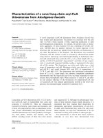
Fig. 1. Proposed mechanism of inhibition of M.EcoRII by 2P-containing DNA duplexes in the absence [6] (A) or in the presence (B) of AdoMet. In the
case of M.EcoRII, the amino acid residue attacking the C6 position is Cys186 [11,12]; the general acid donating a proton to N3 is probably Glu233
[4,28].
2392 O. M. Subach et al. (Eur. J. Biochem. 271) Ó FEBS 2004
were analysed by 8% native PAGE for 3 h at 120 V. The gel
was prerun for 1 h at 100 V. Autoradiographs of the gels
were prepared using Molecular Dynamics Phosphorimager
(Amersham Biosciences, USA). Radioactivities of M.Eco-
RII–DNA complex (cpm
bound
) and free DNA (cpm
free
)
were determined. The ratio of bound to free DNA was
calculated as (cpm
bound
)/(cpm
free
); the concentration of
bound DNA was calculated as [S
0
][(cpm
bound
)/(cpm
bound
+
cpm
free
)]. Data were analyzed by linear regression using the
Microcal
ORIGIN
6.0software.Forallfurtherexperiments
the concentration of active form of M.EcoRII was used.
To determine the apparent dissociation constants (K
app
d
),
1.5–150 n
M
M.EcoRII were incubated with 15 n
M
32
P-
labeled DNA duplexes I–IV in 15 lL of buffer B (40 m
M
Tris/HCl, pH 7.9, 5 m
M
dithiothreitol, 1 m
M
EDTA, con-
taining 8% glycerol and 1 m
M
AdoHcy for 15 min at room
temperature and 15 min at 0 °C. The reaction mixtures were
processed as described above. Radioactivities of M.EcoRII–
DNA complex (cpm
bound
) and free DNA (cpm
free
)were
determined and the fraction of bound DNA was calculated
as (cpm
bound
)/(cpm
bound
+cpm
free
). K
app
d
was calculated by
fitting the data to the following equation derived from a
standard bimolecular binding equilibrium as described [16]:
cpm
bound
ðcpm
bound
þcpm
free
Þ
¼
½ES
½S
0
¼
A
2½S
0
ð½S
0
þ½E
0
þK
app
d
ÞÀ
ffiffiffiffiffiffiffiffiffiffiffiffiffiffiffiffiffiffiffiffiffiffiffiffiffiffiffiffiffiffiffiffiffiffiffiffiffiffiffiffiffiffiffiffiffiffiffiffiffiffiffiffiffiffiffiffiffiffiffiffiffiffiffi
ð½S
0
þ½E
0
þK
app
d
Þ
2
À4Á½E
0
Á½S
0
q
where [S
0
] is total DNA concentration; [E
0
]istotal
M.EcoRII concentration; A is the factor accounting for
nonideal equilibrium conditions during electrophoresis
(cage effect, thermal dissociation). Nonlinear regression
was performed using Microcal
ORIGIN
6.0 software.
Detection of DNA-enzyme adducts
All reactions of
32
P-labeled duplexes I–III (100 n
M
)with
M.EcoRII (200 n
M
) were performed in 15 lL of buffer B
containing 0.5 m
M
AdoMet or 0.5 m
M
AdoHcy. Reaction
mixtures were incubated for 30 min at room temperature
andfor15minoniceandprocessedbyoneofthefollowing
ways: (a) incubated with 1% SDS at room temperature for
10 min and analyzed by 8% native PAGE; (b) incubated
with 1% SDS at 65 °C for 5 min and analyzed by 8% native
PAGE; (c) incubated with 0.8% SDS at room temperature
for 10 min and analyzed by 12% SDS/PAGE (Laemmli
gel).
For autoradiography of the electrophoretic pattern,
Kodak-XOMAT-S film was exposed with an intensifier
screen at )20 °Covernight.
Methylation assay
For determination of initial methylation rates (V
0
), methy-
lation reactions were performed in 26 lL of buffer B,
containing DNA duplexes I–IV or Im–IVm (1 l
M
), M.Eco-
RII (30 n
M
), and [CH
3
–
3
H]-AdoMet (1.3 l
M
). Reactions
were started by adding the enzyme. After a 1, 2, 3, 4 and
5 min incubation at 15 °C, 5 lL of reaction mixture were
pipetted onto DE81 (Whatman) paper disks. The disks were
washed 5· for5minwith50m
M
KH
2
PO
4
,3minwith
water and 3 min with ethanol, air dried and placed into
5 mL of ZHS-106 scintillation fluid. The filter-bound
radioactivity was measured on Tracor Analytic Delta 300
scintillation counter (ThermoQuest/CE Instruments,
Piscataway, USA) and the amount of methylated DNA
was determined as described [17]. Data were analyzed by
linear regression using the Microcal
ORIGIN
6.0 software.
For quantification of the transfer of methyl groups to a
2-pyrimidinone residue in DNA duplexes by M.EcoRII
methylation reactions were performed in 10 lL of buffer B,
containing duplexes I, Im, III, IIIm or V (500–1000 n
M
),
M.EcoRII (12.5–1000 n
M
)and[CH
3
–
3
H]-AdoMet
(1.3 l
M
). In the case of M.HhaI methylation reactions were
performed in 10 lL of buffer C (50 m
M
Tris/HCl, pH 7.5,
5m
M
2-mercaptoethanol, 10 m
M
EDTA, 0.2 mg mL
)1
bovine serum albumin), containing duplexes I, Vm or VIm
(670 n
M
), M.HhaI (20–16 000 n
M
)and[CH
3
–
3
H]-AdoMet
(0.42 l
M
). Reactions were started by adding the enzyme.
After a 30 min incubation at 15 °C (reactions with M.Eco-
RII) or at 20 °C (reactions with M.HhaI), 8 lL of reaction
mixture were pipetted onto DE81 (Whatman) paper disks
and processed as described for determination of V
0
.
The time-dependent methyltransferase assays were per-
formed by incubating M.EcoRII (500 n
M
) with duplexes Im
or IIIm (500 n
M
)and[CH
3
–
3
H]-AdoMet (1.3 l
M
) in buffer
Bat15°C for the indicated time periods. Reaction mixtures
(8 lL) were pipetted onto DE81 (Whatman) paper disks
and processed as described for determination of V
0
.
Results and discussion
To elucidate the mechanism of inhibition of M.EcoRII
by 2-pyrimidinone modified DNA, and to understand the
role of functional groups of pyrimidine bases of the
recognition sequence in specific DNA–M.EcoRII inter-
action, a series of 2P-containing substrate analogues
was synthesized (duplexes II–IV, Table 1 and duplexes
Iim–IVm, Table 2).
Thermodynamics of formation of the
2P
-containing DNA
Insertion of 2P in place of C or T resulted in a marked
destabilization of DNA duplexes [18–21]. To ascertain
whether the 2P-containing DNA duplexes II–IV had a
double helix structure under conditions of methylation by
M.EcoRII, the thermodynamic stability of these duplexes
was evaluated (Table 1). The values of the free energy of
transition, as well as of the transition temperature (melting
temperature, T
m
), point to the following duplex stabilities:
I > II > III > IV. In order to elucidate the contribution
of a single nucleotide substitution, the value of the transition
free energy of the nonmodified duplex I was subtracted
from that of the modified duplexes II–IV (Table 1, DDG).
The greatest energy penalty, DDG, for substitution of 2P
for T in the TA base pair was 4.4 kcalÆmol
)1
.The
substitution of 2P for C appeared to be much more
energetically disadvantageous in the base context C2PT
(III) than in the context A2PC (II). The presence of only
two H-bonds in the 2PG base pair vs. the three H-bonds in
the CG base pair [19] as well as electrostatic interactions of
2P in the duplexes [21] may largely contribute to the
destabilization of the duplexes.
Ó FEBS 2004 2-Pyrimidinone as a probe for DNA methyltransferase (Eur. J. Biochem. 271) 2393
Conformation of the
2P
-containing DNA
To answer the question of whether 2P substitution for
pyrimidines led to a marked distortion of DNA conforma-
tion, we compared CD spectra of duplexes I–IV (Fig. 2).
There are minor changes in the CD spectra, predominantly
an intensity drop in the longwave CD band. This may be
attributed to a strongly reduced absorption of the 2P base at
260 nm [18,19], which eliminates a potential contribution of
the nearest-neighbor stacking contacts involving the 2PG
or 2PA base pair to the CD signals in the corresponding
spectral region. The difference in the CD spectra (Fig. 2,
open symbols) reflects the decreased contribution of 2P to
the conservative CD spectrum in comparison with the
contributions of cytosine and thymine. Thus, the observed
minor dissimilarity in the CD spectra of duplexes II–IV
from CD spectrum of duplex I can be attributed to the
distinctive optical features of the 2P analog, rather than to a
marked distortion of the DNA conformation.
Determination of concentration of active form
of M.EcoRII
It is known that the concentration of the active form of
DNA methyltransferases does not correspond accurately to
the total protein concentration [5,22]. The concentration of
active form of M.EcoRII was estimated by titration of the
enzyme (20 n
M
) with M.EcoRII substrate in the presence of
AdoHcy (Fig. 3, inset). The ratio of bound to free DNA
was plotted vs. concentration of bound DNA (a Scatchard
plot, Fig. 3) [23]. An 18-mer DNA duplex 5¢-GAG
CCAACCTGGCTCTGA-3¢/3¢-CTCGGTTGGACCGAG
ACT-5¢ (I¢) was used as M.EcoRII substrate. The horizontal
axis intercept gives the total concentration of DNA binding
sites (n[E
0
]) equal to 2.35 ± 0.12 n
M
(Fig. 3) or
2.04 ± 0.25 (data not shown). As the number of DNA
binding sites per molecule of enzyme (n) is 1 for M.EcoRII,
the average concentration of M.EcoRII active form is
2.2 ± 0.2 n
M
. So, the amount of enzyme bound to DNA
was assumed to be only 11 ± 1% of the total enzyme
molecules. For all further experiments the concentration of
active form of M.EcoRII was used.
Binding and methylation of
2P
-containing DNA
by M.EcoRII
Different heterocyclic base analogues have proven to be
useful for studies of DNA–enzyme interactions [24]. In
order to investigate the influence of introducing 2P into the
different positions of the substrate DNA on the sequence-
specific interaction of M.EcoRII with DNA, we studied
Table 2. Relative initial methylation rates of hemimethylated 2P-con-
taining DNA duplexes by M.EcoRII. Relative initial methylation rates
(V
rel
0
) were calculated as ratio of V
0
of duplexes Iim–IVm to V
0
of
duplex Im. M, 5-methylcytosine. Recognition sequence is in bold; 2P is
underlined.
No. DNA duplex V
0
(n
M
Æmin
)1
)V
rel
0
(%)
Im
5¢-GCCAACCTGGCTCT-3¢/ 172.0 ± 34.4 100 ± 20
3¢-CGGTTGGAMCGAGA-5¢
IIm
5¢-GCCAA
2P
CTGGCTCT-3¢/ 197.8 ± 53.3 115 ± 31
3¢-CGGTT-GGAMCGAGA-5¢
IIIm
5¢-GCCAAC
2P
TGGCTCT-3¢/0 0
3¢-CGGTTG-GAMCGAGA-5¢
IVm
5¢-GCCAACC
2P
GGCTCT-3¢/ 34.4 ± 3.4 20 ± 2
3¢-CGGTTGG-AMCGAGA-5¢
Fig. 2. CD spectra of DNA duplexes I–IV. ––,I;–d–, II; –m–, III;
–j–, IV. Difference of CD spectra of I and duplexes: –s–, II; –n–, III;
–h–, IV. Temperature was 20 °C.
Table 1. Thermodynamic parameters of formation of 2P-containing DNA duplexes determined from thermal denaturation curves. Thermodynamic
parameters and their standard deviations were determined from fitting the theoretical melting curves to experimental curves (see Materials and
methods). Standard deviation of DS was less than 0.1 calÆmol
)1
ÆK
)1
. Experimental conditions see Materials and methods. 2P, 2-pyrimidinone;
T
m
, melting temperature. Recognition sequence is in bold; 2P is underlined.
No. DNA duplex
T
m
(°C)
DH
(kcalÆmol
)1
)
DS
(kcalÆmol
)1
ÆK
)1
)
DG(T ¼ 20 °C)
(kcalÆmol
)1
)
DDG(T ¼ 20 °C)
(kcalÆmol
)1
)
I
5¢-GCCAACCTGGCTCT-3¢/ 52.6 ± 0.1 )65.0 ± 0.6 )173 )14.3 ± 0.6 –
3¢-CGGTTGGACCGAGA-5¢
II 5¢-GCCAA
2P
CTGGCTCT-3¢/ 46.0 ± 0.1 )43.9 ± 0.7 )111 )11.5 ± 0.7 2.8
3¢-CGGTT-GGACCGAGA-5¢
III
5¢-GCCAAC
2P
TGGCTCT-3¢/ 41.4 ± 0.1 )36.3 ± 0.4 )88 )10.3 ± 0.4 4
3¢-CGGTTG-GACCGAGA-5¢
IV
5¢-GCCAACC
2P
GGCTCT-3¢/ 35.4 ± 0.1 )41.7 ± 0.7 )106 )9.9 ± 0.7 4.4
3¢-CGGTTGG-ACCGAGA-5¢
2394 O. M. Subach et al. (Eur. J. Biochem. 271) Ó FEBS 2004
binding and methylation of canonical (I) and the 2P-
containing DNA duplexes (II-IV) with M.EcoRII (Table 3).
The formation of complexes was monitored by gel mobility-
shift assays in the presence of AdoHcy because of the
known increase in the affinity of M.EcoRII for DNA in the
presence of the cofactor [12]. DNA duplexes were incubated
with increasing M.EcoRII concentrations at saturating
AdoHcy concentrations. A binding isotherm and corres-
ponding autoradiograph of a typical experiment are shown
in Fig. 4. The calculated apparent dissociation constants
(K
app
d
) are summarized in Table 3.
The methylation reactions were performed under steady-
state conditions (Tables 2 and 3). Reactions of M.EcoRII
with hemimethylated duplexes Im–IVm were performed in
order to determine the influence of the 2P on methylation of
2P-containing strands of the DNA duplexes (Table 2).
M.EcoRII binds to the substrate analogue II with the
same affinity as to the parent duplex I (Table 3). Replace-
ment of the outer C (duplexes II and IIm) by 2P does not
affect methylation of either strand (II) or the 2P-containing
strand (IIm) (Tables 2 and 3). According to the thermo-
dynamic and conformational analysis of the 2P-containing
duplexes, the substitution of 2P for C could be a good probe
for DNA–protein interactions in the major groove of DNA.
Substitution of 2P for C appears to cause a small
destabilizing effect and duplex II is conformationally similar
to duplex I. The explanation of the observed equal binding
affinities and methylation rates can be attributed to the
difference in the chemical structure between 2P and C.
Therefore, the 4-NH
2
group of the outer cytosine residue in
the recognition sequence is not likely to be essential for
sequence-specific DNA binding by M.EcoRII. In contrast,
2P substitution for both nontarget cytosine residues in the
recognition sequence of MspI and HpaII C5 MTases
prevents these MTases from binding, and the C4 amino
functional groups of the nontarget cytosine residues are
essential for DNA binding by these MTases [25].
M.EcoRII binds to duplex III containing 2P in place of
the target cytosine with a K
app
d
similar to that of duplex I
(Table 3). It could be that the 4-NH
2
group of the target
cytosine is not essential for recognition of DNA by
M.EcoRII as was suggested for several other MTases, as
base flipping probably occurs with any base at the target
position [26]. However, in the case of the M.EcoRII complex
with duplex III such a simple conclusion is ambiguous
because in addition to the noncovalent complex a stable
Fig. 3. Scatchard plot of the ratio of bound to
free DNA substrate vs. concentration of bound
DNA substrate. Inset: autoradiograph of gel
shift assay of M.EcoRII with DNA substrate
I¢. Lanes 1–4: 20 n
M
M.EcoRII with 1 m
M
AdoHcy and increasing concentrations of
duplex I¢ (1,2,3.5and10n
M
).
Table 3. Binding and substrate properties of 2P-containing DNA duplexes. Apparent dissociation constants (K
app
d
) of complex M.EcoRII–DNA–
AdoHcy were calculated as described in Materials and methods. Relative K
app
d
[K
app
d
(rel.)] were calculated as ratio of K
app
d
of duplexes II–IV to
K
app
d
of duplex I. Relative initial methylation rates (V
rel
0
) were calculated as ratio of V
0
of duplexes II–IV to V
0
of duplex I. Recognition sequence is
in bold; 2P is underlined.
No DNA duplex K
app
d
(n
M
) K
app
d
(rel.) (%) V
0
(n
M
Æmin
)1
) V
rel
0
(%)
I
5¢-GCCAACCTGGCTCT-3¢/ 4.9 ± 1.8 100 ± 37 195.6 ± 39.1 100 ± 20
3¢-CGGTTGGACCGAGA-5¢
II
5¢-GCCAA
2P
CTGGCTCT-3¢/ 3.9 ± 1.4 80 ± 28 170.2 ± 56.7 87 ± 29
3¢-CGGTT-GGACCGAGA-5¢
III
5¢-GCCAAC
2P
TGGCTCT-3¢/ 5.3 ± 2.4 108 ± 49 3.5 ± 0.3 1.8 ± 0.1
3¢-CGGTTG-GACCGAGA-5¢
IV
5¢-GCCAACC
2P
GGCTCT-3¢/ 96.0 ± 35.6 1959 ± 726 41.1 ± 11.7 21 ± 6
3¢-CGGTTGG-ACCGAGA-5¢
Ó FEBS 2004 2-Pyrimidinone as a probe for DNA methyltransferase (Eur. J. Biochem. 271) 2395
covalent M.EcoRII complex with duplex III can be formed
(see below). Thus, the K
app
d
obtained does not represent true
binding affinity of M.EcoRII to duplex III. In the case of
duplexes III and IIIm, methylation was essentially not
detected under steady-state conditions. The transfer of a
methyl group is blocked or occurs at very low levels.
In the case of duplex IV containing 2P in place of the
central T in the recognition sequence, we observed a
substantial increase in K
app
d
(Table 3). Initial methylation
rates of both strands (IV) or 2P-containing strand (IVm)
were decreased (Tables 2 and 3). It is probable that the
decrease in V
0
is attributed to the weak binding affinity of
these substrate analogues to the enzyme. Recently, it was
suggested that AT vs. GC discrimination is achieved by
interactions between the large domain of M.EcoRII and the
minor groove of DNA [13]. M.EcoRII did not bind to a
substrate analogue with 2-aminopurine having been substi-
tuted for adenine (M. G. Brevnov, O. A. Rechkoblit and
E. S. Gromova, unpublished results). Hence, the appear-
ance of the amino group in the minor groove in the case of
2-aminopurine for adenine substitution, disturbs the recog-
nition of the specific DNA sequence by M.EcoRII. This is in
agreement with the role of the minor groove in substrate
recognition by M.EcoRII [13]. In the case of a substitution
of T by 2P (duplex IV), the pattern of functional groups
exposed into the minor groove remains the same, with the
groups of the central thymine residue exposed into the
major groove being disturbed. Therefore, it is likely that
weak binding of duplex IV to M.EcoRII may be attributed
to the elimination of some DNA–protein contacts in the
major groove of the double helix. Alternatively, this effect
may be caused by a greater destabilization of duplex IV in
comparison with duplexes II and III (Table 1). However,
the conformations of duplexes I–IV are similar. It has also
been shown that substitution of AT by CI in the 5¢…GGT/
ACC…3¢ sequence for SinI C5 MTase led to a considerable
increase in K
m
[13]. This observation corresponds to our
suggestion that specific contacts of C5 MTases with the
central base pair could be mediated by contacts not only in
the minor but also in the major groove.
Comparison of methylation of unmethylated and hemi-
methylated DNA duplexes (Tables 2 and 3) permits us to
speculate about influence of 2P on methylation of unmodi-
fied DNA strand in duplexes II–IV. Equal methylation rates
of duplexes II and IIm allow us to suggest that rates of
methylation of unmodified and 2P-containing strands in
duplex II are virtually the same. Analogously, we suppose
equal methylation rates of unmodified and 2P-containing
strands in duplex IV. The unmodified strand in duplex III
was not methylated under steady-state conditions – prob-
ably due to formation of the stable covalent adduct of
M.EcoRII with 2P-containing strand.
Mechanism-based inhibition of M.EcoRII
by
2P
-containing DNA
To examine the possibility of covalent adduct formation
between M.EcoRII and DNA containing 2P in place of the
target C in the presence of AdoMet or AdoHcy, duplex III
was incubated with the enzyme. The resulting samples were
analyzed under different conditions. First, the enzyme was
denaturated by adding SDS to a final concentration of 1%
and subjected (or not) to heating with a subsequent analysis
by 8% native PAGE (Fig. 5A). Without heating in the
presence of AdoHcy, a small part of the noncovalent
complex remains in the case of canonical duplex I and
duplex II in which the outer cytosine residue of the
recognition sequence was replaced by 2P, however, this
complex is absent after heating. We did not observe the
formation of the covalent adduct in the case of duplex II in
the presence of AdoMet. Figure 5A (lanes 4, 5 and 9, 10)
demonstrates that duplex III forms in the presence or in the
absence of AdoMet a covalent intermediate with M.EcoRII
stable to heating at 65 °C for 5 min. Thus, 2-pyrimidinone
for the target C substitution results in the inhibition of
M.EcoRII.
The adducts of 2P-containing duplex III with M.EcoRII
obtained in the presence of AdoHcy or AdoMet are not
resistant to heating in SDS solution at 90 °Cfor5 min(data
not shown) or to the addition of SDS and analysis by SDS/
PAGE (Fig. 5B, lanes 6 and 10). However, we observed
products moving faster than the protein and slower than the
oligonucleotides. These products seem to be oligonucleo-
tides generated from the duplex III–M.EcoRII adducts. The
SDS gel (Laemmli) exhibits two components at different
pH: an upper part at pH 6.8 (stacking gel) and a lower part
at pH 8.8 (separating gel) (Fig. 5B). Due to a pH change
from 6.8 to 8.8, b-elimination of the proton from the C5
position of 2P and dissociation of the covalent intermediates
of M.EcoRII and duplex III take place. The appearance of
the slowly moving oligonucleotides is attributed to retarda-
tion of the duplex III–M.EcoRII covalent intermediates in
the upper part of the gel before dissociation.
It is interesting to compare the stabilities of the adducts of
C5 MTases with DNA duplexes containing AzaC, FC or
2P in place of the target C in the presence of AdoMet. The
adducts of M.EcoRII with AzaC DNA [5] and M.HhaI
with FC DNA [27] are resistant to heating in SDS solution
Fig. 4. Binding of M.EcoRII to DNA duplex I in the presence of
AdoHcy. Relative amount of M.EcoRII–DNA–AdoHcy complex
obtained from the gel-shift autoradiograph vs. protein concentration is
plotted. M.EcoRII (1.5–94 n
M
) was incubated with duplex I (15 n
M
)in
the presence of AdoHcy (1 m
M
). Inset, autoradiograph of gel-shift
assay of M.EcoRII with duplex I. Lanes: 1–8, duplex I with 1 m
M
AdoHcy and increasing concentrations of M.EcoRII (5, 6, 7, 10, 40,
50, 62.5 and 78 n
M
); 9, duplex I.
2396 O. M. Subach et al. (Eur. J. Biochem. 271) Ó FEBS 2004
and analysis by SDS/PAGE. In contrast, the covalent
intermediates of M.EcoRII with 2-pyrimidinone (duplex
III) dissociate upon heating in SDS solution or during
analysis by SDS/PAGE, probably because of b-elimination
of the proton from the C5 position of 2-pyrimidinone.
Analysis of possibility of 2-pyrimidinone methylation
To clarify the role of AdoMet in the formation of the
covalent adduct between 2P-containing DNA and M.Eco-
RII it is important to examine the possibility of a methyl
group transfer to the 2P residue. 2P-modified DNA was
not methylated by MspIandHhaI C5 MTases [6]. We
also did not observe the methylation of duplex III under
steady-state conditions (Table 3). However, the proposed
mechanism of inhibition of C5 MTases by 2-pyrimidinone
[6] does not contradict the transfer of a methyl group to
the 2P residue.
The possibility of the methylation of DNA duplexes
containing 2P in place of the target cytosine (III and IIIm)
by M.EcoRII was tested at different enzyme concentrations
inthepresenceofAdoMetat15°C for 30 min (Fig. 6).
Hemimethylated duplex IIIm was used to exclude methy-
lation of the unmodified strand. Under the same conditions,
methylation of canonical duplexes I and Im was performed.
The increase of enzyme concentration favoured methylation
of 2P-containing DNA duplexes III and IIIm (Fig. 6). No
methyl transfer was detected at all enzyme concentrations in
the case of duplex 5¢-GAGCCAAGCGCACTCTGA-3¢/
3¢-CTCGGTTCGCGTGAGACT-5¢(V) lacking the EcoR-
II recognition sequence (Fig. 6). There was also no methy-
lation in a control sample containing the same amount of
enzyme and AdoMet but no DNA. Methylation of duplex
III may be due to methyl transfer to the target unmethylated
cytosine residue. However, this is impossible in the case of
duplex IIIm. Hence, one can suggest that a methyl group
transfer occurs to the 2-pyrimidinone base.
The methylation of duplex IIIm may be stopped at the
stage of formation of the covalent intermediate (Fig. 1B,
step1) or may proceed with dissociation of the covalent
intermediate and release of the methylated 2P-containing
DNA (Fig. 1B, step2). In the first case, the quantity of methyl
groups incorporated into duplex IIIm should correspond to
the quantity of methyl groups incorporated into canonical
DNA after the first turnover of the methylation reaction. In
the second case, we should observe more than one turnover
of the methylation reaction for duplex IIIm. To clarify the
nature of this new effect, we compared the dependence of
methylation of duplexes Im and IIIm on an enzyme
concentration (Fig. 6). Complete methylation of duplex Im
was observed even at low enzyme concentration. M.EcoRII
transfers the methyl group to unmodified DNA strand, turns
Fig. 6. Dependence of methylation of unmethylated (III), hemimethyl-
ated (Im and IIIm) and nonspecific (V) DNA duplexes on concentration of
M.EcoRII. M.EcoRII was incubated with indicated duplexes (500 n
M
)
in buffer B in the presence of [CH
3
–
3
H]AdoMet (1.3 l
M
)at15°Cfor
30 min. Relative methylation was calculated as the ratio of radio-
activities of duplexes Im, III, IIIm and V to the radioactivity of duplex I.
Methylation of duplex I (not shown) was accepted as 100%. s,
Canonical duplex Im; j, duplex III; d, duplex IIIm and m, duplex V.
Fig. 5. Covalent adduct formation of M.EcoRII with DNA duplexes
I–III in the presence of AdoMet or AdoHcy. M.EcoRII (200 n
M
)was
incubated with indicated duplexes (100 n
M
) in buffer B in the presence
of AdoHcy or AdoMet (0.5 m
M
). (A). Autoradiograph of 8% native
PAGE.Reactionswereincubatedwith1%SDSandheatedfor5min
at 65 °C prior to electrophoresis if indicated. Lanes: 1, without
M.EcoRII; 2–11, with M.EcoRII. (B). Autoradiograph of 12% SDS/
PAGE (Laemmli). Reactions were incubated with 0.8% SDS prior to
electrophoresis. After autoradiography gel was stained with Coomas-
sie Blue G-250, protein band is indicated by arrow; Enz is for
M.EcoRII. Composition of the reaction mixtures is indicated on the
top of the gels; III
T
(lane 1) is the upper strand of the duplex III.
Stacking (upper) and separating (lower) components of the gel are
shown schematically.
Ó FEBS 2004 2-Pyrimidinone as a probe for DNA methyltransferase (Eur. J. Biochem. 271) 2397
over several times and, as a result, methylates all target
cytosine residues for 30 min. The observed level of methy-
lation of duplex IIIm was low. There was a linear increase of
methylation with the increase of the enzyme concentration.
This effect may be due to the arrest of the reaction after one
turnover. One can suggest that the stable covalent adduct
between M.EcoRII and 2P residue in DNA was formed. Its
amount grew with the increase of the enzyme concentration.
Therefore, for duplex IIIm, inhibition of M.EcoRII by 2P-
containing DNA (i.e. the covalent intermediate is formed)
occurs with methyl group transfer to the C5 position of 2P,
and all active enzyme molecules become covalently bound to
2P-containing DNA (Fig. 1B, step 1). In duplex III, only one
strand is modified. However, the level of methylation of
duplex III was unexpectedly low (Fig. 6). We suppose that
formation of the stable covalent adduct with strand
containing 2P prevents effective methylation of the duplex
III unmodified strand.
The time dependence of methyl transfer to duplexes Im
and IIIm was studied (data not shown). Most of the methyl
groups were transferred to duplexes Im and IIIm by
M.EcoRII within 1–2.5 min. During the remainder of the
time there was very little or no further methyl transfer to
DNA. We suggest that duplex IIIm forms a covalent adduct
with M.EcoRII within the first few minutes of the reaction.
Taken together, the results obtained suggest that the
mechanism of C5 MTases inhibition by 2P in the presence
of AdoMet involves methyl group transfer to the C5
position of 2P. The methylation of duplex IIIm is attributed
to the formation of the stable covalent intermediate
(Fig. 1B, step 1).
To ascertain whether other C5 MTases can methylate 2P,
methyl transfer reactions by M.HhaI to hemimethylated
duplexes 5¢-GAGCCAAGCGCACTCTGA-3¢(Vm)/3¢-
CTCGGTTCGMGTGAGACT-5¢ or 5¢-GAGCCAA
G2PGCACTCTGA-3¢(VIm)/3¢-CTCGGTTCGMGT
GAGACT-5¢ were performed at different enzyme concen-
trations (Fig. 7). Duplex VIm contained 2P in place of the
target cytosine in the HhaI recognition sequence (GCGC).
We did not observe methylation of 2P-containing duplex
VIm at low enzyme concentrations as was mentioned
earlier [6]. However, methylation of duplex VIm took
place at increased M.HhaI concentrations. No methyl
transfer was detected in the case of duplex I lacking the
HhaI recognition sequence (data not shown). Therefore, as
in the case of M.EcoRII inhibition of M.HhaI by 2P was
accompanied by a methyl group transfer to the 2-pyrimid-
inone base.
Our study allows us to assume that there are two ways of
formation of covalent adducts between C5 MTases and
2P-containing DNA. In the absence of AdoMet, proton
transfer to the C5 position of 2-pyrimidinone occurs
(Fig. 1A) [6]. In the presence of AdoMet, methyl transfer
to the C5 position of 2-pyrimidinone occurs (Fig. 1B).
Similar complexes of M.EcoRII with AzaC containing
DNA were reported [5]. The formation of the stable
covalent intermediate between M.EcoRII and 2P-contain-
ing DNA in the presence of AdoMet causes the inhibition
of methylation. One can suggest that the potency of
2-pyrimidinone as an inhibitor arises from the retardation
of proton elimination from the covalent intermediate in the
course of catalysis as a consequence of the absence of the N4
amino group in the pyrimidinone ring.
In summary, our data suggest that the conformation of
DNA is not markedly affected by substitution of 2P for
C or T in the sequences studied. 2-Pyrimidinone signifi-
cantly destabilizes the DNA double helix in the order of
sequence contexts: ACCTG > A2PCTG > AC2PTG>
ACC2PG. The amino group of the outer cytosine residue
in the recognition sequence does not take part in the
recognition of DNA by M.EcoRII. Functional groups of
the central thymine exposed in the major groove are
probably involved in the recognition by the enzyme.
EcoRII C5 MTase is inhibited by DNA containing
2-pyrimidinone instead of the target cytosine, two types
of covalent intermediates are possible depending on the
presence of AdoMet or AdoHcy. Both types of adducts
undergo decomposition under heating in the presence of
SDS or under analysis by SDS/PAGE. The revised
mechanism of inhibition of C5 MTases by 2-pyrimidinone
containing DNA may be useful in the application of
2-pyrimidinone containing DNA as a MTase inhibitor.
2-pyrmidinone incorporation in DNA sequences may also
serve as a specific probe for studying discrimination
contacts formed by proteins and functional groups of
pyrimidine bases exposed in the major groove of DNA.
Acknowledgements
The research was supported by a US Public Health Service grant
from the Fogarty International Center (No. TW05689) grants from
the Russian Foundation for Basic Research (01-04-48637, 01-04-
48561, 02-04-48790 and 02-04-06804). We thank S. N. Mikhailov for
preparation of 2-pyrimidinone phosphoramidite, S. Mu
¨
ller for oligo-
nucleotide synthesis, S. Klimas
ˇ
auskas for M.HhaI, A. S. Bhagwat for
plasmid pT71 used for construction of a hybrid plasmid carrying the
gene for M.EcoRII and O. V. Kirsanova for help on purification of
M.EcoRII. We are grateful to N. E. Geacintov, C. Crean and
A. Kolbanovskiy for critically reading the manuscript and to V. L.
Florentiev for helpful discussion.
Fig. 7. Dependence of methylation of hemimethylated DNA duplex VIm
on concentration of M.HhaI. M.HhaI was incubated with indicated
duplex (670 n
M
) in buffer B in the presence of [CH
3
–
3
H]AdoMet
(420 n
M
)at20°C for 30 min. Relative methylation was calculated as
the ratio of radioactivity of duplex VIm to the radioactivity of duplex
Vm. Methylation of duplex Vm (not shown) was accepted as 100%.
2398 O. M. Subach et al. (Eur. J. Biochem. 271) Ó FEBS 2004
References
1. Chen, L., MacMillan, A.M., Chang, W., Ezaz-Nikpay, K., Lane,
W. & Verdine, G.L. (1991) Direct identification of the active-site
nucleophile in a DNA (cytosine-5)methyltransferase. Biochemistry
30, 11018–11025.
2. Jeltsch, A. (2002) Beyond Watson and Crick: DNA methylation
and molecular enzymology of DNA methyltransferases. Chembi-
ochem. 3, 274–293.
3. Santi,D.V.,Garrett,C.E.&Barr,P.J.(1983)Onthemechanism
of inhibition of DNA-cytosine methyltransferases by cytosine
analogs. Cell 33, 9–10.
4. Klimasauskas, S., Kumar, S., Roberts, R.J. & Cheng, X. (1994)
HhaI methyltransferase flips its target base out of the DNA helix.
Cell 76, 357–369.
5. Gabbara, S. & Bhagwat, A. (1995) The mechanism of inhibition of
DNA (cytosine-5-)-methyltransferases by 5-azacytosine is likely
involve methyl transfer to the inhibitor. Biochem. J. 307, 87–92.
6. Zhou, L., Cheng, X., Connoly, B.A., Dickman, M.J., Hurd, P.J. &
Hornby, D.P. (2002) Zebularine: a novel DNA methylation
inhibitor that forms a covalent complex with DNA methyl-
transferases. J. Mol. Biol. 321, 591–599.
7. Hurd, P.J., Whitmarsh, A.J., Baldwin, G.S., Kelly, S.M., Waltho,
J.P., Price, N.C., Connoly, B.A. & Hornby, D.P. (1999)
Mechanism-based inhibition of C5-cytosine DNA methyl-
transferases by 2-H pyrimidinone. J. Mol. Biol. 286, 389–401.
8. Taylor, C., Ford, K., Connoly, B.A. & Hornby, D.P. (1993)
Determination of the order of substrate addition to MspI DNA
methyltransferase using a novel mechanism-based inhibitor. Bio-
chem. J. 291, 493–504.
9. Baldwin, G.S., Kelly, S.M., Price, N.C., Wilson, G.W., Connoly,
B.A., Artymiuk, P.J. & Hornby, D.P. (1994) Ligand-induced
conformational states of the cytosine-specific DNA methyl-
transferase M.HgaI-2. J. Mol. Biol. 235, 545–553.
10. Driscoll, J.S., Marquez, V.E., Plowman, J., Liu, P.S., Kelley, J.A.
& Barchi, J.J. Jr (1991) Antitumor properties of 2 (1H) -pyri-
midinone riboside (zebularine) and its fluorinated analogues.
J. Med. Chem. 34, 3280–3284.
11. Friedman, S. & Ansari, N. (1992) Binding of the EcoRII
methyltransferase to 5-fluorocytosine-containing DNA. Isolation
of a bound peptide. Nucleic Acids Res. 20, 3241–3248.
12. Wyszynski, M.W., Gabbara, S., Kubareva, E.A., Romanova,
E.A., Oretskaya, T.S., Gromova, E.S., Shabarova, Z.A. &
Bhagwat, A.S. (1993) The cysteine conserved among DNA cyto-
sine methylases is required for methyl transfer, but not for specific
DNA binding. Nucleic Acids Res. 21, 295–301.
13. Kiss,A.,Posfai,G.,Zsurka,G.,Rasko,T.&Venetianer,P.(2001)
Role of DNA minor groove interactions in substrate recognition
by the M.SinI and M.EcoRII DNA (cytosine-5) methyl-
transferases. Nucleic Acids Res. 29, 3188–3194.
14. Babkina, O.V., Evstafieva, A.G., Chichkova, N.V., Vartapetian,
A.B., Mu
¨
ller, S., Baskunov, V.B., Petrauskene, O.V., Kochetkov,
S.N. & Gromova, E.S. (2000) Recombinant components of the
EcoRII restriction-modification system. Ability of restriction
endonucleases to interact with DNA-RNA duplexes. Mol Biol.
(Russ.) 34, 1065–1073.
15. Zhou, Y. & Ts’o, P.O. (1996) Solid-phase synthesis of oligo-2-
pyrimidinone-2¢-deoxyribonucleotides and oligo-2-pyrimidinone-
2¢-deoxyriboside methylphosphonates. Nucleic Acids Res. 24,
2652–2659.
16. Gowher, H. & Jeltsch, A. (2000) Molecular enzymology of the
EcoRV DNA-(Adenine-N(6)-methyltransferase: kinetics of DNA
binding and bending, kinetic mechanism and linear diffusion of
the enzyme on DNA. J. Mol. Biol. 303, 93–110.
17. Brennan, C.A., Van Cleve, M.D. & Gumport, R.I. (1986) The
effects of base analogue substitutions on the methylation by the
EcoRI modification methylase of octadeoxyribonucleotides con-
taining modified EcoRI recognition sequences. J. Biol. Chem. 261,
7279–7286.
18. Connolly, B.A. & Newman, P.C. (1989) Synthesis and properties
of oligonucleotides containing 4-thiothymidine, 5-methyl-2-pyri-
midinone-1-b-
D
-(2¢-deoxyriboside) and 2-thiothymidine. Nucleic
Acids Res. 17, 4957–4974.
19. Gildea, B. & McLaughlin, L.W. (1989) The synthesis of 2-pyri-
midinone nucleosides and their incorporation into oligodeoxy-
nucleotides. Nucleic Acids Res. 17, 2261–2281.
20. Rappaport, H.P. (1988) The 6-thioguanine/5-methyl-2-pyrimidi-
none base pair. Nucleic Acids Res. 16, 7253–7267.
21. Kaluzhny, D.N., Mikhailov, S.N., Efimtseva, E.V., Borisova,
O.F., Florentiev, V.L., Shchyolkina, A.K. & Jovin, T.M. (2003)
Fluorescent 2-pyrimidinone nucleoside in parallel-stranded DNA.
Nucleosides Nucleotides Nucleic Acids 22, 1499–1503.
22. Karyagina, A., Shilov, I., Tashlitskii, V., Khodoun, M., Vasil’ev,
S., Lau, P.C.K. & Nikolskaya, I. (1997) Specific binding of SsoII
DNA methyltransferase to its promoter region provides the reg-
ulation of SsoII restriction-modification gene expression. Nucleic
Acids Res. 25, 2114–2120.
23. Scatchard, G. (1949) The attraction of proteins for small mole-
cules and ions. Ann. NY Acad. Sci. 51, 660–672.
24. Aiken, C.R. & Gumport, R.I. (1991) Base analogs in study of
restriction enzyme–DNA interactions. Methods Enzymol. 208,
433–457.
25. Ford,K.,Taylor,C.,Connolly,B.&Hornby,D.P.(1993)Effects
of co-factor and deoxycytidine substituted oligonucleotides upon
sequence–specific interactions between MspI DNA methyltrans-
ferase and DNA. J. Mol. Biol. 230, 779–786.
26. Cheng, X. & Roberts, R.J. (2001) AdoMet-dependent methyla-
tion, DNA methyltransferases and base flipping. Nucleic Acids
Res. 29, 3784–3795.
27. Osterman, D.G., DePillis, G.D., Wu, J.C., Matsuda, A. & Santi,
D.V. (1988) 5-fluorocytosine in DNA is a mechanism-based
inhibitor of HhaI methylase. Biochemistry 27, 5204–5210.
28. Sharath, A.N., Weinhold, E. & Bhagwat, A.S. (2000) Reviving a
dead enzyme: cytosine deaminations promoted by an inactive
DNA methyltransferase and S-adenosylmethyonine analogue.
Biochemistry 39, 14611–14616.
Ó FEBS 2004 2-Pyrimidinone as a probe for DNA methyltransferase (Eur. J. Biochem. 271) 2399