Báo cáo khoa học: The role of histones in chromatin remodelling during mammalian spermiogenesis pot
Bạn đang xem bản rút gọn của tài liệu. Xem và tải ngay bản đầy đủ của tài liệu tại đây (406.46 KB, 11 trang )
REVIEW ARTICLE
The role of histones in chromatin remodelling during mammalian
spermiogenesis
Je
´
ro
ˆ
me Govin, Ce
´
cile Caron, Ce
´
cile Lestrat, Sophie Rousseaux and Saadi Khochbin
Laboratoire de Biologie Mole
´
culaire et Cellulaire de la Diffe
´
renciation, INSERM U309, E
´
quipe Chromatine et Expression des ge
`
nes,
Institut Albert Bonniot, Faculte
´
de me
´
decine, La Tronche, France
One of the most dramatic chromatin remodelling processes
takes place during mammalian spermatogenesis. Indeed,
during the postmeiotic maturation of male haploid germ
cells, o r s permiogenesis, histones are replaced by small basic
proteins, which in mammals are transition proteins and
protamines. However, nothing is k nown of the mechanisms
controlling the process of histone replacement. Two h ints
from the literature could help to shed light on the underlying
molecular events: one is the massive synthesis of histone
variants, including testis-specific members, and the second is
a stage specific post-translational modification of histones. A
new testis-specific Ôhistone codeÕ can therefore be generated
combining both histone variants and h istone post-transla-
tional modifications. This review will detail these two phe-
nomena and discuss possible functional significance of the
global chromatin alterations occurring prior to histone
replacement during spermiogenesis.
Keywords: bromodomain; chromodomain; epigenetics;
histone chaperone; histone structure.
Introduction
The basic unit of chromatin is the nucleosome, which
consists of 146 base pairs of DNA wrapped around an
octamer of core histones, including two molecules of H2A,
H2B, H3 and H4 [1]. A fifth histone, H1, protects additional
DNA fragments linking neighbouring nucleosomes [2]. The
nucleosomes are also the building blocks of a complex
organization of chromatin, which adopts different architec-
tures i n r esponse to specific stimuli. These i nclude organ-
ization states going from a Ôbeads-on-a-stringÕ structure to
the highly condensed mitotic chromosomes. Because of the
specific nature of gene expression during development and
in various adult tissues, the chromatin structure also has to
undergo local structural alterations.
Three major strategies contributing t o l ocal and specific
chromatin remodelling have so far been identified. ATP
utilizing complexes act directly on nucleosomes to modify
the a ccessibility of factors to limited DNA regions present
in a nucleosome [3]. Histone modifying enzymes dictate
combinations of post-translational modifications of
histones to create specific signals defining the Ôhistone codeÕ,
which in turn induces localized alterations of the c hromatin
structure and function. The histone code hypothesis postu-
lates that specific factors can act on chromatin by recog-
nizing and binding particular histone modifications [4–6].
This hypothesis is so far supported by the discovery of
chromatin interacting modules present in various factors,
specifically recognizing methylated or acetylated lysines of
histones [7].
Finally, variants of histones H2A, H2B, H3 and H1 have
been identified. Some of these variants have a lready been
shown to mediate specific functions such as DNA repair in
response to genotoxic treatments [8].
In somatic c ells, these three mechanisms act together to
locally induce alterations of the chromatin structure and to
maintain a region-dependent differentiation of chromatin
over generations of cells, although many questions remain
unanswered on the molecular basis of their action. An
extreme case of chromatin remodelling occurs during
spermatogenesis, where histones are massively removed
and replaced [9]. Although n othing is known o f the
underlying me chanisms, one can e xpect a major participa-
tion of the three chromatin modifying mechanisms already
known to a ct in somatic cells. Indeed, disparate data from
the literature suggest that histone removal during
spermiogenesis is preceded by a massive incorporation of
histone variants associated with the i nduction of different
types of histone modifications (Fig. 1).
In this review, data from the literature are analysed in
order to finally discuss the functional significance of histone
variants, as well a s of histone post-translational modifica-
tions, during spermiogenesis.
The main histone variants
Histone variants are nonallelic forms of the conventional
histones [8]. Conventional histones are mostly synthesized
and a ssembled into nucleosomes during S phase
Correspondence to S. Khochbin, Laboratoire de Biologie Mole
´
culaire
et Cellulaire de la Diffe
´
renciation, INSE RM U309, E
´
quipe Chroma-
tine et Expression des ge
`
nes, Institut Albert Bonniot, Faculte
´
de
me
´
decine, Domaine de la Merci, 38 706 La Tronche, France.
Fax: +33 0474549595, Tel.: +33 0474549583,
E-mail:
(Received 14 May 2004, revised 16 June 2004, accepted 23 June 2004)
Eur. J. Biochem. 271, 3459–3469 (2004) Ó FEBS 2004 doi:10.1111/j.1432-1033.2004.04266.x
progression, whereas replacement histones can be produced
and incorporated throughout the cell c ycle. Testis specific
variants have been described [9], but many nontissue s pecific
histone variants are also e xpressed and incorporated into
chromatin during spermatogenesis (Fig. 1).
Linker histone variants
In mammals, at least six somatic subtypes (H1.1–H1.5 and
H1°), one oocyte-specific and two testis-specific linker
histones (H1t and HILS1) are expressed [2,10,11].
H1t contains the usual tripartite structure of linker
histones, but is highly divergent in its primary structure
compared to the other five members H1.1–H1.5 (Fig. 2). Its
expression has been characterized in the mouse [10] as well
as in the rat [12]. In situ hybridization detects the RNA in
mid-pachytene s permatocytes, a nd immunodetection indi-
cates the presence of the p rotein from the stage of pachytene
spermatocytes until round and elongating spermatids
[10,13]. At this stage, the H1t amount constitutes up to
55% of t he total linker histones. Mice bearing invalidated
H1t gene display no phenotype [14–16], but the analysis of
enriched populations of pachytene s permatocytes and
round spermatids in these mice has shown that its absence
is partially compensated by the other H1s, still permissive to
end maturation and fertilization [14,15]. Interestingly, other
groups have shown that the interaction of H1t with
nucleosomes leads to a less compact structure than that of
other H1 subtypes [17,18], suggesting that this variant may
help chromatin de-compaction, giving accessibility to other
chromatin remodelling factors.
Among the somatic linker histones, H1.1 (H1a) is
present at a high level in spermatogonia and then
decreases upon further development during mitotic and
meiotic cell d ivisions [19,20]. Neverth eless H1.1 disru pted
mice display no significant phenotype, and show normal
spermatogenesis, fertility and testicular morphology [21].
It has been shown recently that in the absence of H1t,
H1.1 is o ver-expressed to m aintain the normal ratio
of H1 to core histone [22]. Interestingly, the elimination
of both H1.1 and H1t led to a s ignificant d ecrease of
H1/core histone ratio (75% of the normal r atio) w ith-
out any defect in spermatogenesis [22]. These findings
suggest that male germ cell development can normally
proceed in the presence of reduced ratio of H1 to core
histones.
A last H 1 variant, named HILS1 (H1-like protein in
spermatids 1), has been found recently in human and mouse
[11,23]. Whereas H1t is essentially present until the round/
elongating spermatids stages, HILS is detected later in
elongating a nd condensing spermatids nucleus, suggesting a
sequential action of linker histones during chromatin
remodelling.
H3 variants
At least five H3 variants have been described, of which one
seems to be testis specific.
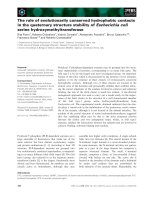
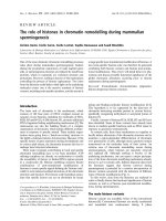
Fig. 1. Chromatin components during spermatogenesis. The major chromatin components and their post-translational modifications are presented.
Histone variants are incorpora ted during meiosis, except linker variant HILS, which shows a delayed expression. Highly basic proteins, transition
proteins and protamines, replace histones during late spermiogenesis. The temporal distribution of the main post-translational histone modifi-
cations is also presented (A, ac etylation; U, u biquitination ; M, m ethylation; P, phosphorylation). Spermatogenesis, the differentiation of male
germinal cells, is characterized by three major stages: preme iotic, m eiotic and postmeiotic. Pre-meiotic spermagogonia divide b y mitosis. They then
enter meiosis by the formation of preleptotene primary spermatocytes, which replicate DNA and subsequently go through the leptotene, zygotene,
pachytene and diplotene stages of the first meiotic division prophase. Meiotic I division yields secondary spermatocytes which then rapidly go
through m eiotic II division, generating haploid round spermatids. During its postmeiotic maturation, the spermatid undergoes a global remodelling
of its nucleus, which e longates and compacts into the very un ique nucleus structure of the sperm atozoa.
3460 J. Govin et al.(Eur. J. Biochem. 271) Ó FEBS 2004
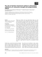
Fig. 2. Sequence analysis of known histone variants expressed during spermatogenesis. The sequences of conventional histones and their sperma-
togenic variants are aligned in (A), (B), (C) and (D). All sequences are murine, as most of the sequence data are available for this species, except for
TH2A (rat), and hTH2B and H3t (human sequences). Con ventional histone sequences were chosen on the basis of work by M arzluff and colleagues
[94]. Alignments were perf ormedwiththealgorithm
CLUSTALW
on the web interface o f the PBIL at and coloured with
ESPRIT
at Pript/[96]. Some of the histone modifications d iscussed are indicated [67,91]. Modification
cassettes (amino acids Thr/Ser-Lys or Lys-Thr/Ser) [91] were searched in conventional histones and variants, and are rep resented by small
rectangles, underneath the corresponding sequenc es. Black r ectan gles underline cassettes p resent in conventional histones. Some cassettes are not
conserved in variants and arrows indicate changes leading to the cassette disappearance in the variants. Open rectangles underline new cassettes
specific t o a variant and absent in conventional his tones. Original crystallographic data were used for the representat ion of the secondary structu res
[1,97]. Sequence accession numbers: H3.1 (P16106); H3.3 (P06351); H3t (Q16695); H2A (NP_783591); TH2A (Q00728); H2B (NP_835502); TH2B
(Q00729); hTSH2B (NP_733759); H1.1 (P43275); H1t ( Q07133); HILS1 (Q9QYL0).
Ó FEBS 2004 Chromatin code and spermatogenesis (Eur. J. Biochem. 271) 3461
A testis-specific H3 variant, only detected in the human,
has been isolated in 1996 [24,25]. This variant, named H3t,
differs from the canonical H3 by only four residues
(Fig. 2A). The RNA of this variant was only detected in
primary spermatocytes. T he experimental sequencing
helped to identify another t estis specific variant, n amed
TH3 in rat [26]. However, no gene or sequen ce information
is available on this putative histone variant and no
corresponding genes have b een found in known mammalian
genomes [25].
More data is available on nontestis specific H3 variants.
CENP-A is a centromeric specific variant, and unique by its
N-terminal amino-ac id composition [27]. In somatic cells,
CENP-A is deposited on newly duplicated centromeres, and
is required for the recruitment of other proteins to
centromeres and kinetochores. A similar function in germ
cells would imply its involvement during mitotic and/or
meiotic segregation.
The other H3 variant is H3.3, which differs b y 4–5 amino
acids from H3, depending on the allelic form considered
(Fig. 2A). The two H3.3 genes, H3.3A and H3.3B,are
expressed in mouse testis [28,29]. H3.3A mRNA was
detected before and after meiosis while the expression of
H3.3B gene was found to be restricted to cells of the meiotic
prophase [29]. Interestingly, H3.3A was identified by a gene
trap strategy as a gene expressed in spermatocytes, a nd of
which homozygous disruption caused partial neonatal
lethality and, in surviving mutants, reduced growth, neuro-
muscular deficits and male subfertility [30]. The number of
copulations per male, as well as the number of pregnancies
per copulatory p lug, were significantly lower for H3.3A–/–
mutants than for non mutants. No obvious differences in
the testis, epididymis, vas deferens, or sperm numbers were
reported in this study, suggesting that spermatogenesis was
not quantitatively affected.
Akhmanova and colleagues [31] have sho wn that Dro -
sophila H3.3 is incorporated during the first meiotic
prophase, then concentrated in a limited number of
chromatin regions and further disappears with the other
core histones during the elongation of spermatids. In
somatic cells, a ctively t ranscribing regions have been shown
to be enriched in H3.3 [32], suggesting that the replacement
of H3 by H3.3 in spermatocytes could also be linked to the
very active transcription that takes place during meiosis [9].
H2B variants
Rat, mouse and human TH2B have been cloned, showing
very high levels of conservation [ 33–35]. The main differ-
ences between H2B and TH2B a re in the N -terminal, and to
a lesser extent, the histone fold domain (Fig. 2C). Most of
these differences are c onserved between the three species,
suggesting a conserved role for this variant during sperma-
togenesis (see below).
In rat, TH2B is actively expressed in early primary
spermatocytes until mid–late pachytene [19] and then
remains the major form o f H2B in round and elongating
spermatids. Using an antibody that, luckily, cross-reacts
with TH2B, it has been shown in human testis that TH2B
first appears in spermatogonia, is maximal in round
spermatids, and then gradually disappears during the
elongation of spermatids [36]. In contrast, the human
TH2B, hTSH2B, was retained in mature sperms and
presented a specific nuclear localization only in 20% of
sperm populations [35].
There is also apparently a nonchromatin function for
histones during spermatogenesis. Indeed, recently, in bull
somatic type core histones h ave been foun d associated with
the perinuclear theca, which is a layer surrounding the
nucleus of mammalian sperms [37]. A h istone H2B variant,
named SubH2Bv, has also been found associated with the
theca in bull sperm [38]. The function of these non-nuclear
histones has not been defined.
H2A variants
Only one testis specific H2A variant has been characterized
and named TH2A, which differs from somatic H2A in
several residues l ocated in its histone fold domain as well as
in its N- and C-term inal tails (Fig. 2B). T H2A is actively
expressed a nd incorporated in the chromatin of pachytene
spermatocytes [19,39].
The expression of nontestis specific H2A variants have
been studied in more detail. Mainly, t wo H2A v ariants
are expressed during spermatogenesis, H2A.X, and mac-
roH2A. In somatic cells, H2A.X is involved in DNA
double strand b reaks (DSB) surveillance and repair [9,40].
H2A.X disruption leads to male sterility with abnormal
spermatogenesis. Indeed, in the male mutants, no DNA
alignment for synapsis is observed at zygotene and early
pachytene stages. In the spermatocytes that progress into
mid-pachytene M1h1, a mismatch repair protein, do not
display t he foci characteristic of recombined DNA
strands, and chromosomes X and Y are abnormally
paired with autosomes, leading to apoptosis of mid
pachytene spermatocytes [41].
MacroH2A is a long variant of H2A, containing a
large C-terminal nonhistone region [42]. Two allelic
forms, macroH2A1 and macroH2A2, are expressed. They
are 80% identical [43,44]. The macroH2A1 gene encodes
two proteins generated by an alternative splicing mech-
anism, macroH2A1.1 and macroH2A1.2 [43]. In somatic
cells of female mammals , the inactive X c hromosome has
been shown marked by a high concentration of histone
macroH2A [43,45,46], forming a dense structure, referred
to as the macrochromatin body. MacroH2A1.2 is found
at high concentrations in mice testis [47,48]. During
spermatogenesis, it has been observed in the nuclei of
germ cells, with a localization that is largely to the
developing XY-body in early pachytene spermatocytes
[49,50]. Hence, the process of X-inactivation in XX
somatic cells [51] and that in XY spermatocytes show
some similarities, including a heterochromatinization of
the region which is densely stained (forming, respectively,
the Barr Body or the S ex Vesicle) and a coating of the
X with the Xist RNA, a non coding RNA specifically
associated w ith the inactive X chromosome [52]. Interest-
ingly, a potential relationship has been discovered
between macroH2A1.2 and the mammalian HP1-like
heterochromatin p rotein M31 (HP1beta or MOD1)
during meiosis. The HP1-like protein M31 was found
initially to colocalize with heterochromatic regions in
Sertoli cells, in mid-stage pachytene spermatocytes, a s well
as in round spermatids (where it localized with the
3462 J. Govin et al.(Eur. J. Biochem. 271) Ó FEBS 2004
centromeric chromocenter) [53]. Both macroH2A1.2 and
M31 were found to colocalize in a time-dependent
manner at specific nuclear regions, including the pseudo-
autosomal region (PAR) of the sex body [50], suggesting
a role for this heterochromatic region in preventing
precocious desynapsis of the terminally associated X and
Y chromosomes prior to a naphase I. According to the
data described above, the large histone H2A variant,
macroH2A1.2, along with the HP1-like protein M31,
could be involved in the partial pairing of X and Y
chromosomes and the formation of th e s ex vesicle, which,
although of unknown function, is an indispensable f eature
of a successful male meiotic division. Indeed, meiotic
studies in men presenting an impaired spermatogenesis in
the context of a constitutional chromosomal abn ormality
have suggested that the presence of a sex vesicle is crucial
for the achievement of meiosis.
In one stu dy, macroH2A1.2 has also been found in
murine spermatozoa, suggesting that it may be important
for other functions besides meiotic recombination [49].
However, according to another stud y, macroH2A was not
found among sperm nuclear proteins, not even in species
fully retaining the histones in mature sperm such as catfish
and bullfrog [54].
Histones and post-translational modifications
The histone code hypothesis proposes that combinations of
histone modifications could define specific signals, and serve
as an interface languag e b etween hist ones a nd chroma tin
modifying activities, to assign particular structure and
function to specific chromatin domains [5,6]. In fact each
histone has several sites of potential modifications including
acetylation, methylation, phosphorylation, etc… Assuming
that the eight core histones of each nucleosome could have
different associations of modifications, their combination in
a multinucleosomal microenvironment would create a
tremendously complex epigenetic code. This hypothesis
stands only if experimental data support the existence o f a
machinery capable of specifically re cognizing and reading
the histone c ode. The existe nce of cellular factors recogni-
zing and binding to specifically modified histones is i n
support of this hypothesis [7,55].
The histone code is probably in a ction in spermatogenic
cells as stage-specific histone modifications have been
reported to occur during the postmeiotic genome reorgan-
ization phase. However, despite detailed descriptions of
some histone modifications [9], nothing is known about
their potential function in chromatin reorganization and
histone replacement in elongating spermatids (Fig. 1).
Histone acetylation
Acetylated forms of histones have been found during
spermatogenesis in various species including, trout [56], rat
[57] and rooster [58]. The use of antibodies, specifically
recognizing individual acetylated residues, has allowed a
more precise characterization of histone acetylation pattern
during spermatogenesis [59]. Spermatogonia and prelepto-
tene spermatocytes contain acetylated H2A H2B and H4,
whereas histones are underacetylated during meiosis and in
round spermatids. The replication-dependent acetylation of
H4 and H 3 [60] can partially explain the acetylation signal
detected in DNA replicating cells.
Interestingly, these data also showed that in elon gating
spermatids, histones become hyperacetylated in the total
absence of DNA replication. In the case of histone H4, this
acetylation was shown to follow a stage-specific distribution
[59,61]. Indeed, the H4 hyperacetylation observed in the
early elongating spermatids affects the nucleus in a g lobal
manner. This distribution then changes during the elonga-
tion and condensation s tages and finally acetylated H4
disappears following an antero-caudal movement in con-
densing spermatids.
This replication and transcription-independent hist one
acetylation seems to be tightly linked to histone replace-
ment. Indeed, histones remain under-acetylated in species
where histones remain all through spermiogenesis such as
winter flounder and carp [62,63]. However, the role of
acetylation of core histones in their replacement remains
largely unknown. Some in vitro experiments suggest that
histone acetylation could facilitate their displacement by
protamines [64,65], but there is no hint in the literature on
how it could affect in vivo chromatin remodelling in
spermatids. The recent identification of a new bromo-
domain-containing testis specific factor capable of cond en-
sing acetylated chromatin suggests t hat histone acetylation
could primarily be a signal for chromatin condensation
[66].
Histone methylation
Suv39h1 and Suv39h2 a re two histone methyltransferases
(HMTs) responsible for methylating Lys9 of H3 in hetero-
chromatic regions, in somatic cells [67]. Suv39h2 is
over-expressed in the testis [68], where it is enriched in
heterochromatic regions from leptotene spermatocytes to
round spermatids stages. The H3 Lys9 methylation pattern
colocalizes with Suv39h2 [69]. A disruption of heterochro-
matic H MT activities (double knockout of Suv39h1 and h2)
leads to c hromosomal instability, impaired homologous
interactions and meiosis defects.
Histone phosphorylation
Ser10 and 28 of H3, both very conserved in the H3 family,
are phosphorylated during mitotic chromosome formation.
The mitotic-specific phosphorylation of histone H3 Ser10
has also been shown to occur during meiosis very probably
associated with chromosome condensation [70]. However,
no information is available a bout the phosphorylation of
Ser28 during spermatogenesis.
Site-specific phosphorylations of H2A [71], H2AX [40]
and H2B [72] have also been reported. While nothing is
known a bout the phosphorylation of H2A and H2B
during spermatogenesis, that of H2AX may play a c rucial
role as it is tightly linked to the function of H2AX in
DNA double strands breaks repair [40]. Indeed, a
transient phosphorylation of H2AX on Ser139 accom-
panies double strand break damage repair, as well as
DNA cleavage events such as those associated with
meiotic recombination [73].
Ó FEBS 2004 Chromatin code and spermatogenesis (Eur. J. Biochem. 271) 3463
Histone ubiquitination
Ubiquitination is a modification known to b e a mark for
protein degradation via the proteasome pathway. How-
ever, the function of protein ubiquitination is not
restricted to degradation, and data from the literature
suggest its involve ment in DNA repair, cell cyc le control,
cellular response to stress, as well as in the histone code
[74].
H1 and H 3 have been found occasionally ubiquitinated
in vivo, but H2A and H2B appear to be the predominant
forms of ubiquitinated histones i n e ukaryotes, encompas-
sing 5–15% of H2A and 1–2% of H2B [75].
Histone ubiquitination has been described du ring sper-
matogenesis in many species, including rat, mouse, trout
and rooster [75]. In the mouse, a high proportion of
ubiquitinated H 2A (uH2A) is detected by immunochemis-
try in the specific chromatin domain formed by the sex body
in pachytene spermatocytes. uH2A becomes depleted from
round spermatids, but reappears in elongating spermatids
[74]. In elongating spermatids H2A, H 2A.Z, H2B, H3 and
TH3 were found mono and poly ubiquitinated in t he rat
[74,76].
HR6, a ubiquitin-conjugating enzyme, homolo gous to
the yeast RAD6 protein, ubiquitinates H2B in vivo and is
strongly expressed in the testis [77]. A disruption of the
HR6-encoding gene induces a spermatogenesis arrest at
the round/elongating spermatids st age [78] pointing to the
fundamental role of histone ubiquitination during spermio-
genesis.
All these data suggest that histone ubiquitination can be
considered as one of the important epigenetic mark involved
in chromatin remodelling in postmeiotic male germ cells.
Histone variants
Functional significance of sequence divergence in
chromatin remodelling. One of the most distinctive
characteristics of chromatin remodelling during
spermatogenesis is the expression of a large number of
histone variants. Indeed, in ad dition to all the somatic-
type histone variants, spermatogenic cells express t estis-
specific histones corresponding t o three of the four core
histones. Nevertheless, understanding how each variant
specifically acts on chromatin s tructure and function is a
real challenge. The fundamental structu ral basis of a
nucleosome is very well conserved during evolution. The
incorporation of histone variants could lead to the
formation of nucleosomes with altered structure and
modified properties.
Histone variants incorporated during spermatogenesis,
although showing only small changes in their primary
structures, could therefore bring major changes in the
nucleosome function and stability.
A detailed analysis of testis-specific histone variants
shows that the histone fold is usually well conserved
between variants (Fig. 2A,B,C). The N-terminal region of
H3 is very similar between the variants, including H3.3 and
H3t, whereas the N-terminal regions of TH2A and TH2B
present several differences with their somatic c ounterparts,
which m ay potentially affect residues modified by known
histone post-translational modifications (Fig. 2).
Interestingly, the comparison of H2A/TH2A sequence
shows three amino acid changes in a region covering the end
of alpha1, loop1 and the beginning of alpha2. As a
structural analysi s has a lready shown that H2A Loop1 is
the only area of contact between the two (H2A–H2B)
dimers within the nucleosome core particle [1], the m inor
sequence changes observed in TH2A could have important
functional consequences, as a lready established in t he case
of H2A.Z by crystallographic data [79]. The structural
analysis also showed that the incorporation of two
heterodimers of H2A–H2B and H2A.Z–H2B within the
same nucleosome is unlikely, suggesting that the incorpor-
ation of the first (H2A.Z–H2B) dimer could facilitate t he
recruitment o f another H2A.Z-containing dimer [79,80].
Similarly, the i ncorporation of given testis-specific histone
variants might facilitate the incorporation of other variants,
creating highly specialized nucleosomes.
Moreover, the H2A.Z containing nucleosomes display an
altered surface, with the possible incorporation of a metal
divalent ion, which could lead to changes of higher order
structures or modify the recruitment of specific factors [79].
It could be assumed that similar properties associated with
testis specific histones would lead to an altered chromatin
structure and facilitate the recruitment of testis-specific
chromatin remodelling factors.
The centromere specific histone variant, CENP A, has
been shown to be retained in mature spermatozoa,
suggesting that it could have a role in organizing the
centromeres during the final stages of spermiogenesis and/or
the paternal genome during early embryogenesis [81].
A role for specific histone chaperones. Cellular machinery
containing histone chaperon e HIRA, has recently been
discovered that is capable of uniquely assembling histone
H3 variant, H3.3, in specialized nucleosomes [82,83]
enriched in transcriptionally active regions [32]. The
localization of H 3.3-containing chromatin has not yet
been determined in mammalian germ cells, but in
Drosophila, H3.3 is incorporated in chromatin during first
meiotic prophase [31]. It remains concentrated in specific
regions (compared to H3, which is evenly distributed) in
round and elongating spermatids, and disappears in
condensed spermatids like other histones. H3.3 is
therefore present in haploid male g erm cells in the total
absence of transcription. One possible function of this
specific H3 variant could be linked to the massive histone
replacement, taking place i n elongating s permatids where
HIRA, or m aybe other spermatid-specific factors, could
recognize H3.3 and dismantle the nucleosomes. Histone
removal by HIRA may also occur in somatic cells but to a
much lesser extent than in spermatids. Therefore the
identification of HIRA partners in spermatids w ould be of
great interest in understanding the molecular b asis of
histone replacement during spermiogenesis and furthermore
in that of nucleosome disassembly in gene ral.
Recently, a histone variant exchanger that specifically
replaces co nventional H2A by H2A.Z has been identified in
yeast [ 84,85] showing that H3 and also H2A variants c an be
deposited by specific factors.
Recent work showed that in yeast, a protein identified as
Hif1p is a histone H3 and H4 chaperone involved in
chromatin assembly [86]. Interestingly, Hif1p is the
3464 J. Govin et al.(Eur. J. Biochem. 271) Ó FEBS 2004
homologue of a H1 chaperone, known as NASP, which has
a testis-specific variant expressed in different species of
mammals and is present all through spermiogenesis [87]. It
has been proposed that tNASP may bind and t ranslocate
testicular histone variants to nucleosomes [87]. Its presence
during late spermiogenesis suggests that the protein may
also function as a histone remover, as no chromatin
assembly occurs during these stages.
It is therefore very possible that the enrichment of
spermatid chromatin with different histone variants would
first increase the accessibility of chromatin to various factors
(such as those involved in recombination in pachytene cells
or histone modifying enzymes in spermatids) and then
facilitate histone replacement. Moreover, histone modifica-
tions, such as acetyla tion, may mediate the action of more
specialized chaperones (see below). It would therefore be
important to investigate the structural characteristics of
testis-specific histone v ariants, and to e xplore whether the
testis-specific and somatic histone chaperones expressed in
spermatogenic cells are capable of exchanging TH2A,
TH2B as well as H3t with transition proteins.
Histone acetylation – a signal for histone replacement?
As mentioned previously, postmeiotic histone hyperacety-
lation has not been observed in species where somatic
histones are retained completely in spermatozoa. This
specific histone modification therefore appears to be tightly
associated with histone replacement. Moreover, the obser-
vation that in mice spermatids, acetylated H4 disappearance
follows an antero-caudal pattern similar to that of chroma-
tin condensation [59], reinforces the h ypothesis of a direct
link between histone acetylation, th eir replacement and
nucleus condensation.
The m echanisms under lying this sudden histone hyper-
acetylation in early elongating spermatids are unknown.
However, a recent work showed that it is associated with the
degradation of the major cellular histone deacetylases [88],
a phenomenon that is able to play an important role by
disrupting the cellular acetylation equilibrium.
According to the histone code hypothesis, histone
hyperacetylation in elongating spermatids would serve as
a signal for the recruitment of specific machinery acting on
acetylated histones. Such machinery probably contains
factors such a s bromodomain-bearing p roteins, enab ling
them to bind acetylated chromatin (Fig. 3).
Bromodomains are acetyl-lysine binding modules present
in ATP-dependent chromatin remodelling factors as well as
in some HATs and other nuclear protein s of unknown
function [89]. B romodomain-containing proteins therefore
appear to be excellent candidates to i nterpret the signal
generated by the global histone acetylation taking place
during spermiogenesis. Recently, a testis-specific double
bromodomain-containing protein, named BRDT, was
shown to be capable of inducin g a dramatic condensation
of chromatin strictly dependent o n histone hyp eracetylation
[66]. These data present a new scenario regarding the
significance of histone acetylation during spermiogenesis: it
could primarily act as a signal for chromatin condensation.
In support of this hypothesis, nuclear domains containing
condensed chromatin in elongating spermatids also corres-
pond to regions enriched in acetylated histone H4 (J. Go vin,
C. Caron, C. Lestrat, S. Ro usseaux and S. Khochbin,
unpublished results).
Bromodomain-containing factors, such as BRDT,
upon their interaction with acetylated histones, could
also recruit testis-specific chaperones t o mediate histone
removal. In fact, a new bromodomain-interacting chap-
erone, CIA-II, highly expressed in t he testis, also interacts
with histon e H3 in vivo andwithhistonesH3/H4in vitro
[90]. Such fac tors may establish a link between an
acetylation-dependent chromatin compaction mediated by
bromodomain p roteins and histone displacement. More-
over, it has recently been shown in yeast that Hat1p/
Hat2p/Hif1p specifically binds acetylated histones H4 and
H3 [86]. A s mentioned a bove, the testis-specific homo-
logue of H if1p, tNASP, is present all through spermio-
genesis, and may also provide a link between histone
acetylation and histone removal (Fig. 3B).
Chromatin r emodelling that o ccurs during spermiogen-
esis seems to d epend simultaneously on histone variants and
histone modifications (histone code). It is therefore very
likely that the combination of histone variants and partic-
ular histone modifications generate a testis-specific Ôchro-
matin codeÕ (Fig. 3A).
It is noteworthy that all the sites in histon es potentially
involved in generating the histone code are conserved
between histone variants expressed during spermiogenesis,
with the exception of the H2B phospho-acceptor site S14,
which is not conserved in TH2B. This sequence divergence
signifies a modification of the TH2B related histone code in
spermatogenic cells, as for H2B, Ser14 phosphorylation has
been shown to play an essential role in somatic cell
apoptosis [72]. In contrast, compared to H2B, hTSH2B
has gained four potentially new phosphorylation sites
(Fig. 2 C).
The observation of a p air of neighbouring amino acids
both targets of post-translational modifications has
recently led to the proposal of the Ôbinary switchesÕ
hypothesis modulating the readout of specific marks such
as lysine methylation [91]. In fact the phophorylation of
Thr/Ser in Thr/Ser-Lys or Lys-Thr/Ser pairs found in the
four histones may negatively regulate the binding of
chromodomains to methylated lysines. Indeed, chromo-
domain-containing proteins are involved in a variety of
functions, but all seem to deal with chromatin. In
some of these proteins, such as heterochromatin protein 1
(HP1), the chromodomain has been shown to specifically
interact with histone tails bearing methylated lysines [7].
In order to assess the potential function of these binary
switches during spermatogenesis, they were searched for
on the primary sequences of the different histone
variants. Among the three testis-specific core histones,
TH2B seems t o be the on ly variant which presents
significantly divergent binary cassettes compared to its
somatic counterpart. Indeed, in testis-specific H2Bs, in
three cases the Thr/Ser residues occurring in somatic type
H2B next to a Lys residue were replaced by nonphos-
pho-acceptor residues, and three new binary cassettes
were created (Fig. 2C).
These analyses show that, on top of a structural role,
sequence divergence in testis-specific histone variants
may participate in increasing the complexity of the histone
code.
Ó FEBS 2004 Chromatin code and spermatogenesis (Eur. J. Biochem. 271) 3465
Concluding remarks
After analysing all the available data i t clearly appears that a
massive chromatin alteration occurs before histone replace-
ment due to an extensive incorporation of histone variants
as well as to globally specific histone modifications.
Recruitment of histone variants in nucleosomes may have
two general effects on chromatin structure and function.
First, subtle sequence divergences can have important
consequences on the stability of the nucleosome. Second,
these sequence divergences may change t he potential of core
histones to b e modified. A testis-specific histone code can
therefore be generated directing chromatin compaction,
histone removal and degradation. Very little i s known on
the nature of this s pecific histone code and t he way it directs
chromatin remodelling in spermatids.
Recently, two factors expressed in spermatids and
potentially capable of participating in chromatin
remodelling have been identified [66,88,92]. One of these
factors containing two bromodomains, BRDT, has been
shown to have the ability to induce in vitro and in vivo an
histone acetylation-dependent chromatin compaction. His-
tone H4 acetylation o ccurring in elongating s permatids
might primarily be a signal for chromatin c ondensation.
However, more investigations are required to link this
acetylation-dependent chromatin compaction to histone
removal. With this regard, histone chaperones may play a
crucial role. Indeed, it is very plausible that specific
chaperones identified to mediate nucleosome a ssembly [93]
may reverse their function and control the dismantlement of
nucleosomes in spermatids.
Spermatogenic cells would therefore constitute an excel-
lent source for the discovery of a nucleosome disassembly
machinery. The identification of such factors would not
only shed light on the molecular basis of chromatin
reorganization during spermiogenesis but also give valuable
Fig. 3. Integrative model for chromatin remodelling during spermatogenesis. (A) Chromatin remodelling combines h istone variants (1) and the
histone code ( 2, 3). In the late stages of spermiogenesis, transition proteins and protamines participate in constituting the fin al sperm chro matin
structure (4). (B ) Putative factors involved in the spermatogeni c rem odelling process. Brdt is prob ably on e o f th e histo ne c ode ÔreadersÕ, binding
acetylated histones, and conden sing acetylated chromatin [66]. HIRA, Hif1p (also nam ed NASP) an d tNASP are suspected to behave as histon e
chaperones during t his re modelling process, with some histone specificity (see text for more det ails).
3466 J. Govin et al.(Eur. J. Biochem. 271) Ó FEBS 2004
information on the yet unkn own mechanism of nucleosome
disassembly.
Acknowledgements
This work was supported by ‘‘Re
´
gion Rhoˆ ne-Alpes’’ emergence pro-
gram. C.L. i s supported by ‘‘Re
´
gion Rhoˆ ne-Alpes’’ PhD fellowship.
References
1. Luger, K., Mader, A.W., Richmond, R.K., Sargent, D.F. &
Richmond, T.J. (1997) Crystal structure of the nucleosome core
particle at 2.8 A
˚
resolution. Nature 389, 251–260.
2. Kh ochbin, S. (2001) Histone H1 diversity: b ridging regulatory
signals to linker histone function. Gene 271, 1–12.
3. Lusser, A. & Kadonaga, J.T. (2003) Chromatin remodeling by
ATP-dependent molecular machines. Bioessays 25, 1192–1200.
4. Turner, B .M. (1993) Decoding the nucleosome. Cell 75, 5–8.
5. Strahl, B.D. & Allis, C.D. (2000) The l anguage of covalent histone
modifications. Nature 403, 41–45.
6. Turner, B.M. (2002) Cellular memory and the histone code. Cell
111, 285–291.
7. Khorasanizadeh, S. (2004) The nucleosome: from genomic
organization t o ge nomic regulation. Cell 116, 259–272.
8. Malik, H.S. & Henikoff, S. (2003) Phylogenomics of the nucleo-
some. Nat. Struct. Biol. 10, 882–891.
9. Lewis, J.D., Abbott, D.W. & Ausio, J. (2003) A haploid affair:
core histone t ransitions du rin g sp ermatogene sis. B ioc hem. Cell
Biol. 81, 131–140.
10. Drabent, B., Bode, C., Bramlage, B. & Doenecke, D. (1996)
Expression of the mouse testicular histon e g ene H1t d uring sper-
matogenesis. Histochem. Cell Biol. 106, 247–251.
11. Iguchi, N., Tanaka, H., Yomogida, K. & Nishimune, Y. (2003)
Isolation and characterization of a novel cDNA encoding a DNA-
binding prote in (H ils1) s pecificallyexpressedintesticularhaploid
germ cells. Int. J. Androl. 26, 354–365.
12. Grime s, S.R., Wo lfe, S.A., A nderson, J.V., Stein, G .S. & Stein,
J.L. (1990) Structu ral and fun ctional analysis of the rat testis-
specific histone H1t gene. J. Cell Biochem. 44, 1–17.
13. Steger, K., Klonisch, T., Gavenis, K., Drabent, B., Doenecke, D .
& Bergmann, M. (1998) E xp ressi on of mRNA and p rotein of
nucleoproteins durin g human spe rmiogenesis. Mol. Hum. Reprod.
4, 939–945.
14. Drabent, B., Saftig, P., Bode, C. & Doenecke, D. (2000) Sper-
matogenesis proceeds n ormally in m ice wit hout l inker h istone
H1t. Histochem. Cell Biol. 113, 433–442.
15. Lin, Q., Sirotkin, A. & Skoultchi, A.I. (2000) Normal spermato-
genesis in mice lacking the testis-specific linker h istone H 1t. Mol.
Cell Biol. 20 , 2122–2128.
16. Fantz, D.A., Hatfield, W.R., Horvath, G., Kistler, M.K. &
Kistler, W.S. (2001) Mice with a targeted disruption of the H1t
gene are fertile and undergo normal changes i n structural chro-
mosomal proteins during spermiogenesis. Biol. Reprod. 64,425–
431.
17. De Lucia, F., Faraone-Mennella, M.R., D’Erme, M., Q uesada, P.,
Caiafa,P.&Farina,B.(1994)Histone-induced condensation of
rat testis chromatin: testis-specific H1t versus somatic H1 variants.
Biochem. Biophys. Res. Commun. 19 8, 32–39.
18. Khadake, J.R. & Rao, M.R. (1995) DNA- and chromatin-con-
densing properties of rat testes H1a and H1t compared to those
of rat live r H1bdec; H1t is a poor condenser of chromatin.
Biochemistry 34, 15792–15801.
19. Meistrich, M.L., Bucci, L.R., Trostle-Weige, P.K. & Brock, W.A.
(1985) Histone variants in rat spermatogonia a nd primary sper-
matocytes. Dev. Biol. 112, 230–340.
20. Franke, K ., Drabent, B. & Doeneck e, D. ( 199 8) Testicular
expression of the mouse histon e H1.1 gene. Histochem. Cell Biol.
109, 383–390.
21. Rabini, S., Franke, K., S aftig, P., Bode, C., D oenecke, D. &
Drabent, B. (2000) Spermatogenesis in mice is not affected by
histone H1.1 deficiency. Exp. Cell Res. 255, 114–124.
22. Lin, Q., Inselman, A., Han, X., Xu, H., Zhang, W., Handel, M.A.
& Sko ultchi, A.I. (2004) Red uctions in linker histone levels are
tolerated in developing spermatocytes but cause chan ges in specific
gene expression. J. Biol. Chem. 279, 23525–23532.
23. Yan, W., Ma, L., Burns, K.H. & Matzuk, M.M. (2003) HILS1 is a
spermatid-specific linker histone H1-like p rotein implicated in
chromatin r emodeling during mammalian spermiogenesis. Proc.
NatlAcad.Sci.USA100, 10546–10551.
24. Albig, W., Ebentheuer, J., Klobeck, G., Kunz, J. & Doenecke, D.
(1996) A solitary human H3 histone gene on chromosome 1. Hum.
Genet. 97, 486–491.
25.Witt,O.,Albig,W.&Doenecke,D.(1996)Testis-specific
expression of a novel human H3 h istone gene. Exp. Cell Res. 229,
301–306.
26. Trostle-Weige, P.K., Meistrich, M.L., Brock, W.A. & Nishioka,
K. (1984) Isolation and characterization of T H3, a germ cell-
specific variant of histone 3 in rat testis. J. Biol. Chem. 25 9, 8769–
8776.
27. Sm ith, M.M. (2002) Centromeres and variant histones: what,
where, when and why? Curr. Opin. Cell Biol. 14, 279–285.
28. Albig,W.,Bramlage,B.,Gruber,K.,Klobeck,H.G.,Kunz,J.&
Doenecke, D. (1995) The human replacement histone H3.3B gene
(H3F3B). Genomics 30, 264–272.
29. Bramlage, B., Kosciessa, U. & Doenecke, D. (1997) Differential
expression of the murine histone genes H3.3A and H 3.3B. Dif-
ferentiation 62, 13–20.
30. Cou ldrey, C., Carlton, M.B ., Nolan, P.M., Colledge, W.H. &
Evans, M.J. (1999) A retroviral gene trap insertion into the histone
3.3A ge ne causes partial neo natal lethality, stunted growth,
neuromuscular deficits and male sub-fertility in transgenic mice.
Hum. Mol. Genet. 8, 2489–2495.
31. Akhmanova, A., Miedema, K., Wang, Y., van Bruggen, M.,
Berden, J.H., Moudrianakis, E.N. & Hennig, W. (1997) The lo-
calization of histone H3.3 in germ lin e chromatin of Drosophila
males as established with a histoneH3.3-specificantiserum.
Chromo so ma 106, 335–347.
32. McKittrick, E., Gafken, P.R., Ahmad, K. & Henikoff, S. (2004)
Histone H3.3 is enriched in covalent modifications associated with
active chromatin. Proc. Natl Acad. Sci. USA 101, 1525–1530.
33. Hwang, I. & Chae, C.B. (1989) S-phase-specific transcription
regulatory elem ents are present in a replication-independent testis-
specific H2 B histon e gene. Mol. Cell. B iol. 9, 1005–1013.
34. Choi, Y.C., Gu, W., Hecht, N.B., Feinberg, A.P. & Chae, C.B.
(1996) Molecular cloning of mouse somatic and testis-specific H2B
histone genes containin g a meth ylated CpG island. DNA Cell Biol.
15, 495–504.
35. Zalensky, A.O., Siino, J.S., Gineitis, A.A., Zalenskaya, I.A.,
Tomilin, N.V., Yau, P. & Bradbury, E.M. (2002) Human testis/
sperm-specific histone H2B (hT SH2B). Molecular cloning and
characterization. J. Biol. Chem. 277, 43474–43480.
36. van Roijen, H.J., O oms, M.P., Spaargaren, M.C., Baarends,
W.M.,Weber,R.F.,Grootegoed,J.A.&Vreeburg,J.T.(1998)
Immunoexpression of testis-specific histone 2B in human spe r-
matozoa and testis tissue. Hum. Reprod. 13, 1559–1566.
37. Tovich, P.R. & Oko, R.J. (2003) Somatic histones are components
of the perinuclear theca in bovine spermatozoa. J. Biol. Chem. 278,
32431–32438.
38. Aul, R.B. & Oko, R.J. (2001) The major subacrosomal occupant
of bull spermatozoa is a novel histone H2B variant associated with
Ó FEBS 2004 Chromatin code and spermatogenesis (Eur. J. Biochem. 271) 3467
the forming acrosome during spermiogenesis. Dev. Biol. 239, 376–
387.
39. Rao , B.J., Brahmachari, S.K. & Rao, M.R. (1983) Structural or-
ganization of the m eiotic prophase chromatin in the rat testis.
J. Biol. Chem. 258, 13478–13485.
40. Rogakou, E.P., Pilch, D.R., Orr, A.H., Ivanova, V.S. & Bonner,
W.M. (1998) DNA double-stranded breaks induce histone
H2AX phosphorylation on serine 139. J. Biol. Chem. 273, 5858–
5868.
41. Celeste, A., Petersen, S., Romanienko, P.J., Fernandez-Capetillo,
O., Chen, H.T., Sedelnikova, O.A., Reina-San-Martin, B., Cop-
pola, V., Meffre, E., Difilippantonio, et al. (2002) Genomic
instability in m ice lacking histone H2AX. Science 29 6 , 922–927.
42. Pehrson, J.R. & Fried, V.A. (1992) MacroH2A, a core histone
containing a large nonhistone re gion. Science 257, 1398–1400.
43. Costanzi, C. & Pehrson, J.R. (2001) MACROH2A2, a new
member of the MACROH2A core histone family. J. Biol. Chem.
276, 21776–21784.
44. Chadwick, B.P., Vall ey, C.M. & Willard, H .F. (2001) Histone
variant macroH2A contains two distinct macrochromatin
domains capable of directing macroH2A to the inactive X chro-
mosome. Nucleic Acids Res. 29, 2699–2705.
45. Costanzi, C. & Pehrson, J.R. (1998) Histone macroH2A1 is con-
centrated in the inactive X chromosome of female mammals.
Nature 393, 599–601.
46. Perc he, P., Vourc’h, C., Konecny, L., Souc hier, C., Robert-
Nicoud,M.,Dimitrov,S.&Khochbin,S.(2000)Highercon-
centrations of histone macroH2A in the Barr body are correlated
with higher nucleosome density. Curr. Biol. 10, 1531–1534.
47. Pehrson, J.R., Costanzi, C. & Dharia, C. (1997) Developmental
and tissue expression patterns of histone macroH2A1 subtypes.
J. Cell Biochem. 65, 107–113.
48. Rasm ussen, T.P., Huang, T., Mastrangelo, M.A., Loring, J.,
Panning, B. & Jaenisch, R. (1999) Messen ger RNAs encoding
mouse histone macro H2A1 isoforms are expressed a t similar levels
in male and female cells and result from alternative splicing.
Nucleic Acids Res. 27, 3685–3689.
49. HoyerFender, S., Costanzi, C. & Pehrson, J.R. (2000) Histone
MacroH2A1.2 is concen trated in the XY-body by the early
pachytene stage of spermatogenesis. Exp. Cell Res. 258, 254–260.
50. Tu rner, J.M., Burgoyne, P.S. & Singh, P.B. (2001) M31 and
macroH2A1.2 colocalise at the pseudoautosomal region during
mouse meiosis. J. Cell Sci. 114, 3367–3375.
51. Avner, P. & Heard, E. (2001) X-chromosome inactivation:
counting, choice and initiation. Nat. Rev. Genet. 2, 59–67.
52.Ayoub,N.,Richler,C.&Wahrman,J.(1997)XistRNAis
associated with the transcriptionally inactive XY body in mam-
malian male meiosis. Chromosoma 106, 1–10.
53. HoyerFender,S.,Singh,P.B.&Motzkus, D. (2000) The murine
heterochromatin protein M31 is associated with the chrom ocenter
in round spermatids and is a component of mature spermatozoa.
Exp. Cell Res. 254, 72–79.
54. Abbott, D.W., Laszczak, M., Lewis, J.D., Su, H., Moore, S.C.,
Hills, M ., Dimitrov, S. & Ausio, J. (2004) Structural character-
ization of mac roH2A containing chromatin. Biochemistry 43,
1352–1359.
55. Jenuwein, T. & Allis, C.D. (2001) Translating the histone code.
Science 293, 1074–1080.
56. Candido, E.P. & Dixon, G.H. (1972) Amino-terminal sequences
and s ites of in vivo acetylation of trout-testis histones 3 and IIb 2.
Proc.NatlAcad.Sci.USA69, 2015–2019.
57. Grimes,S.R.Jr,Platz,R.D.,Meistrich,M.L.&Hnilica,L.S.
(1975) Partial characterization of a new basic nuclear protein from
rat testis elongated spermatids. Biochem. Biophys. Res. Commun.
67, 182–189.
58. Oliva, R. & Mezquita, C. (1982) Histone H4 hyperacetylation and
rapid turnover of its acetyl groups in transcriptionally inactive
rooster testis s permatids. Nucleic Acids Res. 10, 8049–8059.
59. Hazzouri, M., Pivot-Pajot, C., Faure, A.K., Usson, Y., Pelletier,
R.,Sele,B.,Khochbin,S.&Rousseaux,S.(2000)Regulated
hyperacetylation of core histones d uring mouse sp ermatogenesis:
involvement of histone deacetylases. Eur. J. Cell Biol. 79, 950–960.
60. Verreault, A . (2000) De novo nucleosome assembly: new pieces in
an old p uzzle. Genes Dev. 14, 1430–1438.
61. Marcon, L. & Boissonneault, G. (2004) Transient D NA strand
breaks during mo use an d hum an spermioge nesis: new insights in
stage specificity and link to chromatin remodeling. Biol. Reprod.
70, 910–918.
62. Kennedy, B.P. & Davies, P.L. (1980) Acid-soluble nuclear pro-
teins of the testis during spermatogenesis in the winter flounder.
Loss of th e high mobility group p roteins. J. Biol. Chem. 255, 2533–
2539.
63. Kennedy, B.P. & Davies, P.L. (1981) Phosphorylation of a group
of high molecular weight basic nuclear proteins during sperma-
togenesis in the winter flounder. J. Biol. Chem. 256 , 9254–9259.
64. Oliva, R. & Mezquita, C. (1986) Marked differences in the ability
of distinct protamines to disassemb le nucleosomal co re particles i n
vitro. Biochemistry 25, 6508–6511.
65. Oliva, R., Bazett-Jones, D., Mezquita, C. & Dixon, G.H. (1987)
Factors affecting nucleosome disassembly by protamines in vitro.
Histone hyperacetylation a nd chromatin structure, t ime depen-
dence, and the size of the sperm nuclear proteins. J. Biol. Chem.
262, 17016–17025.
66. Pivot-Pajot, C ., Caron, C., G ovin, J., Vion , A., Rou sseaux, S . &
Khochbin, S. (2003) Acetylation-dependent chromatin reorgani-
zation by BRDT, a testis-specific bromodomain-containing pro-
tein. Mol. Cell. Biol. 23, 5354–5365.
67. Sims, R.J., Nishioka, K. & Reinberg, D. (2003) Histone lysine
methylation: a signature for chromatin fu nction. Trends Genet. 19 ,
629–639.
68. O’Carroll, D., Scherthan, H., Peters, A.H., Opravil, S., Haynes,
A.R., Laible, G., Rea, S., Schmid, M., Lebersorger, A.,
Jerratsch,M.,Sattler,L.,Mattei,M.G.,Denny,P.,Brown,
S.D., Schweizer, D. & Jenuwein, T. (2000) Isolation and char-
acterization of Suv39h2, a second h istone H3 methyltransferase
gene that displays testis-specifi c expression. M ol. Cell. Biol. 20,
9423–9433.
69. Peters, A.H., O’Carroll, D., Scherthan, H., Mechtler, K., Sauer,
S., Schofer, C., We ipoltshamme r, K., Pagani, M., L achner, M.,
Kohlmaier, A., Opravil, S., Doyle, M., Sibilia, M. & Jenuwein, T.
(2001) Loss of the s uv39h histone methyltransferases i mpairs
Mammalian heterochromatin and genome stability. Cell 107,
323–337.
70. Prigent, C. & Dimitrov, S. (2003) Phosphorylation of serine 10 in
histone H3, what for? J. Cell Sci. 116, 3677–3685.
71. Zhang, Y., Griffin, K., Mondal, N. & Parvin, J.D. (2004) Phos-
phorylation of histone H2A inhib its transcription on chromatin
templates. J. Biol. Chem. 279, 21866–21872.
72. Cheung, W.L., Ajiro, K., Samejima, K., Kloc, M., Cheung, P.,
Mizzen, C.A., Be eser, A ., E tkin, L.D ., Ch ernoff, J., Earnshaw,
W.C. & Allis, C.D. (2003) Apoptotic phosphorylation of histone
H2B is m ediated by mammalian sterile twenty kinase. Cell 113,
507–517.
73. Mahade vaiah, S.K., T urner, J.M., Baudat, F., Rogakou, E.P.,
de Boer, P., Blanco-Rodriguez, J., Jasin, M., Keeney, S.,
Bonner, W.M. & Burgoyne, P.S. (2001) Recombinational DNA
double-strand breaks i n mice precede synapsis. Nat. Genet. 27,
271–276.
74. Baarends, W.M., Hoogerbrugge, T .W., Roest, H.P., Ooms, M.,
Vreeburg, J., H oeijm akers, J.H.J. & Grootegoed, J.A. (1999)
3468 J. Govin et al.(Eur. J. Biochem. 271) Ó FEBS 2004
Histone ubiquitination a nd ch romatin r emodelin g in mou se
spermatogenesis. Dev. Biol. 207, 322–333.
75. Jason,L.J.,Moore,S.C.,Lewis,J.D.,Lindsey,G.&Ausio,J.
(2002) Histone ubiquitination: a tagging tail unfolds? Bioessays 24,
166–174.
76. Chen,H.Y.,Sun,J.M.,Zhang,Y.,Davie,J.R.&Meistrich,M.L.
(1998) Ubiquitination of histone H3 in elongating spermatids of
rat testes. J. Biol. Chem. 273, 13165–13169.
77. Sung, P., Prakash, S. & Prakash, L. (1988) The RAD6 protein of
Saccharomyce s c erevisia e polyubiquitinates histones, and its acidic
domain mediates this activity. Genes Dev. 2, 1476–1485.
78. Roest, H.P., van Klaveren, J., de Wit, J., van Gurp, C.G.,
Koken, M.H., Vermey, M., van Roijen, J.H., Hoogerbrugge,
J.W.,Vreeburg,J.T.,Baarends,W.M.,Bootsma,D.,Grootegoed,
J.A. & Hoeijmakers, J.H. (1996) Inactivation of the HR6B ubi-
quitin-conjugating D NA repair enzyme in mice causes
male sterility associated with chromatin modification. Cell 86,
799–810.
79. Suto, R.K., Clarkson, M.J., Tremethick, D.J. & Luger, K. (2000)
Crystal structure of a nucleosome core particle containing the
variant h istone H2A.Z. Nat. Struct. Biol. 7, 1 121–1124.
80. Korber, P. & Horz, W. (2004) SWRred not shaken; mixing the
histones. Cell 117, 5–7.
81. Palmer, D.K., O’Day, K. & Margolis, R.L. (1990) The centromere
specific histone CENP-A is selectively retained in discrete foci in
mammalian s perm nuclei. Chromosoma 100, 32–36.
82. Ray-Gallet, D., Quivy, J.P., Scamps, C., Martini, E.M., Lipinski,
M. & A lmouzni, G. (2002) HIRA is critical for a nucleosome
assembly p athway independent of DNA synthe sis. Mol. Cell 9,
1091–1100.
83. Tagami, H., Ray-Gallet, D., Almouzni, G. & Nakatani, Y. (2004)
Histone H3.1 and H3.3 com plexe s m ediate nucleosome assembly
pathways dependent or independent of DNA synthesis. Cell 116,
51–61.
84. Mizuguchi,G.,Shen,X.,Landry,J.,Wu,W.H.,Sen,S.&Wu,C.
(2004) ATP-driven exchange of histone H2AZ. variant catalyzed
by SWR1 chromatin remodeling complex. Science 303, 343–348.
85. Ko bor, M .S., Venk atasubrah manyam, S., M eneghini, M.D., Gin,
J.W.,Jennings,J.L.,Link,A.J.,Madhani,H.D.&Rine,J.(2004)
A protein complex containing the conserved Swi2/Snf2-Related
ATPase swr1p deposits histone variant H2A.Z into Euc hromatin.
PLoS Biol. 2, online publication E131.
86. Ai, X. & Parthun, M.R. (2004) The nuclear Hat1p/Hat2p com-
plex; a molecular link between type B histone acetyltransferases
and chromat in assembly. Mol. Cell 14, 195–205.
87. Lee, Y.H. & O’Rand, M.G. (1993) Ultrastructural localization o f
a nuclear auto antige nic sperm protein in spermatogenic cells and
spermatozoa. Anat. Rec. 236, 442–448.
88. Caro n, C., Pivot-Pajot, C., Van Grunsven, L.A., Col, E.,
Lestrat, C., Rousseaux, S. & Khochbin, S. (2003) Cd yl: a new
transcriptional co-repressor. EMBO Report 4, 877–882.
89. Zeng, L. & Zhou, M.M. (2002) Bromodomain: an acetyl-lysine
binding do main. FEBS Lett. 513, 124–128.
90. Umehara, T. & Horikoshi, M. (2003) Transcription initiation
factor IID-interactive histone chaperone CIA-II implicated in
mammalian s permatoge nesis. J. Biol. Chem. 278, 35660–35667.
91. Fischle, W., Wang, Y. & Allis, C.D. (2003) Binary switches and
modification cassettes in histone biology and beyond. Nature 425,
475–479.
92. Lahn , B.T., Tang, Z.L., Zhou, J ., Barndt, R .J., Parvinen, M.,
Allis, C.D. & Page, D.C. (2002) Previously uncharacterized his-
tone acetyltransferases implicated in mammalian spermatogenesis.
Proc.NatlAcad.Sci.USA99, 8707–8712.
93. Akey, C.W. & Luger, K. (2003) Histone chaperones and nucleo-
some assembly. Curr. Opin. Struct. Biol. 13 , 6–14.
94. Marzluff, W.F., Gongidi, P., Woods, K.R., Jin, J. & Maltais, L.J.
(2002) The human and m ouse replication-dependen t histone
genes. Genomics 80, 487–498.
95. Combet, C., Blanchet, C ., Geourjon, C . & Deleage, G. (2000)
NPS@: network protein sequence analysis. Trends Biochem. Sci.
25, 147–150.
96. Gouet,P.,Courcelle,E.,Stuart,D.I.&Metoz,F.(1999)ESPript:
analysis of multiple sequence alignments in PostScript. Bioinfor-
matics 15, 305–308.
97. Ramakrishnan, V., Finch, J.T., Graziano, V., Lee, P.L. &
Sweet, R.M. (1993) Crystal s tructure of globular d omain of his-
tone H5 and i ts implications for nucleosome binding. Nature 362,
219–223.
Ó FEBS 2004 Chromatin code and spermatogenesis (Eur. J. Biochem. 271) 3469